Introduction
Poor blood sugar control can lead to numerous
complications of diabetes, with diabetic cardiomyopathy (DCM) being
one of the common causes of death associated with diabetes. DCM is
a prevalent cardiovascular disease with a heavy medical burden,
expected to affect 783.2 million people by 2045, with healthcare
costs reaching 1.054 trillion USD (1). Systemic metabolic disorders,
inflammation, and oxidative stress are key pathophysiological
factors in DCM. These factors lead to ferroptosis and apoptosis,
resulting in impaired myocardial contraction and relaxation, left
ventricular hypertrophy, and ultimately heart failure (2). Among these pathological factors,
oxidative stress plays a crucial role. Current treatments for DCM
primarily focus on controlling the underlying disease, and there is
a lack of specific cardioprotective drugs (3). Therefore, it is particularly
important to investigate the mechanisms of DCM and find appropriate
drugs to modulate redox balance, apoptosis, and ferroptosis.
Heart is an energy-intensive organ that requires
substantial energy for contraction. As a result, cardiomyocytes
typically contain a large number of mitochondria. However, in
environments with high blood glucose and insulin resistance, the
microenvironment of cardiomyocytes is disrupted, leading to damage
of the mitochondrial respiratory chain. This accelerates the
imbalance of mitochondrial fission and fusion, resulting in
prolonged production of reactive oxygen species (ROS) and
subsequent oxidative stress (4,5).
Oxidative stress and mitochondrial dysfunction are mutually
reinforcing; sustained ROS production causes mitochondrial damage,
disruption of protein post-translational modifications,
inflammation, myocardial fibrosis and hypertrophy, ultimately
leading to cardiomyocyte apoptosis and heart failure (6). Additionally, the generation of ROS
and mitochondrial damage are key mechanisms of ferroptosis.
Ferroptosis is characterized by the accumulation of iron and ROS,
and glutathione peroxidase 4 (GPX4) is a marker of ferroptosis
(7), playing a crucial role in
antioxidant defense. Ferroptosis has been confirmed to be
associated with various diseases (8,9).
Therefore, inhibiting ferroptosis may be a new target for DCM.
Emodin (PubChem CID: 3220;
1,3,8-Trihydroxy-6-methylanthraquinone, emo) is an anthraquinone
compound found in various traditional Chinese medicines, including
aloe, cassia seeds and rhubarb. Researchers have discovered that
anthraquinone compounds not only possess antitumor,
anti-inflammatory, immunosuppressive and analgesic effects, but
also are a new type of ferroptosis inhibitor (10-12). Increasing evidence shows that emo
has also made significant breakthroughs in the field of
cardiovascular diseases. Previous research by the authors indicates
that emo inhibits doxorubicin-induced heart failure by modulating
the gut microbiota (13).
Additionally, emo can improve LPS-induced myocardial injury by
inhibiting NLRP3 inflammasome activation (14). In hypoxia models, emo protects
against hypoxia-induced damage by upregulating miR-138 (15). He et al (12) found that aloin can activate the
Nrf2 pathway to increase downstream antioxidant genes, thereby
alleviating DOX-induced oxidative stress injury and inhibiting
ferroptosis in cardiomyocytes. However, research on emo in DCM is
very limited. Currently, only a previous study has confirmed that
emo prevents the progression of DCM by upregulating the AKT/GSK-3β
signaling pathway (16). There
is a lack of research on emo's regulation of high glucose-induced
ferroptosis and mitochondrial dysfunction.
The aryl hydrocarbon receptor (AHR) is a
ligand-dependent transcription factor. In the absence of a ligand,
AHR resides in the cytoplasm. Upon ligand binding, AHR translocates
to the nucleus, where it interacts with AHR nuclear translocator to
regulate the expression of target genes (17,18). AHR has been shown to be closely
related to various physiological processes such as immune
regulation, intestinal barrier function and tumor microenvironment.
Crucially, literature evidence has established that AHR can
directly function as an upstream transcription factor for Nrf2,
thereby activating its transcription and driving antioxidant
defense (19). Nrf2 is a
transcription factor with antioxidant properties, playing a crucial
role in numerous disease models by exerting antioxidant effects. In
diabetic retinopathy, the Keap1-Nrf2 pathway provides significant
antioxidant protection (20). In
the nervous system, activating Nrf2 and inhibiting the NF-kB
pathway can improve Alzheimer's disease (21). Previous research by the authors
indicates that emo can reshape the gut microbiota structure
disrupted by DOX, and the beneficial metabolites produced activate
Nrf2 to exert cardioprotective effects. Whether emo can also
protect the heart in DCM by activating the AHR-Nrf2 pathway is a
question worth investigating (13).
In the present study, it was hypothesized that emo
can alleviate ferroptosis and mitochondrial fission-fusion dynamics
disturbances in cardiomyocytes, thereby protecting the heart under
high glucose stress, and that these effects are mediated by the
AHR-Nrf2 pathway.
Materials and methods
Animal experiments
All animal procedures followed the 'Guidelines for
Animal Care and Use' and received approval from the Shaoxing
People's Hospital Ethics Committee for Experimental Research
(approval no. 2022C020; Shaoxing, China). In total, 24 male
C57BL/6J mice (5 weeks old, weight from 18 to 25 g) were purchased
from Zhejiang Chinese Medical University. Before the experiment
began, the mice were housed for one week at 25±2°C with free access
to standard rodent chow and water. After the acclimation period,
the mice were randomly divided into four groups: Control group, DCM
group, DCM + low dose group, and DCM + high dose group. Throughout
the experiment, mice in the control group received a standard diet,
while all other groups were fed a high-sugar, high-fat and
high-cholesterol diet. After 8 weeks, all groups except for the
control group received continuous intraperitoneal injection of
streptozotocin (STZ, 55 mg/kg body weight, 0.1 mol/l citric acid
carrier, pH 4.5) for 3 days. Mice with random blood glucose levels
reaching 11.1 mmol/l or higher for three consecutive days were
considered to have successfully induced diabetes (22,23). Following successful induction of
diabetes, and while continuing on the high-fat diet (HFD), mice in
the DCM + low dose group were gavaged daily with 10 mg/kg/day of
emo (MedChemExpress), and mice in the DCM + high dose group were
gavaged daily with 30 mg/kg/day of Emo, last for 16 weeks.
Cardiac ultrasound imaging
A total of 1 week before the end of the experiment,
cardiac ultrasound imaging was performed. Mice were anesthetized
with isoflurane (3% for induction and 1-2% for maintenance), and
left ventricular end-systolic diameter, left ventricular
end-diastolic diameter, left ventricular ejection fraction (LVEF)
and left ventricular fractional shortening (LVFS) were measured
using the VisualSonics Vevo 2100 Imaging System.
Sample collection
For each mouse, nasal hair was removed, and 1 ml of
peripheral blood was collected from the retro-orbital venous sinus
under isoflurane anesthesia (5% for induction, delivered at an
oxygen flow rate of 2 l/min). The mice were then euthanized by
cervical dislocation. Subsequently, the skull and cervical
vertebrae were rapidly dissected. The carcasses were immersed in
75% ethanol, and the sternum was opened to quickly collect the
hearts. The hearts were rinsed in pre-cooled PBS, flash-frozen in
liquid nitrogen, and stored at −80°C until analysis.
Histopathological examination
Hearts were perfused with physiological saline to
remove red blood cells and then immediately fixed in 10% formalin
at 4°C for 48 h. After the tissue underwent gradient dehydration,
it was embedded in a paraffin machine to prepare 5-μm
paraffin sections. Hematoxylin and eosin staining was performed
using a staining kit (Beyotime Institute of Biotechnology) to
assess myocardial morphology. Masson's trichrome stain (Wuhan
Servicebio Technology Co., Ltd.) was be applied to observe fibrotic
myocardial interstitium. The cross-sectional area of cardiomyocytes
was observed by staining cell membranes with FITC-labeled WGA
(Wuhan Servicebio Technology Co., Ltd.). Images of the stained
slides were captured using a Leica DM3000 microscope (Leica
Microsystems GmbH), and image quantification was performed using
Image-ProPlus 6 (Media Cybernetics, Inc.).
Transmission electron microscopy (TEM)
and Flameng Scoring
Apical heart fragments ~1 mm in length were
sequentially double-fixed with 2.5% glutaraldehyde and 1% osmium
tetroxide, dehydrated through a graded acetone series, infiltrated
with propylene oxide and embedded in epoxy resin. After ultrathin
sectioning, the grids were counter-stained with 2% uranyl acetate
and 0.2% lead citrate. Mitochondrial images were finally acquired
on a spherical-aberration-corrected transmission electron
microscope (Titan G2 60-300). The degree of mitochondrial
ultrastructural injury in myocardial tissues was blindly evaluated
under TEM using the 0-4 scale Flameng grading system (0, normal
mitochondrial structure with intact granules; 1, normal structure
with granule loss; 2, swollen mitochondria with a clear matrix; 3,
disrupted cristae and transparent matrix; 4, ruptured inner and
outer membranes). Five fields were randomly selected per sample,
and 20 mitochondria were evaluated per field to calculate the mean
score for statistical analysis.
Blood glucose and biomarker
detection
During the final week of the experiment, random
blood glucose levels were measured for three consecutive days using
a Yuwell blood glucose meter (Yuwell). For each measurement, ~5
μl of blood was collected from the tail vein. At the end of
the experiment, terminal blood samples were collected from the
retro-orbital venous sinus and centrifuged to obtain serum. The
concentrations of glutathione (GSH), lactate dehydrogenase (LDH),
malondialdehyde (MDA) and superoxide dismutase (SOD) in serum were
detected using ELISA kits (GSH, cat. no. A006-2-1; LDH, cat. no.
A020-2-2; MDA, cat. no. A003-1-2; SOD, cat. no. A001-2-2; all from
Nanjing Jiancheng Bioengineering Institute). Serum triglyceride
(TG) levels were determined using a tissue TG enzymatic assay kit
(Applygen Technologies, Inc.). Myocardial iron content was measured
using an Iron Assay Kit (Beyotime Institute of Biotechnology)
according to the manufacturer's instructions.
Cell culture and treatment
H9C2 cells were obtained from the Cell Bank of the
Chinese Academy of Sciences and cultured in Dulbecco's Modified
Eagle's Medium (DMEM) containing 10% fetal bovine serum (Gibco;
Thermo Fisher Scientific, Inc.) at 37°C in a humidified atmosphere
with 5% CO2. Cells were cultured to 70-80% confluence
before experiments. To establish a high-glucose (HG) toxicity
model, cells were incubated in HG medium (33 mM) for 48 h. To
validate the role of AHR in vitro, AHR-targeted small
interfering RNA (si-AHR) (Obio Technology Corp., Ltd.) was used to
interfere with AHR expression. The sequences of the small
interfering RNAs used for RNA interference are detailed in Table SI.
Five groups were established: Control, HG, HG + emo,
siNC + HG + emo, and siAHR + HG + emo. The Control group received
no treatment, with a medium glucose concentration of 5.5 mM. The
other groups underwent HG stimulation for 48 h as described. The HG
+ emo group was then treated with 40 μM Emo and incubated
for 24 h. For the siNC + HG + emo and siAHR + HG + emo groups, 30
nM of siRNA was applied to 60% confluent cells using Lipofectamine
3000 (Invitrogen; Thermo Fisher Scientific, Inc.) in Opti-MEM
medium (Gibco; Thermo Fisher Scientific, Inc.). Transfection was
performed at 37°C for 6 h, after which the transfection medium was
replaced with fresh culture medium. After 48 h, HG stimulation and
40 μM emo incubation were conducted as aforementioned.
Ferrostatin-1 (Fer-1) treatment and cell
viability assay
For ferroptosis inhibition experiments, H9C2 cells
were pretreated with Fer-1 (cat. no. HY-100579; MedChemExpress) at
different concentrations for 2 h prior to HG stimulation.
Subsequently, cells were exposed to HG medium and treated with
Emodin as aforementioned. Cell viability was assessed using a Cell
Counting Kit-8 (CCK-8) assay (cat. no. HY-K0301; MedChemExpress).
Briefly, H9C2 cells were seeded into 96-well plates at a density of
2,000 cells/well in 100 μl of culture medium and subjected
to the indicated treatments. After treatment, 10 μl of CCK-8
reagent was added to each well and incubated at 37°C for 2 h
according to the manufacturer's instructions. The absorbance at 450
nm was measured using a microplate reader, and cell viability was
expressed relative to the control group.
Immunofluorescence staining of Nrf2 in
vivo and in vitro
H9C2 cells were fixed with 4% paraformaldehyde at
room temperature for 15 min and permeabilized using 0.1% Triton.
Following permeabilization, the cells were blocked with 10% BSA
(cat. no. HY-D0842; MedChemExpress) at 37°C for 30 min. They were
then incubated with the Nrf2 antibody (1:200; cat. no. AF0639;
Affinity Biosciences) overnight at 4°C. Afterward, the cells were
treated for 1 h with Alexa Fluor 594-conjugated goat anti-rabbit
secondary antibody (1:200; cat. no. SA00013-4; Proteintech Group,
Inc.). Finally, nuclear staining was performed with DAPI (0.005
mg/ml), and images were acquired using a confocal microscope (Leica
Biosystems).
Detection of cellular ROS
Intracellular ROS levels were assessed using a ROS
assay kit (cat. no. S0033S; Beyotime Institute of Biotechnology).
Following the manufacturer's protocol, the cell culture medium was
removed, and an appropriate volume of diluted DCFH-DA staining
solution was added. Cells were incubated at 37°C in a cell culture
incubator for 20 min. After incubation, the cells were washed three
times with serum-free medium to eliminate any excess fluorescent
dye that had not penetrated the cells. Images were captured using a
confocal microscope (Leica Biosystems).
Lipid peroxidation assay
Lipid peroxidation during ferroptosis was assessed
using BODIPY lipid peroxidation fluorescent probes (Beyotime
Institute of Biotechnology). According to the manufacturer's
instructions, cells at the appropriate density were incubated with
the probe in the dark for 1 h, washed twice with PBS, and observed
under a confocal laser microscope at excitation wavelengths of 488
and 565 nm.
Mitochondrial morphology imaging and
quantification
Mitochondria of viable H9C2 cells were specifically
labeled using 200 nM Mito-Tracker Red (cat. no. C1035; Beyotime
Institute of Biotechnology), and nuclei were stained with Hoechst
(cat. no. C1018; Beyotime Institute of Biotechnology). Images of
mitochondrial morphology were acquired using a laser confocal
scanning microscope (Leica Biosystems). Morphometric analysis was
performed in ImageJ software (version 1.54t; National Institutes of
Health); the mitochondrial channel was background-subtracted,
thresholded, and analyzed with Analyze Particles to derive mean
mitochondrial size (fold of control) and number of mitochondria per
cell. Six cells per group were analyzed by a blinded
investigator.
In vivo transfection of adeno-associated
virus
The cTnT promoter-driven AAV9 vector was constructed
to package small hairpin RNA (shRNA) targeting AHR (AAV9-shAHR) or
its negative control (AAV9-shNC), which were supplied by Hanbi
Biotechnology Co., Ltd. In total, 24 5-week-old male C57BL/6J mice
were purchased from Zhejiang Chinese Medical University. After one
week of acclimation, all mice were fed HFD. A total of 1 week
before model induction, each mouse was injected via the tail vein
with AAV9-shNC or AAV9-shAHR at a dose of 2×10¹¹ viral
genomes (v.g.) per mouse, diluted in 150 μl of sterile
normal saline. After successful model induction, emodin was
administered by gavage at a dose of 30 mg/kg/day or an equal volume
of normal saline was given for 16 weeks. The mice were divided into
four groups: AAV9-NC + DCM group, AAV9-NC + DCM + emo group,
AAV9-shAHR + DCM group, and AAV9-shAHR + DCM + emo group.
Western blotting
Total protein was extracted from heart tissues or
H9C2 cells using RIPA lysis buffer (Beyotime Institute of
Biotechnology) containing phosphatase inhibitor cocktail II
(MedChemExpress). Protein concentrations were determined using an
enhanced bicinchoninic acid (BCA) protein assay kit (cat. no.
P0010; Beyotime Institute of Biotechnology) according to the
manufacturer's instructions. Equal amounts of protein (20 μg
per lane) were separated by SDS-PAGE using 10% polyacrylamide gels
and transferred onto PVDF membranes. After blocking with 5% non-fat
milk for 1 h at room temperature, the membranes were incubated
overnight at 4°C with specific primary antibodies, followed by
incubation with HRP-conjugated secondary antibodies at room
temperature for 1 h. Protein bands were visualized using enhanced
chemiluminescence (Beyotime Institute of Biotechnology) and
quantified with Image-Pro Plus 6 software (Media Cybernetics,
Inc.). The specific primary antibodies used in this study included:
Anti-dynamin-related protein 1 (DRP1; 1:2,000; cat. no. ab184247;
Abcam), anti-mitochondrial fusion protein 1 (MFN1; 1:2,000; cat.
no. ab126575; Abcam), anti-MFN2 (1:2,000; cat. no. ab124773;
Abcam), anti-optic atrophy protein 1 (OPA1; 1:1,500; cat. no.
ab119685; Abcam), anti-GPX4 (1:1,000; cat. no. DF6701; Affinity
Biosciences), anti-acyl-CoA synthase long chain family member 4
(ACSL4; 1:1,000; cat. no. 38493; Cell Signaling Technology, Inc.),
anti-solute carrier family 7 member 11 (SLC7A11; 1:1,000; cat. no.
98051; Cell Signaling Technology, Inc.), anti-AHR (1:1,000; cat.
no. AF6278; Affinity Biosciences), anti-Nrf2 (1:2,000; cat. no.
ab62352; Abcam), anti-heme oxygenase 1 (HO-1; 1:1,000; cat. no.
AF5393; Affinity Biosciences), anti-quinone oxidoreductase 1 (NQO1;
1:1,000; cat. no. DF6437; Affinity Biosciences), TfR1 (1:1,000;
cat. no. 10084-e2-AP; Proteintech Group, Inc.), FTH1 (1:1,000; cat.
no. 11682-1-AP; Proteintech Group, Inc.) and anti-β-actin (1:1,000;
cat. no. AF7018; Affinity Biosciences).
Assessment of mitochondrial membrane
potential (MMP)
The MMP was measured using a JC-1 assay kit (Beijing
Solarbio Science & Technology Co., Ltd.). After the specified
interventions, 2.5 mmol/l JC-1 dye was added to the H9C2 cells and
incubated in the dark at 37°C for 30 min. Images were captured
using a laser confocal scanning microscope (Leica Biosystems). Red
fluorescence indicates JC-1 aggregates representing normal, highly
polarized membrane potential, while green fluorescence indicates
JC-1 monomers representing the loss of MMP.
Reverse transcription-quantitative PCR
(RT-qPCR)
Total RNA from mouse myocardial tissues was
extracted using the FastPure Complex Tissue/Cell Total RNA
Isolation Kit (cat. no. RC113-01; Vazyme Biotech Co., Ltd.).
Subsequently, RT-qPCR amplification was performed using the
HiScript II One Step RT-qPCR SYBR Green Kit (cat. no. Q221-01;
Vazyme Biotech Co., Ltd.) to detect the relative mRNA expression of
AHR in myocardial tissues. The reaction conditions were set as
follows: i) 95°C for 10 min, ii) 40 cycles of 95°C for 10 sec and
60°C for 30 sec, iii) 72°C for 15 sec. GAPDH was used to normalize
the target genes via the 2−ΔΔCq method (24). The primers used in the present
study were shown in Table
SII.
Molecular docking
Molecular docking was performed to investigate the
potential interaction between emodin and AHR. The three-dimensional
structure of emodin was obtained from PubChem (CID: 3220), and the
AHR structure was retrieved from the Protein Data Bank (PDB ID:
5NJ8). The protein and ligand structures were prepared using PyMOL
and AutoDockTools, respectively. Molecular docking was performed
using AutoDock Vina (version 1.1.2; The Scripps Research
Institute). The predicted binding pose and protein-ligand
interactions were analyzed using PyMOL and the Protein-Ligand
Interaction Profiler (PLIP).
Statistical analysis
All data were presented as the mean ± SD (standard
deviation). Statistical analysis was performed using SPSS 22.0 (IBM
Corp.), and graphs were generated using Prism 8 software (GraphPad;
Dotmatics). A two-tailed unpaired Student's t-test was used for
comparisons between two groups, and one-way or two-way ANOVA
followed by Tukey's multiple comparisons test was used for
comparisons among more than two groups. P<0.05 was considered to
indicate a statistically significant difference.
Results
Emo improves cardiac ejection dysfunction
induced by high glucose
To investigate the protective effect and
dose-dependence of emo on DCM, emo was administered by gavage for
16 weeks to C57BL/6J mice induced by a high-sugar, high-fat and
high-cholesterol diet combined with STZ injection (Fig. 1A). Throughout the experimental
period, the metabolic characteristics were evaluated. Compared with
the control group, mice in the DCM group exhibited metabolic
alterations, characterized by a decrease in body weight alongside
increases in blood glucose and serum TG levels (Fig. S1A-C, P<0.01), indicating the
induction of the diabetic phenotype. The cardiac functions of each
group were further evaluated by echocardiography. Compared with the
control group, the DCM mice showed significant cardiac function
impairment, manifested as significantly reduced LVEF and LVFS
(P<0.01). Meanwhile, echocardiography showed that LVIDd and
LVIDs in the DCM group were significantly enlarged, suggesting
marked ventricular dilation (Fig.
1B-F). By contrast, after 16 weeks of emo treatment, cardiac
function was significantly improved in a dose-dependent manner.
Low-dose treatment increased LVEF and LVFS to a certain extent,
while high-dose treatment demonstrated a stronger protective
effect. In addition, high-dose treatment significantly reversed
ventricular remodeling, and LVIDd and LVIDs were significantly
smaller than those in the DCM group (P<0.01) (Fig. 1B-F). The dose-dependent decrease
in serum creatine kinase-MB levels (Fig. 1G) further confirmed that emo
treatment could alleviate diabetic myocardial injury.
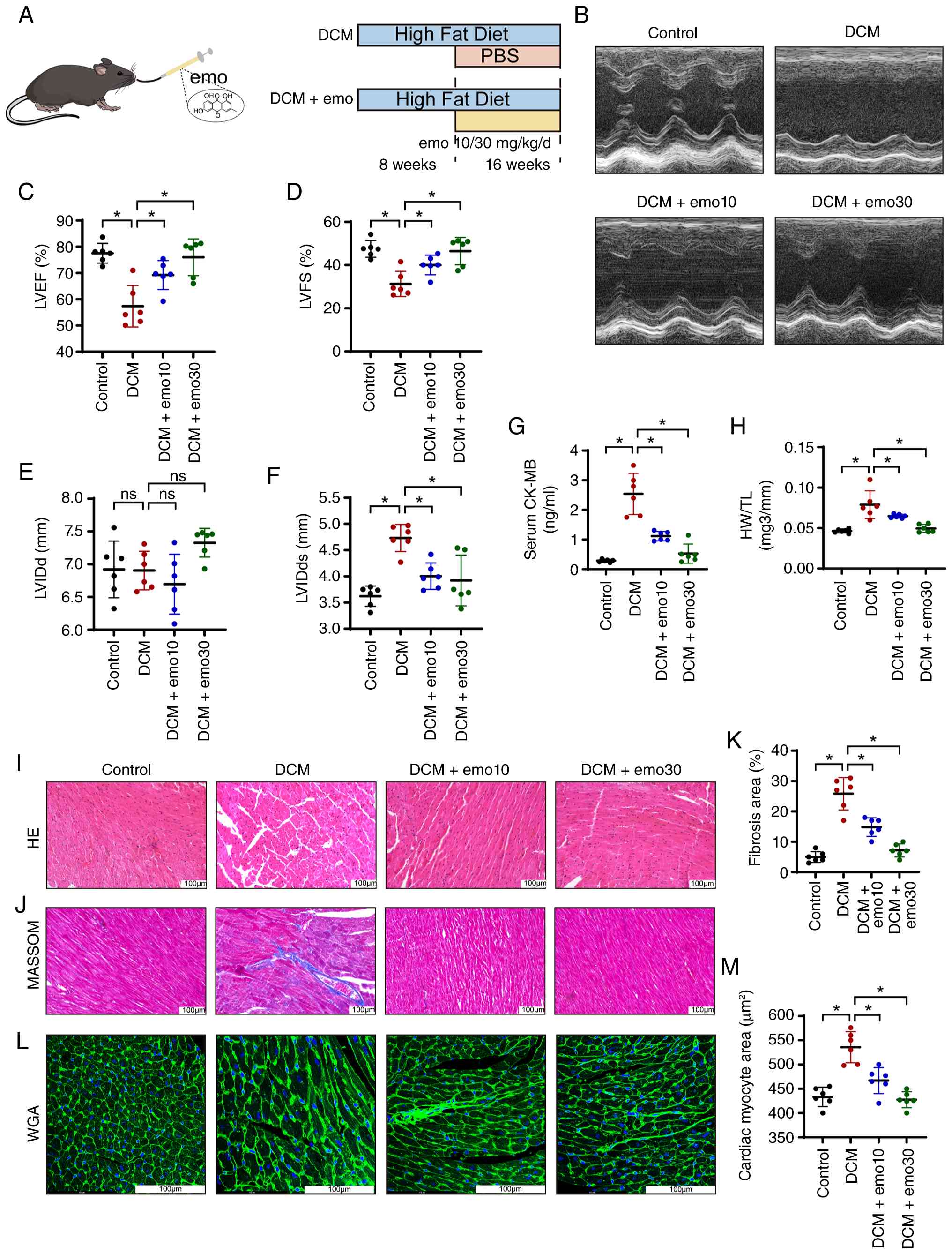 | Figure 1Emodin improves cardiac ejection
function and reduces myocardial injury in DCM. (A) Schematic
diagram of the animal experimental protocol. (B) M-mode
echocardiography. (C-F) LVEF, LVFS, LVIDd and LVIDs measured by
M-mode echocardiography. (G) Serum cardiac injury markers CK-MB
measured by ELISA. (H) HW/TL ratio for each group of mice. (I)
H&E staining to assess myocardial morphology. (J) Masson
staining to detect myocardial fibrosis. (K) Quantification of
fibrosis area in Masson staining. (L) WGA staining to evaluate
myocardial hypertrophy. (M) Quantification of average myocardial
cell area. Data are presented as the mean ± SD (n=6 per group).
*P<0.05. DCM, diabetic cardiomyopathy; LVEF, left
ventricular ejection fraction; LVFS, left ventricular fractional
shortening; LVID, left ventricular internal dimension; LVIDd, LVID
at end-diastole; LVIDs, LVID at systole; CK-MB, creatine kinase-MB;
HW/TL, heart weight/tibial length; ns, not statistically
significant. |
Cardiac hypertrophy and interstitial fibrosis are
the characteristic pathological changes of DCM. As shown in
Fig. 1H-M, compared with the
control group, the hearts of the DCM group exhibited typical
pathological changes. The increase in the ratio of heart weight to
tibial length (Fig. 1H), the
disordered and broken arrangement of central muscle fibers in
H&E staining (Fig. 1I), the
intensified interstitial fibrosis shown by MASSON staining
(Fig. 1J and K), and the
increase in the cross-sectional area of myocardial cells indicated
by WGA staining (Fig. 1L and M)
were verified. Emo treatment improved the above-mentioned cardiac
pathological changes caused by diabetes, especially the high-dose
treatment group effectively alleviated the myocardial damage caused
by long-term diabetes. These findings suggested that emo
significantly alleviated myocardial hypertrophy and interstitial
fibrosis caused by diabetes in a dose-dependent manner, thereby
improving cardiac remodeling in DCM.
Emo alleviates mitochondrial dynamic
imbalance in DCM mice and reduces ferroptosis
To explore whether emo exerts a protective effect by
regulating mitochondrial division and fusion. The mitochondrial
structure was observed by electron microscopy (Fig. 2A). The results demonstrated that
compared with the control group, the mice treated with DCM had
mitochondrial abnormalities, including mitochondrial crest
destruction, increased porosity, increased membrane density and
membrane rupture, which was indicated by an increase in Flameng
scores (Fig. S1F). In addition,
the average size of individual mitochondria in mice treated with
DCM decreased and the mitochondrial density per unit area increased
compared with the control group (Fig. 2B and C). The enzymatic activity
of the mitochondrial complex was additionally measured (Fig. 2D). The results showed that
compared with the control group, the activities of mitochondrial
complexes I and III in the DCM group were impaired, while complexes
II and IV were basically unaffected. The aforementioned results
suggest that the mitochondrial oxidative respiratory chain is
damaged, the generation of superoxides increases, and tissue
oxidative stress intensifies. However, the aforementioned results
were reversed after emo intervention. Emo exerts a protective
effect by regulating mitochondrial division and fusion. The
aforementioned conjecture was verified by detecting the relevant
proteins (Fig. 2E-I). Compared
with the control group, the mitochondrial dynamics in the DCM group
were severely imbalanced, among which the expression of the key
protein DRP1 mediating mitochondrial division was significantly
upregulated. Conversely, the expression levels of MFN1 and MFN2 for
outer mitochondrial membrane fusion and OPA1 for inner membrane
fusion were significantly downregulated. This change pattern
indicates excessive mitochondrial division in DCM cardiomyocytes.
Emo treatment effectively reversed this imbalance. The low-dose of
emo slightly reversed the expression of the aforementioned
proteins, and the high-dose of emo strongly inhibited the
expression of DRP1 and increased the protein levels of MFN1, MFN2
and OPA1.
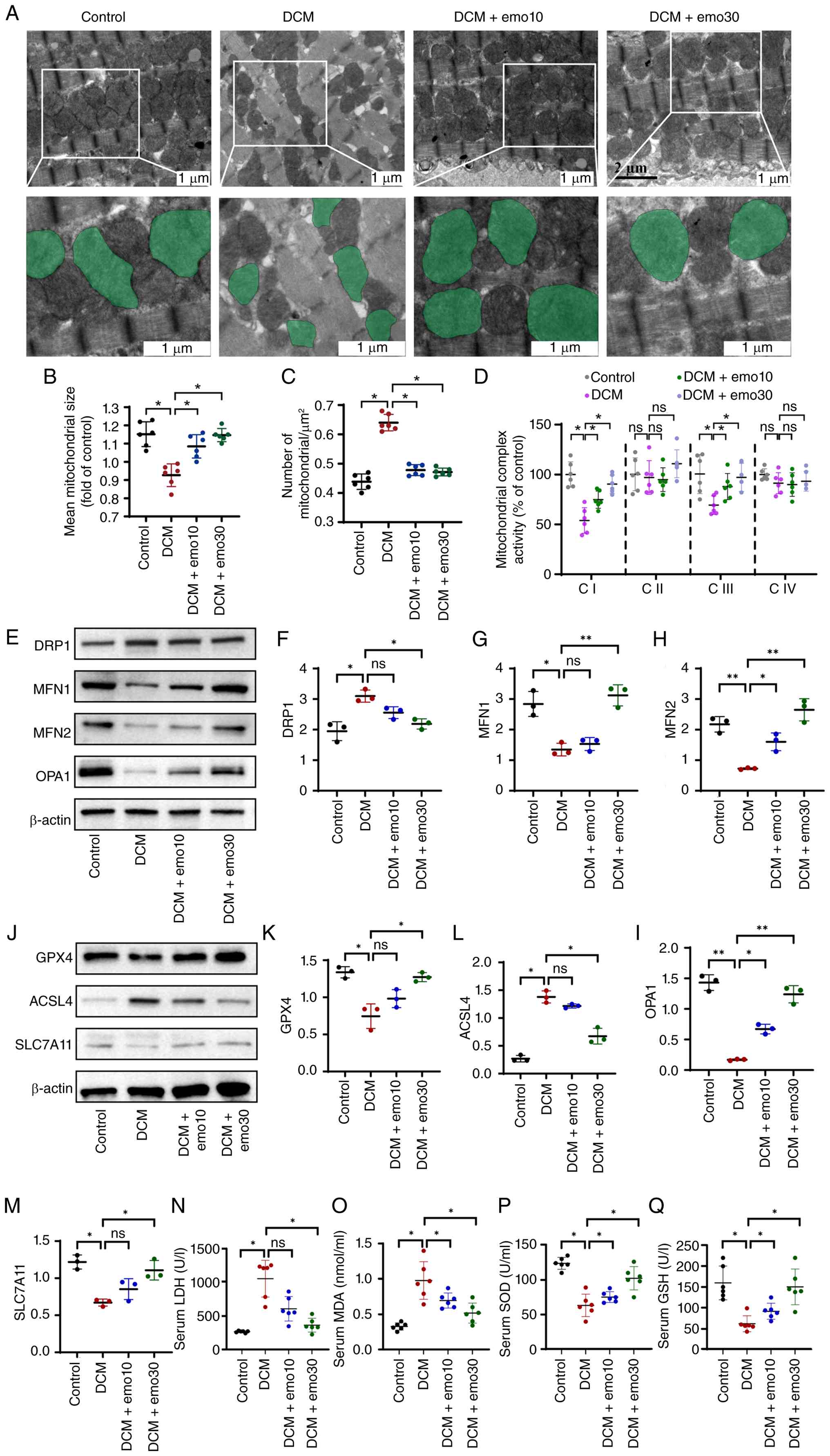 | Figure 2Emodin alleviates mitochondrial
dynamic imbalance in the myocardium of DCM mice and reduced
ferroptosis. (A) Representative mitochondrial images obtained by
transmission electron microscopy, with the bottom images (magnified
from the top images) showing representative mitochondria marked in
light green pseudocolor to highlight mitochondrial changes. (B and
C) Quantitative data on average mitochondrial size and the number
of mitochondria per μm2. (D) Quantitative
assessment of mitochondrial respiratory chain complex activity.
(E-I) Western blot analysis of the relative protein expression
levels of DRP1, MFN1, MFN2 and OPA1 in vivo. (J-M) Western
blot analysis of the relative protein expression levels of GPX4,
ACSL4 and SLC7A11 in vivo. (N-Q) Serum redox markers LDH,
MDA, SOD and GSH measured by ELISA. Data are presented as the mean
± SD (n=6 independent animal experiments; n=3 independent western
blot experiments). *P<0.05 and
**P<0.01. DCM, diabetic cardiomyopathy; DRP1,
dynamin-related protein 1; MFN, mitochondrial fusion protein; OPA1,
optic atrophy protein 1; GPX4, glutathione peroxidase 4; ACSL4,
acyl-CoA synthase long chain family member 4; SLC7A11, solute
carrier family 7 member 11; MDA, malondialdehyde; SOD, superoxide
dismutase; GSH, glutathione; ns, not statistically significant. |
Mitochondrial dynamics imbalance can lead to the
production of a large amount of ROS. The accumulated ROS can damage
cellular proteins, lipids and DNA, cause lipid peroxidation, damage
cells and tissues, and eventually lead to ferroptosis. As an
iron-dependent form of cell death, ferroptosis is characterized by
iron overload. Consistent with this, colorimetric absolute
quantification revealed an iron accumulation in the myocardial
tissues of DCM mice (Fig. S1D).
Immunoblotting analysis revealed that the expression level of GPX4
in the DCM group was significantly lower than that in the control
group. Moreover, the expression of SLC7A11, a key transporter for
synthesizing glutathione, decreased significantly, while the
expression of ACSL4, which promotes lipid peroxidation, was
significantly upregulated (Fig.
2J-M). It is worth noting that emo treatment inhibited
ferroptosis in DCM mice, reversed the aforementioned results, and
counterbalanced the cardiac ferroptosis damage induced by high
sugar and high fat, with the high-dose group having the strongest
protective effect. The relevant serological indicators also
verified the aforementioned results (Fig. 2N-Q). Compared with the control
group, the serum LDH activity and MDA content in the DCM group were
significantly increased, while the SOD activity and GSH level were
significantly decreased. Emo treatment could dose-dependently
reduce LDH and MDA, increase SOD and GSH, and the high-dose group
had the strongest improvement effect. It is suggested that emo can
alleviate ferroptosis in DCM and protect cardiomyocytes. The
aforementioned results clearly show that there is a significant
mitochondrial dynamic imbalance in DCM. Emo effectively inhibits
ferroptosis in cardiomyocytes in DCM by reshaping mitochondrial
dynamic balance (inhibiting division and promoting fusion) and
improving mitochondrial function, thereby exerting a protective
effect on the heart. To investigate the role of ferroptosis
inhibition in the effects of emo and to evaluate the expression of
iron metabolism-related proteins, in vitro treatment was
subsequently conducted utilizing the specific ferroptosis inhibitor
Ferrostatin-1 (Fer-1).
Emo regulates ferroptosis in DCM mice
through the AHR-Nrf2 pathway
To reveal the specific mechanism by which emo
regulates ferroptosis, focus was addressed on the key role of AHR
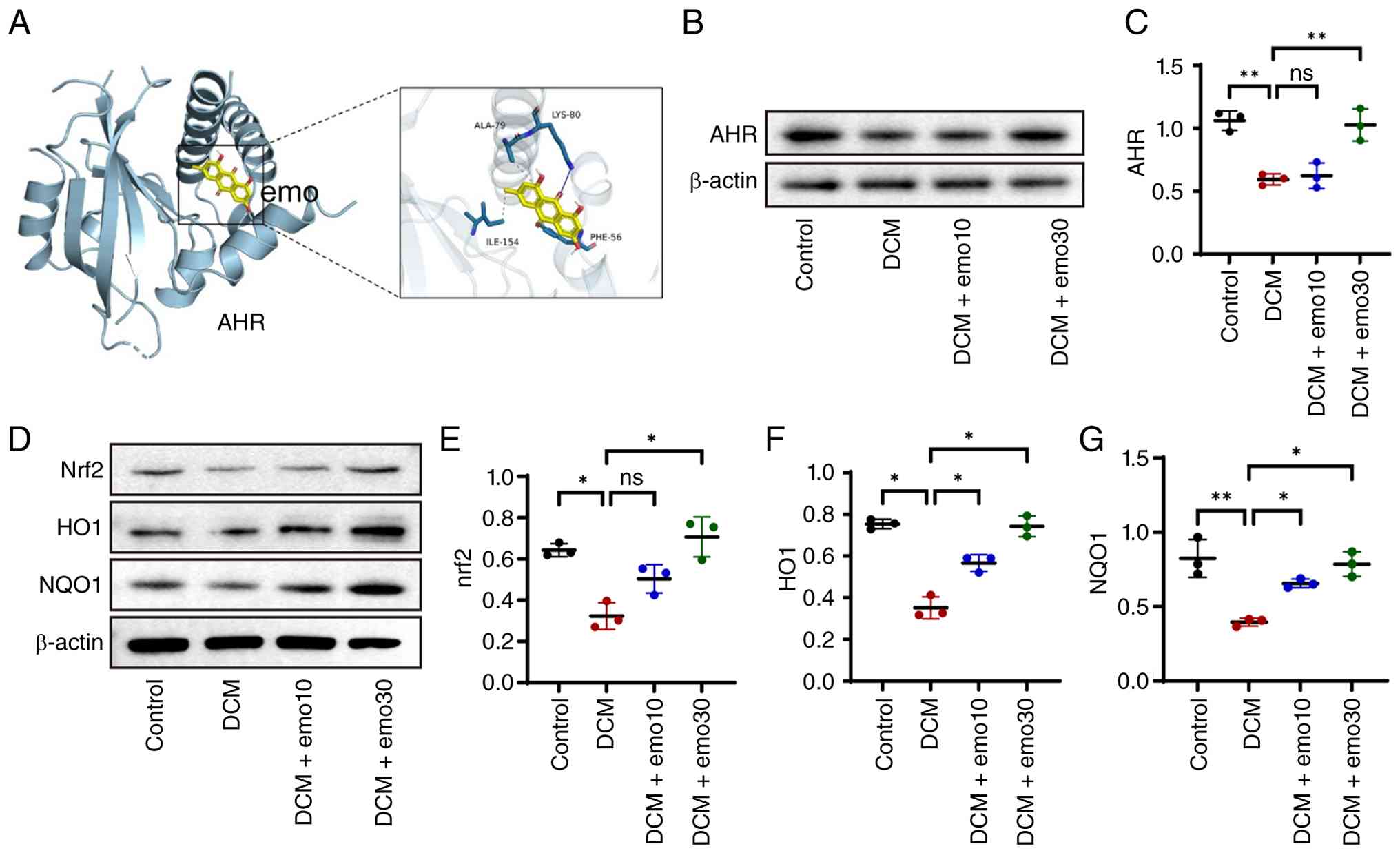
in metabolic disorders. Molecular docking prediction indicates that
emo can specifically bind to AHR (Fig. 3A). Consistently, RT-qPCR revealed
that the relative mRNA expression of AHR in the myocardial tissues
of the DCM group was decreased compared with the control group, an
effect that was dose-dependently increased by emo treatment
(Fig. S1E). The expression of
AHR was detected by western blotting, which was consistent with the
predicted results of molecular docking (Fig. 3B and C). The expression of AHR in
the DCM group was inhibited compared with that in the control
group. After emo treatment, the expression of AHR increased in a
dose-dependent manner. Given that the activation of AHR regulates
Nrf2, the expression of Nrf2 was further examined (Fig. 3D and E). The results revealed
that compared with the control group, the protein level of Nrf2 in
the cell nucleus of the DCM group was significantly decreased.
Correspondingly, the protein expression levels of its key
downstream antioxidant enzymes, HO-1 and NQO1, were also
significantly downregulated. This indicates that in the DCM state,
the core antioxidant defense system within the cells is impaired in
function. However, emo treatment effectively reversed this
phenomenon. In the emo treatment group, it could be observed that
the level of Nrf2 protein in the cell nucleus and the expression
levels of HO-1 and NQO1 increased significantly (Fig. 3D-G). These results indicate that
emo can dose-dependently activate the AHR-Nrf2 signaling pathway,
thereby enhancing the endogenous antioxidant capacity of
cardiomyocytes, inhibiting the production of ROS, and alleviating
ferroptosis.
Emo alleviates high glucose-induced
ferroptosis via an AHR dependent way
The cytoprotective mechanism of emo against
HG-elicited ferroptosis in H9C2 cardiomyoblasts was investigated in
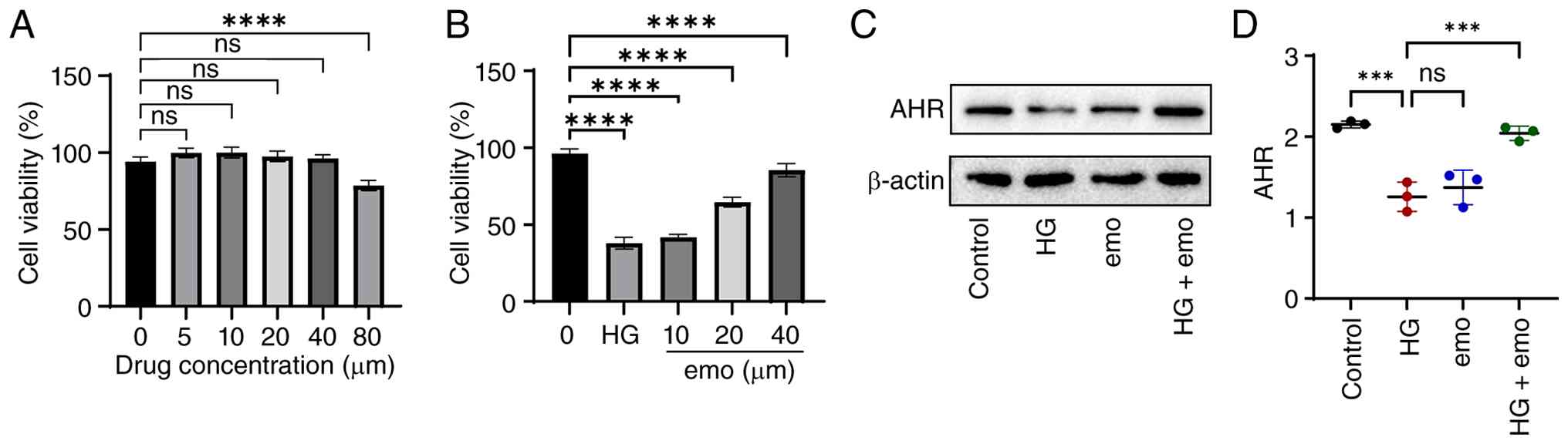
the present study. In the absence of HG stress, emo up to 40
μM did not alter basal cell viability (Fig. 4A). CCK-8 assays revealed that emo
dose-dependently attenuated HG-induced H9C2 injury, conferring
significant cytoprotection (Fig.
4B). Based on these viability data, while 20 μM showed
substantial protection, 40 μM emo elicited the maximum
cytoprotective effect without causing basal cytotoxicity.
Therefore, 40 μM was selected as the optimal effective
concentration for subsequent mechanistic experiments. Western blot
analysis demonstrated that HG significantly downregulated AHR
protein expression relative to control, whereas concomitant emo
treatment restored AHR levels above those observed in the HG group
(P<0.05), indicating that emo reverses HG-mediated suppression
of AHR (Fig. 4C and D).
To verify the crucial role of the AHR-mediated
transcriptional network in emo protection, a specific AHR siRNA was
used to knockdown its expression in H9C2 cells. Among them, AHR
siRNA3 had the best knockdown effect and was selected for
subsequent application (Fig. 5A and
B). The localization of Nrf2 under different interventions was
observed by fluorescence staining. Compared with the control, HG
stimulation led to a weakened fluorescence intensity of Nrf2.
However, after emo treatment, a significant increase in Nrf2
nuclear translocation could be clearly observed. Importantly, the
emo-induced promotion of Nrf2 nuclear translocation was
significantly abolished following the knockdown of AHR (Fig. 5C). The results of western
blotting found that compared with the control group, the expression
levels of Nrf2, NQO1 and HO-1 proteins in the HG group were
significantly decreased. The aforementioned results were reversed
after treatment with emo. However, the emo-induced upregulation of
Nrf2, NQO1 and HO-1 was significantly suppressed when AHR was
silenced (compared with the HG + emo group, P<0.05, Fig. 5D-G). These findings demonstrate
that emo promotes Nrf2 activation and its downstream antioxidant
defense in an AHR-dependent manner.
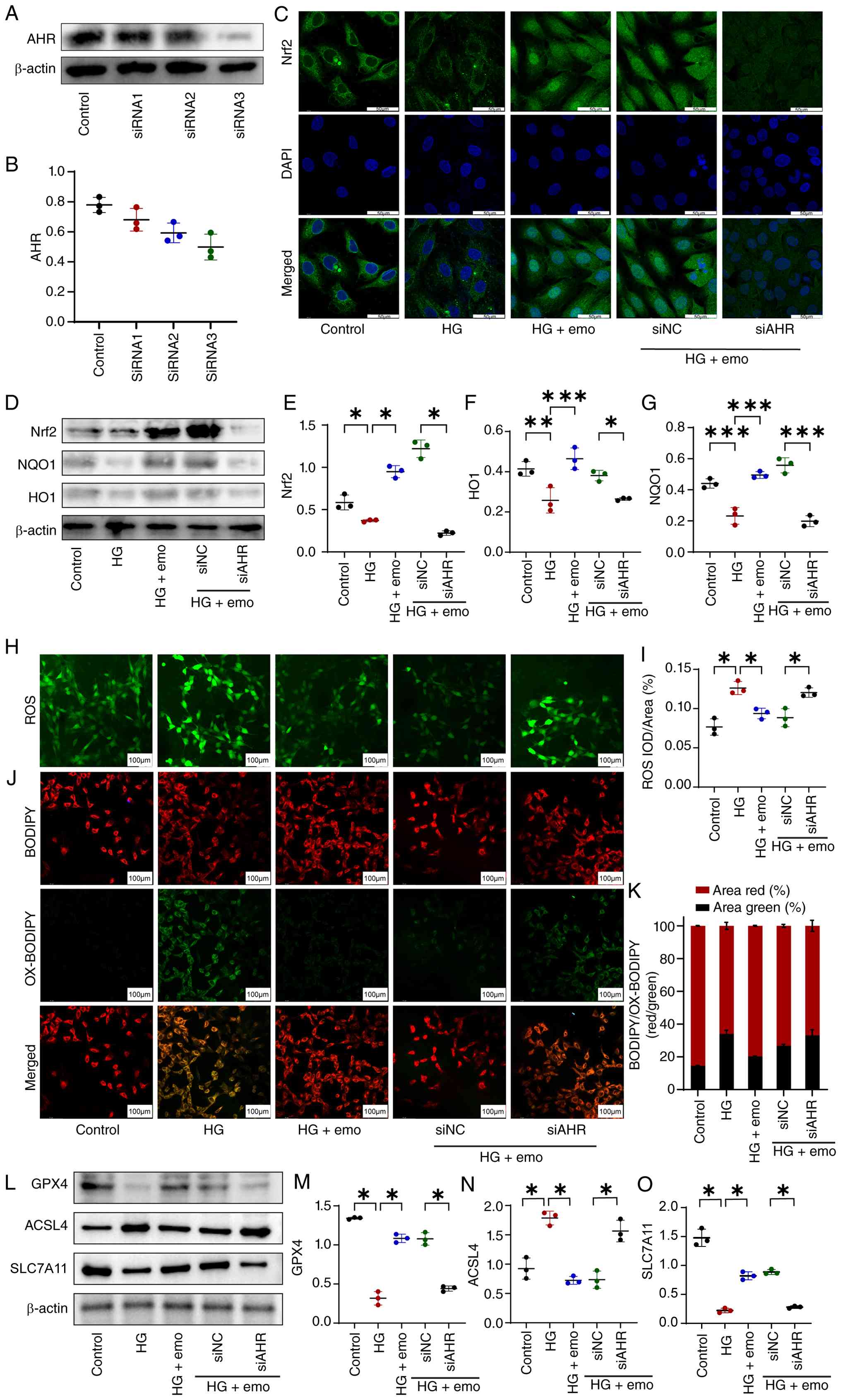 | Figure 5Emodin improves lipid peroxidation in
cardiomyocytes induced by high glucose in vitro and
alleviates ferroptosis, and this effect requires AHR mediation. (A
and B) Western blot analysis to verify the efficiency of
siRNA-mediated AHR knockdown. (C) Representative images showing
Nrf2 distribution in cultured H9C2 cells. (D-G) Western blot
analysis of Nrf2 and downstream antioxidant molecules NQO1 and HO-1
expression in H9C2 cells in each group. (H and I) DCFH-DA
fluorescent probe observation of ROS production in H9C2 cells
across different groups. (J and K) BODIPY staining to detect lipid
peroxidation in H9C2 cells (red: reduced BODIPY; green: oxidized
BODIPY). (L-O) Western blot analysis of the relative protein
expression of GPX4, ACSL4 and SLC7A11 in H9C2 cells. Data are
presented as the mean ± SD (n=3). *P<0.05,
**P<0.01 and ***P<0.001. AHR, aryl
hydrocarbon receptor; siRNA, small interfering RNA; NQO1, quinone
oxidoreductase 1; HO-1, heme oxygenase 1; ROS, reactive oxygen
species; NC, negative control; GPX4, glutathione peroxidase 4;
ACSL4, acyl-CoA synthase long chain family member 4; SLC7A11,
solute carrier family 7 member 11; HG, high-glucose. |
Subsequently, the intracellular ROS levels were
detected. Compared with the Control group, the ROS fluorescence
intensity and IOD/Area value in the HG group were significantly
increased. However, the ROS level was significantly reversed after
emo intervention. Notably, the inhibitory effect of emo on ROS
production was largely neutralized in the siAHR + HG + emo group
(Fig. 5H and I). Similarly,
BODIPY staining on H9C2 cells was used to detect the level of lipid
ROS. The results demonstrated that compared with the HG group, the
green fluorescence intensity of intracellular lipid peroxidation
decreased after emo treatment, suggesting that emo alleviated lipid
peroxidation in HG cardiomyocytes induced by high glucose. However,
in AHR-deficient cardiomyocytes, the protective effect of emo
against lipid peroxidation was markedly diminished (Fig. 5J and K). Western blotting
revealed that the HG-induced downregulation of GPX4 and SLC7A11 and
upregulation of ACSL4 were reversed by emo. Crucially, the
anti-ferroptotic effect of emo was significantly attenuated after
AHR knockdown (Fig. 5L-O).
Collectively, these results indicate that emo exerts its
antioxidant and anti-ferroptotic effects by activating the AHR-Nrf2
signaling axis.
To evaluate the potential involvement of ferroptosis
in HG-induced cardiomyocyte injury, H9C2 cells were treated with
different concentrations of Fer-1. As revealed in Fig. S2A, Fer-1 treatment increased the
viability of HG-stimulated H9C2 cells. Therefore, 10 μM
Fer-1 was used in subsequent experiments. Furthermore, BODIPY
staining showed that both Emodin and Fer-1 reduced HG-induced lipid
peroxidation (Fig. S2B and C).
Subsequently, the protein expression of the iron-uptake marker
TfR1, the iron-storage protein FTH1, the core anti-ferroptotic
enzyme GPX4 and the pro-ferroptotic lipid metabolism enzyme ACSL4
were assessed (Fig. S2D-H).
Consistent with previous observations, HG stimulation significantly
upregulated the pro-ferroptotic drivers TfR1 and ACSL4 while
suppressing the defense proteins FTH1 and GPX4 (P<0.05),
indicating a pathological state of intracellular iron overload and
unchecked lipid peroxidation. Notably, emo treatment effectively
reversed these changes, mirroring the molecular profile of Fer-1
intervention. Specifically, both emo and Fer-1 restored GPX4 levels
and normalized iron metabolism by downregulating TfR1 and
upregulating FTH1. These findings provide compelling functional
evidence that emo protects H9C2 cells against HG-induced injury by
maintaining iron homeostasis and inhibiting the ferroptotic
pathway.
Emo activates AHR to reshape the
mitochondrial dynamic balance of cardiomyocytes induced by HG
Previous studies demonstrated that mitochondrial
dynamics imbalance plays a key role in cell damage induced by HG
(25). To verify that emo
reshapes the mitochondrial dynamic balance of cardiomyocytes
induced by HG through AHR mediation, the effect of AHR knockdown on
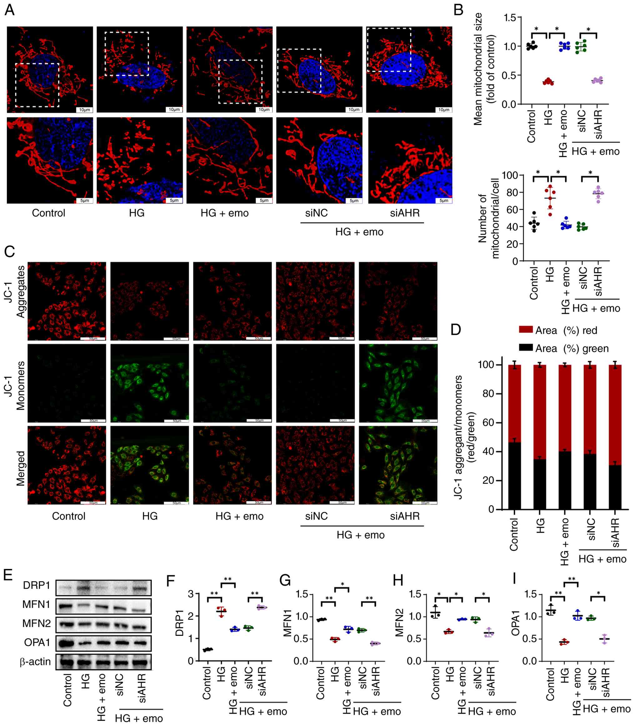
mitochondrial dynamics was studied in vitro. Mitochondria
were labeled using mitochondrial probes and images were captured
under confocal microscopy. The results showed that HG caused marked
mitochondrial fragmentation, whereas emo reversed this phenotype
and restored elongated rod-like mitochondria; these effects were
lost after AHR knockdown (Fig. 6A
and B). The changes in MMP were evaluated using the JC-1
detection kit. The results revealed that emo also normalized
HG-induced MMP loss, which was again abolished by AHR silencing
(Fig. 6C and D). Consistently,
emo decreased DRP1 and increased MFN1, MFN2 and OPA1 expression,
shifting mitochondrial dynamics from fission toward fusion, while
AHR inhibition reversed these changes (Fig. 6E-I). This indicates that in in
vitro experiments, emo intervention to activate the AHR pathway
effectively reshaped the mitochondrial dynamic balance of H9C2
cells induced by HG. Importantly, to investigate whether the
mitochondrial dynamic alterations and ferroptosis are causally
linked or merely parallel outcomes, mitochondrial morphology was
evaluated following ferroptosis inhibition. Strikingly, the
pharmacological inhibition of ferroptosis by Fer-1 decreased
HG-induced mitochondrial fragmentation, maintaining the elongated
rod-like mitochondrial networks (Fig. S2I-K). At the molecular level,
western blot analysis revealed that Fer-1 treatment reversed the
HG-induced molecular imbalance of mitochondrial dynamics, evidenced
by the downregulation of the fission mediator DRP1 and the
restoration of core fusion proteins, including MFN1, MFN2 and OPA1
(Fig. S2L-P). These critical
findings unequivocally demonstrate that mitochondrial dynamic
collapse is not an isolated parallel event but is intimately and
sequentially coupled with the ferroptotic cascade. Therefore,
Emodin-mediated AHR activation regulates mitochondrial dynamics,
which is associated with the inhibition of ferroptosis.
Emo activates AHR to ease the imbalance
mitochondrial dynamics in DCM mice
To verify whether emo alleviates DCM-induced
mitochondrial dynamic imbalance in an AHR-dependent manner,
cardiac-specific AHR knockdown was achieved via tail-vein injection
of AAV9-shAhR. Successful viral transduction and efficient AHR
silencing were confirmed by myocardial EGFP fluorescence and a
significant reduction in AHR expression compared with the AAV9-shNC
control (Fig. 7A and B; Fig. S3A). AHR silencing largely
abolished the cardioprotective effects of emo, as reflected by
reduced LVEF and LVFS, increased LVIDd and LVIDs, and marked
ventricular dilatation (Fig.
7C-G). Consistently, H&E, Masson and WGA staining showed
aggravated myofiber disarray, interstitial fibrosis and
cardiomyocyte hypertrophy in the AAV9-shAHR group compared with
AAV9-sh-NC (Fig. 7H-L),
indicating that AHR knockdown blunts Emo-mediated functional and
structural benefits. Electron microscopy further revealed that AHR
silencing negated the corrective effects of emo on mitochondrial
dynamics, leading to severe ultrastructural damage (cristae
disruption, matrix vacuolization, membrane rupture), reduced mean
mitochondrial volume, and increased mitochondrial number per unit
area (Fig. 7M). To quantify
these ultrastructural pathologies, mitochondrial Flameng scoring
was performed, which showed that the suppressive effect of emo on
mitochondrial injury was attenuated following AHR knockdown
(Fig. S3B). Furthermore, AHR
silencing impaired the ability of emo to restore mitochondrial
morphology, as evidenced by reduced mean mitochondrial volume and
increased mitochondrial number per unit area (Fig. 7N and O). Given that mitochondrial
dynamic disruption is associated with local ferroptotic cascades
and iron pool dysregulation in the heart, tissue iron levels were
also monitored and it was found that the regulatory effect of emo
on limiting cardiac iron overload was suppressed in the AAV9-shAHR
group, manifesting as a recurrence of myocardial iron accumulation
(Fig. S3C). At the molecular
level, western blotting confirmed a shift toward excessive
mitochondrial fission, with DRP1 upregulation and MFN1, MFN2 and
OPA1 downregulation (Fig. 7P-T).
Collectively, these data demonstrate that emo exerts its
anti-cardiac injury effects in DCM by engaging cardiac AHR
transcriptional network to reshape mitochondrial dynamics and
preserve myocardial iron homeostasis.
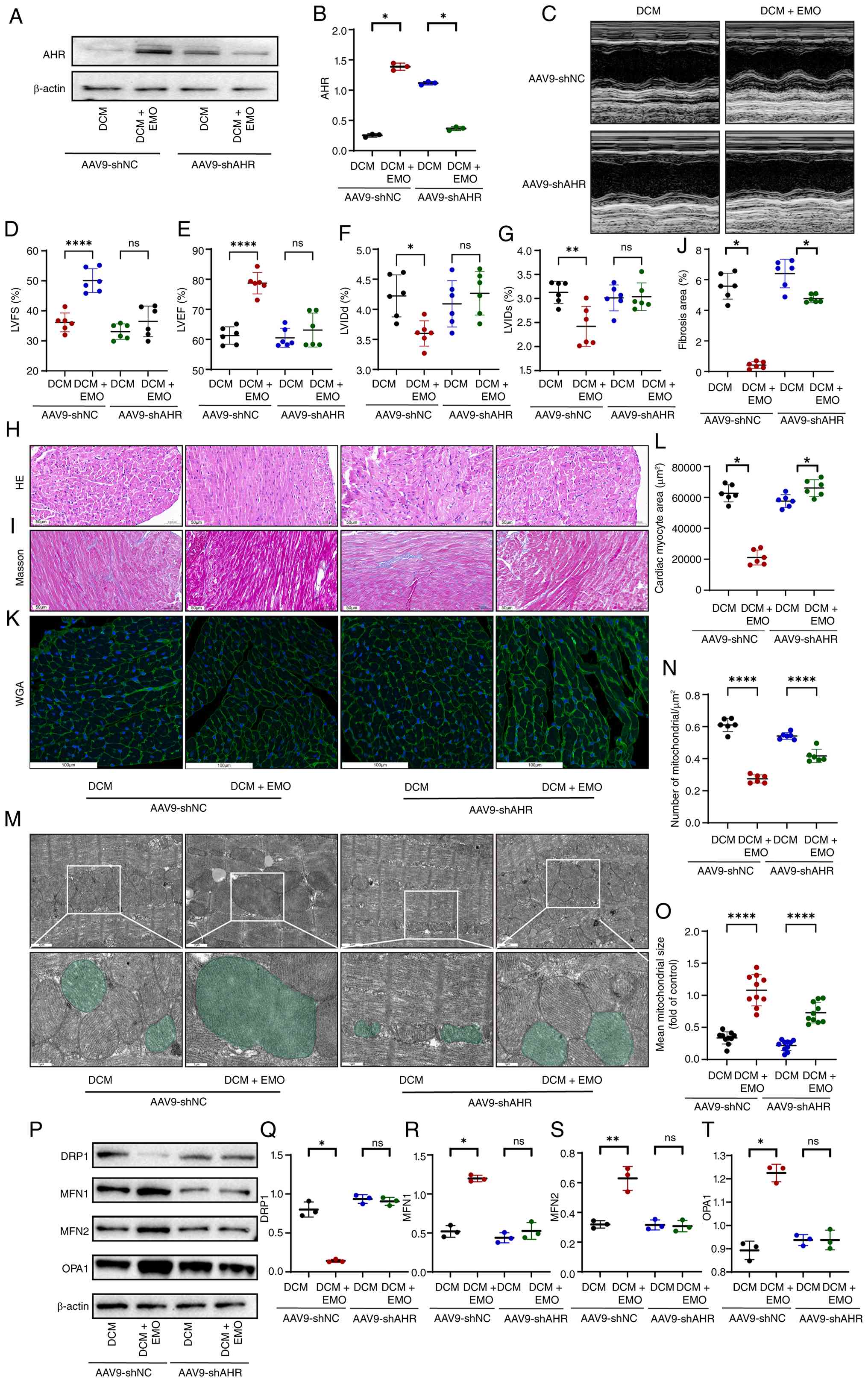 | Figure 7AHR knockdown attenuates the
protective effects of emodin on cardiac function and mitochondrial
dynamics in DCM mice. (A and B) AHR protein expression in heart
tissues and validation of successful cardiac-specific knockdown by
AAV9-cTnT-shAHR (labeled as AAV9-shAHR). (C-G) Assessment of
cardiac function by echocardiography. (H-L) Histological evaluation
of myocardial pathology and fibrosis. (M-O) Ultrastructural and
quantitative analysis of mitochondria by transmission electron
microscopy. (P-T) Protein levels of mitochondrial fission (DRP1)
and fusion (MFN1, MFN2 and OPA1) regulators. Data are presented as
the mean ± SD (n=6 independent animal experiments; n=3 independent
western blot experiments). *P<0.05,
**P<0.01 and ****P<0.0001. AHR, aryl
hydrocarbon receptor; sh-, short hairpin; DRP1, dynamin-related
protein 1; MFN, mitochondrial fusion protein; OPA1, optic atrophy
protein 1; DCM, diabetic cardiomyopathy; NC, negative control;
LVFS, left ventricular fractional shortening; LVID, left
ventricular internal dimension; LVIDd, LVID at end-diastole; LVIDs,
LVID at systole; ns, not statistically significant. |
Discussion
In the present study, it was found that emo can
inhibit cardiomyocyte apoptosis and myocardial fibrosis under high
glucose stress, thereby effectively protecting cardiac ejection
function in DCM. Further research revealed that emo can effectively
restore mitochondrial respiratory chain enzyme activity and inhibit
excessive mitochondrial fission, which is crucial for protecting
cardiac function. Additionally, in vitro studies
demonstrated that the aforementioned effects of emo require AHR
mediation and are achieved by promoting Nrf2 nuclear translocation
and activating the expression of downstream antioxidant
molecules.
DCM is a condition affecting cardiac structure and
function in patients with diabetes, independent of coronary heart
disease, valvular heart disease and other cardiovascular diseases
(26). It is characterized by
metabolic disturbances, abnormalities in subcellular components and
immunological changes, leading to local cardiac inflammation,
oxidative stress, mitochondrial dysfunction and cell apoptosis.
These changes cause impaired cardiac systolic and diastolic
function, left ventricular hypertrophy and dilated cardiomyopathy,
eventually leading to heart failure (2). Currently, the main treatments for
DCM include controlling blood glucose, regulating blood lipids,
improving microcirculation, and managing heart failure. However,
these treatments primarily focus on managing the primary disease
and alleviating complications, and there is a lack of effective
specific therapies for DCM. Emo, as an antioxidant molecule, has
been reported to have therapeutic effects in lung injury, cancer
and diabetic nephropathy (10,27,28). Moreover, it has also been
reported to significantly alleviate atherosclerosis in the
cardiovascular system. The present study revealed that emo
significantly alleviates cardiac damage caused by high blood
glucose, primarily by inhibiting myocardial fibrosis, apoptosis and
hypertrophy. By assessing oxidative stress-related markers such as
SOD and MDA, it was found that the effects of emo are likely
closely related to its antioxidant properties.
Mitochondria are essential organelles for energy
production and maintaining normal cellular physiological functions,
and their damage plays a significant role in the pathogenesis of
DCM. In diabetic patients with impaired myocardial contraction,
mitochondria often exhibit swelling and fragmentation, accompanied
by impaired mitochondrial dynamics, suggesting that decreased
myocardial contraction in diabetes may be related to deteriorated
mitochondrial function (29).
Damaged mitochondria, due to increased membrane permeability,
release pro-apoptotic factors into the cytoplasm, contributing to
cardiomyocyte apoptosis. Additionally, mitochondria are closely
linked to oxidative stress. ROS are primarily produced through
mitochondrial pathways, and there is a mutual promotion
relationship between mitochondria and ROS (4,5).
ROS are mainly generated by the electron transport chain located in
the mitochondrial inner membrane, and excessive ROS can further
damage mitochondrial DNA, proteins and membranes (6), exacerbating the dysfunction of the
electron transport chain and leading to mitochondrial homeostasis
disruption and ROS accumulation. Thus, excessive mitochondrial
fission, due to its functional impairment, can be considered an
upstream event of ROS production (30). Therefore, inhibiting pathological
mitochondrial overfission and reducing cardiac oxidative stress are
crucial for protecting cardiac function. The results of the present
study indicated that emo significantly inhibits excessive
mitochondrial fission and regulates the expression levels of
mitochondrial dynamics proteins such as DRP1 and MFN2, shifting the
mitochondrial dynamics towards fusion. Surprisingly, it was also
found that emo significantly protects the enzyme activity of
mitochondrial complexes, which is important for maintaining normal
mitochondrial energy metabolism and reducing ROS production.
AHR, a member of the basic helix-loop-helix
transcription factor family, regulates various systemic diseases,
including skin inflammation and Parkinson's disease (11,13). It has been indicated that AHR can
act as a transcription factor for Nrf2, modulating Nrf2
transcription (19). The
AHR-Nrf2 pathway plays a crucial regulatory role in various
inflammation and oxidative stress-related diseases (31,32). The present findings identified
that AHR expression is impaired under high glucose stress, but emo
can increase AHR levels in myocardial tissue. Importantly,
molecular docking predictions revealed that emo has a strong
binding affinity with AHR, indicating it as a potential ligand for
AHR. Furthermore, fluorescence staining revealed that emo promotes
the nuclear translocation of Nrf2 and increases the expression of
downstream antioxidant molecules NQO1 and HO-1. Mechanistically,
although AHR and Nrf2 are classically governed by distinct
pathways, AHR-ARNT binding to XRE elements and Keap1-mediated
post-translational control of Nrf2, a non-canonical transcriptional
link between them has been documented: Functional XRE-like elements
within the Nrf2 promoter allow ligand-activated AHR to
transactivate Nrf2 transcription, placing the Nrf2-ARE program
downstream of AHR-XRE activation as a complement to the canonical
Keap1 pathway (33). Consistent
with this framework and with a recent study showing that the AHR
ligand indole-3-lactic acid activates Nrf2 to inhibit ferroptosis
in cardiomyocytes (34), the
present findings indicated that Emodin drives Nrf2 activation in an
AHR-dependent manner. Functionally, nuclear translocation of Nrf2,
a definitive marker of its activation and a prerequisite for ARE
binding and induction of target genes such as HO-1, NQO1 and GPX4,
was robustly enhanced by Emodin. Although AHR-reporter and
Nrf2-reporter assays were not performed, the coordinated nuclear
enrichment of Nrf2, the upregulation of its downstream effectors,
and the loss of these effects upon AHR silencing together provide
strong functional evidence that Emodin engages an AHR-dependent
Nrf2 antioxidant program. It is acknowledged that direct AHR
occupancy on the Nrf2 promoter was not examined in the current
cardiac model and represents a worthwhile direction for future
mechanistic study. Subsequent siRNA-mediated AHR interference
showed that the cardioprotective effects of emo significantly
diminished. These lines of evidence collectively suggest that emo
exerts its effects through the AHR-Nrf2 pathway. Furthermore,
although direct pharmacokinetic translation between in vitro
and in vivo models is complex, the selected in vivo
dose of 30 mg/kg was proven highly effective in engaging this
myocardial AHR pathway and reversing structural remodeling,
aligning with the robust cytoprotection observed at 40 μM
in vitro.
The limitations and strengths of the present study
also warrant consideration. First, regarding the animal model, a
combination of HFD feeding and low-dose STZ injection was utilized
in wild-type C57BL/6J mice. The unique advantage of this
non-genetic model is its ability to rapidly and stably induce
severe glucolipid metabolic disorders alongside partial pancreatic
β-cell decompensation, thereby effectively mimicking the core
pathophysiological trajectory of advanced human type 2 DCM. This
approach provides a highly reproducible platform for evaluating
early pharmacological interventions. However, a notable limitation
is that this accelerated chemical induction creates an
exceptionally severe diabetic phenotype, which may not entirely
capture the decades-long, gradual pathological evolution typical of
human DCM. Furthermore, because of inherent genetic and
cardiovascular differences between mice and humans, clinical
studies are still required to validate these findings. Second,
although the present study focused on oxidative stress,
mitochondrial dynamics, ferroptosis and lipid peroxidation,
numerous additional mechanisms underlying the onset and progression
of DCM remain undefined. Future work will further dissect its
pathogenesis and seek drug candidates with more precise, targeted
regulatory effects.
In conclusion, it was first demonstrated that emo
alleviates DCM-related myocardial injury by activating the AHR/Nrf2
pathway to concurrently inhibit ferroptosis and excessive
mitophagy. Mechanistically, emo triggers the AHR-Nrf2 axis, thereby
interrupting HG-induced mitochondrial over-fission and lipid
peroxidation, which in turn suppresses cardiomyocyte ferroptosis
and interstitial fibrosis. These effects substantially restore
cardiac contractile performance in DCM mice and are completely
abolished when AHR is silenced by siRNA, confirming pathway
dependence. Emo represents a first-in-class, AHR-directed
ferroptosis inhibitor ready for pre-clinical optimization and
offers a promising therapeutic candidate for DCM and other
ferroptosis-driven cardiomyopathies.
Supplementary Data
Availability of data and materials
The data generated in the present study may be
requested from the corresponding author.
Authors' contributions
XY managed data and wrote the original manuscript.
HaL developed methodology, provided resources and conducted
software analysis. JY managed data, conducted investigation, and
wrote, reviewed and edited the manuscript. JZ conducted formal
analysis, provided resources, and wrote and edited the manuscript.
JZ and HaL developed methodology, provided resources, and wrote and
edited the manuscript. JY and HuL managed data, conducted formal
analysis, developed methodology, supervised the study, and wrote,
reviewed and edited the manuscript. JC conceived and designed the
study, acquired funding, and contributed to writing, reviewing, and
editing the manuscript. JY and JC confirm the authenticity of all
the raw data. All authors read and approved the final version of
the manuscript.
Ethics approval and consent to
participate
All animal protocols were approved by the Animal
Care and Use Committee of Shaoxing People's Hospital (approval no.
2022C020; Shaoxing, China).
Patient consent for publication
Not applicable.
Competing interests
The authors declare that they have no competing
interests.
Acknowledgements
Not applicable.
Funding
The present study was supported by The Shaoxing Municipal Health
Science and Technology Program Project (grant no. 2023SKY124), The
Zhejiang Provincial Traditional Chinese Medicine Science and
Technology Plan Project (grant no. 2025ZX043), The Zhejiang
Provincial Medical and Health Science and Technology Program (grant
no. 2025KY392) and the National Natural Science Foundation of China
(grant no. 82200390).
References
|
1
|
Sun H, Saeedi P, Karuranga S, Pinkepank M,
Ogurtsova K, Duncan BB, Stein C, Basit A, Chan JCN, Mbanya JC, et
al: IDF diabetes atlas: Global, regional and country-level diabetes
prevalence estimates for 2021 and projections for 2045. Diabetes
Res Clin Pract. 183:1091192022. View Article : Google Scholar
|
|
2
|
Palomer X, Pizarro-Delgado J and
Vázquez-Carrera M: Emerging actors in diabetic cardiomyopathy:
Heartbreaker biomarkers or therapeutic targets? Trends Pharmacol
Sci. 39:452–467. 2018. View Article : Google Scholar : PubMed/NCBI
|
|
3
|
Nakamura K, Miyoshi T, Yoshida M, Akagi S,
Saito Y, Ejiri K, Matsuo N, Ichikawa K, Iwasaki K, Naito T, et al:
Pathophysiology and treatment of diabetic cardiomyopathy and heart
failure in patients with diabetes mellitus. Int J Mol Sci.
23:35872022. View Article : Google Scholar : PubMed/NCBI
|
|
4
|
Sapian S, Taib IS, Latip J, Katas H, Chin
KY, Mohd Nor NA, Jubaidi FF and Budin SB: Therapeutic approach of
flavonoid in ameliorating diabetic cardiomyopathy by targeting
mitochondrial-induced oxidative stress. Int J Mol Sci.
22:116162021. View Article : Google Scholar : PubMed/NCBI
|
|
5
|
Tsutsui H, Kinugawa S and Matsushima S:
Oxidative stress and heart failure. Am J Physiol Heart Circ
Physiol. 301:H2181–H2190. 2011. View Article : Google Scholar : PubMed/NCBI
|
|
6
|
Guo C, Sun L, Chen X and Zhang D:
Oxidative stress, mitochondrial damage and neurodegenerative
diseases. Neural Regen Res. 8:2003–2014. 2013.PubMed/NCBI
|
|
7
|
Galaris D, Barbouti A and Pantopoulos K:
Iron homeostasis and oxidative stress: An intimate relationship.
Biochim Biophys Acta Mol Cell Res. 1866:1185352019. View Article : Google Scholar : PubMed/NCBI
|
|
8
|
Yang X, Kawasaki NK, Min J, Matsui T and
Wang F: Ferroptosis in heart failure. J Mol Cell Cardiol.
173:141–153. 2022. View Article : Google Scholar : PubMed/NCBI
|
|
9
|
Zhang R, Chen J, Wang S, Zhang W, Zheng Q
and Cai R: Ferroptosis in cancer progression. Cells. 12:18202023.
View Article : Google Scholar : PubMed/NCBI
|
|
10
|
Zhang Q, Chen WW, Sun X, Qian D, Tang DD,
Zhang LL, Li MY, Wang LY, Wu CJ and Peng W: The versatile emodin: A
natural easily acquired anthraquinone possesses promising
anticancer properties against a variety of cancers. Int J Biol Sci.
18:3498–3527. 2022. View Article : Google Scholar : PubMed/NCBI
|
|
11
|
Semwal RB, Semwal DK, Combrinck S and
Viljoen A: Emodin-A natural anthraquinone derivative with diverse
pharmacological activities. Phytochemistry. 190:1128542021.
View Article : Google Scholar
|
|
12
|
He Y, Xi J, Fang J, Zhang B and Cai W:
Aloe-emodin alleviates doxorubicin-induced cardiotoxicity via
inhibition of ferroptosis. Free Radic Biol Med. 206:13–21. 2023.
View Article : Google Scholar : PubMed/NCBI
|
|
13
|
Hu S, Zhou J, Hao J, Zhong Z, Wu H, Zhang
P, Yang J, Guo H and Chi J: Emodin ameliorates doxorubicin-induced
cardiotoxicity by inhibiting ferroptosis through the remodeling of
gut microbiota composition. Am J Physiol Cell Physiol.
326:C161–C176. 2024. View Article : Google Scholar
|
|
14
|
Dai S, Ye B, Chen L, Hong G, Zhao G and Lu
Z: Emodin alleviates LPS-induced myocardial injury through
inhibition of NLRP3 inflammasome activation. Phytother Res.
35:5203–5213. 2021. View
Article : Google Scholar : PubMed/NCBI
|
|
15
|
Zhang X, Qin Q, Dai H, Cai S, Zhou C and
Guan J: Emodin protects H9c2 cells from hypoxia-induced injury by
up-regulating miR-138 expression. Braz J Med Biol Res.
52:e79942019. View Article : Google Scholar : PubMed/NCBI
|
|
16
|
Wu Z, Chen Q, Ke D, Li G and Deng W:
Emodin protects against diabetic cardiomyopathy by regulating the
AKT/GSK-3β signaling pathway in the rat model. Molecules.
19:14782–14793. 2014. View Article : Google Scholar : PubMed/NCBI
|
|
17
|
Chaffin CL, Trewin AL and Hutz RJ: Estrous
cycle-dependent changes in the expression of aromatic hydrocarbon
receptor (AHR) and AHR-nuclear translocator (ARNT) mRNAs in the rat
ovary and liver. Chem Biol Interact. 124:205–216. 2000. View Article : Google Scholar : PubMed/NCBI
|
|
18
|
Liu JR, Miao H, Deng DQ, Vaziri ND, Li P
and Zhao YY: Gut microbiota-derived tryptophan metabolism mediates
renal fibrosis by aryl hydrocarbon receptor signaling activation.
Cell Mol Life Sci. 78:909–922. 2021. View Article : Google Scholar
|
|
19
|
Wang K, Lv Q, Miao YM, Qiao SM, Dai Y and
Wei ZF: Cardamonin, a natural flavone, alleviates inflammatory
bowel disease by the inhibition of NLRP3 inflammasome activation
via an AhR/Nrf2/NQO1 pathway. Biochem Pharmacol. 155:494–509. 2018.
View Article : Google Scholar : PubMed/NCBI
|
|
20
|
Chen J, Wang Q, Li R, Li Z, Jiang Q, Yan F
and Ye J: The role of Keap1-Nrf2 signaling pathway during the
progress and therapy of diabetic retinopathy. Life Sci.
338:1223862024. View Article : Google Scholar : PubMed/NCBI
|
|
21
|
He Z, Botchway BOA, Zhang Y and Liu X:
Triptolide activates the Nrf2 signaling pathway and inhibits the
NF-κB signaling pathway to improve Alzheimer disease. Metab Brain
Dis. 39:173–182. 2024. View Article : Google Scholar
|
|
22
|
Bauer BM, Bhattacharya S, Bloom-Saldana E,
Irimia-Dominguez JM and Fueger PT: Dose-dependent progression of
multiple low-dose streptozotocin-induced diabetes in mice. Physiol
Genomics. 55:381–391. 2023. View Article : Google Scholar : PubMed/NCBI
|
|
23
|
Parilla JH, Willard JR, Barrow BM and
Zraika S: A mouse model of beta-cell dysfunction as seen in human
type 2 diabetes. J Diabetes Res. 2018:61060512018. View Article : Google Scholar : PubMed/NCBI
|
|
24
|
Livak KJ and Schmittgen TD: Analysis of
relative gene expression data using real-time quantitative PCR and
the 2(-Delta Delta C(T)) method. Methods. 25:402–408. 2001.
View Article : Google Scholar
|
|
25
|
Cao Y, Chen Z, Hu J, Feng J, Zhu Z, Fan Y,
Lin Q and Ding G: Mfn2 regulates high glucose-induced MAMs
dysfunction and apoptosis in podocytes via PERK pathway. Front Cell
Dev Biol. 9:7692132021. View Article : Google Scholar
|
|
26
|
Rawshani A, Rawshani A, Franzén S, Sattar
N, Eliasson B, Svensson AM, Zethelius B, Miftaraj M, McGuire DK,
Rosengren A and Gudbjörnsdottir S: Risk factors, mortality, and
cardiovascular outcomes in patients with type 2 diabetes. N Engl J
Med. 379:633–644. 2018. View Article : Google Scholar : PubMed/NCBI
|
|
27
|
Liu Y, Shang L, Zhou J, Pan G, Zhou F and
Yang S: Emodin attenuates LPS-induced acute lung injury by
inhibiting NLRP3 inflammasome-dependent pyroptosis signaling
pathway in vitro and in vivo. Inflammation. 45:753–767. 2022.
View Article : Google Scholar :
|
|
28
|
Ji J, Tao P, Wang Q, Cui M, Cao M and Xu
Y: Emodin attenuates diabetic kidney disease by inhibiting
ferroptosis via upregulating Nrf2 expression. Aging (Albany NY).
15:7673–7688. 2023.PubMed/NCBI
|
|
29
|
Montaigne D, Marechal X, Coisne A, Debry
N, Modine T, Fayad G, Potelle C, El Arid JM, Mouton S, Sebti Y, et
al: Myocardial contractile dysfunction is associated with impaired
mitochondrial function and dynamics in type 2 diabetic but not in
obese patients. Circulation. 130:554–564. 2014. View Article : Google Scholar : PubMed/NCBI
|
|
30
|
Yu T, Robotham JL and Yoon Y: Increased
production of reactive oxygen species in hyperglycemic conditions
requires dynamic change of mitochondrial morphology. Proc Natl Acad
Sci USA. 103:2653–2658. 2006. View Article : Google Scholar : PubMed/NCBI
|
|
31
|
Cui Q, Zhang Z, Tian X, Liang X, Lu Y, Shi
Y, Kuerman M, Wang R, Zhou Y, Gong P, et al: Bifidobacterium
bifidum ameliorates DSS-induced colitis in mice by regulating
AHR/NRF2/NLRP3 inflammasome pathways through indole-3-lactic acid
production. J Agric Food Chem. 71:1970–1981. 2023. View Article : Google Scholar : PubMed/NCBI
|
|
32
|
Li H, Yuan Y, Chen H, Dai H and Li J:
Indoleamine 2,3-dioxygenase mediates the therapeutic effects of
adipose-derived stromal/stem cells in experimental periodontitis by
modulating macrophages through the kynurenine-AhR-NRF2 pathway. Mol
Metab. 66:1016172022. View Article : Google Scholar : PubMed/NCBI
|
|
33
|
Miao W, Hu L, Scrivens PJ and Batist G:
Transcriptional regulation of NF-E2 p45-related factor (NRF2)
expression by the aryl hydrocarbon receptor-xenobiotic response
element signaling pathway: Direct cross-talk between phase I and II
drug-metabolizing enzymes. J Biol Chem. 280:20340–20348. 2005.
View Article : Google Scholar : PubMed/NCBI
|
|
34
|
Lian J, Lin H, Zhong Z, Song Y, Shao X,
Zhou J, Xu L, Sun Z, Yang Y, Chi J, et al: Indole-3-lactic acid
inhibits doxorubicin-induced ferroptosis through activating Aryl
hydrocarbon receptor/Nrf2 signalling pathway. J Cell Mol Med.
29:e703582025. View Article : Google Scholar : PubMed/NCBI
|