Introduction
Liver transplantation remains the definitive
therapeutic intervention for acute liver failure, end-stage liver
disease and advanced hepatocellular carcinoma. However, the growing
demand for transplantation is constrained by an acute shortage of
donor organs (1,2). Driven by the global escalation of
obesity and metabolic syndrome, the prevalence of metabolic
dysfunction-associated steatotic liver disease (MASLD) has
increased, leading to a steady increase in the proportion of
steatotic 'marginal' donor livers (grafts outside ideal criteria
that remain usable after risk assessment) (3,4).
Steatosis sensitizes the hepatic parenchyma to ischemia/reperfusion
(I/R), markedly reducing graft tolerance and increasing the risk of
early graft dysfunction or primary nonfunction post-transplantation
(5). While innovative
strategies, including new preservation solutions and machine
perfusion techniques, have been explored to mitigate damage to
steatotic livers (6,7), their widespread clinical
implementation remains restricted by logistical and technical
barriers (8,9). Thus, elucidating the molecular
mechanisms governing I/R injury in the context of steatosis is
imperative for developing targeted interventions to optimize graft
quality and expand the donor pool.
Activation of apoptosis, a markedly regulated form
of programmed cell death essential for maintaining tissue
homeostasis, plays a central role in the pathogenesis of hepatic
I/R injury (10-12). Notably, steatotic livers exhibit
a notably heightened apoptotic response during I/R, yet the precise
regulatory networks and the specific reasons why steatosis
exacerbates this process remain poorly understood (6). Apoptosis is orchestrated by a
complex interplay of extrinsic and intrinsic signals, both of which
converge on the activation of the Caspase protease family (13,14). Specifically, the executioner
caspase-3/7 is activated by the upstream initiators caspase-8 and
caspase-9 (15). The intrinsic
pathway is governed primarily by the BCL-2 family, which dictates
mitochondrial outer membrane permeabilization and the subsequent
release of cytochrome c to trigger the caspase-9 cascade (16-18). Despite these fundamental
insights, the specific molecular switches that sensitize steatotic
livers to I/R-induced apoptosis have yet to be fully
identified.
Orosomucoid 1 (Orm1), also known as α1-acid
glycoprotein, has emerged as a potential candidate for modulating
hepatic resilience. Orm1 is a prominent acute-phase protein
synthesized predominantly by the liver, although it can be induced
in various extrahepatic tissues (19,20). It exerts pleiotropic biological
functions, ranging from immune modulation and capillary barrier
maintenance to the regulation of sphingolipid metabolism and the
neutralization of reactive oxygen species (ROS) (21). Orm1 promotes NF-κB-mediated
inflammation in chronic kidney transplant rejection (22) and serves as a marker of psoriasis
and sepsis-related neuroinflammation (23,24); however, its role in acute hepatic
I/R injury under metabolic stress remains unclear. Therefore, the
present study aimed to investigate the role of Orm1 in steatotic
liver I/R injury and to elucidate the underlying Orm1-Prdx1-Akt
regulatory mechanism.
In the current study, it was demonstrated that Orm1
expression is significantly upregulated in response to steatotic
liver I/R injury. Functionally, Orm1 deficiency exacerbates hepatic
damage, whereas Orm1 overexpression confers protection against
hypoxia-induced death in steatotic hepatocytes. Mechanistically, it
was revealed that Orm1 directly interacts with and stabilises the
peroxiredoxin 1 (Prdx1) protein by shielding it from
ubiquitination-mediated degradation. This stabilisation
subsequently sustains the activation of the Pi3k/Akt survival
signalling pathway, ultimately inhibiting apoptosis. Collectively,
the present findings uncovered a novel Orm1-Prdx1-Akt regulatory
axis that safeguards steatotic livers from I/R injury, providing a
promising theoretical foundation for optimizing the utilisation of
'marginal' graft and improving transplantation outcomes.
Materials and methods
Animals and study approval
C57BL/6 mice (Beijing Huafukang Biotechnology Co.,
Ltd.) were housed under a standard 12 h light/dark cycle with ad
libitum access to food and water. The mice were fed a high-fat
diet (HFD, 60% fat) for 12 weeks to induce hepatic steatosis. All
animal protocols were approved by the Animal Ethics Committee of
Qingdao University (approval no. 20260401C57BL/6452026 0601066) and
conducted in accordance with NIH guidelines (publication no. 88-23,
revised 1996).
Cell culture and treatment
AML12 cells (The Cell Bank of Type Culture
Collection of The Chinese Academy of Sciences; https://www.cellbank.org.cn/search-detail.php?id=893)
were cultured in RPMI 1640 medium (GUMD-B303; HyCyte™; Suzhou
Haixing Biotechnology Co., Ltd.) supplemented with 10% foetal
bovine serum (FCP-X310; HyCyte™; Suzhou Haixing Biotechnology Co.,
Ltd.) and 1% penicillin-streptomycin at 37°C in a humidified 5%
CO2 atmosphere. To establish an in vitro MASLD
model, AML12 cells were treated with a 750 μM free fatty
acid mixture composed of palmitic acid (PA; 12 mM) and oleic acid
(OA; 12 mM) at a 1:2 molar ratio at 37°C for 24 h. This specific
concentration and OA-enriched ratio was selected based on a
previously established protocol to robustly induce hepatic lipid
accumulation while minimising lipotoxicity (25).
For hypoxia/reoxygenation (H/R) experiments, the
cells were subjected to 6 h of hypoxia (37°C, 1% O2, 5%
CO2) in a gas-controlled incubator, followed by standard
12 h of reoxygenation (37°C, 21% O2, 5% CO2)
for the indicated durations.
Drug treatments
HY-149394 (cat. no. HY-149394), HY-10249A (cat. no.
HY-10249A), HY-18749 (cat. no. HY-18749), MG132 (cat. no. HY-13259)
and chloroquine (CQ; cat. no. HY-17589A) were all purchased from
MedChemExpress. For in vitro experiments (cell H/R
experiments), cells were treated with HY-149394 (50 μM),
HY-10249A (5 μM), HY-18749 (10 μM), MG132 (20
μM) or CQ (50 μM) for 10 h at 37°C. In the H/R model,
these compounds were added at the onset of reoxygenation (following
6 h of hypoxia) and maintained throughout the 12 h reoxygenation
period. For in vivo experiments, mice were administered
HY-149394 (10 mg/kg), HY-10249A (55 mg/kg) or HY-18749 (10 mg/kg)
via intraperitoneal injection at 1 h prior to the induction of
ischaemia.
Oil Red O staining
For lipid accumulation analysis, cells cultured in
confocal dishes or tissue sections were washed with PBS and fixed
with 4% paraformaldehyde for 30 min at room temperature. After
washing with PBS, the samples were stained with Oil Red O working
solution (cat: BL941A; Biosharp Life Sciences) for 20 min at room
temperature, followed by differentiation with 60% isopropanol for 5
sec. The samples were then washed with distilled water,
counterstained with hematoxylin (cat: BL702B; Biosharp Life
Sciences) for 10 min at room temperature and examined under a CKX53
cell culture microscope (Olympus Corporation).
Detection of ROS
The intracellular ROS level was determined using
DCFH-DA (cat: S0033S, Beyotime Biotechnology). After overnight
seeding of the cells, the cells were subjected to H/R treatment at
37°C. Subsequently, the cells were washed with PBS and incubated
with 10 μM DCFH-DA in serum-free medium at 37°C in the dark
for 20 min. After three washes with PBS, the fluorescence signal
was detected by fluorescence microscopy.
Animal model
Male C57BL/6 mice (aged 8 weeks, weighing 22-25 g)
were randomly allocated to experimental groups. Following an
overnight fast (~12 h) with ad libitum access to water, the
mice were anaesthetised via intraperitoneal injection of
pentobarbital sodium (50 mg/kg). Subcutaneous injection of
buprenorphine (0.1 mg/kg) was administered for postoperative
analgesia. During the surgical procedure, core body temperature was
continuously monitored and maintained at 37°C using a
feedback-controlled heating pad.
A standard 70% hepatic I/R model was established.
After midline laparotomy, systemic heparinisation was achieved via
intravenous injection of heparin (100 U/kg). The blood supply to
the left and median liver lobes was occluded using an atraumatic
microvascular clamp. Successful occlusion was immediately confirmed
by visual blanching (pale discolouration) of the targeted liver
lobes.
After 60 min of ischaemia, the clamp was removed to
initiate reperfusion, which was confirmed by the restoration of
colour in the ischaemic lobes. Sham-operated mice underwent
identical surgical procedures, including laparotomy and vessel
dissection, but without vascular occlusion. The 6 h reperfusion
time points were selected based on preliminary experiments and
previous literature (25),
establishing these time points as the peak of acute liver injury
and inflammatory responses in this model.
At the end of the designated reperfusion period (6
h), mice were euthanized with an intraperitoneal overdose of
pentobarbital (150 mg/kg) for collection of blood and liver tissue
samples. To minimize potential bias, all subsequent histological
and biochemical assessments were performed by investigators blinded
to the experimental groups.
Intravenous adenoviral injection and
treatment
Mice were intravenously injected via the tail vein
with 100 μl of recombinant adenovirus encoding Orm1 (the
adenovirus delivers shRNA targeting Orm1) (1.0×10¹²
viral particles/ml (GIMRA Genomics; Shanghai GenePharma Co., Ltd.)
or an equal volume of empty control adenovirus (NC). At 3 days
postinjection, the mice were subjected to the aforementioned
hepatic I/R procedure. shRNA primers can be found in Table SI.
In vitro hepatocyte transfection
The overexpression plasmid (100 μg/μl,
GIMRA Genomics; Shanghai GenePharma Co., Ltd.) and transfection
reagent (Lipofectamine™ 3000; Invitrogen; Thermo Fisher Scientific,
Inc.) were mixed and incubated at room temperature for 15 min. The
mixture was then added dropwise to the cultured cells and incubated
at 37°C for 48 h (Table SI).
The mixture was then added dropwise to the cultured cells and
incubated at 37°C for 48 h (Table
SI), after which the cells were harvested and subjected to
subsequent experiments.
Liver function
Serum levels of alanine aminotransferase (ALT),
aspartate aminotransferase (AST) were measured using an automatic
chemistry analyser (AU5800; Olympus Corporation) and serum
concentrations of triglyceride (TG) was determined using the TG
assay kit (cat. no. A110; Nanjing Jiancheng Bioengineering
Institute).
Immunohistochemical staining
Liver tissue samples were fixed in 4%
paraformaldehyde at room temperature for 48 h and then embedded in
paraffin for immunohistochemical analysis. Following
deparaffinisation in xylene and rehydration through a graded series
of ethanol (100, 95, 80, and 70%), antigen retrieval was carried
out by heating the sections in citrate buffer (pH 6.0) at 95°C for
20 min, the tissue sections were blocked and permeabilised with a
solution containing 3% bovine serum albumin (cat. no. IA0910;
Beijing Solarbio Science & Technology Co., Ltd.) at room
temperature for 2 h and 0.4% Triton X-100 (cat. no. T8200; Beijing
Solarbio Science & Technology Co., Ltd.) at room temperature
for 1 h. The sections were then incubated with the corresponding
primary antibodies (Orm1; 1:100; cat. no. 16439-1-AP; Proteintech
Group, Inc.) overnight (12 h) at 4°C, followed by incubation (2 h)
at room temperature with the appropriate secondary antibodies
(Anti-rabbit IgG H+L; 1:5,000; cat. no. RGAM009; Proteintech Group,
Inc.). Antibody details can be found in Table SII.
Cellular immunofluorescence
At room temperature, AML12 cells cultured in
confocal dishes were fixed with 4% paraformaldehyde (30 min),
permeabilised with 0.1% Triton X-100 (2 h) and blocked with 5% goat
serum (30 min) (cat: BL1097A; Biosharp Life Sciences) for
immunofluorescence analysis. Following an overnight incubation with
specific primary antibodies for Orm1 (1:400; cat. no. 16439-1-AP;
Proteintech Group, Inc.) and Prdx1 (1:200; cat. no. 15816-1-AP;
Proteintech Group, Inc.) at 4°C, the cells were then labelled with
Alexa Fluor 488-(1:100; cat no. SA00013-2; Proteintech Group, Inc.)
and 594-(1:100; cat. no. SA00013-3; Proteintech Group, Inc.)
conjugated secondary antibodies at room temperature for 2 h and
counterstained with DAPI (cat no. 28718-90-3; Beijing Solarbio
Science & Technology Co., Ltd.) at room temperature for 10 min.
Images were acquired using a Leica STELLARIS 5 (Leica Microsystems
GmbH) confocal laser scanning microscope. Antibody details can be
found in Table SII.
PI/Hoechst staining assay
Hoechst/PI staining was performed to assess cell
apoptosis and necrosis. AML12 cells were seeded in 12-well plates
and subjected to the indicated treatments. The staining was
performed according to the manufacturer's protocol (cat. no. C1056;
Beyotime Biotechnology), and images were captured using a
fluorescence microscope.
Transcriptome RNA sequencing
(RNA-seq)
Total RNA was extracted from frozen samples and
cultured cells using TRIzol reagent (cat. no. 15596018; Invitrogen;
Thermo Fisher Scientific, Inc.) according to the manufacturer's
protocol (n=3 per group). RNA quality was assessed using a NanoDrop
2000 spectrophotometer (Thermo Fisher Scientific, Inc.), 1% agarose
gel electrophoresis and an Agilent 5300 Fragment Analyzer (Agilent
Technologies, Inc.) to determine RIN/RQN values; only samples with
≥1 μg total RNA, ≥30 ng/μl concentration, RQN >6.5
and OD260/280 of 1.8-2.2 were used. Library construction and
paired-end (PE150) sequencing were performed by Shanghai Majorbio
Bio-Pharm Technology Co., Ltd. using an Illumina NovaSeq 6000
platform with a NovaSeq 6000 SP reagent kit (100 cycles; cat. no.
2002746; Illumina, Inc.), with the final library loaded at 1-20 pM
as quantified by Qubit 4.0 and Agilent 5300 (Agilent Technologies,
Inc.). Differentially expressed genes (DEGs) were identified using
the DESeq2 package (version 1.36.0; https://bioconductor.org/packages/DESeq2/) in R
software (version 4.2.1; RStudio, Inc.), with significance
thresholds of |log2 fold change|>1 and adjusted
P-value (false discovery rate) <0.05. Kyoto encyclopaedia of
genes and genomes (KEGG; https://www.kegg.jp/kegg/pathway.html) pathway
enrichment analysis was performed using the Majorbio Cloud Platform
(Shanghai Majorbio Bio-Pharm Technology Co., Ltd.; https://cloud.majorbio.com)
Silver staining
Gels were silver-stained using a Fast Silver Stain
Kit (cat. no. P0017S; Beyotime Biotechnology) according to the
manufacturer's instructions. Briefly, gels were fixed (50%
ethanol/10% acetic acid, 20 min at room temperature), washed with
30% ethanol (10 min at room temperature) and Milli-Q water (10 min
at room temperature), sensitized (2 min at room temperature),
stained with 1x silver solution (10 min at room temperature) and
developed until protein bands were visible at room temperature. The
reaction was stopped, and gels were washed and stored in Milli-Q
water at room temperature.
Coimmunoprecipitation (Co-IP) and mass
spectrometry
Cells were lysed using NP-40 lysis buffer (cat. no.
P0013F; Beyotime Biotechnology). For immunoprecipitation, the
lysates were incubated overnight at 4°C with specific antibodies
for Orm1 (1:50; cat. no. 16439-1-AP; Proteintech Group, Inc.) or
control IgG (1:50; cat. no. A7028, Beyotime Biotechnology). Protein
A/G magnetic beads (20 μl; cat. no. P1791; Beyotime
Biotechnology) were then added to the lysates and incubated for an
additional 2 h at room temperature with gentle rotation. After
extensive washing, bead-bound immunocomplexes were resolved by
SDS-PAGE, and the subsequent immunoblotting procedure was performed
as described in the 'Western blot analysis' section. Gel bands of
interest were excised and sent to Shanghai Majorbio Bio-Pharm
Technology Co., Ltd. for LC-MS/MS analysis. Samples were reduced
with 10 mM TCEP (37°C, 60 min), alkylated with 40 mM IAM (room
temperature, 40 min in the dark), precipitated with acetone and
digested with trypsin (1:40, m/m) at 37°C overnight. Peptides were
desalted on a C18 column and quantified with a NanoDrop One (cat:
840-317400, Thermo Fisher Scientific, Inc.). A total of 400 ng of
peptides was loaded onto the column for each LC-MS/MS run. Liquid
chromatography-mass spectrometry was performed on a Vanquish Neo
HPLC system (Thermo Fisher Scientific, Inc.) coupled to a timsTOF
HT mass spectrometer (Bruker Corporation) with Compass HyStar
software (Bruker Corporation). Separation was conducted on a
homemade column (15 cm ×100 μm, 1.7 μm) over 8 min at
a flow rate of 300 nl/min using mobile phases A (2%
acetonitrile/0.1% formic acid) and B (80% acetonitrile/0.1% formic
acid). Data were acquired in positive ionisation mode with a
capillary voltage of 1.6 kV, a nitrogen drying gas temperature of
180°C, an ion mobility range of 0.7-1.3 V·sec/cm2, and a
mass scan range of 300-1,500 m/z in data-independent acquisition
mode. As Co-IP magnetic bead samples were used, no additional
quality control was performed prior to mass spectrometry.
Western blot analysis
Total protein was extracted using RIPA lysis buffer
(Beyotime Biotechnology). Protein concentration was determined
using a BCA protein assay kit (cat. no. PC0020; Beijing Solarbio
Science & Technology Co., Ltd.) according to the manufacturer's
instructions. Following quantification, the protein concentrations
were adjusted accordingly, and equal amounts of protein (20
μg per lane) were loaded into SDS-polyacrylamide gels with
varying concentrations (8%, 10% or 12%). These gel samples were
subsequently subjected to electrophoresis, and the proteins
obtained were then transferred onto polyvinylidene fluoride
membranes. The membrane was incubated with specific primary
antibodies overnight at 4°C, followed by incubation with
appropriate horseradish peroxidase (HRP)-conjugated secondary
antibodies (1:50,000; cat no. HA1023; HUABIO) for 2 h at room
temperature. The primary antibodies used in the present study
included anti-Orm1 (1:5,000; cat. 16439-1-AP; Proteintech Group,
Inc.), anti-Bcl2 (1:1,000; cat. no. 3498 T; Cell Signaling
Technology), anti-Bax (1:1,000; cat. no. 2772 T; Cell Signaling
Technology), anti-Pi3k (1:1,000; cat. no. HA722522; HUABIO),
anti-Akt (1:2,000; cat. no. HA721870; HUABIO), anti-P-Pi3k
(1:2,000; cat. no. HA721673; HUABIO), anti-P-Akt (1:1,000; cat. no.
HA724447; HUABIO) and anti-β-actin (1:5,000; cat. no. AB0035;
Abways) antibodies. The protein bands were visualised using an
enhanced chemiluminescence detection system (Tanon Science and
Technology Co., Ltd.). The details of relevant antibodies are
presented in Table SII.
RNA extraction and reverse
transcription-quantitative PCR (RT-qPCR)
Total RNA was extracted using an RNA rapid
extraction kit (Suzhou Xinsaimei Biotechnology Co., Ltd.) according
to the manufacturer's instructions. Reverse transcription was
performed using a PrimeScript RT kit (cat. no. 639505; Takara Bio,
Inc.) according to the manufacturer's instructions. Briefly,
genomic DNA was removed by incubation with gDNA Eraser at 42°C for
2 min, followed by reverse transcription at 37°C for 15 min and
inactivation at 85°C for 5 sec. RT-qPCR was performed using
TransStart® Green qPCR SuperMix UDG (cat. no. AQ111;
TransGen Biotech Co., Ltd.), which contains SYBR Green I as the
fluorescent dye. Each 20 μl reaction mixture contained 10
μl of 2x TransStart® Green qPCR SuperMix UDG, 0.4
μl of forward primer (10 μM), 0.4 μl of
reverse primer (10 μM), 0.4 μl of Passive Reference
Dye (50x; added according to the qPCR instrument used), an
appropriate amount of template and nuclease-free water to a final
volume of 20 μl. The thermocycling conditions were as
follows: 50°C for 2 min (UDG incubation), 94°C for 10 min (UDG
inactivation), followed by 40-45 cycles of 94°C for 5 sec, 60°C for
15 sec and 72°C for 10 sec, followed by a dissociation stage.
Fluorescence signals were acquired during the annealing or
extension step, with acquisition times adjusted according to the
manufacturer's instructions for the specific instrument. β-actin
(Proteintech Group, Inc.) was used as the endogenous control gene.
Relative gene expression was calculated using the 2−ΔΔCq
method (26). The relevant
primers are detailed in Table
SIII.
Cell Counting Kit (CCK)-8 assay
A CCK-8 was obtained from Suzhou Xinsaimei
Biotechnology Co., Ltd. AML12 cells were seeded into 96-well plates
at a density of 1×10⁴ cells per well and cultured in
complete medium for 12 h. Following the indicated treatments and
H/R exposure, 10 μl of CCK-8 solution was added to each
well. After the cells were incubated for an additional 2 h at 37°C,
the absorbance at 450 nm was measured using a microplate
reader.
TUNEL staining
Liver tissues were fixed in 4% paraformaldehyde for
1 h at room temperature, embedded in paraffin and cut into
5-μm-thick sections. Following standard deparaffinisation
and rehydration, the sections were permeabilised with proteinase K
(20 μg/ml) for 20 min at 37°C and washed twice with PBS.
TUNEL staining was performed using a TUNEL apoptosis detection kit
(cat. no. G4890; Beijing Solarbio Science & Technology Co.,
Ltd.) according to the manufacturer's instructions. Briefly,
sections were incubated with 50 μl of TUNEL reaction mixture
containing TdT enzyme and FITC-labelled dUTP at 37°C for 60 min in
the dark. After washing with PBS, nuclei were counterstained with
DAPI (10 μg/ml) for 5-10 min at room temperature in the
dark. Sections were then mounted with an anti-fade mounting medium
and images were captured using a fluorescence microscope (Olympus
Corporation) with excitation at 450-500 nm and emission at 515-565
nm.
Cycloheximide (CHX) chase assay
To assess protein stability, cells were treated with
the protein synthesis inhibitor CHX (50 μM; cat. no.
HY-12320; MedChemExpress) for the indicated durations (37°C; 0, 2,
4 and 8 h). After treatment, the cells were collected and lysed in
RIPA buffer supplemented with protease inhibitors. The lysates were
sonicated and centrifuged at 12,000 × g, for 15 min at 4°C, and the
resulting supernatants were collected. Protein samples were then
mixed with loading buffer, denatured, resolved by SDS-PAGE, and
analysed by western blotting (as aforementioned) using specific
antibodies to evaluate protein expression levels over time.
Data acquisition
In the present study, the GSE193764 dataset was
obtained from the Gene Expression Omnibus (GEO) database
(https://www.ncbi.nlm.nih.gov/geo/query/acc.cgi?acc=GSE193764).
This dataset includes 12 pretransplant and 12 posttransplant
samples from rats with steatotic livers. Differential expression
analysis was performed using R software (version 4.2.1; RStudio,
Inc.). The resulting DEGs were subsequently validated using the
GSE279693 dataset (7), where
genes showing consistent expression trends with our dataset were
considered validated. For single-cell RNA-seq data (CRA004061)
(27), which were downloaded
from the Genome Sequence Archive of the National Genomics Data
Center (https://ngdc.cncb.ac.cn/gsa/),
dimensionality reduction and gene-to-cell mapping analyses were
conducted using Seurat v4 (https://satijalab.org/seurat/).
Statistics
Sample sizes for in vivo experiments (n=4 per
group) were determined based on our preliminary studies and similar
publications in the field to ensure adequate statistical power
(28). The animals were randomly
allocated to different experimental groups. To minimise bias,
histological evaluations and signal quantifications were performed
by investigators who were blinded to the group assignments.
Preestablished exclusion criteria included surgical mortality
before the designated endpoint/severe infection; no other data
points or samples were excluded. All in vitro experiments
were performed with at least three independent biological
replicates, with specific replicate numbers detailed in the
respective figure legends.
Statistical analysis and data visualisation were
performed using GraphPad Prism (version 9.0; Dotmatics). Data are
presented as the mean ± standard deviation (SD). Before applying
the parametric tests, the assumption of normality was assessed
using the Shapiro-Wilk test, and the homogeneity of variance was
evaluated using the Brown-Forsythe test or Levene's test.
Comparisons between two independent groups were
performed using an unpaired two-tailed Student's t test for
normally distributed data with equal variances. Welch's t test was
applied when the assumption of equal variance was violated, and the
Mann-Whitney U test was used for non-normally distributed data.
Experiments involving three or more groups (such as multiarm rescue
experiments and inhibitor/agonist treatments), data were analysed
using one-way analysis of variance (ANOVA) followed by Tukey's post
hoc test. Two-way ANOVA followed by Bonferroni post hoc correction
was employed to analyse data with two independent variables. The
Kruskal-Wallis test followed by Dunn's post hoc test was utilised
for non-parametric data across multiple groups.
Representative images were selected from independent
experiments with results approximating the group mean. P<0.05
was considered to indicate a statistically significant
difference.
Results
Orm1 expression is upregulated during I/R
in steatotic livers
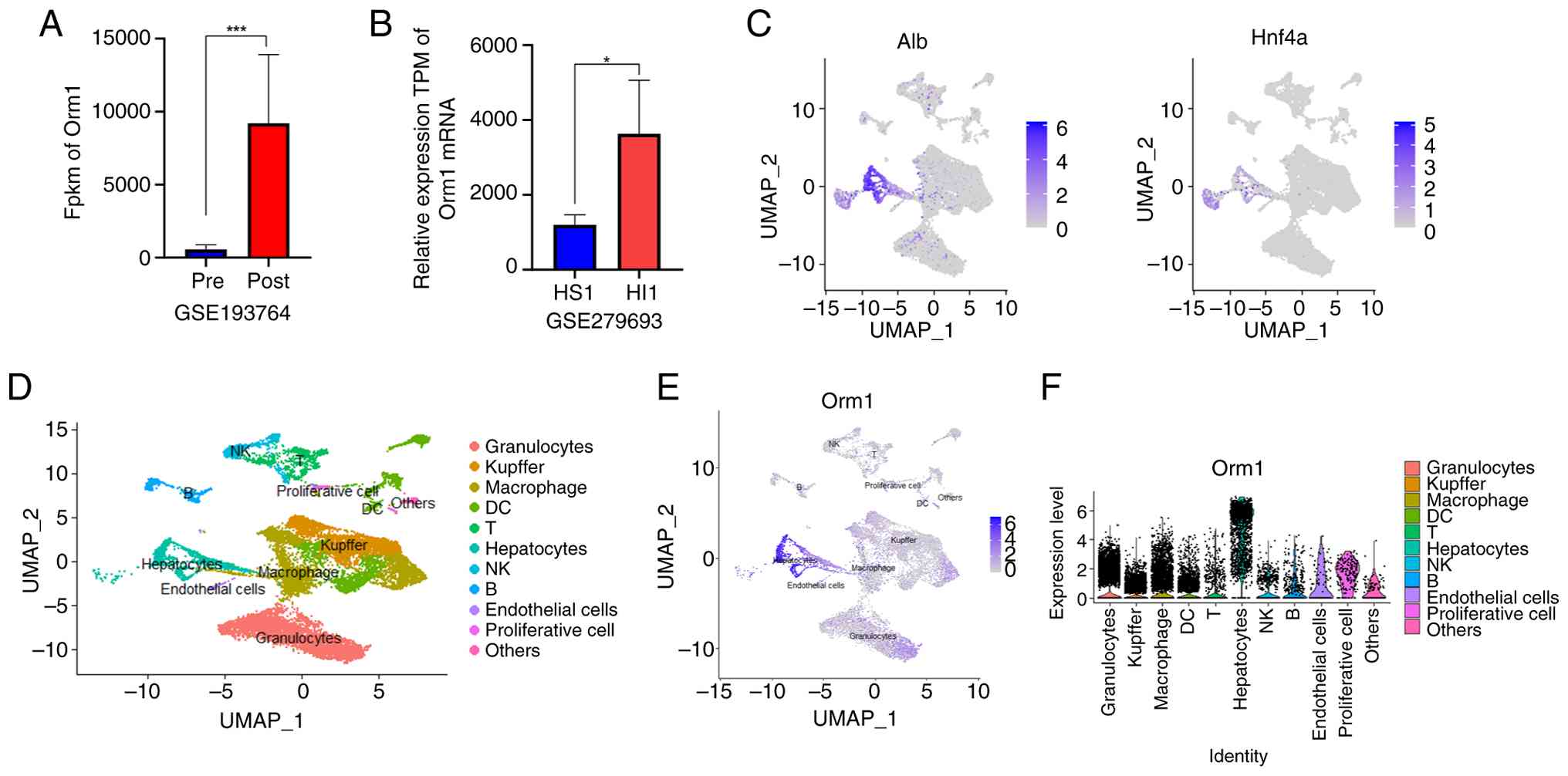
The rat transcriptomic dataset GSE193764 was
analysed to identify key molecular drivers of I/R in steatotic
livers. Differential expression analysis revealed 1,551 DEGs,
including 1,112 upregulated and 439 downregulated genes (Table SIV). Notably, Orm1 emerged as a
significantly upregulated candidate gene following transplantation
(Figs. 1A and S1). To further validate these
findings, the mouse dataset GSE279693 was examined, which confirmed
a consistent and significant increase in Orm1 expression during I/R
in steatotic livers (Fig. 1B).
Given this marked upregulation, the cellular source of Orm1 was
further investigated using the CRA004061 single-cell dataset.
Dimensionality reduction and cluster analysis revealed that Orm1
was predominantly expressed in hepatocytes (Fig. 1C-F). This cell type specificity
is consistent with the findings of previous reports identifying
hepatocytes as the primary site of Orm1 synthesis (29).
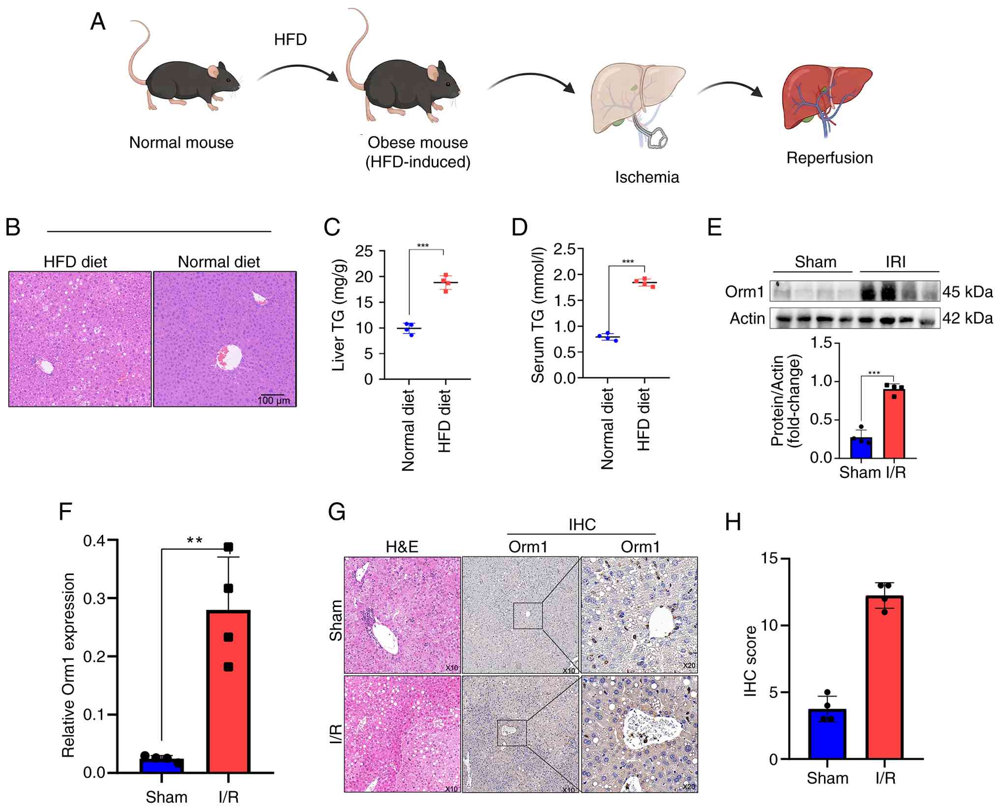
To validate Orm1 expression, a mouse model of
hepatic I/R in steatotic livers was employed (Fig. 2A). After 12 weeks of HFD feeding,
H&E staining revealed moderate hepatic steatosis, characterised
by the presence of lipid droplets (Fig. 2B), in line with prior reports
(30). Serum and liver tissue TG
levels were significantly greater in HFD-fed mice compared with
normal diet-fed mice (Fig. 2C and
D), and their body weights were also significantly greater
(Fig. S2A).
HFD-fed mice were then subjected to 1 h of ischaemia
followed by 6 h of reperfusion. Western blotting and RT-qPCR
analysis demonstrated that I/R significantly upregulated Orm1
expression in steatotic liver tissues (Fig. 2E and F). Immunohistochemical
analysis further confirmed elevated Orm1 levels after I/R, as
reflected by increased immunohistochemical scores (Fig. 2G and H). In parallel, primary
hepatocytes isolated from HFD-fed mice exhibited steatotic
degeneration and were subjected to 6 h of hypoxia followed by 6 h
of reoxygenation. Consistent with the aforementioned findings, both
western blotting and RT-qPCR revealed a marked increase in Orm1
expression following H/R (Fig. S2B
and C).
Collectively, these findings suggest that the
upregulation of Orm1 expression represents an endogenous
compensatory protective response to I/R in steatotic livers.
Orm1 knockdown in mice with diet-induced
steatotic liver aggravated liver damage following I/R injury
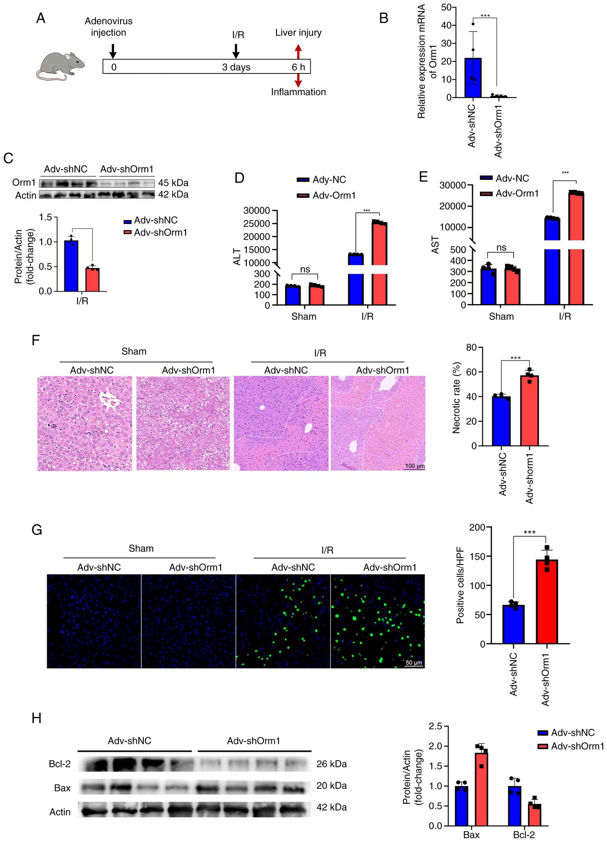
Mice with specific Orm1 knockdown were generated
using a recombinant adenovirus expressing shRNA targeting Orm1
(Adv-shOrm1). These mice were then subjected them to hepatic I/R to
investigate the functional role of Orm1 in steatotic liver I/R
(Fig. 3A). Western blotting and
RT-qPCR confirmed that Orm1 expression was significantly lower in
the liver tissues of Adv-shOrm1-treated mice than in those of
control mice (Fig. 3B and C).
Liver injury was assessed by measuring serum levels of AST and ALT,
as well as by histologically evaluating of necrosis. Notably, Orm1
knockdown resulted in significantly elevated serum AST and ALT
levels (Fig. 3D and E).
Moreover, H&E staining revealed that the area of necrotic
tissue was markedly larger in the Adv-shOrm1 group than in the
Adv-shNC group (Fig. 3F). Given
that apoptosis plays a critical role in the progression of hepatic
I/R (31), the effect of Orm1
knockdown on hepatocyte apoptosis was further examined. TUNEL
staining revealed a significant increase in the number of apoptotic
cells in the livers of Adv-shOrm1 mice (Fig. 3G). Consistent with these
findings, western blot analysis revealed that, compared with the
control treatment, the knockdown of Orm1 led to upregulated
expression of the pro-apoptotic protein Bax and downregulated
expression of the anti-apoptotic protein Bcl-2 (Fig. 3H). These results indicate that
Orm1 deficiency exacerbates liver injury and apoptosis following
steatotic liver I/R.
Hepatocyte-specific Orm1 overexpression
alleviated steatotic liver damage following I/R
An in vitro model of steatotic hepatocytes
was established by treating AML12 cells with a PA:OA mixture to
further clarify the functional role of Orm1 in hepatic I/R
accompanied by steatosis. The oil red staining results revealed
that the liver cells stimulated by the PA:OA mixture exhibited
accumulation of lipids within the cells (Fig. S3A). Following successful
modelling, an Orm1 overexpression plasmid was constructed to
overexpress Orm1 in steatotic hepatocytes. Western blotting and
RT-qPCR revealed that the expression of Orm1 was significantly
increased in liver cells (Fig. S3B
and C). Compared with the control, Orm1 overexpression
significantly reduced the expression of the pro-apoptotic protein
Bax and increased the expression of the anti-apoptotic protein
Bcl-2 (Fig. S3D). Furthermore,
overexpression of Orm1 markedly enhanced cell viability and
decreased the number of PI-positive cells following H/R injury
(Fig. S3E and F). These results
suggest that Orm1 protects steatotic hepatocytes against apoptosis
and cell death under ischaemic stress.
Orm1 regulated I/R in steatotic livers
through the Pi3k/Akt pathway
RNA-seq was performed on liver tissues from Orm1
knockdown and wild-type mice following I/R to investigate the role
of Orm1 in steatotic liver I/R. A total of 3,327 DEGs were
identified, comprising 1,809 upregulated and 1,518 downregulated
genes (Fig. 4A). KEGG pathway
enrichment analysis revealed the top 20 signalling pathways ranked
by P-value, among which the 'PI3K-Akt signalling pathway' was
significantly enriched (Fig.
4B). Western blot analysis revealed that Orm1 knockdown
markedly reduced the phosphorylation levels of Pi3k and Akt
(Figs. 4C and S4A). Conversely, Orm1 overexpression
in steatotic liver cells significantly increased phosphorylation
levels of Pi3k and Akt (Figs. 4D
and S4B).
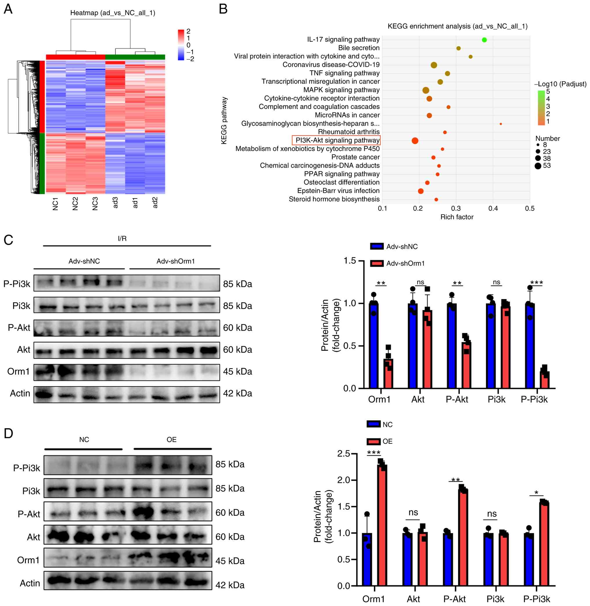 | Figure 4Orm1 participates in the Pi3k/Akt
signalling pathway in steatotic hepatic I/R. (A) Heatmap of 3,327
differentially expressed genes identified by RNA-sequencing. (B)
Top 20 signalling pathway maps enriched in the RNA-sequencing. (C
and D) Western blotting was used to detect the protein expression
levels of Orm1, p-Akt, Akt, p-Pi3k and Pi3k in mouse liver tissue
following Orm1 knockdown and in fatty AML12 cells after Orm1
overexpression. *P<0.05, **P<0.01 and
***P<0.001. ns, not significant; I/R,
ischaemia-reperfusion; shNC, non-coding short hair pin RNA; shOrm1,
Omr1 short hair pin RNA; OE, overexpression. |
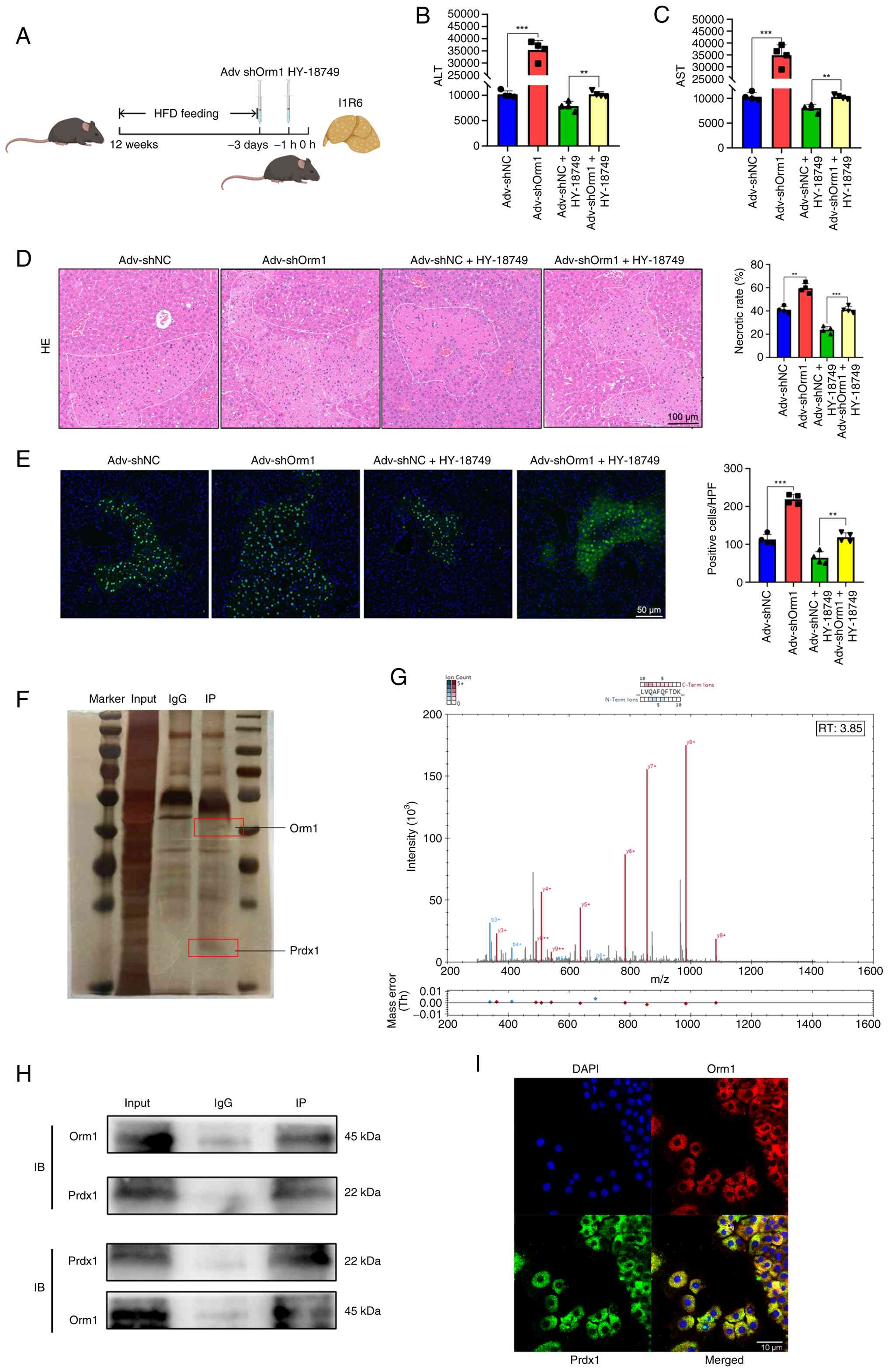
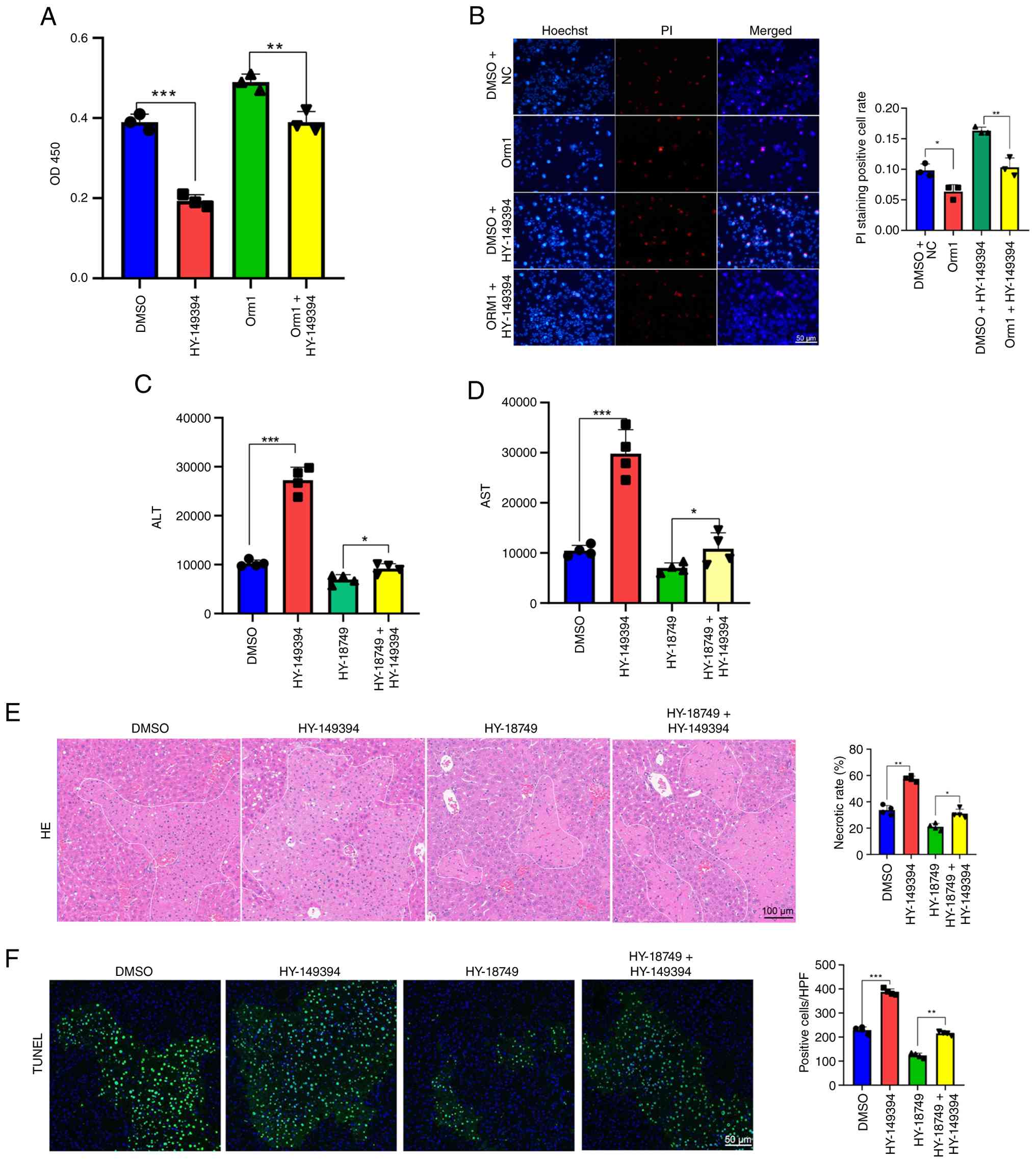
The involvement of the Pi3k/Akt pathway was
subsequently verified in vivo using mice treated with
specific Adv-shOrm1. After 1 h of treatment with HY-18749 (an Akt
agonist), the role of the Pi3k/Akt pathway was assessed in hepatic
I/R induced by Orm1 knockdown in the steatotic liver. HY-18749
significantly reversed the detrimental effects of Orm1 knockdown
following hepatic I/R (Fig.
5A-E). ALT and AST levels were significantly decreased
(Fig. 5B and C), and the area of
necrotic tissue was significantly reduced (Fig. 5D and E). Rescue experiments were
performed using an Akt inhibitor (HY-10249A) to investigate the
role of phosphorylated Pi3k and Akt in Orm1-mediated hepatic I/R in
the steatotic liver. The results showed that treatment with an Akt
inhibitor significantly aggravated liver injury, which was
alleviated by Orm1 overexpression. CCK-8 and Hoechst/PI staining
assays further revealed that, compared with the vector group, the
Akt inhibitor markedly reduced the viability of steatotic liver
cells (Fig. S5A and B). Hence,
these findings suggest that Orm1 regulates hepatic I/R in the
steatotic liver by modulating the activity of the Pi3k/Akt
signalling pathway.
Orm1 interacted and colocalised with
Prdx1
A combined immunoprecipitation-mass spectrometry
(IP-MS) approach was used to identify novel Orm1-interacting
proteins to further elucidate the mechanism by which Orm1 regulates
Pi3k/Akt signalling. Prdx1 was ranked highest among the
differentially enriched proteins associated with the Pi3k/Akt
pathway when Orm1 antibody immunoprecipitates were compared with
IgG controls (Fig. 5F and G and
Table SV) (32,33). To validate the MS data, Co-IP
assays were performed to confirm the physical interaction between
Orm1 and Prdx1 (Fig. 5H).
Additionally, confocal immunofluorescence microscopy revealed
substantial colocalization of endogenous Orm1 and Prdx1 in AML12
cells (Fig. 5I). To further
substantiate the physical interaction between Orm1 and Prdx1, an
exogenous Co-IP assay was performed. Plasmids encoding Flag-tagged
Orm1 (Flag-Orm1) and HA-tagged Prdx1 (HA-Prdx1) were co-transfected
into cells. Subsequent immunoprecipitation using anti-Flag magnetic
beads successfully co-precipitated HA-Prdx1. Notably, no HA signal
was detected in the immunoprecipitates from the negative control
cells transfected with HA-Prdx1 alone (Fig. S3G). This robust interaction
between the exogenously tagged proteins, alongside the endogenous
Co-IP and mass spectrometry data, validates the specific physical
association between Orm1 and Prdx1.
Next, the cells were treated with the Prdx1-specific
inhibitor HY-149394 to explore the functional role of Prdx1 in I/R.
CCK-8 and Hoechst/PI staining revealed that, compared with DMSO
treatment, Prdx1 inhibition significantly reduced hepatocyte
viability and increased cell death (Fig. 6A and B). Furthermore, Prdx1
inhibition markedly suppressed the phosphorylation of Pi3k and Akt,
upregulated the expression of the proapoptotic molecule Bax and
downregulated the expression of the antiapoptotic protein Bcl-2
(Figs. 6C and S4C). Next, it was investigated whether
altered Orm1 expression could concomitantly affect Prdx1
expression. Prdx1 protein levels were upregulated upon Orm1
overexpression and downregulated upon Orm1 knockdown (Fig. 6D and E); however, no significant
changes were observed at the mRNA level (Fig. S5C), suggesting
posttranscriptional or posttranslational regulation. These results
indicate that Prdx1 activation protects hepatocytes against I/R
through the Pi3k/Akt pathway and that Orm1 may regulate hepatocyte
survival via the modulation of Prdx1 protein stability or
activity.
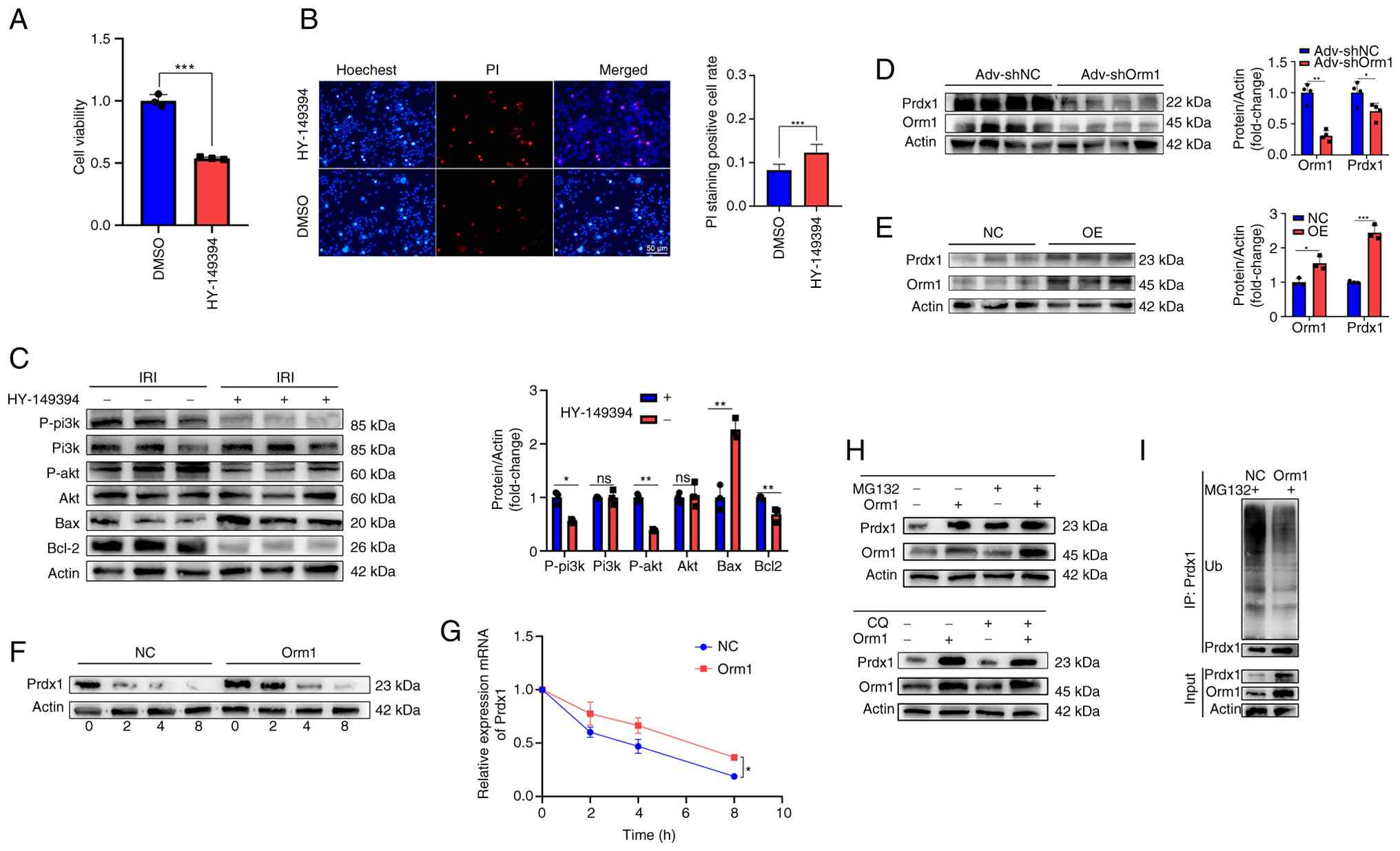 | Figure 6A Prdx1 inhibitor (HY-149394)
decreases the viability of fatty liver cells. The results of the
(A) cell counting kit-8 and (B) Hoechst/PI staining assay revealed
that HY-149394 significantly exacerbated liver cell damage. (C)
Western blotting analysis revealed that the Prdx1 inhibitor
significantly inhibited the phosphorylation of Pi3k/Akt, decreased
the expression level of Bcl-2, and increased the expression of Bax.
(D and E) Western blot analysis revealed that the protein
expression of Prdx1 was positively associated with that of Orm1
demonstrated using (D) knockdown or (E) OE of Orm1. (F and G)
Control and Orm1-overexpressing AML12 cells were treated with CHX
at different time points. (H) Orm1-overexpressing cells were
treated with MG132 (20 μM) or CQ (50 μM) for 10 h,
after which Prdx1 and Orm1 protein levels were determined by
western blotting. (I) Ubiquitination assay. Orm1-overexpressing
AML12 cells were treated with MG132 for 10 h, followed by
immunoprecipitation with anti-Prdx1 and immunoblotting with the
indicated antibodies. *P<0.05, **P<0.01
and ***P<0.001. ns, not significant; OE,
overexpression; shNC, non-coding short hair pin RNA; shOrm1, Omr1
short hair pin RNA; IP, immunoprecipitation; IB, immunoblot. |
Orm1 stabilised Prdx1 by inhibiting its
ubiquitin proteasomal degradation
Previous studies have demonstrated that Prdx1
undergoes constitutive ubiquitination (34,35) and that endogenous protein
degradation primarily occurs through the ubiquitin-proteasome and
lysosomal pathways. CHX chase assays were performed to assess Prdx1
degradation dynamics. Orm1 overexpression significantly attenuated
Prdx1 degradation in AML12 cells (Fig. 6F and G). To elucidate the
degradation pathway, the cells were treated with the proteasome
inhibitor MG132 or the lysosome inhibitor CQ. MG132 treatment
restored Prdx1 levels in Orm1-knockdown cells, whereas CQ had no
significant effect (Fig. 6H),
indicating that Prdx1 is degraded primarily via the proteasomal
pathway. Furthermore, cell-based ubiquitination assays revealed
that Orm1 overexpression markedly reduced Prdx1 ubiquitination
(Fig. 6I), suggesting that Orm1
protects Prdx1 from ubiquitin-mediated proteasomal degradation.
Orm1 regulated I/R in the steatotic liver
via the Prdx1/Pi3k/Akt pathway
The role of Prdx1 in I/R of steatotic liver was
further investigated. CCK-8 and Hoechst/PI staining assays revealed
that the protective effect of Orm1 overexpression on hepatocyte
injury was significantly reversed by a Prdx1 inhibitor (Fig. 7A and B), indicating that Prdx1
mediates the hepatoprotective effects of Orm1 in I/R. Subsequent
in vivo experiments demonstrated that administration of an
Akt agonist alleviated hepatic I/R induced by Prdx1 inhibition.
Compared with the control group, which received the Akt agonist,
the experimental group showed improved liver function (Fig. 7C and D). H&E staining
revealed a reduction in the area of necrosis in the liver (Fig. 7E), along with a decrease in the
proportion of TUNEL-positive cells (Fig. 7F). Overall, these findings
indicate that overexpression of Orm1 alleviates hepatic I/R through
the Prdx1-Pi3k-Akt axis. Prior to the subsequent experiments, the
transfection efficiency of sh-Prdx1 was confirmed by RT-qPCR and
western blotting (Fig. S5D and
E). Furthermore, considering that oxidative stress is one of
the core changes during cell and tissue H/R, the cells were
subjected to H/R treatment. The results revealed that
overexpression of Orm1 reduced the H/R-induced ROS production and
(Figs. S6A-D) and that
inhibition of Prdx1 expression reversed the protective effect of
Orm1 overexpression (Fig.
S6A-D). Subsequently, Prdx1-knockdown cells were treated with
an AKT agonist, and the results revealed that the AKT agonist
reversed the cell damage and ROS production caused by Prdx1
(Fig. S6A-D).
Discussion
Hepatic I/R is a key factor affecting the morbidity
and mortality rates of liver resection and liver transplantation
surgeries (36). In the present
study, Orm1 was discovered as a potential candidate molecule for
treating I/R in the context of a steatotic liver. Although the
current study primarily highlights the protective role of Orm1 in
steatotic livers, its potential hepatoprotective effects in lean
livers during I/R cannot be ruled out. The steatotic model was
specifically focused on because 'marginal' steatotic grafts exhibit
profound hypersensitivity to ischaemic and oxidative stress, making
them more susceptible to primary non-function post-transplantation.
Further studies comparing the differential dynamics of Orm1
expression between lean and steatotic livers will be valuable for
determining whether this protective axis is uniquely amplified
under metabolic stress. The results revealed that the expression of
Orm1 in liver cells significantly increased after liver I/R and
that inhibiting the expression of Orm1 aggravated liver injury,
whereas overexpressing Orm1 had a protective effect on I/R.
Mechanistic studies revealed that Orm1 interacted with Prdx1,
inhibiting its ubiquitination and degradation, thereby activating
the Pi3k/Akt signalling pathway.
Orm1, also known as α1-acid glycoprotein, was first
identified by Shimada and Schmidt more than a century ago (37). As a multifunctional acute-phase
protein, Orm1 not only serves as a disease marker and drug carrier,
but also participates in immune regulation, maintenance of
capillary barrier function and sphingolipid metabolism. The
corresponding receptors have been preliminarily identified in
macrophages (CCR5), neutrophils (Siglec-5) and liver parenchymal
cells (HBB) (29). Numerous
studies have shown that Orm1 has a carcinogenic effect on various
tumours. For instance, the absence of Orm1 can inhibit the
proliferation, migration and invasion of renal clear cell carcinoma
and induce apoptosis (19).
Qiong and Yin (38) reported
that Orm1 can promote the proliferation and migration of breast
cancer cells, and its absence can downregulate the expression of
transfer-related matrix metalloproteinases. In colorectal cancer
and bladder cancer, Orm1 promotes tumour progression by promoting
epithelial-mesenchymal transition and activating the ERK signalling
pathway (39,40). Additionally, its role in
non-tumour diseases has also been investigated. Studies have shown
that Orm1 can improve temporomandibular joint osteoarthritis by
maintaining cartilage homeostasis (41) and mediate LN-IgG podocyte injury
and autophagy regulation through the AMPK/mTOR signalling pathway
(20). In addition, Orm1 can
alleviate doxorubicin-induced oxidative stress and apoptosis in
cardiomyocytes through the Nrf2 signalling pathway (21). Furthermore, a recent study
revealed that the Cebpa-Orm1 axis in liver cells can inhibit
alcohol-related liver diseases (42). Classically, Orm1 is recognised as
a secreted acute-phase glycoprotein involved in systemic
inflammation (43),
immunomodulation and drug binding; however, the present findings
reveal an intracellular regulatory role for Orm1 during hepatic
I/R. The present proposes that under stress, intracellular Orm1
directly regulates protein stability by physically shielding Prdx1
from ubiquitination. This mechanism preserves the cellular
antioxidant capacity to counteract severe oxidative stress. In the
present study, the expression of Orm1 in liver cells significantly
increased after liver I/R; knockdown of Orm1 exacerbated liver
injury, tissue necrosis and cell apoptosis, whereas overexpression
of Orm1 exerted a protective effect.
This dual nature of Orm1, acting as an intracellular
redox stabiliser and a secreted immunomodulator, has profound
clinical implications for steatotic liver transplantation. In
marginal grafts, early oxidative stress triggers apoptosis and
activates innate immune pathways, exacerbating adverse transplant
immunity (44). Therefore,
rather than focusing on its broad role in tumours or other
diseases, the present study specifically highlighted the Orm1-Prdx1
axis as a potential therapeutic target to mitigate oxidative injury
and immune-mediated damage during liver transplantation.
The Pi3k/Akt signalling pathway plays a role in
regulating cell apoptosis, and its phosphorylation activation can
inhibit apoptosis (45-48). In tumour research, apigenin
induces apoptosis and autophagy in liver cancer cells by inhibiting
the Pi3k/Akt/mTOR pathway whereas, Adora2a promotes proliferation
and inhibits apoptosis in colorectal cancer by activating the
Pi3k/Akt pathway. By contrast, in a lipopolysaccharide-induced
acute kidney injury model, rubiogenin alleviated inflammation and
apoptosis by activating the Pi3k/Akt/Nrf2 pathway (47). To further investigate Orm1 in
steatotic liver I/R, RNA-seq identified Prdx1 and Pi3k/Akt as key
regulatory factors, and the function of the Orm1-Prdx1-Pi3k/Akt
axis was further verified. The results of the functional rescue
experiments revealed that Orm1 regulates Pi3k/Akt signalling
through Prdx1, thereby affecting the apoptosis of steatotic liver
cells. Compared with that in the control group, the protein level
of Prdx1 increased after overexpression of Orm1, and the expression
of apoptosis-related molecules in the Bcl-2 family significantly
decreased; in a steatotic liver mouse model, inhibiting Orm1 led to
a significant decrease in Prdx1 expression. Further mechanistic
studies revealed that Orm1 interacts with Prdx1 to inhibit its
ubiquitination and degradation, thereby activating the Pi3k/Akt
signalling pathway and alleviating ischaemic injury to liver cells.
The present study revealed the molecular mechanism underlying
Orm1-mediated regulation of I/R in steatotic livers and expanded
the understanding of its role in liver injury protection. The
present study investigated the potential molecular mechanism of
Orm1 in liver I/R. Ubiquitination, as an important
posttranslational modification method, plays a role in protein
stability and functional regulation. Previous studies have shown
that Prdx1 can be regulated at the transcriptional and
posttranscriptional levels and that its activity is also regulated
by various modifications, such as ubiquitination, acetylation,
phosphorylation, glutathionylation and S-nitrosylation (49,50). For instance, the E3 ligase HECTD3
can mediate the ubiquitination degradation of Prdx1 (51). The present study further
confirmed that in steatotic liver cells, the absence of Orm1 can
promote the degradation of Prdx1 via the ubiquitin-proteasome
pathway.
Notably, although the present study initially
revealed the mechanism of Orm1 in steatotic liver I/R, the I/R
process involves a complex regulatory network of multiple factors
and pathways, and numerous unanswered questions remain. For
instance, whether Orm1 regulates apoptosis through other signalling
pathways or participates in other biological processes remains
unclear. Future studies are needed to comprehensively understand
the function of Orm1 in steatotic liver I/R.
The present study initially revealed the protective
mechanism of Orm1 in steatotic liver I/R; however, the I/R process
involves a complex regulatory network of multiple factors and
pathways, and several limitations remain to be addressed. First,
although the present in vitro models demonstrate the
cytoprotective effects of Orm1 overexpression against H/R injury,
the present study lacks in vivo Orm1 overexpression models.
However, a recent study demonstrated that in vivo
restoration of ORM1 expression via AAV delivery or recombinant
protein administration effectively rescues hepatic lipid
accumulation in alcohol-associated liver disease (42). These in vivo findings are
consistent with the present in vitro results and support the
therapeutic potential of exogenous Orm1 administration, warranting
future in vivo studies to validate its efficacy in
optimizing steatotic liver grafts. Second, regarding the mechanism
of Prdx1 stabilisation, a previous study demonstrated that another
binding protein, IRAK1, can inhibit the ubiquitination of Prdx1 by
blocking the activity of the E3 ubiquitin ligase HECTD3 (51). Given that Orm1 lacks intrinsic
de-ubiquitinase activity, we hypothesise that the physical
interaction between Orm1 and Prdx1 may occur through a similar
mechanism, causing steric hindrance. This spatial shielding may
prevent specific E3 ubiquitin ligases, such as HECTD3, from
accessing the ubiquitination sites on Prdx1. However, since this
was not experimentally validated in the present the study this
spatial shielding effect or the specific involvement of HECTD3,
this mechanism remains strictly hypothetical. Further structural
biology studies and targeted ubiquitination assays are needed to
elucidate the precise interacting domains and to confirm this
model. Finally, whether Orm1 regulates apoptosis through other
signalling pathways or participates in other biological behaviours
remains to be clarified. In summary, the present study revealed the
protective effect of Orm1 in hepatic I/R. The molecular mechanism
by which Orm1 interacts with Prdx1, inhibits the ubiquitination and
degradation of Prdx1, and activates the Pi3k/Akt signalling
pathway, thereby reducing hepatocyte apoptosis, has been
elucidated. Targeting the Orm1-Prdx1-Pi3k/Akt axis is expected to
become a potential therapeutic strategy for hepatic I/R in the
context of steatotic liver.
In conclusion, the present study revealed that Orm1
is a protective factor against steatotic hepatic I/R. Orm1 is
upregulated upon I/R; its loss aggravates hepatocellular damage and
apoptosis, whereas its overexpression confers protection.
Mechanistically, Orm1 binds Prdx1 to shield it from
ubiquitin-proteasome degradation, thereby activating Pi3k/Akt
survival signalling. Collectively, our findings establish the
Orm1-Prdx1-Pi3k/Akt cascade as a promising therapeutic axis for
steatotic liver I/R.
Supplementary Data
Availability of data and materials
The RNA sequencing data generated in the present
study may be found in the National Genomics Data Centre under the
accession number CRA048839 or at the following URL: https://ngdc.cncb.ac.cn/search/specific?db=bioproject&q=CRA048839).
The IP-MS data generated in the present study may be found in the
Integrated Proteome Resources (iProX) database under the accession
number IPX0019637004 or at the following URL: https://www.iprox.cn//page/subproject.html?id=IPX0019637004).
The other data generated in the present study may be requested from
the corresponding author.
Authors' contributions
JC and XC designed the research and helped draft
the manuscript. YuW, HC and RD performed the experiments. YuW, HL,
RD, YiW and HC performed the statistical analyses. YiW and HC
participated in data visualisation, including the generation of
graphs and figures presented in the manuscript. JC and XC confirm
the authenticity of all the raw data. All authors read and approved
the final version of the manuscript.
Ethics approval and consent to
participate
All animal experiments were conducted in accordance
with the guidelines of the Animal Care and Use Committee of Qingdao
University (ethics approval no. 20260401C57BL/64 520260601066).
Patient consent for publication
Not applicable.
Competing interests
The authors declare that they have no competing
interests.
Acknowledgements
The authors acknowledge Dr Jijun Shan at Nanjing
University Drum Tower Hospital for his invaluable guidance on the
research ideas provided to the student Yuming Wang in the present
study.
Funding
The present study was supported by the National Natural Science
Foundation of China (grant nos. 82370666 and 82500972), Shandong
Provincial Natural Science Foundation (grant no. ZR2025QC1668) and
Qingdao Natural Science Foundation (grant no. 25-1-1-144-zyyd
jch).
References
|
1
|
Lucey MR, Furuya KN and Foley DP: Liver
transplantation. N Engl J Med. 389:1888–1900. 2023. View Article : Google Scholar : PubMed/NCBI
|
|
2
|
Song Z, Han H, Ge X, Das S, Desert R,
Athavale D, Chen W, Komakula SSB, Lantvit D and Nieto N: Deficiency
of neutrophil high-mobility group box-1 in liver transplant
recipients exacerbates early allograft injury in mice. Hepatology.
78:771–786. 2023. View Article : Google Scholar : PubMed/NCBI
|
|
3
|
Li Y, Yang P, Ye J, Xu Q, Wu J and Wang Y:
Updated mechanisms of MASLD pathogenesis. Lipids Health Dis.
23:1172024. View Article : Google Scholar : PubMed/NCBI
|
|
4
|
Steinberg GR, Valvano CM, De Nardo W and
Watt MJ: Integrative metabolism in MASLD and MASH: Pathophysiology
and emerging mechanisms. J Hepatol. 83:584–595. 2025. View Article : Google Scholar : PubMed/NCBI
|
|
5
|
Liu J, Pang L, Ng KTP, Chiu TLS, Liu H,
Liu X, Xu A, Lo CM and Man K: Compromised AMPK-PGCIα axis
exacerbated steatotic graft injury by dysregulating mitochondrial
homeostasis in living donor liver transplantation. Ann Surg.
276:e483–e492. 2022. View Article : Google Scholar
|
|
6
|
Liu R, Cao H, Zhang S, Cai M, Zou T, Wang
G, Zhang D, Wang X, Xu J, Deng S, et al: ZBP1-mediated apoptosis
and inflammation exacerbate steatotic liver ischemia/reperfusion
injury. J Clin Invest. 134:e1804512024. View Article : Google Scholar : PubMed/NCBI
|
|
7
|
Liu R, Wang G, Qian Y, Jiang Z, Wang W,
Cai M, Zhang S, Wang G, Wang C, Zou T, et al: Hexosamine
biosynthesis dysfunction-induced LIFR N-glycosylation deficiency
exacerbates steatotic liver ischemia/reperfusion injury.
Metabolism. 168:1562582025. View Article : Google Scholar : PubMed/NCBI
|
|
8
|
Sousa Da Silva RX, Bautista Borrego L,
Lenggenhager D, Huwyler F, Binz J, Mancina L, Breuer E, Wernlé K,
Hefti M, Mueller M, et al: Defatting of human livers during
long-term e x situ normothermic perfusion: Novel strategy to rescue
discarded organs for transplantation. Ann Surg. 278:669–675. 2023.
View Article : Google Scholar : PubMed/NCBI
|
|
9
|
Sousa Da Silva RX, Weber A, Dutkowski P
and Clavien PA: Machine perfusion in liver transplantation.
Hepatology. 76:1531–1549. 2022. View Article : Google Scholar : PubMed/NCBI
|
|
10
|
Elmore S: Apoptosis: A review of
programmed cell death. Toxicol Pathol. 35:495–516. 2007. View Article : Google Scholar : PubMed/NCBI
|
|
11
|
Xu X, Lai Y and Hua ZC: Apoptosis and
apoptotic body: Disease message and therapeutic target potentials.
Biosci Rep. 39:BSR201809922019. View Article : Google Scholar :
|
|
12
|
de Oliveira THC and Gonçalves GKN: Liver
ischemia reperfusion injury: Mechanisms, cellular pathways, and
therapeutic approaches. Int Immunopharmacol. 150:1142992025.
View Article : Google Scholar : PubMed/NCBI
|
|
13
|
Sahoo G, Samal D, Khandayataray P and
Murthy MK: A review on caspases: Key regulators of biological
activities and apoptosis. Mol Neurobiol. 60:5805–5837. 2023.
View Article : Google Scholar : PubMed/NCBI
|
|
14
|
Beltrán-Visiedo M, Soler-Agesta R,
Sarosiek KA, Green DR and Galluzzi L: Regulation of inflammatory
processes by caspases. Nat Rev Mol Cell Biol. 26:884–901. 2025.
View Article : Google Scholar : PubMed/NCBI
|
|
15
|
Budihardjo I, Oliver H, Lutter M, Luo X
and Wang X: Biochemical pathways of caspase activation during
apoptosis. Annu Rev Cell Dev Biol. 15:269–290. 1999. View Article : Google Scholar : PubMed/NCBI
|
|
16
|
Vogler M, Braun Y, Smith VM, Westhoff MA,
Pereira RS, Pieper NM, Anders M, Callens M, Vervliet T, Abbas M, et
al: The BCL2 family: From apoptosis mechanisms to new advances in
targeted therapy. Signal Transduct Target Ther. 10:912025.
View Article : Google Scholar : PubMed/NCBI
|
|
17
|
Singh R, Letai A and Sarosiek K:
Regulation of apoptosis in health and disease: The balancing act of
BCL-2 family proteins. Nat Rev Mol Cell Biol. 20:175–193. 2019.
View Article : Google Scholar : PubMed/NCBI
|
|
18
|
Sora V and Papaleo E: Structural details
of BH3 motifs and BH3-mediated interactions: An updated
perspective. Front Mol Biosci. 9:8648742022. View Article : Google Scholar : PubMed/NCBI
|
|
19
|
Yu G and Gao J, Hu W, Hu D, Wang W, Yang S
and Gao J: ORM1 promotes tumor progression of kidney renal clear
cell carcinoma (KIRC) through CALR-mediated apoptosis. Sci Rep.
13:156872023. View Article : Google Scholar : PubMed/NCBI
|
|
20
|
Chen J, Zou L, Liu L, Wu C and Hu M: ORM1
mediates Ln-IgG-induced podocyte damage and autophagy via the
AMPK/mTOR signaling. Organogenesis. 21:25196142025. View Article : Google Scholar : PubMed/NCBI
|
|
21
|
Cheng X, Liu D, Xing R, Song H, Tian X,
Yan C and Han Y: Orosomucoid 1 attenuates doxorubicin-induced
oxidative stress and apoptosis in cardiomyocytes via Nrf2
signaling. Biomed Res Int. 2020:59235722020. View Article : Google Scholar : PubMed/NCBI
|
|
22
|
Chen X, Liu Y, Mu S, Chen T, Yuan J, Xiao
C, Dong Q, Li Q, Wang Y, Chen Q, et al: Orosomucoid 1 participates
in alveolar hypercoagulation and fibrinolytic inhibition involving
NF-κB signaling pathway in LPS-induced ARDS. FASEB J.
39:e711472025. View Article : Google Scholar
|
|
23
|
Fan C: Orosomucoid types in allergic
contact dermatitis. Hum Hered. 45:117–120. 1995. View Article : Google Scholar : PubMed/NCBI
|
|
24
|
Astrup LB, Skovgaard K, Rasmussen RS,
Iburg TM, Agerholm JS, Aalbæk B, Jensen HE, Nielsen OL, Johansen
FF, Heegaard PMH and Leifsson PS: Staphylococcus aureus infected
embolic stroke upregulates Orm1 and Cxcl2 in a rat model of septic
stroke pathology. Neurol Res. 41:399–412. 2019. View Article : Google Scholar : PubMed/NCBI
|
|
25
|
Wu D, Xu J, Zhang Y, Wang Y, Bai Y, Zhan
X, Gao Y, Zhou H, Hu H, Wang P and Rao Z: tBHQ mitigates fatty
liver ischemia-reperfusion injury by activating Nrf2 to attenuate
hepatocyte mitochondrial damage and macrophage STING activation.
Int Immunopharmacol. 138:1125152024. View Article : Google Scholar : PubMed/NCBI
|
|
26
|
Livak KJ and Schmittgen TD: Analysis of
relative gene expression data using real-time quantitative PCR and
the 2(-Delta Delta C(T)) method. Methods. 25:402–408. 2001.
View Article : Google Scholar
|
|
27
|
Yang X, Lu D, Wang R, Lian Z, Lin Z, Zhuo
J, Chen H, Yang M, Tan W, Yang M, et al: Single-cell profiling
reveals distinct immune phenotypes that contribute to
ischaemia-reperfusion injury after steatotic liver transplantation.
Cell Prolif. 54:e131162021. View Article : Google Scholar : PubMed/NCBI
|
|
28
|
Wu Y, Li C, Lu D, Chen K, Su R, Xu S, Gao
F, Lian Z, Yang F, Chen J, et al: Insulin-induced gene 2 alleviates
ischemia-reperfusion injury in steatotic liver by inhibiting
GPX4-dependent ferroptosis. Cell Death Discov. 11:1272025.
View Article : Google Scholar : PubMed/NCBI
|
|
29
|
Luo Z, Lei H, Sun Y, Liu X and Su DF:
Orosomucoid, an acute response protein with multiple modulating
activities. J Physiol Biochem. 71:329–340. 2015. View Article : Google Scholar : PubMed/NCBI
|
|
30
|
Patrono D, De Stefano N, Vissio E, Apostu
AL, Petronio N, Vitelli G, Catalano G, Rizza G, Catalano S, Colli
F, et al: How to preserve steatotic liver grafts for
transplantation. J Clin Med. 12:39822023. View Article : Google Scholar : PubMed/NCBI
|
|
31
|
Liu H and Man K: New insights in
mechanisms and therapeutics for short- and long-term impacts of
hepatic ischemia reperfusion injury post liver transplantation. Int
J Mol Sci. 22:82102021. View Article : Google Scholar : PubMed/NCBI
|
|
32
|
Xue X, Huang C, Chen J, Ding W, Ren Y,
Zhang W, Li Q, Yang Z and Sun Z: Ginsenoside Rg1 mitigates
sepsis-associated acute respiratory distress syndrome by promoting
autophagy through the Prdx1-PTEN/PI3K/AKT pathway. Phytomedicine.
154:1580272026. View Article : Google Scholar : PubMed/NCBI
|
|
33
|
Zhang T, Li L, Mo X, Xie S, Liu S, Zhao N,
Zhang H, Chen S, Zeng X, Wang S, et al: Matairesinol blunts adverse
cardiac remodeling and heart failure induced by pressure overload
by regulating Prdx1 and PI3K/AKT/FOXO1 signaling. Phytomedicine.
135:1560542024. View Article : Google Scholar : PubMed/NCBI
|
|
34
|
Xiong Y, Zhao X, Qin X, Zhang Y, Wang L,
Zheng Q, Liu X, Liu H and Chen Z: OTUD7B attenuates renal fibrosis
by regulating PRDX1 protein stability. J Adv Res. Mar 5–2026.Epub
ahead of print. View Article : Google Scholar
|
|
35
|
Sun X, Wu T, Chen S, Zhao Z, Jia R, Ma J,
Yin L, Pan X, Ping Y, Mao Y, et al: OTUD1 inhibits osteoclast
differentiation and osteoclastic bone loss through deubiquitinating
and stabilizing PRDX1. Theranostics. 15:6719–6736. 2025. View Article : Google Scholar : PubMed/NCBI
|
|
36
|
Zhang Y, Wang Z, Jia C, Yu W, Li X, Xia N,
Nie H, Wikana LP, Chen M, Ni Y, et al: Blockade of hepatocyte PCSK9
ameliorates hepatic ischemia-reperfusion injury by promoting
pink1-parkin-mediated mitophagy. Cell Mol Gastroenterol Hepatol.
17:149–169. 2024. View Article : Google Scholar
|
|
37
|
Fournier T, Medjoubi NN and Porquet D:
Alpha-1-acid glycoprotein. Biochim Biophys Acta. 1482:157–171.
2000. View Article : Google Scholar : PubMed/NCBI
|
|
38
|
Qiong L and Yin J: Characterization of
alpha-1-acid glycoprotein as a potential biomarker for breast
cancer. Bioengineered. 13:5818–5826. 2022. View Article : Google Scholar : PubMed/NCBI
|
|
39
|
Yue L, Xu X, Dai S, Xu F, Zhao W, Gu J,
Dai X and Qian X: Orosomucoid 1 promotes colorectal cancer
progression and liver metastasis by affecting PI3K/AKT pathway and
inducing macrophage M2 polarization. Sci Rep. 13:140922023.
View Article : Google Scholar : PubMed/NCBI
|
|
40
|
Liu Z and Pu X: Orosomucoid 1 interacts
with S100A12 and activates ERK signalling to expedite the
advancement of bladder cancer. Cell Adh Migr. 19:1–11. 2025.
View Article : Google Scholar
|
|
41
|
Zhang D, Zhang Y, Xia S, Chen L, Shen P
and Yang C: Orosomucoid 1 ameliorates temporomandibular joint
osteoarthritis by maintaining cartilage homeostasis. Adv Sci
(Weinh). 12:e000282025. View Article : Google Scholar : PubMed/NCBI
|
|
42
|
Yan N, Xia Y, Zhang Y, Sawaswong V, Xu X,
Saito Y, Nanduri R, Dai M, Aibara D, Krausz KW, et al: Hepatocyte
CEBPA-ORM1 axis restricts alcohol-associated liver disease.
Hepatology. Mar 9–2026.Epub ahead of print. View Article : Google Scholar
|
|
43
|
Elpek GO: Orosomucoid in liver diseases.
World J Gastroenterol. 27:7739–7747. 2021. View Article : Google Scholar : PubMed/NCBI
|
|
44
|
Elias-Miró M, Jiménez-Castro MB, Rodés J
and Peralta C: Current knowledge on oxidative stress in hepatic
ischemia/reperfusion. Free Radic Res. 47:555–568. 2013. View Article : Google Scholar : PubMed/NCBI
|
|
45
|
Deng RM and Zhou J: The role of PI3K/AKT
signaling pathway in myocardial ischemia-reperfusion injury. Int
Immunopharmacol. 123:1107142023. View Article : Google Scholar : PubMed/NCBI
|
|
46
|
Li Y, Xia J, Jiang N, Xian Y, Ju H, Wei Y
and Zhang X: Corin protects H2O2-induced
apoptosis through PI3K/AKT and NF-κB pathway in cardiomyocytes.
Biomed Pharmacother. 97:594–599. 2018. View Article : Google Scholar
|
|
47
|
Zhang B, Zeng M, Li B, Kan Y, Wang S, Cao
B, Huang Y, Zheng X and Feng W: Arbutin attenuates LPS-induced
acute kidney injury by inhibiting inflammation and apoptosis via
the PI3K/Akt/Nrf2 pathway. Phytomedicine. 82:1534662021. View Article : Google Scholar : PubMed/NCBI
|
|
48
|
Han X, Liu X, Zhao X, Wang X, Sun Y, Qu C,
Liang J and Yang B: Dapagliflozin ameliorates sepsis-induced heart
injury by inhibiting cardiomyocyte apoptosis and electrical
remodeling through the PI3K/Akt pathway. Eur J Pharmacol.
955:1759302023. View Article : Google Scholar : PubMed/NCBI
|
|
49
|
Moretton A, Kourtis S, Gañez Zapater A,
Calabrò C, Espinar Calvo ML, Fontaine F, Darai E, Abad Cortel E,
Block S, Pascual-Reguant L, et al: A metabolic map of the DNA
damage response identifies PRDX1 in the control of nuclear ROS
scavenging and aspartate availability. Mol Syst Biol.
19:e112672023. View Article : Google Scholar : PubMed/NCBI
|
|
50
|
Wang J, Zhang Y, Cao J, Wang Y, Anwar N,
Zhang Z, Zhang D, Ma Y, Xiao Y, Xiao L and Wang X: The role of
autophagy in bone metabolism and clinical significance. Autophagy.
19:2409–2427. 2023. View Article : Google Scholar : PubMed/NCBI
|
|
51
|
Li J, Sun Y, Zhao X, Ma Y, Xie Y, Liu S,
Hui B, Shi X, Sun X and Zhang X: Radiation induces IRAK1 expression
to promote radioresistance by suppressing autophagic cell death via
decreasing the ubiquitination of PRDX1 in glioma cells. Cell Death
Dis. 14:2592023. View Article : Google Scholar : PubMed/NCBI
|