Kidney stone disease ranks among the most prevalent
and painful urological conditions, currently affecting ~11-12% of
the adult population globally, with a >50% recurrence rate
within 5 years, imposing considerable economic strain on
health-care systems (1). Kidney
stone disease has an annual incidence ranging from 114 to 720 per
100,000 individuals across different countries. Over the past
decades, multiple distinct pathways underlying kidney stone
formation have been postulated. Nevertheless, the specific
mechanisms responsible for kidney stone formation and the
associated renal injury remain incompletely understood. Recent
evidence has established ferroptosis as a pivotal driver of
oxalate-crystal nephropathy (2-4).
The present review provides a comprehensive summary of the
bidirectional positive feedback loop between ferroptosis, calcium
oxalate crystal deposition and the associated renal injury. In
addition, promising agents for ferroptosis-targeted therapy are
discussed.
Ferroptosis is a novel, iron-regulated form of
oxidative cell death with potential therapeutic implications for
both cancer and neurodegenerative diseases identified in 2012
(5). Ferroptosis is distinct
from apoptosis, necrosis, and autophagy in morphology,
biochemistry, and genetics. It is characterized by the
iron-dependent accumulation of lipid reactive oxygen species (ROS)
on the cellular membrane. Mitochondria appear shrunken with
increased membrane density; however, mitochondrial electron
transport chain activity is not required (2,4,6-8).
Recently, ferroptosis has been intensively studied in various
diseases, such as cancer (9,10), kidney injury (11,12), nervous system diseases (13,14) and cardiovascular diseases
(15,16), and is suggested to be a valuable
therapeutic target for prevention and treatment.
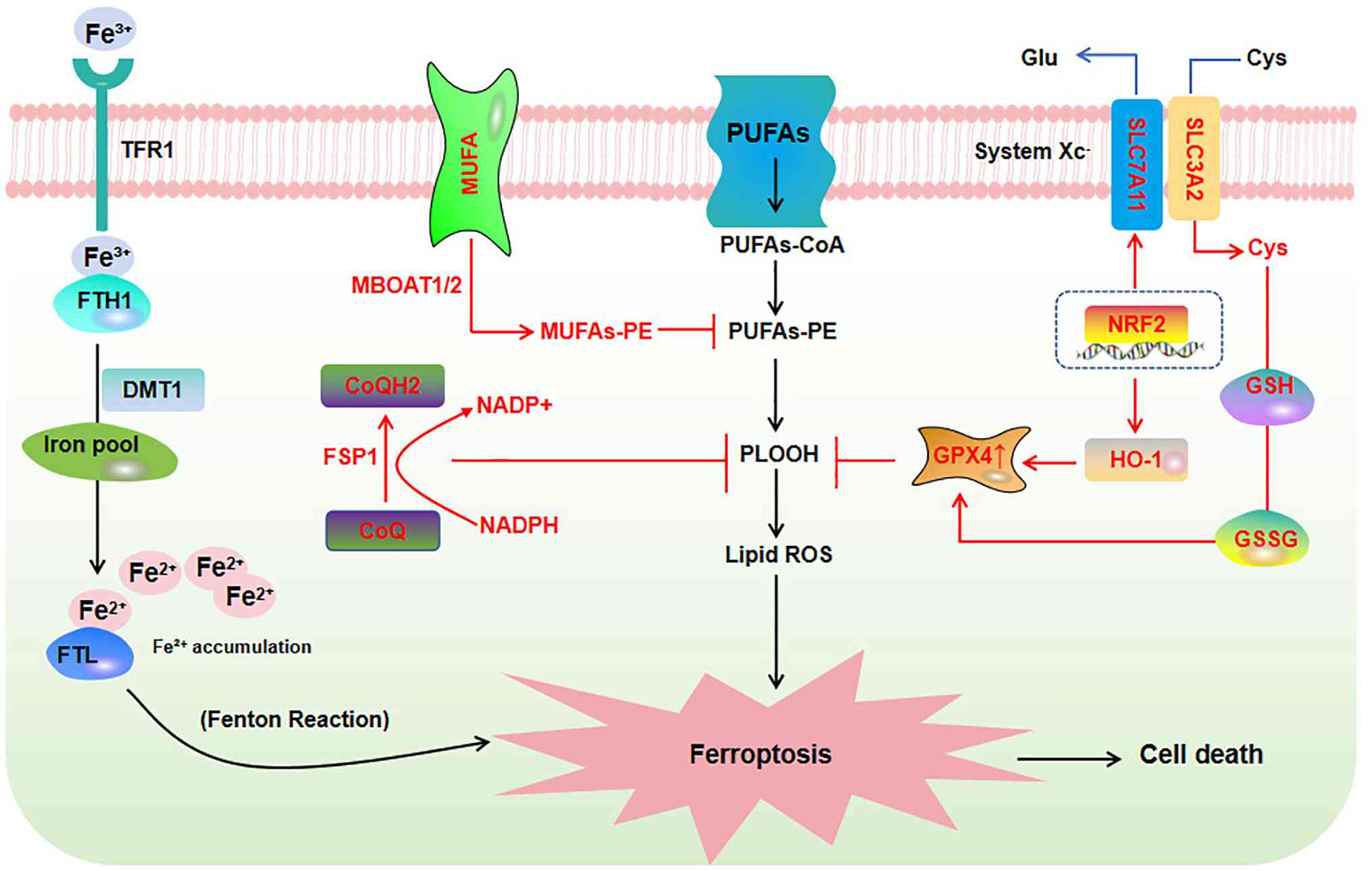
Mechanistically, iron accumulation serves as a
pivotal trigger for the induction of ferroptosis. Extracellular
ferric iron (Fe3+) binds to transferrin (TF), and is
then taken into cells through TFR1, forming the
'TF-Fe3+-TFR1' complex (17). Subsequently, the intracellular
Fe3+ is converted to ferrous iron (Fe2+) and
stored in the cellular iron pool, where excess iron is held in
ferritin. Ferritin is responsible for iron storage, consisting of
two subunits: Ferritin heavy chain (FTH) and ferritin light chain
(FTL). The FTH has ferroxidase activity that converts
Fe2+ into Fe3+, and FTL can facilitate the
iron nuclei formation. The disruption of this balance often results
in the excessive accumulation of Fe2+ ions. This
subsequently triggers the production and accumulation of hydroxyl
radicals via the Fenton reaction; these radicals have potent
oxidizing capabilities, and ultimately induce cellular
ferroptosis.
The execution of ferroptosis is primarily driven by
the iron-dependent lipid peroxidation of polyunsaturated fatty acid
(PUFA)-containing phospholipids, a process tightly regulated by key
enzymes. Specifically, acyl-CoA synthetase long-chain family member
4 (ACSL4) and lysophosphatidylcholine acyltransferase 3 (LPCAT3)
esterify PUFAs (e.g., arachidonic acid and adrenic acid) into
membrane phospholipids, which serve as substrates for peroxidation
by lipoxygenases (LOXs) or the Fenton reaction (18).
The glutathione (GSH)- glutathione peroxidase 4
(GPX4) axis is a central regulatory node in the suppression of
ferroptosis. GPX4 is a plasma-membrane seleno-enzyme, using GSH as
electron donor to reduce membrane lipid hydroperoxides (LOOH →
LOH), halting the radical chain reaction that otherwise ruptures
membranes. GSH availability therefore sets GPX4 activity: Cystine
import via the Xc− system (SLC7A11/SLC3A2) supplies
cysteine for GSH synthesis; Xc− blockade lowers GSH,
inactivates GPX4 and drives ferroptosis. Transcriptional control
reinforces this circuit-nNuclear factor erythroid 2-related factor
2 (Nrf2) upregulates both Xc− subunits and GPX4, whereas
p53 represses SLC7A11, sensitizing cells to ferroptosis (19).
Beyond the classical GSH-GPX4 axis, several
GPX4-independent antioxidant systems have been identified as
critical defense mechanisms against ferroptosis. The ferroptosis
suppressor protein 1 (FSP1)-coenzyme Q10 (CoQ10)-nicotinamide
adenine dinucleotide phosphate [NAD(P)H] axis functions in parallel
with GPX4 to suppress lipid peroxidation by reducing CoQ10 to the
potent lipophilic antioxidant ubiquinol (20). Similarly, the GTP cyclohydrolase
1 (GCH1)-tetrahydrobiopterin (BH4)-dihydrofolate reductase axis
provides another layer of antioxidant defense by generating BH4.
Additionally, membrane-bound O-acyltransferase (MBOAT)1/2 functions
as another GPX4- and FSP1-independent ferroptosis inhibitor system.
MBOAT1/2 belongs to the lysophospholipase family; using
monounsaturated fatty acid (MUFA)-CoA as a substrate, this compound
catalyzes the acylation of lyso-phosphatidylethanolamine (PE) to
generate PE-MUFA, thereby reducing the levels of PE-PUFA, which is
prone to lipid peroxidation, in cell membranes, and consequently
inhibits ferroptosis at the substrate level (21). Furthermore, within the
mitochondria, dihydroorotate dehydrogenase (DHODH) generates
mitochondrial CoQH2 to specifically inhibit
mitochondrial lipid peroxidation (22-24). These interconnected pathways
collectively govern the susceptibility of cells to ferroptosis.
In summary, ferroptosis is orchestrated by iron
overload-driven Fenton chemistry and enzymatic lipid peroxidation,
with the ACSL4/LPCAT3/LOX axis serving as a key executor. The
cellular defense against this death program relies on a
multilayered antioxidant network, including the canonical GSH-GPX4
system and GPX4-independent pathways such as FSP1-CoQ10, GCH1-BH4
and DHODH-CoQH2. The balance between these
pro-ferroptotic and anti-ferroptotic mechanisms ultimately
determines cell fate under various pathological and physiological
conditions (Fig. 1).
Whether ferroptosis serves as either a causative
factor or a downstream consequence of crystal formation in kidney
stones remains a subject of debate. Nonetheless, it is unequivocal
that the induction of ferroptosis actively promotes kidney stone
formation (3,25). Conversely, the deposition of
calcium oxalate (CaOx) crystals, a hallmark pathological feature of
nephrolithiasis, induces substantial renal injury via diverse
mechanisms, including ferroptotic cell death (3). Accordingly, a bidirectional
positive feedback loop is constituted between ferroptosis and CaOx
crystal deposition. In the following section, the upstream
triggers, downstream effectors and feedback amplification of this
loop are elaborated.
Ample evidence have demonstrated that the gut
microbiota is involved in the formation of CaOx crystals (26), and the metabolites of gut
microbiota represent the most critical mechanism underlying this
process.
Specifically, bile acids (BAs), as representative
metabolites of gut microbiota, are closely correlated with kidney
stone formation by inducing ferroptosis by activating the heat
shock protein (HSP)90α/Nrf2/heme oxygenase (HO)-1 signaling pathway
(27). Specifically, BAs are
metabolized into deoxycholic acid (DCA), which upregulates nuclear
Nrf2 and promotes HSP90α expression. On the one hand, Nrf2
transactivates HO-1; sustained high HO-1 expression catalyzes heme
degradation, releasing ferrous ions (Fe2+) and carbon
monoxide, which drive uncontrolled ROS generation via the Fenton
reaction, lipid peroxidation and ferroptosis. On the other hand,
HSP90α further enhances Nrf2 expression and directly interacts with
GPX4 to promote HSP90α-mediated ubiquitination and degradation of
GPX4, thereby promoting ferroptosis (28). Although Nrf2 activation typically
plays a protective role (15,29,30), in this DCA-induced context it
fails to protect GPX4 due to the following: i) The Nrf2/HO-1 axis
amplifies ROS and lipid peroxidation; and ii) HSP90α simultaneously
targets GPX4 for ubiquitin-dependent degradation. The paradoxical
function of Nrf2 arises from its simultaneous transcriptional
regulation of numerous target genes in the three major ferroptosis
pathways, encompassing both ferroptosis-inhibitory factors (GPX4,
SLC7A11, FTH1, NQO1 and FSP1) and ferroptosis-promoting factors
(HMOX1/HO-1 and MRP1/ABCC1) (30). Therefore, Nrf2 functions as a
dynamic regulatory factor, and its functional outcome is jointly
determined by the relative expression balance of its target genes,
cell type, metabolic and iron status, as well as the intensity and
duration of its activation.
Notably, another metabolites of the gut microbiota,
the unconjugated bilirubin (UCB) exerts dual effects on kidney
stone formation and ferroptosis: It promotes CaOx stone formation
by activating caspase-3/PARP-1, upregulating Slc26a6, and reducing
crystal zeta potential (31),
whereas it inhibit ferroptosis via reducing ROS/lipid peroxidation
(32), upregulating Nrf2/HO-1
and GPX4 (32-34), and chelating iron to limit free
iron pools (33). The ultimate
outcome depends on its free concentration, the specific cell type
and the local redox milieu; such context dependency establishes UCB
as a sophisticated and key signaling metabolite along the
gut-kidney axis.
Moreover, gut microbiota dysbiosis reduces the level
of indole-3-acetic acid (IAA), an aryl hydrocarbon receptor (AHR)
ligand that upregulates AHR expression and inhibits NF-κB
phosphorylation (p-p65/p-IκBα), thereby significantly affecting the
oxidative-inflammation-apoptosis network and blocking early stone
formation (35).
Based on those molecular pathways, intervention
strategies targeting the gut microbiota have shown protective
effects (36,37). For instance, healthy human fecal
microbiota transplantation (hFMT) can reduce serum creatinine and
blood urea nitrogen, alleviate tubular injury and upregulate
superoxide dismutase (SOD)/Nrf2 expression, thereby attenuating
oxidative stress and renal CaOx crystal deposition (38).
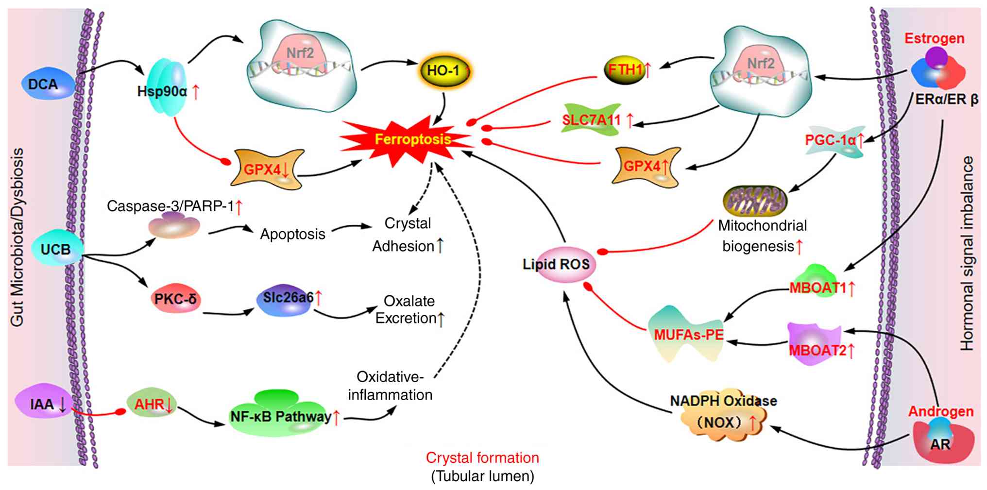
In summary, the gut microbiota metabolites, such as
DCA and UCB induce ferroptosis, apoptosis and crystal adhesion,
while the loss of protective metabolites such as IAA promotes
oxidative stress and inflammation. Conversely, gut microbiota-based
interventions, including hFMT, probiotics and recombinant
probiotics, hold therapeutic potential by restoring microbial
homeostasis, enhancing oxalate degradation, and alleviating renal
injury (Fig. 2). These findings
highlight the gut-kidney axis as a critical regulatory system and a
promising target for the prevention and treatment of
nephrolithiasis.
Hormones function as critical regulators of
ferroptosis, which is involved in the formation of CaOx crystals.
Accumulating evidence indicates that female animals and humans
exhibit less kidney damage in ischemia-reperfusion injury than
males (39,40). Estradiol plays critical roles in
this sex-related protective effect via activating estrogen receptor
(ER)α to inhibit TGF-βRI, thereby blocking SMAD signaling and
reducing inflammation and fibrosis (39). Similarly, epidemiological data
reveal that the prevalence of kidney stones is significantly higher
among adult men than among women (41). With an advancing age, the risk of
developing kidney stones in females continuously rises, while it
exhibits an increasing and then decreasing trend in males; this is
deemed to be associated with the weakening of androgen signaling
(42). These observations
highlight the potential significance of hormones in nephropathy and
renal injury (43).
Ferroptosis is a key driver in the occurrence and
progression of various nephropathies, including acute kidney injury
(AKI) and renal fibrosis (11,44). Therefore, it is reasonable to
speculate that hormones are closely associated with ferroptosis and
may thus contribute to the pathogenesis of renal disorders.
Recently, studies have demonstrated that estrogens inhibit
ferroptosis through both non-genomic and genomic mechanisms,
thereby protecting the kidneys from AKI (45,46). Specifically, 2-hydroxyestradiol
functions as a radical-trapping antioxidant, directly inhibiting
ferroptosis in renal tubules of male mice (45). Additionally, estrogen receptor 1
exerts anti-ferroptotic effects in female tubular cells by
suppressing the ether lipid pathway and enhancing the hydrogen
sulfide system (45). These
findings provide potential therapeutic targets for AKI by
modulating estrogen levels or its receptor activity (47,48).
Beyond AKI, emerging evidence links hormone
signaling to ferroptosis in kidney stone disease. Results from
in vitro and in vivo models have demonstrated that
ERβ protects against renal CaOx crystal deposition by
simultaneously inhibiting hepatic oxalate biosynthesis and renal
oxidative stress-induced injury (49,50). This provides a novel molecular
mechanism for the sex differences observed in the in incidence of
kidney stones and suggests that ERβ and its downstream pathways
(such as NOX2) may serve as potential targets for prevention and
treatment (50). These insights
provide new perspectives for the management of kidney stone disease
mediated by the interplay between hormones and ferroptosis.
Mechanistic studies have demonstrated that estrogen
enhances cellular antioxidant defenses by activating Nrf2 and
upregulating core anti-ferroptosis genes, such as GPX4, SLC7A11 and
FTH1 (51,52). It also promotes mitochondrial
biogenesis and function via the peroxisome proliferator-activated
receptor gamma coactivator 1-α (PGC-1α) signaling axis, reducing
ROS production (53,54). Moreover, estrogen regulates lipid
metabolism to lower low-density lipoprotein and increase
high-density lipoprotein cholesterol, thereby reducing the lipid
substrates for ferroptosis (45,55). By contrast, androgens may
increase ROS production by upregulating NOX activity, deplete GSH,
and weaken the GPX4 system (55,56).
Notably, MBOAT1 and MBOAT2 are transcriptionally
upregulated by ER and androgen receptor, respectively. These
enzymes function to transfer MUFA to lyso-PE, increasing cellular
PE-MUFA, while reducing PE-PUFA, thereby suppressing ferroptosis
(21). Thus, the sex hormone
signaling pathway affects ferroptosis sensitivity by regulating the
expression of MBOAT1 and MBOAT2. Although the involvement of
MBOAT1/2 in kidney stone formation via ferroptosis has not yet been
directly reported, the sex hormone-MBOAT-ferroptosis signaling axis
may provide a novel perspective for the understanding of the
molecular mechanisms underlying sex differences in nephrolithiasis
(Fig. 2).
In summary, these hormone-mediated ferroptosis
regulatory pathways significantly contribute to sex differences in
nephrolithiasis. Future research is warranted ts further elucidate
the crosstalk between hormone signaling and ferroptosis in kidney
stone disease, and evaluate the translational potential of
hormone-based interventions for nephrolithiasis.
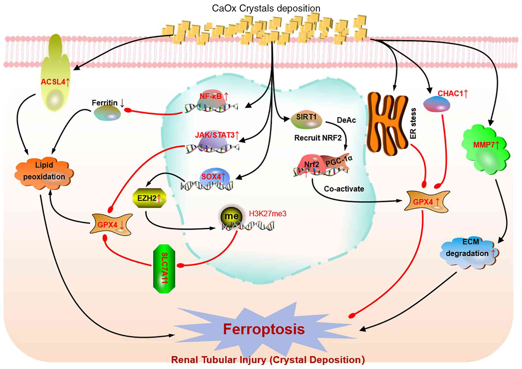
Epigenetics regulation is a key downstream effector
of CaOx crystal initiation and deposition. In the context of
CaOx-induced kidney injury, Yan et al (57) demonstrated that the
SOX4/EZH2/SLC7A11 signaling axis plays a pivotal role in mediating
ferroptosis. Specifically, CaOx crystal deposition significantly
upregulates the expression of SOX4, a transcription factor known
for its involvement in cell fate determination and tissue
development. SOX4 functions as a master regulator in this axis by
directly binding to the promoter region of EZH2, thereby enhancing
its transcription (58). This
upregulation of EZH2 is crucial as EZH2, as a core component of the
polycomb repressive complex 2, catalyzes the trimethylation of
histone H3 lysine 27 (H3K27me3), leading to the epigenetic
silencing of downstream genes (59). One of the key downstream targets
of EZH2 in this context is SLC7A11, a critical component of the
cystine-glutamate antiporter system xc(-) that regulates
intracellular GSH levels and maintains cellular antioxidant
capacity. EZH2-mediated H3K27me3 enrichment at the SLC7A11 promoter
region suppresses its expression, thereby disrupting the
antioxidant defense system and promoting ferroptosis (60). This suppression of SLC7A11
expression results in decreased GSH levels and impaired activity of
GPX4, which is essential for neutralizing lipid peroxides and
maintaining cellular redox balance (61,62). Consequently, the imbalance in
antioxidant defenses leads to increased lipid peroxidation [e.g.,
malondialdehyde (MDA)] and ROS accumulation, and elevated
expression of ferroptosis-related proteins, such as ACSL4,
hallmarks of ferroptosis. In summary, the SOX4/EZH2/SLC7A11
signaling axis is activated in CaOx-induced kidney injury, driving
ferroptosis through epigenetic suppression of SLC7A11 expression
(57)
Inflammation and the aberrant activation of related
signaling pathways are the most common causes of kidney injury and
crystal deposition (67,68). Among these, the JAK/STAT pathway,
particularly STAT3, has been identified as a key regulator of
ferroptosis in kidney cells (69). Mechanistically, GPX4 and SLC7A11,
the core regulatory nodes of ferroptosis, serve as downstream
effectors of STAT3; by modulating their expression, STAT3
suppresses System Xc− function and thereby promotes
ferroptosis (70). Moreover,
oxalate induces osteogenic-like differentiation of exacerbate renal
tubular epithelial cells (RTECs) via JAK2/STAT3 activation, a
recognized initiating step in kidney stone formation (71). Likewise, insulin-like growth
factor 1 receptor promotes CaOx crystal-induced
epithelial-mesenchymal transition through the JAK2/STAT3 signaling
pathway, which in turn facilitates the progression of kidney stones
(72,73).
Furthermore, studies have verified that CaOx
crystals function as potent activators of the renal NF-κB pathway,
which is a core driver of renal tubular injury and inflammatory
responses (6,78,79). The activation of the NF-κB
pathway suppresses ferritin expression and downregulates the
antioxidant system, thereby exacerbating iron overload and lipid
peroxidation (80,81). In models such as endothelial
injury, NF-κB activation can drive ferroptosis-mediated tissue
damage (80).
In summary, an interconnected signaling network
exists, which mediates the crosstalk between inflammation and
ferroptosis in CaOx crystal-induced renal damage. Therapeutically,
targeting these pathways, particularly inhibiting NF-κB or
JAK-STAT3, or enhancing the protective PI3K-AKT-mTOR signaling,
holds promise for mitigating crystal-induced renal injury.
Its well-established that oxalate and CaOx
monohydrate (COM) can induce oxidative stress in renal tubular
cells (6,82-84). For instance, Chuenwisad et
al (85) demonstrated that
oxalate, COM and urine from patients with kidney stones induced
oxidative stress in HK-2 cells by increasing the content of protein
carbonyls and decreasing the total antioxidant capacity. As a
consequence of this oxidative insult, the proportion of senescent
HK-2 cells, as revealed by SA-βgal staining, significantly
increases, while the relative telomere length markedly shortens
(85-87). Moreover, Sun et al
(88) suggested that the
depletion of GSH directly impairs the activity of GPX4, thereby
compromising the cellular capacity to effectively eliminate lipid
peroxides. This in turn leads to the accumulation of lipid ROS and
the onset of ferroptosis (88).
Further studies have demonstrated that in both
oxalate-induced cell models and renal stone mouse models,
ERS-related proteins (e.g., GRP78, ATF4 and CHOP) and
ferroptosis-related proteins (e.g., GPX4, XCT and FTH1) are
significantly upregulated (89-91). The CaOx crystal deposition drives
oxidative stress-dependent ferroptosis through a cascade of ERS,
glutathione-specific gamma-glutamylcyclotransferase 1
(CHAC1)-mediated glutathione depletion, GPX4 inactivation and
subsequent lipid peroxidation. CHAC1 serves as the core molecular
link between crystal stimulation and ferroptosis, represents a
potential interventional target in this process (89,92,93).
To further investigate the kidney stone pathological
environment, researchers exposed HK-2 cells to 2 mM CaOx for 24 h.
Compared with the controls, CaOx exposure significantly increased
the mRNA levels of ACSL4, MMP7, transglutaminase 2,
palmitoyl-protein thioesterase 1 and lactotransferrin, while it
decreased those of protein tyrosine kinase 2, dual specificity
phosphatase 4, secreted protein acidic and rich in cysteine like 1
and pleckstrin homology like domain family B member 2 (94). These key genes associated with
ERS in CaOx kidney stones may play a crucial role in the
pathogenesis of CaOx kidney stones, with their expression levels
closely related to the formation and development of kidney stones
(94). Among these, ACSL4 is a
long-chain fatty acyl-CoA synthetase that can activate fatty acids
into fatty acyl-CoA and is a key initiator of ferroptosis. During
ferroptosis, ACSL4 increases the levels of lipid peroxidation
within cells, thereby promoting cell death (14,95). The significant upregulation of
ACSL4 in CaOx kidney stone samples suggests that it may exacerbate
renal tubular epithelial cell damage by promoting ferroptosis
during the formation of kidney stones.
In summary, accumulating evidence demonstrates that
CaOx crystal deposition triggers oxidative stress-driven
ferroptosis in renal tubular cells through multiple interconnected
pathways. Key molecules, such as ACSL4 is upregulated in CaOx
kidney stones, potentially exacerbating cell injury via
ferroptosis. These findings highlight that ferroptosis is a
critical pathological process in CaOx-induced kidney injury and may
represent a promising therapeutic target for
nephrolithiasis-related renal injury (Fig. 3).
Accumulating evidence demonstrates that the
ferroptosis-kidney stone formation loop is a core pathogenic
mechanism driving kidney injury and disease progression. This loop
has been proposed as a promising therapeutic target, and disrupting
it via various inhibitors may produce protective effects.
First, the lipid peroxidation end-products, MDA and
4-hydroxynonenal (4-HNE), along with DAMPs released from
ferroptotic cells, activate NF-κB signaling, which in turn promotes
the secretion of pro-inflammatory cytokines (TNF-α, IL-6 and
IL-1β). These inflammatory mediators exacerbate RTEC injury, create
conditions for crystal deposition and further lower the ferroptosis
threshold, completing a positive feedback loop (3,25,96). A number of protective agents,
such as lycopene, curcumin and mesenchymal stem cells, exert their
anti-inflammatory, antioxidant and renoprotective effects precisely
by inhibiting NF-κB activation (97-100). Animal studies have indicated
that suppressing the NF-κB signaling pathway represents a
potentially effective therapeutic strategy for CaOx nephrolithiasis
(101,102).
Second, ferroptotic RTECs release DAMPs [high
mobility group box 1 (HMGB1), ATP, DNA, mitochondrial DNA, oxidized
phospholipids and MDA] that activate the innate immune response,
recruiting and activating macrophages. These macrophages secrete
mediators, such as macrophage inhibitory protein-1, monocyte
chemoattractant protein-1 and IL-8, leading to interstitial
inflammation (3). Critically,
inflammatory factors released by macrophages act back on RTECs,
upregulating adhesion molecules (CD44, osteopontin and hyaluronic
acid) on the surface of ferroptosis-susceptible RTECs, promoting
crystal adhesion and disposition. This immune-amplification loop
contributes to Randall's plaque formation and the transition to
fibrosis.
Third, during ferroptosis the 'wave of death'
phenomenon amplifies the loop spatially (3,103). When a proximal RTEC undergoes
ferroptosis, NADPH diffuses from adjacent cells to the dead cell
via gap junctions and tight junctions, reducing the antioxidant
capacity of neighboring cells and rendering them more susceptible
to ferroptosis. This chain reaction causes large-area tubular
damage, releasing more DAMPs and lipid peroxidation products, and
further amplifying inflammation (3), in turns promote kidney stone
formation. GPX4 serves as a critical negative regulator of the
vicious cycle of cellular injury; however, its loss amplifies this
feedback loop. GPX4 deficiency exacerbates TGF-β1 production and
fibrosis, while elevated GPX4 attenuates NF-κB activation (3,29,44).
In summary, these findings indicated that the
ferroptosis-kidney stone formation loop, driven by lipid
peroxidation, inflammatory amplification and intercellular
propagation, constitutes a key pathogenic mechanism in renal injury
and stone disease, and that targeted disruption of this vicious
cycle, particularly by GPX4 restoration or NF-κB inhibition,
represents a promising therapeutic strategy for clinical
intervention.
Ferroptosis can be therapeutically targeted by a
couple of small molecules, including natural products and synthetic
chemical compounds, which are collectively referred to as
ferrostatins. The restoration of this axis via ferrostatin-1, Nrf2
activators or melatonin reinstates GPX4 activity, limits lipid
peroxidation, and attenuates CaOx-induced injury and stone
formation, thereby identifying the GSH-GPX4 axis as a feasible
therapeutic target for nephrolithiasis (2,57,83,84,91,104-106). Several high-quality compounds
with acceptable pharmacodynamic and pharmacokinetic properties are
currently under development (16,107), and the initiation of clinical
trials appears imminent.
Fer-1, as a lead molecule, inhibits ferroptosis by
scavenging initiating alkoxyl radicals produced by ferrous iron
from lipid hydroperoxides (107). In both in vivo and in
vitro models, Fer-1 upregulates the key ferroptosis regulators
GPX4 and SLC7A11, downregulates HO-1 and SOD2, attenuates
mitochondrial damage, reduces ROS levels and lipid peroxidation,
decreases tubular epithelial cell apoptosis and renal tissue
injury, reverses the aberrant expression of fibrosis markers
including E-cadherin, vimentin and α-SMA, and significantly
suppresses CaOx crystal deposition (108). In CaOx models, Fer-1 partially
restores the downregulated expression of the hub
ferroptosis-related genes lysosome-associated membrane protein 2
and mouse double minute 4, which further enhances its protective
effects against oxalate-induced kidney injury and stone formation
(109).
New analogues, such as UAMC-3203, UAMC-3234,
UAMC-3206 and UAMC-3240, have significantly longer microsomal
half-lives and improved solubility, addressing the limitations of
Fer-1. Molecular dynamics simulations suggest that these analogues
rapidly insert into phospholipid bilayers, consistent with their
role in inhibiting lipid peroxidation (111,121). Another Fer-1 analog, Srs11-92,
was found to mitigate brain infarct size, neuronal damage and
neurological deficits in the mouse model of middle cerebral artery
occlusion and reperfusion by inhibiting iron overload, ROS
accumulation and neuroinflammation. This was achieved through the
upregulation of GPX4, Nrf2 and HO-1, while concurrently suppressing
HMGB1 and NF-κB p65 in the injured hippocampus or Alzheimer's
disease models (112,122,123). Beyond the analogs listed above,
numerous Fer-1 derivatives have been comprehensively reviewed and
are summarized in Table I.
Calcium oxalate crystals adhere to renal tubular
epithelial cells, inducing substantial ROS generation, which leads
to lipid peroxidation and the release of iron ions. This stimulus
causes a decrease in GPX4 expression and an upregulation of ACSL4
in renal tubular epithelial cells, ultimately resulting in
ferroptosis (82-85,124). The inhibitory effects of Fer-1
and its analogues on ferroptosis have been well-documented in
various experimental models; however, their roles in
oxalate-induced kidney injury and nephrolithiasis remain largely
understudied, and whether they exert protective effects against
crystal-induced renal damage remains unclear.
Numerous natural products have been reported to
exhibit significant anti-ferroptotic effects, highlighting their
potential as therapeutic agents for diseases involving ferroptosis,
including urolithiasis. For example, ursolic acid (UA) exerts
antitumor effects in malignant tumors of the urinary system
(prostate cancer/bladder cancer/renal cell carcinoma) by inducing
ferroptosis, specifically by promoting SLC7A11-mediated
ferroptosis; however, in subarachnoid hemorrhage, UA inhibits
ferroptosis via brain-targeted nanodelivery, thereby exerting
neuroprotective effects (125,126). Additionally, UA can alleviate
CaOx-induced injury to renal tubular epithelial cells by
suppressing oxidative stress and inflammation (127). UA exerts reverse effects in
different types of disease, which may be attributed to the
expression levels of SLC7A11 or the state of oxidative stress. The
application of nanomaterial-based delivery systems to achieve
neuroprotection may also be related to how these delivery systems
modulate the local concentration, targeting specificity, and
functional selectivity of UA. As an upstream multi-target
protective agent (inhibiting oxidative stress and inflammation), UA
directly alleviate CaOx-induced RTEC injury, suggesting its
potential for further research and clinical application in the
management of renal stone disease.
Similarly, 6-gingerol can promote ferroptosis in
prostate cancer, while inhibiting ferroptosis in inflammatory,
metabolic or vascular diseases (15,125,128,129). As an NRF2 activator, 6-gingerol
ameliorates atherosclerosis and protects against diabetic
cardiomyopathy by inhibiting ferroptosis via NRF2-mediated
anti-ferroptotic pathways (15,128,129). It can also alleviate
inflammation and oxidative stress by suppressing the JAK/STAT
signaling pathway (130),
reduce high glucose-induced inflammation and cytotoxicity in THP-1
cells by inhibiting the TLR4/NF-κB pathway (131), and attenuate NNK-induced
oxidative damage in lung cells by activating the NRF2 pathway
(132).
Talaroconvolutin A inhibits RSL3-induced
ferroptosis, likely by alleviating lipid peroxidation and
upregulating NRF2 expression (133). Baicalin activates signaling
pathways, such as Nrf2/GPX4, PI3K/AKT/GPX4 and SIRT3/GPX4, thereby
reducing lipid peroxidation and ROS accumulation. Additionally,
baicalin can directly chelate iron ions, reducing iron deposition
and oxidative damage (134-138). Kukoamine A has been shown to
exert protective effects in a model of osteoarthritis by inhibiting
inflammation and ferroptosis via the SIRT1/GPX4 signaling pathway,
and to alleviate nephrolithiasis by inhibiting endogenous oxalate
synthesis through the IL-6/JAK/STAT3/D-amino acid oxidase signaling
pathway (139,140).
Collectively, these natural products demonstrate
significant potential in modulating ferroptosis, either by directly
inducing ferroptosis or by protecting cells from ferroptotic
damage. Their mechanisms involve regulating key pathways, such as
iron metabolism, lipid peroxidation and antioxidant defense
systems. These findings provide preliminary evidence that numerous
natural products may hold therapeutic potential for the prevention
and treatment of kidney stone disease by modulating ferroptosis;
however, this remains to be validated by further mechanistic and
clinical studies.
A series of existing drugs exhibit novel therapeutic
indications. For example, mMetformin, an approved and core
first-line medication for type 2 diabetes, can reverse COM-induced
oxidative stress by activating the Nrf2/HO-1 signaling pathway,
thereby simultaneously inhibiting CaOx crystal formation
(dissolving COM and promoting COD conversion) and mitigating renal
tubular injury (106). However,
metformin also exerts antitumor effects in breast cancer cells by
inhibiting the UFMylation of SLC7A11, thereby promoting ferroptosis
(141). These opposite effects
on ferroptosis may be due to differences in SLC7A11 expression
levels across different disease types, as well as the diverse
pharmacological effects of metformin in distinct disease contexts.
Reserpine is a classic sympatholytic agent, belonging to
antihypertensive drugs (142).
Previous research has demonstrated that it exerts anti-ferroptotic
and anti-inflammatory effects in the context of cisplatin-induced
acute kidney injury (143).
Propafenone, a class Ic antiarrhythmic agent
clinically applied for rhythm control in patients with atrial
fibrillation and supraventricular tachycardia (144), exerts sodium channel blocking
(145) and immunomodulatory
effects (146). These effects
may alleviate oxalate-induced cellular stress and inflammatory
responses to a certain degree, thereby indirectly inhibiting
ferroptosis and preventing kidney stone formation. Omeprazole, a
classic proton pump inhibitor that potently suppresses gastric acid
secretion by irreversibly inhibiting the
H+/K+-ATPase in gastric parietal cells.
Recent studies have revealed that omeprazole also possesses
non-antacid effects, including antioxidant activity, free radical
scavenging, regulation of copper metabolism and exhibits
ferroptosis-related activity in various disease models (147,148). Lidocaine attenuates
hypoxia/reoxygenation-induced inflammation and apoptosis, and
inhibits ferroptosis in lung epithelial cells by suppressing the
p38 MAPK pathway, thereby exerting a protective effect (149). Promethazine exhibits
non-histamine receptor-dependent antioxidant and anti-ferroptosis
activities, and can protect multiple organs from
ferroptosis-related damage by scavenging lipid peroxyl radicals
(147,150,151). Reduced vitamin K
(VKH2 and menadiol) possesses potent antioxidant
activity and can inhibit ferroptosis by directly scavenging lipid
peroxidation free radicals through various mechanisms (152).
Collectively, the examples discussed above,
metformin, reserpine, propafenone, omeprazole, lidocaine,
promethazine and reduced VKH2, illustrate a compelling
yet largely unexplored phenomenon: A range of established drugs,
originally developed for distinct therapeutic purposes, may exert
unexpected renoprotective and anti-lithogenic effects via the
common pathway of ferroptosis inhibition. Given the vast arsenal of
clinically approved drugs with previously unrecognized
anti-ferroptotic properties, it is highly probable that a number of
other molecules await identification and systematic evaluation.
Future drug repurposing efforts, leveraging high-throughput
screening, computational approaches and ferroptosis-specific
biomarker assays, are urgently required to uncover additional
candidates that could effectively prevent or treat nephrolithiasis
by targeting ferroptosis. Such discoveries may open new avenues for
non-surgical, mechanism-based intervention in kidney stone
disease.
7-Dehydrocholesterol (7-DHC) is a direct precursor
of cholesterol in the cholesterol biosynthetic pathway, produced
from the catalytic reaction of 7-DHC reductase (DHCR7), and is also
an endogenous sterol possessing a conjugated diene structure.
Recently, Multiple studies have demonstrated that 7-DHC can inhibit
phospholipid autoxidation through its conjugated diene structure,
thereby protecting cells from ferroptosis (153-155). As a naturally occurring potent
ferroptosis inhibitor, 7-DHC has been shown to protect renal
tubular epithelial cells in a renal ischemia-reperfusion injury
model (155). Ferroptosis can
be significantly suppressed by either inhibiting DHCR7 or directly
supplementing 7-DHC (153,154,156,157).
With the rapid advancement of biochemical and
structural biology techniques, novel small molecules and
theoretical concepts for modulating ferroptosis have emerged
continuously, opening up new directions for the treatment of
ferroptosis-related diseases. However, current findings are based
predominantly on in vitro cell lines and in vivo
rodent models, which still have several limitations on their
translational to human kidney stone disease. First, in vitro
experiments rely almost exclusively on proximal tubular epithelial
cell lines, whereas stones develop in the renal papilla, and the
roles of other renal resident cells and infiltrating immune cells
during stone formation remain unexplored (160). Second, current in vivo
models do not fully reproduce human nephrolithiasis: Experimentally
induced hyperoxaluria generates animal models with CaOx crystal
deposition, which may more effectively represent
hyperoxaluria-driven conditions than common idiopathic stone
disease. Third, the association between ferroptosis and stone
pathogenesis remains to be rigorously validated in humans. There is
still a lack of clinical cohort data confirming whether ferroptosis
biomarkers (e.g., GPX4 downregulation and lipid peroxide
accumulation) are genuinely elevated in kidney stone patients, and
whether targeting ferroptosis can reduce stone formation or
recurrence in patients. Collectively, these model limitations
underscore the need for more representative, human-relevant models
for ferroptosis related therapeutic agent selection and
investigation for nephrolithiasis.
Despite the notable progress being made in
identifying the role of ferroptosis in CaOx nephropathy, and the
promising advances on anti-ferroptosis molecules, the clinical
translation of ferroptosis-targeting strategies for kidney stone
disease faces multifaceted challenges that require critical
evaluation.
Foremost among these is the pharmacokinetic
insufficiency of current ferroptosis inhibitors. The conventional
anti-ferroptosis agent, Fer-1, suffers from both poor aqueous
solubility, and a low metabolic and plasma stability (111,161). Its direct analog, SRS11-92,
still retains the ester-based metabolic instability of the Fer-1
core (123). Although
liproxstatin-1 displays a substantial bioavailability, it is
constrained by a short plasma half-life (162). The optimized UAMC series,
UAMC-3240 and UAMC-3234, exhibit marked species-dependent intrinsic
clearance, while their earlier sulfonamide progenitors were limited
by poor solubility in vivo (111). JP4-039 is limited not by
solubility, but by its low mitochondrial enrichment, which markedly
restricts its efficacy (118).
Collectively, these pharmacokinetic and targeting drawbacks impair
the bioavailability and renal tissue penetration.
A second challenge is the off-target toxicity and
systemic safety concerns inherent to systemic ferroptosis
modulation. The complete inhibition of ferroptosis may increase the
risk of tumorigenesis by undermining the surveillance that
ferroptosis exerts over potentially malignant cells, which has not
been addressed in long-term safety assessments (9). Moreover, the complete inhibition of
ferroptosis may disrupt iron homeostasis, and the lack of
renal-specific drug delivery systems exacerbates this detrimental
effect (10,163). The development of
kidney-directed nanocarriers, exosomes, or antibody-drug conjugates
is proposed (164-166), although this remains at the
preclinical stage.
A third challenge is the absence of specific and
standardized biomarkers to guide patient selection and monitor
therapeutic response. Although BODIPY C11 staining combined with
MDA and 4-HNE detection has been used to confirm the association
between COM-induced cytotoxicity and ferroptosis in COM treated
HK-2 cells, these methods are non-specific, as their signals can
also be generated by mitochondrial-derived oxidative stress
(44,64,161). Some emerging candidates, such
as oxidized phospholipids and ferroptosis-related ncRNAs (e.g.,
LINC00239, lncRNA CRCMSL, and lncRNA HMG), lack standardized
detection protocols and clinical validation (44,167).
Finally, the incompletely understood role of
ferroptosis in kidney stone disease complicates rational drug
screen and design. Studies using CaOx models have demonstrated that
ferroptosis, oxidative stress, inflammation, autophagy and
apoptosis function simultaneously, which indicates that
single-target monotherapy may not achieve sufficient therapeutic
effects (2-4,25,89). Current clinical stone therapy
relies on pharmaceuticals that modulate urinary crystalloid
supersaturation, and the integration of ferroptosis-targeting
agents still requires additional clinical evidence verifying their
specific efficacy in reducing stone burden. Overcoming these
barriers, through renal-specific drug delivery systems, rigorous
pharmacokinetic profiling, long-term safety profiling, validated
biomarkers and combination therapeutic strategies, is essential
before ferroptosis-targeting small molecules can become a
cornerstone of kidney stone management.
Ferroptosis functions as a pivotal driver of
oxalate crystal nephropathy by facilitating crystal nucleation and
adhesion. Conversely, oxalate crystals exacerbate ferroptosis,
thereby amplifying renal injury. This bidirectional positive
feedback loop suggests that ferroptosis may represent a promising
pharmacological target for the treatment of crystal nephropathy.
Small molecules, including natural products and synthetic chemical
compounds, have been found to exert certain efficacy in inhibiting
or activating ferroptosis for disease treatment. In kidney stone
disease, although novel findings shed light, considerable efforts
are still warranted, and further evidence is required for the
implication of ferroptosis regulators in disease treatment and
prevention.
Not applicable.
ZW and HL proposed the topic of the review. QD and
CX investigated the literature and prepared the manuscript. ZW and
YZ revised the manuscript. All authors have read and approved the
final manuscript. Data authentication is not applicable.
Not applicable.
Not applicable.
The authors declare that they have no competing
interests.
Not applicable.
The present study was financially supported by the Scientific
and Technological Innovation Fund of Longhua Shenzhen (grant no.
11501A20240704F5EB6FF), and the Construction Funds of Key Clinical
Medical Disciplines in Longhua District Shenzhen (grant no.
MKD202508210305).
|
1
|
Bargagli M, Scoglio M, Howles SA and
Fuster DG: Kidney stone disease: Risk factors, pathophysiology and
management. Nat Rev Nephrol. 21:794–808. 2025. View Article : Google Scholar : PubMed/NCBI
|
|
2
|
Tang K, Ye T, He Y, Ba X, Xia D, Peng E,
Chen Z, Ye Z and Yang X: Ferroptosis, necroptosis, and pyroptosis
in calcium oxalate crystal-induced kidney injury. Biochim Biophys
Acta Mol Basis Dis. 1871:1677912025. View Article : Google Scholar : PubMed/NCBI
|
|
3
|
Yang J, Wu W, Amier Y, Li X, Wan W, Xun Y
and Yu X: Ferroptosis and its emerging role in kidney stone
formation. Mol Biol Rep. 51:3142024. View Article : Google Scholar : PubMed/NCBI
|
|
4
|
Khan SR and Alli AA: Apoptosis,
ferroptosis, necrosis, necroptosis and pyroptosis in the formation
of calcium oxalate kidney stones. Urolithiasis. 53:1532025.
View Article : Google Scholar : PubMed/NCBI
|
|
5
|
Dixon SJ, Lemberg KM, Lamprecht MR, Skouta
R, Zaitsev EM, Gleason CE, Patel DN, Bauer AJ, Cantley AM, Yang WS,
et al: Ferroptosis: An iron-dependent form of nonapoptotic cell
death. Cell. 149:1060–1072. 2012. View Article : Google Scholar : PubMed/NCBI
|
|
6
|
Ming S, Tian J, Ma K, Pei C, Li L, Wang Z,
Fang Z, Liu M, Dong H, Li W, et al: Oxalate-induced apoptosis
through ERS-ROS-NF-kappaB signalling pathway in renal tubular
epithelial cell. Mol Med. 28:882022. View Article : Google Scholar
|
|
7
|
Abate M, Festa A, Falco M, Lombardi A,
Luce A, Grimaldi A, Zappavigna S, Sperlongano P, Irace C, Caraglia
M and Misso G: Mitochondria as playmakers of apoptosis, autophagy
and senescence. Semin Cell Dev Biol. 98:139–153. 2020. View Article : Google Scholar
|
|
8
|
Dixon SJ and Olzmann JA: The cell biology
of ferroptosis. Nat Rev Mol Cell Biol. 25:424–442. 2024. View Article : Google Scholar : PubMed/NCBI
|
|
9
|
Wu K, Vaughan AJ, Bossowski JP, Hao Y,
Ziogou A, Kim SM, Kim TH, Nakamura MN, Pillai R, Mancini M, et al:
Targeting FSP1 triggers ferroptosis in lung cancer. Nature.
649:487–495. 2026. View Article : Google Scholar :
|
|
10
|
Diao J, Jia Y, Dai E, Liu J, Kang R, Tang
D, Han L, Zhong Y and Meng L: Ferroptotic therapy in cancer:
Benefits, side effects, and risks. Mol Cancer. 23:892024.
View Article : Google Scholar : PubMed/NCBI
|
|
11
|
Zhang L, Luo F, Yuan N, Yin J, Shen B,
Chai Y, Sun L, Wang X, Yin L and Luo C: Research progress of
ferroptosis in acute kidney injury. Front Cell Dev Biol.
13:16141562025. View Article : Google Scholar : PubMed/NCBI
|
|
12
|
Ye K, Lan R, Chen Z, Lai K, Song Y, Li G,
Ma H, Chen H and Xu Y: Roles of ACSL4/GPX4 and FSP1 in
oxalate-induced acute kidney injury. Cell Death Discov. 11:2792025.
View Article : Google Scholar : PubMed/NCBI
|
|
13
|
Huang YQ, Huang ZW and Zhang XJ: Targeting
nuclear factor erythroid 2-related factor 2-regulated ferroptosis
to treat nervous system diseases. World J Clin Cases. 12:6655–6659.
2024. View Article : Google Scholar : PubMed/NCBI
|
|
14
|
Jia B, Li J, Song Y and Luo C:
ACSL4-Mediated ferroptosis and its potential role in central
nervous system diseases and injuries. Int J Mol Sci. 24:100212023.
View Article : Google Scholar : PubMed/NCBI
|
|
15
|
Khan SU, Halawi MH, Almehmadi M, Ibrahim
EH, Taha R, Alsyaad KM, Ahmed AE, Al-Doaiss AA and Thornbury W:
NRF2-mediated anti-ferroptotic pathways in diabetic cardiomyopathy:
Mechanistic insights, therapeutic advances, and challenges in
cardiovascular protection. Curr Probl Cardiol. 51:1032512026.
View Article : Google Scholar
|
|
16
|
Ju J, Song YN and Wang K: Mechanism of
ferroptosis: A potential target for cardiovascular diseases
treatment. Aging Dis. 12:261–276. 2021. View Article : Google Scholar : PubMed/NCBI
|
|
17
|
Chang S, Zhang M, Liu C, Li M, Lou Y and
Tan H: Redox mechanism of glycerophospholipids and relevant
targeted therapy in ferroptosis. Cell Death Discov. 11:3582025.
View Article : Google Scholar : PubMed/NCBI
|
|
18
|
Song C, Liu Z, Tang J and Huang Y:
Ferroptosis in cerebral ischemia/reperfusion injury: Mechanistic
drivers and therapeutic frontiers. Neuropsychiatr Dis Treat.
22:5801702026. View Article : Google Scholar : PubMed/NCBI
|
|
19
|
Ru Q, Li Y, Xie W, Ding Y, Chen L, Xu G,
Wu Y and Wang F: Fighting age-related orthopedic diseases: Focusing
on ferroptosis. Bone Res. 11:122023. View Article : Google Scholar : PubMed/NCBI
|
|
20
|
Li C, Deng D, Jiang Q, Shi J, Xu L and Liu
Y: Ferroptosis in NAFLD: Insights and the therapeutic potential of
exercise. Front Med (Lausanne). 12:14621452025. View Article : Google Scholar : PubMed/NCBI
|
|
21
|
Liang D, Feng Y, Zandkarimi F, Wang H,
Zhang Z, Kim J, Cai Y, Gu W, Stockwell BR and Jiang X: Ferroptosis
surveillance independent of GPX4 and differentially regulated by
sex hormones. Cell. 186:2748–2764 e22. 2023. View Article : Google Scholar : PubMed/NCBI
|
|
22
|
Deng H, Zhao L, Ge H, Gao Y, Fu Y, Lin Y,
Masoodi M, Losmanova T, Medová M, Ott J, et al: Ubiquinol-mediated
suppression of mitochondria-associated ferroptosis is a targetable
function of lactate dehydrogenase B in cancer. Nat Commun.
16:25972025. View Article : Google Scholar : PubMed/NCBI
|
|
23
|
Deng R, Fu L, Liang H, Ai X, Liu F, Li N,
Wu L, Li S, Yang X, Lin Y, et al: Inhibition of mitochondrial
complex I induces mitochondrial ferroptosis by regulating CoQH2
levels in cancer. Cell Death Dis. 16:2542025. View Article : Google Scholar : PubMed/NCBI
|
|
24
|
Liang NN, Guo YY, Zhang XY, Ren YH, He YZ,
Liu ZB, Xu DX and Xu S: Mitochondrial dysfunction-evoked DHODH
acetylation is involved in renal cell ferroptosis during
cisplatin-induced acute kidney injury. Adv Sci (Weinh).
11:e24047532024. View Article : Google Scholar : PubMed/NCBI
|
|
25
|
Ran Y, Ma Y, Luo Y and Ruan Y: The
potential functions of ferroptosis on urinary stones: mechanisms
and therapeutic implications. Front Physiol. 16:16334682025.
View Article : Google Scholar : PubMed/NCBI
|
|
26
|
Wang Z, Zhang Y, Zhang J, Deng Q and Liang
H: Recent advances on the mechanisms of kidney stone formation
(Review). Int J Mol Med. 48:1492021. View Article : Google Scholar : PubMed/NCBI
|
|
27
|
Zhou Z, Feng D, Shi D, Gao P, Wang L and
Wu Z: Untargeted and targeted metabolomics reveal bile acid profile
changes in rats with ethylene glycol-induced calcium oxalate
nephrolithiasis. Chem Biol Interact. 381:1105702023. View Article : Google Scholar : PubMed/NCBI
|
|
28
|
Liu L, Ma Y, Jian Z, Liao B, Li Y, Lin L,
Wang M, Chen J, Wei J, Yang M, et al: Gut microbiota-bile acid
crosstalk contributes to calcium oxalate nephropathy through
Hsp90α-mediated ferroptosis. Cell Rep. 44:1159362025. View Article : Google Scholar
|
|
29
|
Han H, Zhang G, Zhang X and Zhao Q:
Nrf2-mediated ferroptosis inhibition: A novel approach for managing
inflammatory diseases. Inflammopharmacology. 32:2961–2986. 2024.
View Article : Google Scholar : PubMed/NCBI
|
|
30
|
de Souza I, da Silva Teixeira AB, Ferreira
Dos Santos A, Friedmann Angeli JP and Ribeiro Reily Rocha C:
Tipping the balance: NRF2's dual role in ferroptotic fate.
Oncogenesis. 15:262026. View Article : Google Scholar : PubMed/NCBI
|
|
31
|
Li S, Wu W, Zhou Y, Zhang S, Wei D, Zhu M,
Ying X, Sun X, Liu H, Zhu W, et al: Gut microbiota-regulated
unconjugated bilirubin metabolism drives renal calcium oxalate
crystal deposition. Gut Microbes. 17:25461582025. View Article : Google Scholar : PubMed/NCBI
|
|
32
|
Manikanta, NaveenKumar SK, Thushara RM,
Hemshekhar M, Sumedini ML, Sunitha K, Kemparaju K and Girish KS:
Counteraction of unconjugated bilirubin against heme-induced
toxicity in platelets. Thromb Res. 244:1091992024. View Article : Google Scholar : PubMed/NCBI
|
|
33
|
Yao Q, Sun R, Bao S, Chen R and Kou L:
Bilirubin protects transplanted islets by targeting ferroptosis.
Front Pharmacol. 11:9072020. View Article : Google Scholar : PubMed/NCBI
|
|
34
|
Qaisiya M, Coda Zabetta CD, Bellarosa C
and Tiribelli C: Bilirubin mediated oxidative stress involves
antioxidant response activation via Nrf2 pathway. Cell Signal.
26:512–520. 2014. View Article : Google Scholar
|
|
35
|
Jing J, Yan X, Wang L, Zhang Y, Qi W, Xi J
and Hao Z: Gut microbiota-derived indole-3-acetic acid ameliorates
calcium oxalate renal stone formation via AHR/NF-ĸB axis.
Urolithiasis. 53:1342025. View Article : Google Scholar
|
|
36
|
Yang J, Li D, Li T and Jia B: Probiotics
in the prevention and treatment of calcium oxalate kidney stones:
Mechanisms and therapeutic potential. Front Microbiol.
16:16631382025. View Article : Google Scholar : PubMed/NCBI
|
|
37
|
Zhang M, Fan L and Li J: Prediction of
calcium oxalate kidney stones: A comprehensive analysis of clinical
and gut microbiome characteristics. Medicine (Baltimore).
104:e431032025. View Article : Google Scholar : PubMed/NCBI
|
|
38
|
Zhou Y, Lei M, Cai W, Chang Z, An L, Zhang
S, Wei D, Jiao R, Gao J, Xu Y, et al: Human fecal microbiota
transplantation attenuates high dietary oxalate-induced renal
calcium oxalate crystal depositions in rats via repairing
Allobaculum-related gut barrier damage. mSystems. 10:e00810252025.
View Article : Google Scholar : PubMed/NCBI
|
|
39
|
Ren L, Li F, Di Z, Xiong Y, Zhang S, Ma Q,
Bian X, Lang Z, Ye Q and Wang Y: Estradiol ameliorates acute kidney
ischemia-reperfusion injury by inhibiting the TGF-βRI-SMAD pathway.
Front Immunol. 13:8226042022. View Article : Google Scholar
|
|
40
|
Park KM, Kim JI, Ahn Y, Bonventre AJ and
Bonventre JV: Testosterone is responsible for enhanced
susceptibility of males to ischemic renal injury. J Biol Chem.
279:52282–52292. 2004. View Article : Google Scholar : PubMed/NCBI
|
|
41
|
Abufaraj M, Xu T, Cao C, Waldhoer T, Seitz
C, D'andrea D, Siyam A, Tarawneh R, Fajkovic H, Schernhammer E, et
al: Prevalence and trends in kidney stone among adults in the USA:
Analyses of National health and nutrition examination survey
2007-2018 data. Eur Urol Focus. 7:1468–1475. 2021. View Article : Google Scholar
|
|
42
|
Xu JZ, Li C, Xia QD, Lu JL, Wan ZC, Hu L,
Lv YM, Lei XM, Guan W, Xun Y and Wang SG: Sex disparities and the
risk of urolithiasis: A large cross-sectional study. Ann Med.
54:1627–1635. 2022. View Article : Google Scholar : PubMed/NCBI
|
|
43
|
Fuster DG, Morard GA, Schneider L,
Mattmann C, Lüthi D, Vogt B and Dhayat NA: Association of urinary
sex steroid hormones with urinary calcium, oxalate and citrate
excretion in kidney stone formers. Nephrol Dial Transplant.
37:335–348. 2022. View Article : Google Scholar
|
|
44
|
Luo Y, Long M, Wu X and Zeng L: Targeting
ferroptosis: Novel therapeutic approaches and intervention
strategies for kidney diseases. Front Immunol. 16:17000042025.
View Article : Google Scholar : PubMed/NCBI
|
|
45
|
Tonnus W, Maremonti F, Gavali S, Schlecht
MN, Gembardt F, Belavgeni A, Leinung N, Flade K, Bethe N, Traikov
S, et al: Multiple oestradiol functions inhibit ferroptosis and
acute kidney injury. Nature. 645:1011–1019. 2025. View Article : Google Scholar : PubMed/NCBI
|
|
46
|
Zhang S, Deng Z and Xu H: Hydroxyestradiol
alleviates acute kidney injury via blockade of ferroptosis. Trends
Endocrinol Metab. 36:869–871. 2025. View Article : Google Scholar : PubMed/NCBI
|
|
47
|
Carney EF: Inhibitory effects of
oestradiol on ferroptosis may underlie sex differences in AKI. Nat
Rev Nephrol. 21:7252025. View Article : Google Scholar : PubMed/NCBI
|
|
48
|
Wu S, Zhuang L and Gan B:
Oestradiol-mediated ferroptosis defense shapes sex differences in
acute kidney injury. Cell Death Differ. 33:428–430. 2026.
View Article : Google Scholar
|
|
49
|
Zhu W, Zhao Z, Chou FJ, Zuo L, Liu T,
Bushinsky D, Chang C, Zeng G and Yeh S: The protective roles of
estrogen receptor beta in renal calcium oxalate crystal formation
via reducing the liver oxalate biosynthesis and renal oxidative
stress-mediated cell injury. Oxid Med Cell Longev.
2019:53050142019. View Article : Google Scholar
|
|
50
|
Gillams K, Juliebo-Jones P, Juliebo SO and
Somani BK: Gender differences in kidney stone disease (KSD):
Findings from a systematic review. Curr Urol Rep. 22:502021.
View Article : Google Scholar : PubMed/NCBI
|
|
51
|
Zhang Y, Lu Q, Yan Y, Zhang J, Liu T, Yu
L, Fan Q and Wei K: Artemisia keiskeana Miq. alleviates oxidative
stress and ferroptosis in APAP-induced liver injury by mediating
Nrf2/GPX4/NF-ĸB signaling pathway through ESR1. Phytomedicine.
148:1574362025. View Article : Google Scholar
|
|
52
|
Lv Y, Zhang S, Weng X, Huang J, Zhao H,
Dai X, Bai X, Bao X, Zhao C, Zeng M, et al: Estrogen deficiency
accelerates postmenopausal atherosclerosis by inducing endothelial
cell ferroptosis through inhibiting NRF2/GPX4 pathway. FASEB J.
37:e229922023. View Article : Google Scholar : PubMed/NCBI
|
|
53
|
Chen GH, Song CC, Pantopoulos K, Wei XL,
Zheng H and Luo Z: Mitochondrial oxidative stress mediated
Fe-induced ferroptosis via the NRF2-ARE pathway. Free Radic Biol
Med. 180:95–107. 2022. View Article : Google Scholar : PubMed/NCBI
|
|
54
|
Tian Y, Hong X, Xie Y, Guo Z and Yu Q:
17β-Estradiol (E(2)) Upregulates the ERα/SIRT1/PGC-1α Signaling
pathway and protects mitochondrial function to prevent bilateral
oophorectomy (OVX)-Induced nonalcoholic fatty liver disease
(NAFLD). Antioxidants (Basel). 12:21002023. View Article : Google Scholar
|
|
55
|
Yu K, Li J, Tian T, Shi R, Deng Y and
Chang L: Sex hormone-specific regulation of ferroptosis in vascular
cells in atherosclerosis: Molecular mechanisms and targeted
strategies. Front Physiol. 16:16806252025. View Article : Google Scholar : PubMed/NCBI
|
|
56
|
Costa TJ, Ceravolo GS, dos Santos RA, de
Oliveira MA, Araújo PX, Giaquinto LR, Tostes RC, Akamine EH, Fortes
ZB, Dantas AP and Carvalho MH: Association of testosterone with
estrogen abolishes the beneficial effects of estrogen treatment by
increasing ROS generation in aorta endothelial cells. Am J Physiol
Heart Circ Physiol. 308:H723–H732. 2015. View Article : Google Scholar : PubMed/NCBI
|
|
57
|
Yan X, Xia Y, Li B, Ye Z, Li L, Yuan T,
Song B, Yu W, Rao T, Ning J, et al: The SOX4/EZH2/SLC7A11 signaling
axis mediates ferroptosis in calcium oxalate crystal
deposition-induced kidney injury. J Transl Med. 22:92024.
View Article : Google Scholar : PubMed/NCBI
|
|
58
|
Hanieh H, Ahmed EA, Vishnubalaji R and
Alajez NM: SOX4: Epigenetic regulation and role in tumorigenesis.
Semin Cancer Biol. 67(Pt 1): 91–104. 2020. View Article : Google Scholar
|
|
59
|
Yu C and Zhuang S: Histone
methyltransferases as therapeutic targets for kidney diseases.
Front Pharmacol. 10:13932019. View Article : Google Scholar : PubMed/NCBI
|
|
60
|
Liu GZ, Xu XW, Tao SH, Gao MJ and Hou ZH:
HBx facilitates ferroptosis in acute liver failure via EZH2
mediated SLC7A11 suppression. J Biomed Sci. 28:672021. View Article : Google Scholar : PubMed/NCBI
|
|
61
|
Mou Y, Wang J, Wu J, He D, Zhang C, Duan C
and Li B: Ferroptosis, a new form of cell death: opportunities and
challenges in cancer. J Hematol Oncol. 12:342019. View Article : Google Scholar : PubMed/NCBI
|
|
62
|
Li J, Cao F, Yin HL, Huang ZJ, Lin ZT, Mao
N, Sun B and Wang G: Ferroptosis: Past, present and future. Cell
Death Dis. 11:882020. View Article : Google Scholar : PubMed/NCBI
|
|
63
|
Ye QL, Wang DM, Wang X, Zhang ZQ, Tian QX,
Feng SY, Zhang ZH, Yu DX, Ding DM and Xie DD: Sirt1 inhibits kidney
stones formation by attenuating calcium oxalate-induced cell
injury. Chem Biol Interact. 347:1096052021. View Article : Google Scholar : PubMed/NCBI
|
|
64
|
Duan C, Li B, Liu H, Zhang Y, Yao X, Liu
K, Wu X, Mao X, Wu H, Xu Z, et al: Sirtuin1 suppresses calcium
oxalate nephropathy via inhibition of renal proximal tubular cell
ferroptosis through PGC-1α-mediated transcriptional coactivation.
Adv Sci (Weinh). 11:e24089452024. View Article : Google Scholar
|
|
65
|
Yao X, Liu H, Duan C, Zhang Y, Wu X, Li B,
Li S, Gong Y, Liu T, Wang X and Xu H: Sirtuin1 mitigation of
calcium oxalate nephropathy via enhancing itaconate abundance
through reduction of histone trimethylation. Clin Transl Med.
15:e704502025. View Article : Google Scholar : PubMed/NCBI
|
|
66
|
Duan C, Liu H, Yang X, Liu J, Deng Y, Wang
T, Xing J, Hu Z and Xu H: Sirtuin1 inhibits calcium oxalate
crystal-induced kidney injury by regulating TLR4 signaling and
macrophage-mediated inflammatory activation. Cell Signal.
112:1108872023. View Article : Google Scholar : PubMed/NCBI
|
|
67
|
Khan SR, Canales BK and
Dominguez-Gutierrez PR: Randall's plaque and calcium oxalate stone
formation: Role for immunity and inflammation. Nat Rev Nephrol.
17:417–433. 2021. View Article : Google Scholar : PubMed/NCBI
|
|
68
|
Capolongo G, Ferraro PM and Unwin R:
Inflammation and kidney stones: Cause and effect? Curr Opin Urol.
33:129–135. 2023. View Article : Google Scholar
|
|
69
|
Dong XQ, Chu LK, Cao X, Xiong QW, Mao YM,
Chen CH, Bi YL, Liu J and Yan XM: Glutathione metabolism rewiring
protects renal tubule cells against cisplatin-induced apoptosis and
ferroptosis. Redox Rep. 28:21526072023. View Article : Google Scholar : PubMed/NCBI
|
|
70
|
Dai Y, Cui C, Jiao D and Zhu X: JAK/STAT
signaling as a key regulator of ferroptosis: Mechanisms and
therapeutic potentials in cancer and diseases. Cancer Cell Int.
25:832025. View Article : Google Scholar : PubMed/NCBI
|
|
71
|
Song Q, Liao W, He Z, Li D, Dong C, Song C
and Yang S: Oxalate induces the ossification of RTECs by activating
the JAK2/STAT3 signaling pathway and participates in the formation
of kidney stones. Arch Biochem Biophys. 727:1093252022. View Article : Google Scholar : PubMed/NCBI
|
|
72
|
Pan J, Zhang Y, Yao R, Yang M, Mao X, Song
Z, Xu Y, Chen Y, Hou B, Liu X, et al: IGF1R enhances calcium
oxalate monohydrate-induced epithelial-mesenchymal transition by
reprogramming metabolism via the JAK2/STAT3 signaling. Int J Biol
Sci. 21:415–432. 2025. View Article : Google Scholar : PubMed/NCBI
|
|
73
|
Convento MB, Pessoa EA, Cruz E, da Gloria
MA, Schor N and Borges FT: Calcium oxalate crystals and oxalate
induce an epithelial-to-mesenchymal transition in the proximal
tubular epithelial cells: Contribution to oxalate kidney injury.
Sci Rep. 7:457402017. View Article : Google Scholar : PubMed/NCBI
|
|
74
|
Wang S, Lei Z, Liu W, Shi Y, Sherryn S,
Gao Q and Zhang B: ACOT4 and ACOT6 Activate Akt-mTOR pathway and
inhibit calcium oxalate-induced renal tubular cell injury. Kidney
Blood Press Res. 50:533–544. 2025. View Article : Google Scholar : PubMed/NCBI
|
|
75
|
Yi J, Zhu J, Wu J, Thompson CB and Jiang
X: Oncogenic activation of PI3K-AKT-mTOR signaling suppresses
ferroptosis via SREBP-mediated lipogenesis. Proc Natl Acad Sci USA.
117:31189–31197. 2020. View Article : Google Scholar : PubMed/NCBI
|
|
76
|
Hirohama D, Abedini A, Moon S, Surapaneni
A, Dillon ST, Vassalotti A, Liu H, Doke T, Martinez V, Md Dom Z, et
al: Unbiased human kidney tissue proteomics identifies matrix
metalloproteinase 7 as a kidney disease biomarker. J Am Soc
Nephrol. 34:1279–1291. 2023. View Article : Google Scholar : PubMed/NCBI
|
|
77
|
Canela VH, Bowen WS, Ferreira RM, Syed F,
Lingeman JE, Sabo AR, Barwinska D, Winfree S, Lake BB, Cheng YH, et
al: A spatially anchored transcriptomic atlas of the human kidney
papilla identifies significant immune injury in patients with stone
disease. Nat Commun. 14:41402023. View Article : Google Scholar : PubMed/NCBI
|
|
78
|
Liang H, Xu Y, Sun S, Chen Y, Wang W and
Hao Z: Based on network pharmacology, the mechanism of Dioscin in
alleviating renal tubular epithelial cell injury induced by calcium
oxalate crystals was explored. Urolithiasis. 53:32024. View Article : Google Scholar : PubMed/NCBI
|
|
79
|
Si Y, Liu L, Cheng J, Zhao T, Zhou Q, Yu
J, Chen W, Ding J, Sun X, Lu H and Guo Z: Oral hydrogen-rich water
alleviates oxalate-induced kidney injury by suppressing oxidative
stress, inflammation, and fibrosis. Front Med (Lausanne).
8:7135362021. View Article : Google Scholar : PubMed/NCBI
|
|
80
|
Wu S, Zhou Y, Liang J, Ying P, Situ Q, Tan
X and Zhu J: Upregulation of NF-ĸB by USP24 aggravates ferroptosis
in diabetic cardiomyopathy. Free Radic Biol Med. 210:352–366. 2024.
View Article : Google Scholar
|
|
81
|
Chen Y, Fang ZM, Yi X, Wei X and Jiang DS:
The interaction between ferroptosis and inflammatory signaling
pathways. Cell Death Dis. 14:2052023. View Article : Google Scholar : PubMed/NCBI
|
|
82
|
Kumar P, Laurence E, Crossman DK, Assimos
DG, Murphy MP and Mitchell T: Oxalate disrupts monocyte and
macrophage cellular function via Interleukin-10 and mitochondrial
reactive oxygen species (ROS) signaling. Redox Biol. 67:1029192023.
View Article : Google Scholar : PubMed/NCBI
|
|
83
|
Liu Y, Sun Y, Kang J, He Z, Liu Q, Wu J,
Li D, Wang X, Tao Z, Guan X, et al: Role of ROS-Induced NLRP3
inflammasome activation in the formation of calcium oxalate
nephrolithiasis. Front Immunol. 13:8186252022. View Article : Google Scholar : PubMed/NCBI
|
|
84
|
Nong WJ, Tong XY and Ouyang JM: Comparison
of endoplasmic reticulum stress and pyroptosis induced by
pathogenic calcium oxalate monohydrate and physiologic calcium
oxalate dihydrate crystals in HK-2 cells: Insights into kidney
stone formation. Cells. 13:20702024. View Article : Google Scholar :
|
|
85
|
Chuenwisad K, More-Krong P, Tubsaeng P,
Chotechuang N, Srisa-Art M, Storer RJ and Boonla C: Premature
senescence and telomere shortening induced by oxidative stress from
oxalate, calcium oxalate monohydrate, and urine from patients with
calcium oxalate nephrolithiasis. Front Immunol. 12:6964862021.
View Article : Google Scholar : PubMed/NCBI
|
|
86
|
Xu Y, You J, Yao J, Hou B, Wang W and Hao
Z: Klotho alleviates oxidative stress and mitochondrial dysfunction
through the Nrf2/HO-1 pathway, thereby reducing renal senescence
induced by calcium oxalate crystals. Urolithiasis. 53:612025.
View Article : Google Scholar : PubMed/NCBI
|
|
87
|
Chu M, Jiang S, Xue J, Li W, Jing G, Li H,
Zhang J and Xu W: Premature renal epithelial cell senescence
promoted by LXN/Rps3/p53 signaling pathway activation increases
calcium oxalate crystal deposition by altering macrophage
polarization. Front Immunol. 16:16589892025. View Article : Google Scholar : PubMed/NCBI
|
|
88
|
Sun Y, Zheng Y, Wang C and Liu Y:
Glutathione depletion induces ferroptosis, autophagy, and premature
cell senescence in retinal pigment epithelial cells. Cell Death
Dis. 9:7532018. View Article : Google Scholar : PubMed/NCBI
|
|
89
|
Dong C, He Z, Liao W, Jiang Q, Song C,
Song Q, Su X, Xiong Y, Wang Y, Meng L and Yang S: CHAC1 mediates
endoplasmic reticulum stress-dependent ferroptosis in calcium
oxalate kidney stone formation. Adv Sci (Weinh). 12:e24039922025.
View Article : Google Scholar : PubMed/NCBI
|
|
90
|
Duan X, Kong Z, Mai X, Lan Y, Liu Y, Yang
Z, Zhao Z, Deng T, Zeng T, Cai C, et al: Autophagy inhibition
attenuates hyperoxaluria-induced renal tubular oxidative injury and
calcium oxalate crystal depositions in the rat kidney. Redox Biol.
16:414–425. 2018. View Article : Google Scholar : PubMed/NCBI
|
|
91
|
He Y, Peng E, Ba X, Wu J, Deng W, Huang Q,
Tong Y, Shang H, Zhong Z, Liu X, et al: ROS responsive cerium oxide
biomimetic nanoparticles alleviates calcium oxalate crystals
induced kidney injury via suppressing oxidative stress and M1
macrophage polarization. Small. 21:e24054172025. View Article : Google Scholar
|
|
92
|
Ying X, Gao K, Luo Z, Ren Y, Shen C, Zhang
H, Zhan C, Xu Z, Wu J and Wu G: paliperidone inhibits ferroptosis
mediated by autophagy in renal tubular epithelial cells by
targeting CHAC1. Adv Biol (Weinh). 9:e24006482025. View Article : Google Scholar : PubMed/NCBI
|
|
93
|
Chen J, Liu D, Lei L, Liu T, Pan S, Wang
H, Liu Y, Qiao Y, Liu Z and Feng Q: CNPY2 aggravates renal tubular
cell ferroptosis in diabetic nephropathy by regulating
PERK/ATF4/CHAC1 pathway and MAM integrity. Adv Sci (Weinh).
12:e24164412025. View Article : Google Scholar : PubMed/NCBI
|
|
94
|
Tan Z, She W, Wang B, Wang X, Guan X, Tao
Z and Deng Y: Identification of key genes associated with
endoplasmic reticulum stress in calcium oxalate kidney stones.
Genes (Basel). 16:13382025. View Article : Google Scholar : PubMed/NCBI
|
|
95
|
Sun K, Shi Y, Yan C, Wang S, Han L, Li F,
Xu X, Wang Y, Sun J, Kang Z and Shi J: Glycolysis-Derived Lactate
Induces ACSL4 Expression and Lactylation to Activate Ferroptosis
during Intervertebral Disc Degeneration. Adv Sci (Weinh).
12:e24161492025. View Article : Google Scholar : PubMed/NCBI
|
|
96
|
Mulay SR, Kulkarni OP, Rupanagudi KV,
Migliorini A, Darisipudi MN, Vilaysane A, Muruve D, Shi Y, Munro F,
Liapis H and Anders HJ: Calcium oxalate crystals induce renal
inflammation by NLRP3-mediated IL-1beta secretion. J Clin Invest.
123:236–246. 2013. View Article : Google Scholar :
|
|
97
|
Zhao H, Zhang Y, Cao Y, Zhou B, Xing M and
Wang Y: Lycopene regulates Nrf2 to Ameliorate Sulfamethoxazole
-induced renal injury and apoptosis via inhibiting oxidative stress
and Endoplasmic Reticulum stress. Aquat Toxicol. 283:1073482025.
View Article : Google Scholar : PubMed/NCBI
|
|
98
|
Zou B, Wang D, Zhong J, He Z, Zhou Y, Yang
H, Liu Y, Zeng G and Duan X: Mesenchymal stem cells attenuate
hyperoxaluria-induced kidney injury and crystal depositions via
inhibiting the activation of NLRP3 inflammasome. Life Sci.
371:1236082025. View Article : Google Scholar : PubMed/NCBI
|
|
99
|
Zhu H, Wang X, Wang X, Liu B, Yuan Y and
Zuo X: Curcumin attenuates inflammation and cell apoptosis through
regulating NF-ĸB and JAK2/STAT3 signaling pathway against acute
kidney injury. Cell Cycle. 19:1941–1951. 2020. View Article : Google Scholar : PubMed/NCBI
|
|
100
|
Cai Y, Huang C, Zhou M, Xu S, Xie Y, Gao
S, Yang Y, Deng Z, Zhang L, Shu J, et al: Role of curcumin in the
treatment of acute kidney injury: Research challenges and
opportunities. Phytomedicine. 104:1543062022. View Article : Google Scholar : PubMed/NCBI
|
|
101
|
Sun Y, Liu Y, Guan X, Kang J, Wang X, Liu
Q, Li D, Xu H, Tao Z and Deng Y: Atorvastatin inhibits renal
inflammatory response induced by calcium oxalate crystals via
inhibiting the activation of TLR4/NF-ĸB and NLRP3 inflammasome.
IUBMB Life. 72:1065–1074. 2020. View Article : Google Scholar : PubMed/NCBI
|
|
102
|
Song Q, Song C, Chen X, Xiong Y, Li L,
Liao W, Xue L and Yang S: FKBP5 deficiency attenuates calcium
oxalate kidney stone formation by suppressing cell-crystal
adhesion, apoptosis and macrophage M1 polarization via inhibition
of NF-kappaB signaling. Cell Mol Life Sci. 80:3012023. View Article : Google Scholar
|
|
103
|
Belavgeni A, Meyer C, Stumpf J, Hugo C and
Linkermann A: Ferroptosis and necroptosis in the kidney. Cell Chem
Biol. 27:448–462. 2020. View Article : Google Scholar : PubMed/NCBI
|
|
104
|
Wu J, Liu M, Gao M, Li Y, Zhang Y, Tang L,
Yu H, Liao Z, Cui Y, Zeng F, et al: Epigallocatechin Gallate
Attenuates CaOx crystal-induced renal tubular injury to inhibit
CaOx nephrolithiasis via GRP94/PI3K/AKT signaling. Biomater Res.
29:02712025. View Article : Google Scholar : PubMed/NCBI
|
|
105
|
Zhang J, Zhang G, Jiang L, Qiu D, Wang G
and Chen C: FGF21 relieves calcium oxalate-induced cell injury,
apoptosis, oxidative damage and ferroptosis of renal tubular
epithelial cells through activating Nrf2 signaling pathway.
Cytotechnology. 76:519–531. 2024. View Article : Google Scholar : PubMed/NCBI
|
|
106
|
Zhang X, Liang F, Li T, Jiang Y and Ren F:
Metformin ameliorates calcium oxalate crystallization and stone
formation by activating the Nrf2/HO-1 signaling pathway: Two birds
with one stone. Arch Biochem Biophys. 739:1095682023. View Article : Google Scholar : PubMed/NCBI
|
|
107
|
Miotto G, Rossetto M, Di Paolo ML, Orian
L, Venerando R, Roveri A, Vučković AM, Bosello Travain V, Zaccarin
M, Zennaro L, et al: Insight into the mechanism of ferroptosis
inhibition by ferrostatin-1. Redox Biol. 28:1013282020. View Article : Google Scholar
|
|
108
|
Xie J, Ye Z, Li L, Xia Y, Yuan R, Ruan Y
and Zhou X: Ferrostatin-1 alleviates oxalate-induced renal tubular
epithelial cell injury, fibrosis and calcium oxalate stone
formation by inhibiting ferroptosis. Mol Med Rep. 26:2562022.
View Article : Google Scholar
|
|
109
|
Hou C, Zhong B, Gu S, Wang Y and Ji L:
Identification and validation of the biomarkers related to
ferroptosis in calcium oxalate nephrolithiasis. Aging (Albany NY).
16:5987–6007. 2024.PubMed/NCBI
|
|
110
|
Scarpellini C, Klejborowska G, Lanthier C,
Hassannia B, Vanden Berghe T and Augustyns K: Beyond ferrostatin-1:
A comprehensive review of ferroptosis inhibitors. Trends Pharmacol
Sci. 44:902–916. 2023. View Article : Google Scholar : PubMed/NCBI
|
|
111
|
Devisscher L, Van Coillie S, Hofmans S,
Van Rompaey D, Goossens K, Meul E, Maes L, De Winter H, Van Der
Veken P, Vandenabeele P, et al: Discovery of novel, drug-like
ferroptosis inhibitors with in vivo efficacy. J Med Chem.
61:10126–10140. 2018. View Article : Google Scholar : PubMed/NCBI
|
|
112
|
Chen Y, He W, Wei H, Chang C, Yang L, Meng
J, Long T, Xu Q and Zhang C: Srs11-92, a ferrostatin-1 analog,
improves oxidative stress and neuroinflammation via Nrf2 signal
following cerebral ischemia/reperfusion injury. CNS Neurosci Ther.
29:1667–1677. 2023. View Article : Google Scholar : PubMed/NCBI
|
|
113
|
Wang H and Xie Y: Advances in ferroptosis
research: A comprehensive review of mechanism exploration, drug
development, and disease treatment. Pharmaceuticals (Basel).
18:3342025. View Article : Google Scholar : PubMed/NCBI
|
|
114
|
Zhang NY, Liu JY, Zheng H, Wang KM, Zhang
J, Meng N and Jiang CS: Design, synthesis, and biological
evaluation of new improved ferrostatin-1 derived ferroptosis
inhibitors. Chem Biodivers. 22:e2024021412025. View Article : Google Scholar
|
|
115
|
Yan J, Bao L, Liang H, Zhao L, Liu M, Kong
L, Fan X, Liang C, Liu T, Han X, et al: A Druglike ferrostatin-1
analogue as a ferroptosis inhibitor and photoluminescent indicator.
Angew Chem Int Ed Engl. 64:e2025021952025. View Article : Google Scholar : PubMed/NCBI
|
|
116
|
Zhao Z, Wu J, Xu H, Zhou C, Han B, Zhu H,
Hu Z, Ma Z, Ming Z, Yao Y, et al: XJB-5-131 inhibited ferroptosis
in tubular epithelial cells after ischemia-reperfusion injury. Cell
Death Dis. 11:6292020. View Article : Google Scholar : PubMed/NCBI
|
|
117
|
Airik M, Clayton K, Wipf P and Airik R:
JP4-039 mitigates cisplatin-induced acute kidney injury by
inhibiting oxidative stress and blocking apoptosis and ferroptosis
in mice. Antioxidants (Basel). 13:15342024. View Article : Google Scholar
|
|
118
|
Krainz T, Gaschler MM, Lim C, Sacher JR,
Stockwell BR and Wipf P: A mitochondrial-targeted nitroxide is a
potent inhibitor of ferroptosis. ACS Cent Sci. 2:653–659. 2016.
View Article : Google Scholar : PubMed/NCBI
|
|
119
|
Yalamanchili K, Broadwin M, Harris DD,
Teixeira RB, Sellke FW, Wipf P and Abid MR: Targeting mitochondrial
reactive oxygen species: JP4-039's potential as a cardiovascular
therapeutic. J Clin Med. 14:64652025. View Article : Google Scholar : PubMed/NCBI
|
|
120
|
Teixeira RB, Albro JH, Sabra M, Abedin T,
Tucker AN, Sidharth R, Sellke FW, Wipf P and Abid MR:
Mitochondria-targeted ROS scavenger JP4-039 improves cardiac
function in a post-myocardial infarction animal model and induces
angiogenesis in vitro. PLoS One. 20:e03207032025. View Article : Google Scholar : PubMed/NCBI
|
|
121
|
Peleman C, Hellemans S, Veeckmans G, Arras
W, Zheng H, Koeken I, Van San E, Hassannia B, Walravens M,
Kayirangwa E, et al: Ferroptosis is a targetable detrimental factor
in metabolic dysfunction-associated steatotic liver disease. Cell
Death Differ. 31:1113–1126. 2024. View Article : Google Scholar : PubMed/NCBI
|
|
122
|
Skouta R, Dixon SJ, Wang J, Dunn DE, Orman
M, Shimada K, Rosenberg PA, Lo DC, Weinberg JM, Linkermann A and
Stockwell BR: Ferrostatins inhibit oxidative lipid damage and cell
death in diverse disease models. J Am Chem Soc. 136:4551–4556.
2014. View Article : Google Scholar : PubMed/NCBI
|
|
123
|
Guo Y, Cao H, Zuo C, Huang Y, Gu Z, Song
Y, Chen X, Jiang Q and Wang F: Neuroprotective effects of SRS11-92
against oxidative stress-induced senescence via Nrf2/HO-1/NF-ĸB in
Alzheimer's disease models. Inflammopharmacology. 34:495–507. 2026.
View Article : Google Scholar :
|
|
124
|
Li L, Ye Z, Xia Y, Li B, Chen L, Yan X,
Yuan T, Song B, Yu W, Rao T, et al: YAP/ACSL4 pathway-mediated
ferroptosis promotes renal fibrosis in the presence of kidney
stones. Biomedicines. 11:26922023. View Article : Google Scholar : PubMed/NCBI
|
|
125
|
Tao TY, Gong H and Yang B: Exploring
natural products to target ferroptosis in urologic malignancies:
Advancements from molecular mechanisms to therapeutic strategies.
Chin J Integr Med. 31:1139–1147. 2025. View Article : Google Scholar : PubMed/NCBI
|
|
126
|
Li Y, Zhu X, Xiong W, Zhao Q, Zhou Y, Guo
Y, Liu B, Li M, Chen Q, Jiang X, et al: Brain-targeted ursolic acid
nanoparticles for anti-ferroptosis therapy in subarachnoid
hemorrhage. J Nanobiotechnology. 22:6412024. View Article : Google Scholar : PubMed/NCBI
|
|
127
|
Jia Z, Li W, Bian P, Yang L, Liu H, Pan D
and Dou Z: Ursolic acid treats renal tubular epithelial cell damage
induced by calcium oxalate monohydrate via inhibiting oxidative
stress and inflammation. Bioengineered. 12:5450–5461. 2021.
View Article : Google Scholar : PubMed/NCBI
|
|
128
|
Wang S, Song X, Gao H, Zhang Y, Zhou X and
Wang F: 6-gingerol inhibits ferroptosis in endothelial cells in
atherosclerosis by activating the NRF2/HO-1 pathway. Appl Biochem
Biotechnol. 197:3890–3906. 2025. View Article : Google Scholar : PubMed/NCBI
|
|
129
|
He X, Shang Y, Liao X, Liu G, Yang Q, Liu
J, Xu X and Liu X: 6-Gingerol ameliorates endothelial injury in
hyperlipidemia mice via Nrf2 activation and inflammation
modulation. Food Funct. 17:1007–1017. 2026. View Article : Google Scholar : PubMed/NCBI
|
|
130
|
Zhang Y, Lin J, Xu X, Lu X, Li L, Yang Y
and Lin W: Neocnidilide and 6-gingerol as key bioactives in fresh
and dried centipeda minima: distinct Th1/Th2 modulation via
NF-ĸB/JAK-STAT pathways for allergic rhinitis therapy. Int J Mol
Sci. 26:86782025. View Article : Google Scholar
|
|
131
|
Kang DY, Chi WJ, Cho J and Jang KJ:
6-Gingerol alleviates high glucose-induced inflammation and
cytotoxicity in THP-1 cells by inhibiting TLR4 signaling. Sci Rep.
16:40692026. View Article : Google Scholar : PubMed/NCBI
|
|
132
|
Qu Z, Ding Y, Zhang L, Sun J, Wang T and
Zhuang C: 6-Gingerol alleviates NNK-induced lung carcinogenesis by
boosting antioxidation and reducing inflammation. Free Radic Res.
60:1–15. 2026. View Article : Google Scholar
|
|
133
|
Tian W, Chen X, Zhang G, Gong L, Lin J,
Hong B, Li L and Niu S: Talarotetramates A-F, decalin-containing
tetramic acids as ferroptosis inhibitors from the deep-sea-derived
fungus talaromyces sp. gb505. J Nat Prod. 88:1858–1867. 2025.
View Article : Google Scholar : PubMed/NCBI
|
|
134
|
Gou X, Tang X, Liu C and Chen Z:
Ferroptosis: A new mechanism of traditional Chinese medicine for
treating hematologic malignancies. Front Oncol. 14:14691782024.
View Article : Google Scholar : PubMed/NCBI
|
|
135
|
Li M, Meng Z, Yu S, Li J, Wang Y, Yang W
and Wu H: Baicalein ameliorates cerebral ischemia-reperfusion
injury by inhibiting ferroptosis via regulating GPX4/ACSL4/ACSL3
axis. Chem Biol Interact. 366:1101372022. View Article : Google Scholar : PubMed/NCBI
|
|
136
|
Chen Y, Wang S, Mao C, Lu Q, Zhu X, Fan D,
Liu Y, Chen X, Zhan J, Yang Z, et al: 5-ALA photodynamic
metabolite-powered zero-waste ferroptosis amplifier for enhanced
hypertrophic scar therapy. Nat Commun. 16:83212025. View Article : Google Scholar : PubMed/NCBI
|
|
137
|
Shi Y, Ma C, Tang X, Su H, Lu Y, Wei J, Li
L, Huang T, Wang X, Qin X, et al: Ursolic acid induces colorectal
cancer cells ferroptosis via regulation of system xc(-) and
miR-214-3p/Stat3/GPX4 axis. Front Immunol. 16:16743212025.
View Article : Google Scholar : PubMed/NCBI
|
|
138
|
Yu H, Xu T, Feng Q, Wang Y, Zou J and Ma
X: Baicalin protects retinal ganglion cells by regulating
microglial polarization and ferroptosis via the JAK2/STAT3 pathway.
Front Pharmacol. 16:16602942025. View Article : Google Scholar : PubMed/NCBI
|
|
139
|
Ju Y, Du M, Wang Z, Mu X, Miao Y, Guo Z,
Wang D, Wang S, Ding J, Jin G, et al: Kukoamine A alleviates
nephrolithiasis by inhibiting endogenous oxalate synthesis via the
IL-6/JAK/STAT3/DAO signaling pathway. Phytomedicine.
135:1561452024. View Article : Google Scholar : PubMed/NCBI
|
|
140
|
Sun J, Zhang Y, Wang C and Ruan Q:
Kukoamine A protects mice against osteoarthritis by inhibiting
chondrocyte inflammation and ferroptosis via SIRT1/GPX4 signaling
pathway. Life Sci. 332:1221172023. View Article : Google Scholar : PubMed/NCBI
|
|
141
|
Yang J, Zhou Y, Xie S, Wang J, Li Z, Chen
L, Mao M, Chen C, Huang A, Chen Y, et al: Metformin induces
Ferroptosis by inhibiting UFMylation of SLC7A11 in breast cancer. J
Exp Clin Cancer Res. 40:2062021. View Article : Google Scholar : PubMed/NCBI
|
|
142
|
Shamon SD and Perez MI: Blood
pressure-lowering efficacy of reserpine for primary hypertension.
Cochrane Database Syst Rev. 12:CD0076552016.PubMed/NCBI
|
|
143
|
Xu N, Mu R, Deng S, Han Y, Shi Y, Fu X, Li
H and Yao Q: Reserpine alleviates cisplatin-induced acute kidney
injury via anti-ferroptosis and cGAS/STING pathway. Ren Fail.
46:24063952024. View Article : Google Scholar : PubMed/NCBI
|
|
144
|
Hindricks G, Potpara T, Dagres N, Arbelo
E, Bax JJ, Blomström-Lundqvist C, Boriani G, Castella M, Dan GA,
Dilaveris PE, et al: 2020 ESC Guidelines for the diagnosis and
management of atrial fibrillation developed in collaboration with
the European Association for Cardio-Thoracic Surgery (EACTS): The
Task Force for the diagnosis and management of atrial fibrillation
of the European Society of Cardiology (ESC) Developed with the
special contribution of the European Heart Rhythm Association
(EHRA) of the ESC. Eur Heart J. 42:373–498. 2021. View Article : Google Scholar
|
|
145
|
Seki M, Iinuma A, Shiota Y, Hamaguchi S,
Namekata I and Tanaka H: Na(+)/Ca(2+) Exchanger-mediated negative
inotropy of Na(+) channel blockers pilsicainide, cibenzoline, and
propafenone in guinea pig ventricular myocardial tissue
preparations. Biol Pharm Bull. 49:196–200. 2026. View Article : Google Scholar : PubMed/NCBI
|
|
146
|
Korkmaz TB, Servi H and Ayaz F:
Immunomodulatory activity of propafenone hydrochloride on mammalian
macrophages in the presence of LPS. Cell Biochem Biophys.
83:4749–4754. 2025. View Article : Google Scholar : PubMed/NCBI
|
|
147
|
Mishima E, Sato E, Ito J, Yamada KI,
Suzuki C, Oikawa Y, Matsuhashi T, Kikuchi K, Toyohara T, Suzuki T,
et al: Drugs repurposed as antiferroptosis agents suppress organ
damage, including AKI, by functioning as lipid peroxyl radical
scavengers. J Am Soc Nephrol. 31:280–296. 2020. View Article : Google Scholar
|
|
148
|
Gu L, Sun Y, Bai T, Shao S, Tang S, Xue P,
Cai W, Qin X, Zeng X and Yan S: Functional nanozyme system for
synergistic tumor immunotherapy via cuproptosis and ferroptosis
activation. J Nanobiotechnology. 23:2122025. View Article : Google Scholar : PubMed/NCBI
|
|
149
|
Ma X, Yan W and He N: Lidocaine attenuates
hypoxia/reoxygenation-induced inflammation, apoptosis and
ferroptosis in lung epithelial cells by regulating the p38 MAPK
pathway. Mol Med Rep. 25:1502022. View Article : Google Scholar
|
|
150
|
Wang BQ, Ma YF, Chen R, Zhang GQ, Xie Q,
Gong CX, Cheng XF, Zhang Q, Zhao Y, Zhang S, et al: A novel
ferroptosis inhibitor phenothiazine derivative reduces cell death
and alleviates neurological impairments after cerebral hemorrhage.
Neuropharmacology. 271:1103992025. View Article : Google Scholar : PubMed/NCBI
|
|
151
|
Yang W, Liu X, Song C, Ji S, Yang J, Liu
Y, You J, Zhang J, Huang S, Cheng W, et al: Structure-activity
relationship studies of phenothiazine derivatives as a new class of
ferroptosis inhibitors together with the therapeutic effect in an
ischemic stroke model. Eur J Med Chem. 209:1128422021. View Article : Google Scholar
|
|
152
|
Zhang M, Chen X and Zhang Y: Mechanisms of
vitamins inhibiting ferroptosis. Antioxidants (Basel). 13:15712024.
View Article : Google Scholar :
|
|
153
|
Li Y, Ran Q, Duan Q, Jin J, Wang Y, Yu L,
Wang C, Zhu Z, Chen X, Weng L, et al: 7-Dehydrocholesterol dictates
ferroptosis sensitivity. Nature. 626:411–418. 2024. View Article : Google Scholar : PubMed/NCBI
|
|
154
|
Yamada N, Karasawa T, Ito J, Yamamuro D,
Morimoto K, Nakamura T, Komada T, Baatarjav C, Saimoto Y, Jinnouchi
Y, et al: Inhibition of 7-dehydrocholesterol reductase prevents
hepatic ferroptosis under an active state of sterol synthesis. Nat
Commun. 15:21952024. View Article : Google Scholar : PubMed/NCBI
|
|
155
|
Freitas FP, Alborzinia H, Dos Santos AF,
Nepachalovich P, Pedrera L, Zilka O, Inague A, Klein C, Aroua N,
Kaushal K, et al: 7-Dehydrocholesterol is an endogenous suppressor
of ferroptosis. Nature. 626:401–410. 2024. View Article : Google Scholar : PubMed/NCBI
|
|
156
|
Li Y, Li Z, Ran Q and Wang P: Sterols in
ferroptosis: From molecular mechanisms to therapeutic strategies.
Trends Mol Med. 31:36–49. 2025. View Article : Google Scholar
|
|
157
|
Cui S and Ye J: 7-Dehydrocholesterol: A
sterol shield against an iron sword. Mol Cell. 84:1183–1185. 2024.
View Article : Google Scholar : PubMed/NCBI
|
|
158
|
Doll S, Freitas FP, Shah R, Aldrovandi M,
da Silva MC, Ingold I, Goya Grocin A, Xavier da Silva TN, Panzilius
E, Scheel CH, et al: FSP1 is a glutathione-independent ferroptosis
suppressor. Nature. 575:693–698. 2019. View Article : Google Scholar : PubMed/NCBI
|
|
159
|
Nakamura T, Hipp C, Santos Dias Mourao A,
Borggräfe J, Aldrovandi M, Henkelmann B, Wanninger J, Mishima E,
Lytton E, Emler D, et al: Phase separation of FSP1 promotes
ferroptosis. Nature. 619:371–377. 2023. View Article : Google Scholar : PubMed/NCBI
|
|
160
|
Wang Z, Deng Q, Gu Y, Li M, Chen Y, Wang
J, Zhang Y, Zhang J, Hu Q, Zhang S, et al: Integrated
single-nucleus sequencing and spatial architecture analysis
identified distinct injured-proximal tubular types in calculi rats.
Cell Biosci. 13:922023. View Article : Google Scholar : PubMed/NCBI
|
|
161
|
Mishima E, Nakamura T, Doll S, Proneth B,
Fedorova M, Pratt DA, Friedmann Angeli JP, Dixon SJ, Wahida A and
Conrad M: Recommendations for robust and reproducible research on
ferroptosis. Nat Rev Mol Cell Biol. 26:615–630. 2025. View Article : Google Scholar : PubMed/NCBI
|
|
162
|
Degterev A and Linkermann A: Generation of
small molecules to interfere with regulated necrosis. Cell Mol Life
Sci. 73:2251–2267. 2016. View Article : Google Scholar : PubMed/NCBI
|
|
163
|
Liu J, Kang R and Tang D: Adverse effects
of ferroptotic therapy: Mechanisms and management. Trends Cancer.
10:417–429. 2024. View Article : Google Scholar : PubMed/NCBI
|
|
164
|
Lyu G, Liao H and Li R: Ferroptosis and
renal fibrosis: Mechanistic insights and emerging therapeutic
targets. Ren Fail. 47:24986292025. View Article : Google Scholar : PubMed/NCBI
|
|
165
|
Li Z, Sun S, Zhao Z, Guo Y, He Q, Yang M,
Zheng J, Li J, Xue W and Ding C: Renal-targeted exosomes inhibiting
miR-182-5p for treatment of renal ischemia-reperfusion injury.
Bioeng Transl Med. 11:e700812025. View Article : Google Scholar
|
|
166
|
Qin J, Li Z, Feng Y, Guo Y, Zhao Z, Sun S,
Zheng J, Zhang M, Zhang J, Zhang Y, et al: Reactive oxygen
species-scavenging mesoporous poly(tannic acid) nanospheres
alleviate acute kidney injury by inhibiting ferroptosis. ACS
Biomater Sci Eng. 10:5856–5868. 2024. View Article : Google Scholar : PubMed/NCBI
|
|
167
|
Zhao YP, Liu JL, Wang S and Li X: Role of
non-coding RNA-regulated ferroptosis in colorectal cancer. Cell
Death Discov. 11:3152025. View Article : Google Scholar : PubMed/NCBI
|
|
168
|
Zilka O, Shah R, Li B, Friedmann Angeli
JP, Griesser M, Conrad M and Pratt DA: On the mechanism of
cytoprotection by ferrostatin-1 and liproxstatin-1 and the role of
lipid peroxidation in ferroptotic cell death. ACS Cent Sci.
3:232–243. 2017. View Article : Google Scholar : PubMed/NCBI
|
|
169
|
Zhang Y, Sun C, Zhao C, Hao J, Zhang Y,
Fan B, Li B, Duan H, Liu C, Kong X, et al: Ferroptosis inhibitor
SRS 16-86 attenuates ferroptosis and promotes functional recovery
in contusion spinal cord injury. Brain Res. 1706:48–57. 2019.
View Article : Google Scholar
|
|
170
|
Li W, Zhang Y, Wang Q, Wang Y, Fan Y,
Shang E, Jiang S and Duan J: 6-Gingerol ameliorates ulcerative
colitis by inhibiting ferroptosis based on the integrative analysis
of plasma metabolomics and network pharmacology. Food Funct.
15:6054–6067. 2024. View Article : Google Scholar : PubMed/NCBI
|
|
171
|
Wu S, Zhu J, Wu G, Hu Z, Ying P, Bao Z,
Ding Z and Tan X: 6-Gingerol alleviates ferroptosis and
inflammation of diabetic cardiomyopathy via the Nrf2/HO-1 pathway.
Oxid Med Cell Longev. 2022:30275142022. View Article : Google Scholar
|
|
172
|
Xiang Y, Chen X, Wang W, Zhai L, Sun X,
Feng J, Duan T, Zhang M, Pan T, Yan L, et al: Natural product
erianin inhibits bladder cancer cell growth by inducing ferroptosis
via NRF2 inactivation. Front Pharmacol. 12:7755062021. View Article : Google Scholar : PubMed/NCBI
|
|
173
|
Shen H, Geng Z, Nie X and Liu T: Erianin
induces ferroptosis of renal cancer stem cells via promoting
ALOX12/P53 mRNA N6-methyladenosine modification. J Cancer.
14:367–378. 2023. View Article : Google Scholar : PubMed/NCBI
|
|
174
|
Fan C, Peng M and Zhang W: Erianin induces
ferroptosis in ovarian cancer cells by upregulating PDP2 and
activating the JNK signaling pathway. Biochem Biophys Res Commun.
804:1533532026. View Article : Google Scholar : PubMed/NCBI
|
|
175
|
Qu X, He B, Zhang Y, Hang L and Lu P:
Erianin suppresses pancreatic cancer progression by inducing cell
cycle arrest and ferroptosis. Cancer Manag Res. 17:1657–1666. 2025.
View Article : Google Scholar : PubMed/NCBI
|
|
176
|
Yu B, Feng S, Li Z, Wang B, Gao K and Zhou
L: Erianin induces apoptosis of osteosarcoma cells through
ferroptosis signaling pathway, and inhibits proliferation and
migration of osteosarcoma cells. Cytotechnology. 77:1562025.
View Article : Google Scholar : PubMed/NCBI
|
|
177
|
Xia Y, Liu S, Li C, Ai Z, Shen W, Ren W
and Yang X: Discovery of a novel ferroptosis
inducer-talaroconvolutin A-killing colorectal cancer cells in vitro
and in vivo. Cell Death Dis. 11:9882020. View Article : Google Scholar : PubMed/NCBI
|
|
178
|
Xia Y, Xiang L, Yao M, Ai Z, Yang W, Guo
J, Fan S, Liu N and Yang X: Proteomics, transcriptomics, and
phosphoproteomics reveal the mechanism of talaroconvolutin-A
suppressing bladder cancer via blocking cell cycle and triggering
ferroptosis. Mol Cell Proteomics. 22:1006722023. View Article : Google Scholar : PubMed/NCBI
|
|
179
|
Wang M, Wang Y, Chen L, Yu S, Li X, Huang
G and Jin M: Baicalein suppresses the malignant progression of
acute myeloid leukemia via ROS-dependent metabolic reprogramming:
Mechanisms of differentiation induction and ferroptosis activation.
Eur J Pharmacol. 1002:1778392025. View Article : Google Scholar : PubMed/NCBI
|
|
180
|
Lai JQ, Zhao LL, Hong C, Zou QM, Su JX, Li
SJ, Zhou XF, Li ZS, Deng B, Cao J and Qi Q: Baicalein triggers
ferroptosis in colorectal cancer cells via blocking the
JAK2/STAT3/GPX4 axis. Acta Pharmacol Sin. 45:1715–1726. 2024.
View Article : Google Scholar : PubMed/NCBI
|
|
181
|
Yang R, Gao W, Wang Z, Jian H, Peng L, Yu
X, Xue P, Peng W, Li K and Zeng P: Polyphyllin I induced
ferroptosis to suppress the progression of hepatocellular carcinoma
through activation of the mitochondrial dysfunction via
Nrf2/HO-1/GPX4 axis. Phytomedicine. 122:1551352024. View Article : Google Scholar
|
|
182
|
Feng Z, Luan M, Zhu W, Xing Y, Ma X, Wang
Y and Jia Y: Sanguinarine inhibits gastric cancer progression by
targeting the NOS2/SOD1 axis to promote ferroptosis. Am J Chin Med.
53:1545–1571. 2025. View Article : Google Scholar : PubMed/NCBI
|
|
183
|
Liu S, Tao Y, Wu S, Lin J, Fu S, Lu J,
Zhang J, Fu B, Zhang E, Xu J, et al: Sanguinarine chloride induces
ferroptosis by regulating ROS/BACH1/HMOX1 signaling pathway in
prostate cancer. Chin Med. 19:72024. View Article : Google Scholar : PubMed/NCBI
|
|
184
|
Xu W, Ding J, Li B, Sun T, You X, He Q and
Sheng W: Effects of icariin and curcumol on autophagy, ferroptosis,
and lipid metabolism based on miR-7/m-TOR/SREBP1 pathway on
prostate cancer. Biofactors. 49:438–456. 2023. View Article : Google Scholar : PubMed/NCBI
|
|
185
|
Sheng W, Li B, Sun T, Zhu C, Li Y and Xu
W: Icariin-curcumol promotes ferroptosis in prostate cancer cells
through Nrf2/HO-1 signaling. Exp Ther Med. 27:2322024. View Article : Google Scholar
|
|
186
|
Luo H and Zhang R: Icariin enhances cell
survival in lipopolysaccharide-induced synoviocytes by suppressing
ferroptosis via the Xc-/GPX4 axis. Exp Ther Med. 21:722021.
View Article : Google Scholar
|
|
187
|
Fu W, Xu L, Chen Y, Zhang Z, Chen S, Li Q
and You X: Luteolin induces ferroptosis in prostate cancer cells by
promoting TFEB nuclear translocation and increasing ferritinophagy.
Prostate. 84:223–236. 2024. View Article : Google Scholar
|
|
188
|
Ye Z, Yang S, Chen L, Yu W, Xia Y, Li B,
Zhou X and Cheng F: Luteolin alleviated calcium oxalate crystal
induced kidney injury by inhibiting Nr4a1-mediated ferroptosis.
Phytomedicine. 136:1563022025. View Article : Google Scholar
|
|
189
|
Ooko E, Saeed ME, Kadioglu O, Sarvi S,
Colak M, Elmasaoudi K, Janah R, Greten HJ and Efferth T:
Artemisinin derivatives induce iron-dependent cell death
(ferroptosis) in tumor cells. Phytomedicine. 22:1045–1054. 2015.
View Article : Google Scholar : PubMed/NCBI
|
|
190
|
Deng PX, Silva M, Yang N, Wang Q, Meng X,
Ye KQ, Gao HC and Zheng WH: Artemisinin inhibits neuronal
ferroptosis in Alzheimer's disease models by targeting KEAP1. Acta
Pharmacol Sin. 46:326–337. 2025. View Article : Google Scholar
|
|
191
|
Wang Y, Li Y, Qiu Y, Shen M, Wang L, Shao
J, Zhang F, Xu X, Zhang Z, Guo M and Zheng S: Artesunate induces
ferroptosis in hepatic stellate cells and alleviates liver fibrosis
via the ROCK1/ATF3 A artesunate induces ferroptosis in hepatic
stellate cells and alleviates liver fibrosis via the ROCK1/ATF3
axis. J Clin Transl Hepatol. 12:36–51. 2024. View Article : Google Scholar : PubMed/NCBI
|
|
192
|
Wong YK, Xu C, Kalesh KA, He Y, Lin Q,
Wong WSF, Shen HM and Wang J: Artemisinin as an anticancer drug:
Recent advances in target profiling and mechanisms of action. Med
Res Rev. 37:1492–1517. 2017. View Article : Google Scholar : PubMed/NCBI
|
|
193
|
Wu Z, Su M, Chen H, Chen X, Chen CY, An L,
Shao Z, Liu X, Lin Y, OuYang AJ and Liu CM: Sinularin exerts
anti-cancer effects by inducing oxidative stress-mediated
ferroptosis, apoptosis, and autophagy in prostate cancer cells.
Anticancer Agents Med Chem. 23:1457–1468. 2023. View Article : Google Scholar : PubMed/NCBI
|
|
194
|
Li L, Lu J, Fu S, Li W, Wang Y, Wang K,
Tao Y and Liu S: Evodiamine induces ferroptosis in prostate cancer
cells by inhibiting TRIM26-mediated stabilization of GPX4. Chin
Med. 20:712025. View Article : Google Scholar : PubMed/NCBI
|
|
195
|
Xu Y, Tong Y, Lei Z, Zhu J and Wan L:
Abietic acid induces ferroptosis via the activation of the HO-1
pathway in bladder cancer cells. Biomed Pharmacother.
158:1141542023. View Article : Google Scholar
|
|
196
|
Hao X, Fan H, Yang J, Tang J, Zhou J, Zhao
Y, Huang L and Xia Y: Network pharmacology research and Dual-omic
analyses reveal the molecular mechanism of natural product nodosin
inhibiting muscle-invasive bladder cancer in vitro and in vivo. J
Nat Prod. 85:2006–2017. 2022. View Article : Google Scholar : PubMed/NCBI
|
|
197
|
Su C, Liu Z, Liu L, Xiong Z, Xu T, Zhang
S, Chen Y and Jiang Y: Protective effects of nodosin against
lipopolysaccharide-induced acute kidney injury through regulation
of oxidative stress, inflammation, and ferroptosis in rats. Naunyn
Schmiedebergs Arch Pharmacol. 397:8009–8022. 2024. View Article : Google Scholar : PubMed/NCBI
|
|
198
|
Wang Z, Zhu M, Li Q, Cao J, Zhong Q, Jin
Z, Huang Y, Lan Q, Gao Y and Xiong Z: Lycorine ameliorates liver
steatosis, oxidative stress, ferroptosis and intestinal homeostasis
imbalance in MASLD mice. Mol Med. 30:2352024. View Article : Google Scholar : PubMed/NCBI
|
|
199
|
Yu R, Zhou Y, Shi S, Wang X, Huang S and
Ren Y: Icariside II induces ferroptosis in renal cell carcinoma
cells by regulating the miR-324-3p/GPX4 axis. Phytomedicine.
102:1541822022. View Article : Google Scholar : PubMed/NCBI
|
|
200
|
Ma WQ, Sun XJ, Zhu Y and Liu NF: Metformin
attenuates hyperlipidaemia-associated vascular calcification
through anti-ferroptotic effects. Free Radic Biol Med. 165:229–242.
2021. View Article : Google Scholar : PubMed/NCBI
|
|
201
|
Wu Z, Bai Y, Qi Y, Chang C, Jiao Y, Bai Y
and Guo Z: Metformin ameliorates ferroptosis in cardiac ischemia
and reperfusion by reducing NOX4 expression via promoting AMPKα.
Pharm Biol. 61:886–896. 2023. View Article : Google Scholar : PubMed/NCBI
|
|
202
|
Zhou Q, Dian Y, He Y, Yao L, Su H, Meng Y,
Sun Y, Li D, Xiong Y, Zeng F, et al: Propafenone facilitates
mitochondrial-associated ferroptosis and synergizes with
immunotherapy in melanoma. J Immunother Cancer. 12:e0098052024.
View Article : Google Scholar : PubMed/NCBI
|
|
203
|
Sun D, Li YC and Zhang XY: Lidocaine
promoted ferroptosis by targeting miR-382-5p/SLC7A11 axis in
ovarian and breast cancer. Front Pharmacol. 12:6812232021.
View Article : Google Scholar
|
|
204
|
Le CK, Stevens CA, Park JH and Clark RF:
Promethazine: A review of therapeutic uses and toxicity. J Emerg
Med. 70:127–133. 2025. View Article : Google Scholar : PubMed/NCBI
|
|
205
|
Southard BT and Al Khalili Y:
Promethazine. StatPearls [Internet] Treasure Island (FL):
StatPearls Publishing; 2024
|
|
206
|
Genaro-Mattos TC, Korade Z, Sahar NE,
Angeli JPF, Mirnics K and Peeples ES: Enhancing
7-dehydrocholesterol suppresses brain ferroptosis and tissue injury
after neonatal hypoxia-ischemia. Sci Rep. 14:79242024. View Article : Google Scholar : PubMed/NCBI
|
|
207
|
Qin Q, Zhang H, Lai M, Wei J, Qian J, Chen
X, Wang X and Wang Y: Sulfasalazine induces ferroptosis in
osteosarcomas by regulating Nrf2/SLC7A11/GPX4 signaling axis. Sci
Rep. 15:301972025. View Article : Google Scholar : PubMed/NCBI
|
|
208
|
Yan X, Li Q, Xiao S, Chen J and Song W:
Sulfasalazine-loaded nanoframes: A new frontier in bladder cancer
therapy through ferroptosis induction. Colloids Surf B
Biointerfaces. 246:1143942025. View Article : Google Scholar
|
|
209
|
Shaw AT, Winslow MM, Magendantz M, Ouyang
C, Dowdle J, Subramanian A, Lewis TA, Maglathin RL, Tolliday N and
Jacks T: Selective killing of K-ras mutant cancer cells by small
molecule inducers of oxidative stress. Proc Natl Acad Sci USA.
108:8773–8778. 2011. View Article : Google Scholar : PubMed/NCBI
|
|
210
|
Pedre B, Barayeu U, Ezerina D and Dick TP:
The mechanism of action of N-acetylcysteine (NAC): The emerging
role of H(2)S and sulfane sulfur species. Pharmacol Ther.
228:1079162021. View Article : Google Scholar : PubMed/NCBI
|
|
211
|
Wu Y, Li Y, Huang Y, Li Q, Li Z, Lv L,
Yuan Y, Zhang K, Liu Y and Zheng L: Nobiletin promotes ferroptosis
in breast cancer through targeting AKR1C1-mediated ubiquitination
and degradation of GPX4. Phytomedicine. 146:1570742025. View Article : Google Scholar : PubMed/NCBI
|
|
212
|
Yuk H, Abdullah M, Kim DH, Lee H and Lee
SJ: Necrostatin-1 prevents ferroptosis in a RIPK1- and
IDO-independent manner in hepatocellular carcinoma. Antioxidants
(Basel). 10:13472021. View Article : Google Scholar : PubMed/NCBI
|
|
213
|
Li Q, Liao J, Chen W, Zhang K, Li H, Ma F,
Zhang H, Han Q, Guo J, Li Y, et al: NAC alleviative ferroptosis in
diabetic nephropathy via maintaining mitochondrial redox
homeostasis through activating SIRT3-SOD2/Gpx4 pathway. Free Radic
Biol Med. 187:158–170. 2022. View Article : Google Scholar : PubMed/NCBI
|
|
214
|
Kagan VE, Straub AC, Tyurina YY, Kapralov
AA, Hall R, Wenzel SE, Mallampalli RK and Bayir H: Vitamin
E/Coenzyme Q-Dependent 'Free Radical Reductases': Redox regulators
in ferroptosis. Antioxid Redox Signal. 40:317–328. 2024. View Article : Google Scholar
|
|
215
|
Luo S, Zeng Y, Chen B, Yan J, Ma F, Zhuang
G, Hao H, Cao G, Xiao X and Li S: Vitamin E and GPX4 cooperatively
protect treg cells from ferroptosis and alleviate intestinal
inflammatory damage in necrotizing enterocolitis. Redox Biol.
75:1033032024. View Article : Google Scholar : PubMed/NCBI
|
|
216
|
Lei L, Yuan J, Dai Z, Xiang S, Tu Q, Cui
X, Zhai S, Chen X, He Z, Fang B, et al: Targeting the labile iron
pool with engineered DFO nanosheets to inhibit ferroptosis for
Parkinson's disease therapy. Adv Mater. 36:e24093292024. View Article : Google Scholar : PubMed/NCBI
|
|
217
|
Yao X, Zhang Y, Hao J, Duan HQ, Zhao CX,
Sun C, Li B, Fan BY, Wang X, Li WX, et al: Deferoxamine promotes
recovery of traumatic spinal cord injury by inhibiting ferroptosis.
Neural Regen Res. 14:532–541. 2019. View Article : Google Scholar :
|
|
218
|
Zhang L, Gong F, Zhang F, Ma J, Zhang P
and Shen J: Targeted therapy for human hepatic carcinoma cells
using folate-functionalized polymeric micelles loaded with
superparamagnetic iron oxide and sorafenib in vitro. Int J
Nanomedicine. 8:1517–1524. 2013. View Article : Google Scholar : PubMed/NCBI
|
|
219
|
Lachaier E, Louandre C, Godin C, Saidak Z,
Baert M, Diouf M, Chauffert B and Galmiche A: Sorafenib induces
ferroptosis in human cancer cell lines originating from different
solid tumors. Anticancer Res. 34:6417–6422. 2014.PubMed/NCBI
|
|
220
|
Kang YP, Mockabee-Macias A, Jiang C,
Falzone A, Prieto-Farigua N, Stone E, Harris IS and DeNicola GM:
Non-canonical glutamate-cysteine ligase activity protects against
ferroptosis. Cell Metab. 33:174–189 e7. 2021. View Article : Google Scholar
|
|
221
|
Zhang X, Yu K, Ma L, Qian Z, Tian X, Miao
Y, Niu Y, Xu X, Guo S, Yang Y, et al: Endogenous glutamate
determines ferroptosis sensitivity via ADCY10-dependent YAP
suppression in lung adenocarcinoma. Theranostics. 11:5650–5674.
2021. View Article : Google Scholar : PubMed/NCBI
|
|
222
|
Yang WS, SriRamaratnam R, Welsch ME,
Shimada K, Skouta R, Viswanathan VS, Cheah JH, Clemons PA, Shamji
AF, Clish CB, et al: Regulation of ferroptotic cancer cell death by
GPX4. Cell. 156:317–331. 2014. View Article : Google Scholar : PubMed/NCBI
|
|
223
|
Laleu B, Gaggini F, Orchard M,
Fioraso-Cartier L, Cagnon L, Houngninou-Molango S, Gradia A, Duboux
G, Merlot C, Heitz F, et al: First in class, potent, and orally
bioavailable NADPH oxidase isoform 4 (Nox4) inhibitors for the
treatment of idiopathic pulmonary fibrosis. J Med Chem.
53:7715–7730. 2010. View Article : Google Scholar : PubMed/NCBI
|
|
224
|
Zhan P, Lu X, Li Z, Wang WJ, Peng K, Liang
NN, Wang Y, Li J, Fu L, Zhao H, et al: Mitoquinone alleviates
bleomycin-induced acute lung injury via inhibiting mitochondrial
ROS-dependent pulmonary epithelial ferroptosis. Int
Immunopharmacol. 113(Pt A): 1093592022. View Article : Google Scholar : PubMed/NCBI
|
|
225
|
Yagoda N, von Rechenberg M, Zaganjor E,
Bauer AJ, Yang WS, Fridman DJ, Wolpaw AJ, Smukste I, Peltier JM,
Boniface JJ, et al: RAS-RAF-MEK-dependent oxidative cell death
involving voltage-dependent anion channels. Nature. 447:864–868.
2007. View Article : Google Scholar : PubMed/NCBI
|
|
226
|
Li LY, Zhou CX, Han B, Elsheikha HM, Qiu
HJ, An XD, Zeng T, Liu DA, Yang Q, Zhu XQ and Zhou HY: Ferroptosis
is important for Toxoplasma gondii replication and virulence in
vitro and in vivo. Virulence. 16:25301642025. View Article : Google Scholar : PubMed/NCBI
|
|
227
|
Ghoochani A, Hsu EC, Aslan M, Rice MA,
Nguyen HM, Brooks JD, Corey E, Paulmurugan R and Stoyanova T:
Ferroptosis inducers are a novel therapeutic approach for advanced
prostate cancer. Cancer Res. 81:1583–1594. 2021. View Article : Google Scholar : PubMed/NCBI
|
|
228
|
Mo H, Zhang T, Zhang J, Peng S, Xiang F,
Li H, Ge Y, Yao L and Hu L: Ferrous sulphate triggers ferroptosis
in Candida albicans and cures vulvovaginal candidiasis in a mouse
model. Microbiol Res. 283:1277042024. View Article : Google Scholar : PubMed/NCBI
|
|
229
|
Liang Z, Gu Y, Li Y, Wu Z, Xu X, Wei G,
Wang N, Zhang Z, Hao Q, Peng Y, et al: FGF21-engineered ADSCs
promote diabetic wound healing by mitigating ferroptosis and
oxidative stress via the SIRT1/NRF2/GPX4 signaling pathway. Stem
Cell Res Ther. 16:6502025. View Article : Google Scholar : PubMed/NCBI
|