Introduction
Pancreatic cancer is one of the most lethal human
malignancies (1). It was estimated
that this disease caused more than 37,000 deaths in the United
States in 2011 (2). Despite
efforts in the past 50 years, conventional treatment approaches,
such as surgery, radiation, chemotherapy, or combinations of these,
have had little impact on the course of this aggressive neoplasm.
Five-year survival rates remain at approximately 5% and a median
survival of <6 months has remained unchanged for the last three
decades (3–5). Surgical resection is still the only
curative therapy, although in 80% of patients the tumor is already
unresectable at diagnosis due to metastasis or local invasion
(6). Even in 15 to 20% of patients
undergoing potentially curative resection, the five-year survival
is only 20% (6,7). Almost 100% of patients with
pancreatic cancer develop metastases and succumb to the disease due
to the debilitating metabolic effects of the unrestrained growth.
Therefore, it is imperative to develop novel anticancer agents and
effective combination therapies for the treatment of pancreatic
cancer.
Gemcitabine (2,2-difluorodeoxycytidine) is a
deoxycytidine-analog antimetabolite with broad activity against a
variety of solid tumors and lymphoid malignancies (8). Gemcitabine is an important anticancer
drug that has been approved for the treatment of non-small cell
lung, pancreatic, bladder and breast cancers (9). This drug was approved as the standard
of care in patients with pancreatic cancer over a decade ago
(10). Treatment with gemcitabine
improves clinical benefit responses, such as pain reduction,
improvement in Karnofsky performance status and increase in body
weight. However, the benefit of single-agent gemcitabine treatment
in advanced and metastatic pancreatic cancer is inadequate.
Combinations of gemcitabine and novel anticancer agents that target
critical survival pathways in pancreatic cancer cells may have
synergistic effects on growth inhibition and apoptosis and improve
the therapeutic efficacy of gemcitabine.
The phorbol ester,
12-O-tetradecanoylphorbol-13-acetate (TPA) is a major active
constituent of the seed oil of Croton tiglium L., a leafy
shrub of the Euphorbiaceae family that is native to Southeastern
Asia. In a preliminary study, our laboratory together with
colleagues in China demonstrated pharmacological activity for
intravenously administered TPA for the treatment of seriously ill
myeloid leukemia patients refractory to other therapy (11). The results obtained in this study
and data from a phase I trial with TPA at the Cancer Institute of
New Jersey (12,13) indicated an acceptable toxicity
profile. The study in the Cancer Institute of New Jersey using a
different dosage than that used in China failed to find any
positive effects of TPA on myeloid leukemia patients (12,13).
In additional studies, we found that a low clinically achievable
concentration of TPA (0.16 nM) in combination with all-trans
retinoic acid (ATRA), 1α,25-dihydroxyvitamin D3, sodium
butyrate, or an NF-κB inhibitor
(E)3-[(4-methylphenyl)-sulfonyl]-2-propenenitrile (BAY 11-7082)
synergistically inhibited the growth and stimulated the
differentiation of cultured HL-60 myeloid leukemia cells (14,15).
Studies from our laboratory and from other investigators have shown
that TPA inhibits growth and induces apoptosis in cultured
pancreatic cancer cells (16–19).
In a previous study, we also found that TPA alone or in combination
with ATRA inhibited the growth of Panc-1 and BxPC-3 pancreatic
tumor xenografts in immunodeficient mice (16).
In the present study, we determined the effects of
TPA alone or in combination with gemcitabine on the growth and
apoptosis of Panc-1 pancreatic cancer cells cultured in
vitro or Panc-1 tumors grown in immunodeficient mice. We also
determined the effect of these drugs alone or in combination on the
activation of c-Jun NH2-terminal kinase (JNK). We found that TPA in
combination with gemcitabine more potently inhibited the growth and
induced apoptosis in cultured Panc-1 cells than either agent alone.
We also found that the combination of TPA and gemcitabine had a
stronger inhibitory effect on the growth of Panc-1 tumors in
immunodeficient mice than either agent alone.
Materials and methods
Cell culture and reagents
Panc-1 cells were obtained from Dr Pamela Crowell
(Indiana University-Purdue University Indianapolis, Indianapolis,
IN). TPA was obtained from Alexis Co. (San Diego, CA). Gemcitabine
was provided by the Eli Lilly Co. (Indianapolis, IN). Propylene
glycol, polysorbate 80, benzyl alcohol, ethanol and DMSO were
purchased from Sigma (St. Louis, MO). Matrigel was obtained from BD
Biosciences (Bedford, MA). Dulbecco’s modified Eagle’s medium
(DMEM) tissue culture medium, penicillin-streptomycin, L-glutamine
and fetal bovine serum (FBS) were from Gibco (Grand Island, NY).
Panc-1 cells were maintained in DMEM culture medium containing 10%
FBS that was supplemented with penicillin (100 U/ml)-streptomycin
(100 μg/ml) and L-glutamine (300 μg/ml). Cultured
cells were grown at 37°C in a humidified atmosphere of 5%
CO2 and were passaged twice a week. Panc-1 cells were
initially seeded at a density of 0.2×10⁵ cells/ml in
35-mm tissue culture dishes (2 ml/dish) for the proliferation and
apoptosis assays, and seeded at a density of 1×10⁵
cells/ml of medium in 100 mm culture dishes (10 ml/dish) for the
western blot analysis. TPA and gemcitabine were dissolved in DMSO,
and the final concentration of DMSO in all experiments was
0.2%.
Determination of the number of viable
cells
The number of viable cells after each treatment was
determined using a hemacytometer under a light microscope (Nikon
Optiphot, Hyogo, Japan). Cell viability was determined by the
trypan blue exclusion assay, which was carried out by mixing 80
μl of cell suspension and 20 μl of 0.4% trypan blue
solution for 2 min. Blue cells were counted as dead cells and the
cells that did not absorb dye were counted as live cells.
Morphological assessment of apoptotic
cells
Apoptosis was determined by morphological assessment
in the cells stained with propidium iodide (20,21).
Briefly, cytospin slides were prepared after each experiment and
the cells were fixed with acetone/methanol (1:1) for 10 min at room
temperature, followed by 10 min with propidium iodide staining (1
μg/ml in PBS) and analyzed using a fluorescence microscope
(Nikon Eclipse TE200; Nikon, Tokyo, Japan). Apoptotic cells were
identified by classical morphological features including nuclear
condensation, cell shrinkage and the formation of apoptotic bodies
(20,21). At least 200 cells were counted in
each sample and the percentage of apoptotic cells was
presented.
Western blot analysis
After treatment, Panc-1 cells were washed with
ice-cold PBS and lysed with 800 μl of lysis buffer (10 mM
Tris-HCl, pH 7.4, 50 mM sodium chloride, 30 mM sodium
pyrophosphate, 50 mM sodium fluoride, 100 μM sodium
orthovandate, 2 mM iodoacetic acid, 5 mM ZnCl2, 1 mM
phenylmethylsulfonyl fluoride and 0.5% Triton X-100). The
homogenates were centrifuged at 12,000 × g for 15 min at 4°C. The
protein concentration of whole cell lysates was determined with a
Bio-Rad protein assay kit (Bio-Rad, Hercules, CA). Equal amounts
(20 μg) of protein were then resolved on a 10% Criterion
precast gel (Bio-Rad) and transferred onto a PVDF membrane using a
semi-dry transfer system. The membrane was then probed with
anti-phosphorylated JNK primary antibodies (Cell Signaling
Technology, Beverly, MA). After hybridization with primary
antibody, the membrane was washed with Tris-buffered saline three
times, then incubated with horseradish peroxidase-conjugated
secondary antibody (Santa Cruz Biotechnology, Santa Cruz, CA) and
washed with Tris-buffered saline three times. Final detection was
performed with enhanced chemiluminescent reagents. The extent of
protein loading was determined by blotting for β-actin. The
membrane was incubated in stripping buffer (100 mM
β-mercaptoethanol, 2% SDS, and 62.5 mM Tris-HCl at pH 6.7) at 50°C
for 30 min with occasional agitation before incubating in blocking
buffer and re-probing using anti-β-actin antibody (Santa Cruz
Biotechnology).
Panc-1 tumor xenografts in NCr nude
mice
Female NCr nude mice (6–7 weeks old) were obtained
from Taconic Farms Inc. (Germantown, NY). The animals were housed
in sterile filter-capped microisolator cages and provided with
sterilized food and water. For subcutaneous tumor xenografts,
Panc-1 pancrea tic cancer cells (2×10⁶ cells/0.1
ml/mouse) suspended in 50% Matrigel (Collaborative Research,
Bedford, MA) in DMEM medium were injected subcutaneously into the
right flank of the mice (22).
When the tumors reached a moderate size (0.6–1.0 cm wide and
0.6–1.0 cm long), the mice received daily intraperitoneal (i.p.)
injections with TPA (50 ng/g body weight/day), gemcitabine (0.5
μg/g body weight/day) or a combination of TPA (50 ng/g/day)
and gemcitabine (0.5 μg/g/day) for 26 days. Each group
consisted of eight animals. In all the experiments, animals in the
different experimental groups received the same amount of the
vehicle (5 μl/g body weight) which consisted of propylene
glycol, polysorbate 80, benzyl alcohol, ethanol and water
(40:0.5:1:10:48.5) (21). Tumor
size (length × width) and body weight were measured three times a
week. The animal experiments were carried out under an
Institutional Animal Care and Use Committee (IACUC)-approved
protocol.
Statistical analyses
The potential synergistic effect of TPA and
gemcitabine was assessed by the isobole method (23), using the equation Ac/Ae + Bc/Be =
combination index (CI). Ac and Bc represent the concentration of
drug A and B used in the combination, and Ae and Be represent the
concentration of drug A and B that produced the same magnitude of
effect when administered alone. If the CI is <1, then the drugs
are considered to act synergistically. If the CI is >1 or =1,
then the drugs are considered to act in an antagonistic or additive
manner, respectively. The analysis of variance (ANOVA) model with
Tukey-Kramer adjustment (24) was
used for the comparison of tumor size and body weight among the
different treatment groups at the end of the treatment period.
Results
Effects of TPA and gemcitabine on growth
and apoptosis of Panc-1 pancreatic cancer cells
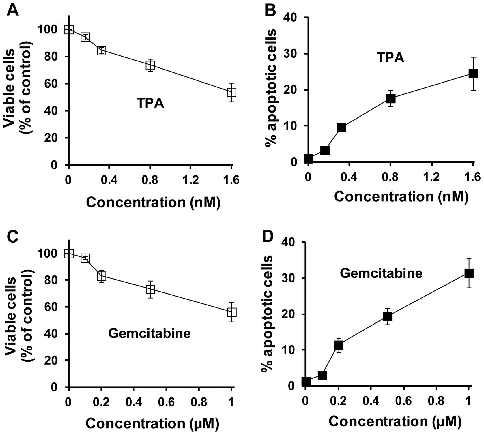
The in vitro effects of TPA and gemcitabine
alone or in combination on the growth and apoptosis of pancreatic
cancer cells were determined using the Panc-1 human pancreatic
cancer cell line. In our experiments, Panc-1 cells were treated
with TPA or gemcitabine alone or in combination for 96 h. As shown
in Fig. 1, the treatment of Panc-1
cells with TPA (0.16–1.6 nM) or gemcitabine (0.1–1.0 μM)
alone resulted in a concentration-dependent decrease in the number
of viable cells. Combinations of TPA and gemcitabine more potently
inhibited the growth of Panc-1 cells than either agent alone
(Table I). The CI for
IC50 was calculated as 0.83, indicating the synergistic
effect of TPA and gemcitabine in combination in the inhibition of
the growth of cultured Panc-1 cells. Treatment with TPA (0.16–1.6
nM) or gemcitabine (0.1–1.0 μM) resulted in 3–25 and 3–32%
apoptosis, respectively (Fig. 1).
Various combinations of TPA and gemcitabine at different
concentrations all had stronger effects on the stimulation of
apoptosis than either agent alone (Table I). The CI for 50% apoptosis was
calculated as 0.69. Our results indicated that a combination of TPA
and gemcitabine had synergistic effects on growth inhibition and
apoptosis stimulation in Panc-1 cells.
 | Table IEffects of TPA and/or gemcitabine on
the growth and apoptosis of Panc-1 cells. |
Table I
Effects of TPA and/or gemcitabine on
the growth and apoptosis of Panc-1 cells.
| Treatment | Viable cells (% of
control) | Apoptosis (% of
cells) |
|---|
| Control | 100 | 1.1±0.1 |
| TPA (0.16 nM) | 94.5±1.9 | 3.3±0.5 |
| Gemcitabine (0.1
μM) | 96.7±1.2 | 3.0±1.1 |
| TPA (0.16 nM) +
gemcitabine (0.1 μM) | 77.5±3.7 | 12.8±1.7 |
| TPA (0.80 nM) | 73.7±4.4 | 17.6±2.3 |
| Gemcitabine (0.5
μM) | 73.3±6.5 | 19.4±2.3 |
| TPA (0.80 nM) +
gemcitabine (0.5 μM) | 31.6±3.2 | 50.7±3.3 |
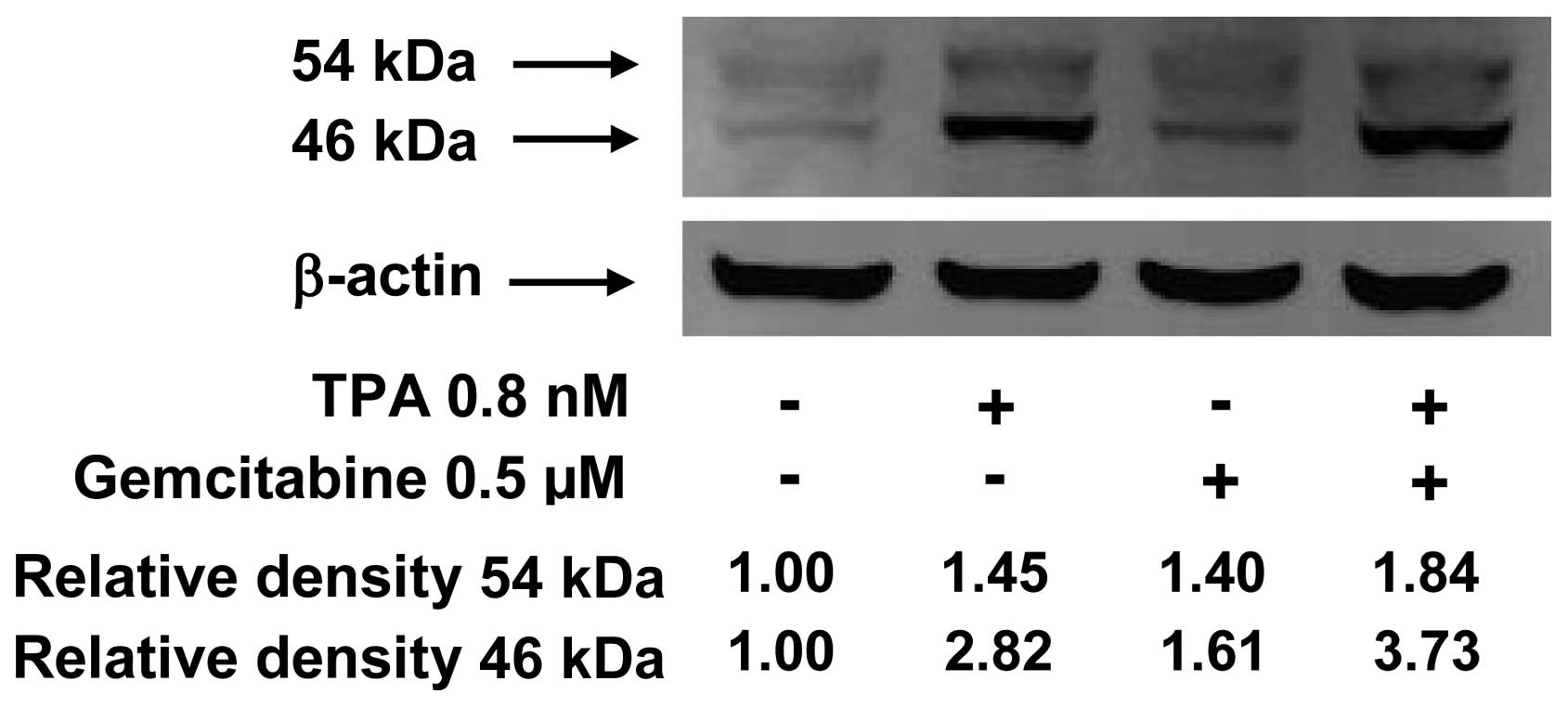
Activation of JNK in Panc-1 cells treated
with TPA or gemcitabine alone or in combination
The effect of TPA and gemcitabine on activation of
JNK was determined by western blot analysis using an
anti-phosphorylated JNK antibody that detects active,
phosphorylated JNK (25). Panc-1
cells were treated with TPA (0.8 nM) or gemcitabine (0.5 μM)
alone or in combination for 2 h and analyzed by western blot
analysis. The treatment of Panc-1 cells with TPA (0.8 nM) resulted
in a significant increase in the level of phosphorylated JNK, while
gemcitabine alone at 0.5 μM caused a slight increase in the
level of phosphorylated JNK (Fig.
2). Treatment of the cells with a combination of TPA (0.8 nM)
and gemcitabine (0.5 μM) caused a further increase in the
level of phosphorylated JNK when compared to either agent alone
(Fig. 2).
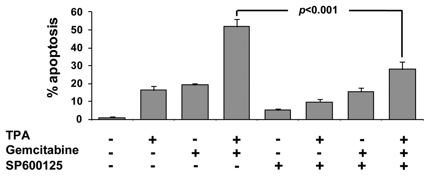
Effects of a JNK inhibitor on apoptosis
in Panc-1 cells induced by TPA or gemcitabine alone or in
combination
To further investigate whether the activation of JNK
is required for apoptosis in Panc-1 cells treated with TPA or
gemcitabine alone or in combination, we determined the effect of a
selective JNK inhibitor (SP600125, Sigma) on apoptosis in these
cells. In these experiments, Panc-1 cells were seeded at a density
of 0.2×10⁵ cells/ml of culture medium. The cells were
treated with TPA (0.8 nM) or gemcitabine (0.5 μM) alone or
in combination in the presence or absence of SP600125 (10
μM) for 96 h. Apoptotic cells were determined by
morphological assessment. As shown in Fig. 3, the addition of SP600125 alone had
a modest effect on apoptosis in Panc-1 cells. Treatment with
SP600125 decreased apoptosis in the Panc-1 cells induced by TPA
alone or in combination with gemcitabine (Fig. 3). Our results suggest that
apoptosis in Panc-1 cells treated with TPA in combination with
gemcitabine is at least partially mediated by the activation of
JNK. Since SP600125 only partially abrogated the effect of TPA and
gemcitabine on apoptosis, other factors may also be involved.
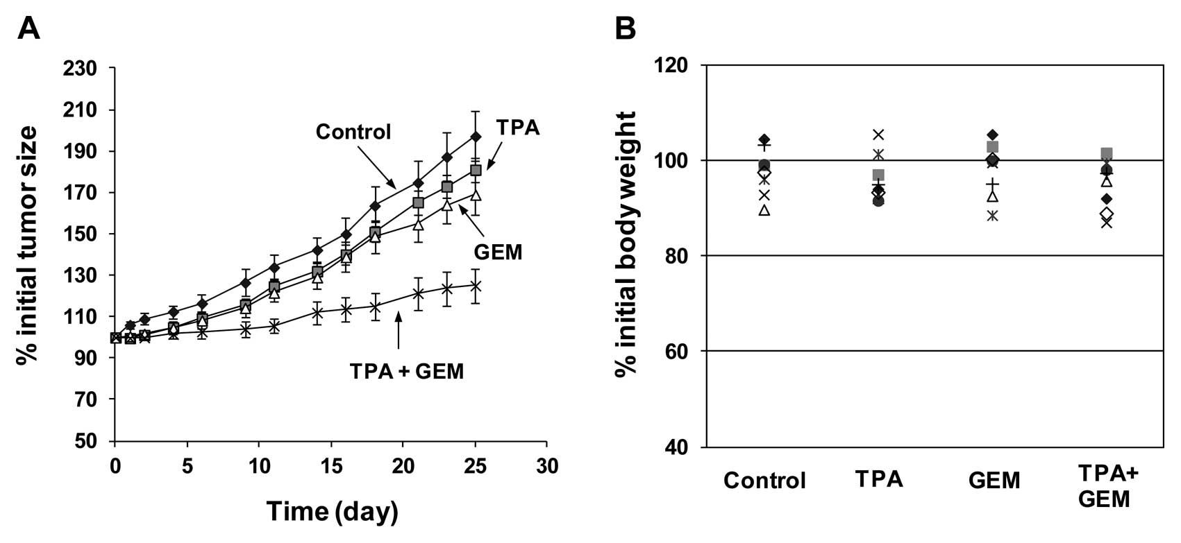
Inhibitory effect of TPA or gemcitabine
alone or in combination on the growth of Panc-1 tumor xenografts in
NCr nude mice
Male NCr nude mice with subcutaneous Panc-1 tumors
were treated with i.p. injections of TPA or gemcitabine alone or in
combination once a day as described in Fig. 4. As shown in Fig. 4A, tumor growth was observed in the
vehicle-treated control group. Treatment with i.p. injections of
low concentrations of TPA or gemcitabine had a modest inhibitory
effect on the growth of the tumors (Fig. 4A). The combination of low
concentrations of TPA and gemcitabine more potently inhibited the
growth of Panc-1 tumors than either agent used individually
(Fig. 4A). Statistical analysis
using ANOVA with the Tukey-Kramer multiple comparison test showed
that the difference in the percentage of initial tumor size at the
end of the experiment between the control group and the combination
group was statistically significant (p<0.001), and that the
differences between the TPA-treated group and the combination group
(p<0.01) or between the gemcitabine-treated group and the
combination group were also statistically significant (p<0.05).
As shown in Fig. 4B, treatment
with TPA or gemcitabine alone or in combination did not affect the
body weight of the animals. Statistical analysis using ANOVA with
the Tukey-Kramer multiple comparison test showed that the
difference in the percentage of initial body weight between any two
groups was not statistically significant (p>0.05).
Discussion
In the present study, we demonstrate that TPA in
combination with gemcitabine synergistically inhibits the growth
and stimulates apoptosis in cultured Panc-1 human pancreatic cancer
cells. We also show that a combination of low concentrations of TPA
and gemcitabine more potently inhibits the growth of Panc-1 tumors
than either agent used individually. To the best of our knowledge,
this is the first study indicating the synergistic effect of TPA
and gemcitabine on growth and apoptosis in human pancreatic cancer
cells.
TPA is an irritant and inflammatory agent that has
been used in previous studies as a tumor promoter (usual dose =
5–16 nmol twice a week) on the skin of mice previously initiated
with 7,12-dimethylbenz[a]anthracene or other polycyclic aromatic
hydrocarbons (26,27). In contrast to the irritant effects
of TPA at high concentrations on mouse skin, at a 10,000-fold lower
concentration TPA is a potent stimulator of differentiation of
cultured myeloid leukemia cells (28–30).
Previous studies from our laboratory have demonstrated that TPA at
clinically achievable concentrations inhibits the growth of
cultured human prostate and pancreatic cancer cells (16,21).
The peak blood concentrations of TPA ± SD in several patients who
received an intravenous infusion of TPA (0.125 mg/m2)
was 1.75 ± 0.55 ng/ml and ranged between 0.3 and 5.2 ng/ml
(13,31). The concentrations of TPA used to
obtain a synergistic effect in the present study (0.1–1 ng/ml;
0.16–1.6 nM) are clinically achievable (13,31).
Gemcitabine is the standard first-line
chemotherapeutic agent for the treatment of advanced pancreatic
cancer. The majority of pancreatic cancer patients treated with
gemcitabine do not have an objective response to treatment and only
a minority obtain stabilization of disease or a partial response
(9,32). Due to the inadequate benefit and
significant toxic side-effects of gemcitabine as a single-agent
(9,32), efforts have been devoted to
developing effective combinations of gemcitabine with other
anticancer agents (33). However,
the combination of other chemotherapeutic agents with gemcitabine
has not resulted in a meaningful improvement in survival (34–38).
Combinations of gemcitabine and novel anticancer agents that target
critical survival pathways in pancreatic cancer cells may have
synergistic effects on growth inhibition and apoptosis. Our
previous study demonstrated that a combination of TPA and ATRA
(16) enhanced the anticancer
activity of TPA. The present study demonstrates that low
concentrations of gemcitabine in combination with TPA
synergistically inhibit the growth and increase apoptosis in Panc-1
pancreatic cancer cells. These results indicate that the anticancer
activity of gemcitabine may be increased while the toxic
side-effects of gemcitabine may be reduced when a low dose is used
in combination with TPA.
The mechanisms for the synergistic effect of TPA and
gemcitabine on pancreatic cancer Panc-1 cells are not yet clear.
TPA is known to activate protein kinase C (PKC) leading to the
activation of JNK and apoptosis in several experimental systems
(25,39–42).
In the present study, we found that TPA strongly activated JNK in
Panc-1 cells, and that the addition of a selective inhibitor of JNK
partially abrogated the effect of TPA on growth inhibition and
apoptosis stimulation. These results suggest that JNK activation is
required for TPA-induced growth inhibition and apoptosis
stimulation in cultured Panc-1 cells. The main action of
gemcitabine is thought to be the competitive incorporation of
gemcitabine diphosphate and triphosphate into DNA, after which DNA
polymerase is able to add only one more nucleotide before DNA
fragmentation and cell death occur (43,44).
This so-called ‘masked chain termination’ prevents exonuclease
recognition and the excision of gemcitabine. Gemcitabine induces an
S-phase arrest (45,46) and stimulates apoptosis in cancer
cells (47,48). The activation of JNK is required
for gemcitabine-induced apoptosis in human lung cancer cells
(49). Gemcitabine alone or in
combination with other agents has been shown to induce the
activation of JNK in pancreatic cancer cells (50,51).
In the present study, we found that the treatment of Panc-1 cells
with a low dose of gemcitabine resulted in a small increase in the
level of phosphorylated-JNK. A combination of low doses of TPA and
gemcitabine further increased the level of phosphorylated JNK in
the cells. Moreover, we found that the addition of a selective JNK
inhibitor partially abrogated the stimulatory effect of TPA in
combination with gemcitabine on apoptosis in Panc-1 cells. These
results suggest that apoptosis in Panc-1 cells induced by TPA in
combination with gemcitabine is at least partially mediated by the
activation of JNK.
In conclusion, in the present study, we show that
TPA in combination with gemcitabine synergistically inhibits growth
and induces apoptosis in Panc-1 human pancreatic cancer cells. The
concentrations of TPA and gemcitabine required for these effects
are clinically achievable. In addition, we found that the treatment
of NCr nude mice with a combination of TPA and gemcitabine
inhibited the growth of Panc-1 tumor xenografts. Clinical studies
with TPA in combination with gemcitabine on patients with
pancreatic cancer are warranted in order to confirm our
results.
Acknowledgements
The present study was supported by
departmental funds from the Department of Chemical Biology in the
Ernest Mario School of Pharmacy at Rutgers University. The authors
thank Ms. Annette Dionisio for her excellent assistance in the
preparation of this manuscript.
References
|
1.
|
Zavoral M, Minarikova P, Zavada F, Salek C
and Minarik M: Molecular biology of pancreatic cancer. World J
Gastroenterol. 17:2897–2908. 2011. View Article : Google Scholar
|
|
2.
|
Siegel R, Ward E, Brawley O and Jemal A:
Cancer statistics, 2011: the impact of eliminating socioeconomic
and racial disparities on premature cancer deaths. CA Cancer J
Clin. 61:212–236. 2011. View Article : Google Scholar : PubMed/NCBI
|
|
3.
|
Li D, Xie K, Wolff R and Abbruzzese JL:
Pancreatic cancer. Lancet. 363:1049–1057. 2004. View Article : Google Scholar
|
|
4.
|
Sultana A, Tudur Smith C, Cunningham D,
Starling N, Neoptolemos JP and Ghaneh P: Meta-analyses of
chemotherapy for locally advanced and metastatic pancreatic cancer:
results of secondary end points analyses. Br J Cancer. 99:6–13.
2008. View Article : Google Scholar : PubMed/NCBI
|
|
5.
|
Zalatnai A: Pancreatic cancer - a
continuing challenge in oncology. Pathol Oncol Res. 9:252–263.
2003. View Article : Google Scholar : PubMed/NCBI
|
|
6.
|
Ahrendt SA and Pitt HA: Surgical
management of pancreatic cancer. Oncology. 16:725–734.
2002.PubMed/NCBI
|
|
7.
|
Wray CJ, Ahmad SA, Matthews JB and Lowy
AM: Surgery for pancreatic cancer: recent controversies and current
practice. Gastroenterol. 128:1626–1641. 2005. View Article : Google Scholar : PubMed/NCBI
|
|
8.
|
Hilbig A and Oettle H: Gemcitabine in the
treatment of metastatic pancreatic cancer. Expert Rev Anticancer
Ther. 8:511–523. 2008. View Article : Google Scholar : PubMed/NCBI
|
|
9.
|
Toschi L, Finocchiaro G, Bartolini S,
Gioia V and Cappuzzo F: Role of gemcitabine in cancer therapy.
Future Oncol. 1:7–17. 2005. View Article : Google Scholar
|
|
10.
|
Burris HA III, Moore MJ, Andersen J, Green
MR, Rothenberg ML, Modiano MR, Cripps MC, Portenoy RK, Storniolo
AM, Tarassoff P, Nelson R, Dorr FA, Stephens CD and von Hoff DD:
Improvements in survival and clinical benefit with as first-line
therapy for patients with advanced pancreas cancer: a randomized
trial. J Clin Oncol. 15:2403–2413. 1997.PubMed/NCBI
|
|
11.
|
Han ZT, Zhu XX, Yang RY, Sun JZ, Tian GF,
Liu XJ, Cao GS, Newmark HL, Conney AH and Chang RL: Effect of
intravenous infusions of 12-0-tetradecanoyl-phorbol-13-acetate
(TPA) in patients with myelocytic leukemia: Preliminary studies on
therapeutic efficacy and toxicity. Proc Natl Acad Sci USA.
95:5357–5361. 1998. View Article : Google Scholar
|
|
12.
|
Strair RK, Schaar D, Goodell L, Aisner J,
Chin KV, Eid J, Senzon R, Cui XX, Han ZT, Knox B, Rabson AB, Chang
R and Conney A: Administration of a phorbol ester to patients with
hematological malignancies: preliminary results from a phase I
clinical trial of 12-O-tetradecanoylphorbol-13-acetate. Clin Cancer
Res. 8:2512–2518. 2002.
|
|
13.
|
Schaar D, Goodell L, Aisner J, Cui XX, Han
ZT, Chang R, Martin J, Grospe S, Dudek L, Riley J, Manago J, Lin Y,
Rubin EH, Conney A and Strair RK: A phase I clinical trial of
12-O-tetradecanoylphorbol-13-acetate for patients with
relapsed/refractory malignancies. Cancer Chemother Pharmacol.
57:789–795. 2006. View Article : Google Scholar : PubMed/NCBI
|
|
14.
|
Zheng X, Chang RL, Cui XX, Kelly KA, Shih
WJ, Lin Y, Strair R, Suh J, Han ZT, Rabson A and Conney AH:
Synergistic effects of clinically achievable concentrations
of12-O-tetradecanoylphorbol-13-acetate in combination with
all-trans retinoic acid, 1alpha,25-dihydroxyvitamin D3, and sodium
butyrate on differentiation in HL-60 cells. Oncol Res. 12:419–427.
2000.
|
|
15.
|
Hansson A, Marín YE, Suh J, Rabson AB,
Chen S, Huberman E, Chang RL, Conney AH and Zheng X: Enhancement of
TPA-induced growth inhibition and apoptosis in myeloid leukemia
cells by BAY 11-7082, an NF-κB inhibitor. Int J Oncol. 27:941–948.
2005.PubMed/NCBI
|
|
16.
|
Avila GE, Zheng X, Cui XX, Ryan AD,
Hansson A, Suh J, Rabson AB, Chang RL, Shih WJ, Lin Y, Crowell P,
Lu YP, Lou YR and Conney AH: Inhibitory effects of
12-O-tetradecanoylphorbol-13-acetate alone or in combination with
all-trans retinoic acid on the growth of cultured human pancreas
cancer cells and pancreas tumor xenografts in immunodeficient mice.
J Pharmacol Exp Ther. 315:170–187. 2005. View Article : Google Scholar
|
|
17.
|
Bond JA, Gescher AJ, Verschoyle RD,
Lemoine NR, Errington R, Wiltshire M, Smith PJ and Wynford-Thomas
D: Cytotoxic action of phorbol esters on human pancreatic cancer
cells. Int J Cancer. 121:1445–1454. 2007. View Article : Google Scholar : PubMed/NCBI
|
|
18.
|
Salabat MR, Ding XZ, Flesche JB, Ujiki MB,
Robin TP, Talamonti MS, Bell RH Jr and Adrian TE: On the mechanisms
of 12-O-tetradecanoylphorbol-13-acetate-induced growth arrest in
pancreatic cancer cells. Pancreas. 33:148–155. 2006. View Article : Google Scholar : PubMed/NCBI
|
|
19.
|
Detjen KM, Brembeck FH, Welzel M, Kaiser
A, Haller H, Wiedenmann B and Rosewicz S: Activation of protein
kinase Calpha inhibits growth of pancreatic cancer cells via
p21(cip)-mediated G(1) arrest. J Cell Sci. 113:3025–3035.
2000.PubMed/NCBI
|
|
20.
|
Ploszaj T, Motyl T, Orzechowski A,
Zimowska W, Wareski P, Skierski J and Zwierzchowski L:
Antiapoptotic action of prolactin is associated with up-regulation
of Bcl-2 and down-regulation of Bax in HC11 mouse mammary
epithelial cells. Apoptosis. 3:295–304. 1998. View Article : Google Scholar : PubMed/NCBI
|
|
21.
|
Zheng X, Chang RL, Cui XX, Avila GE, Lee
S, Lu YP, Lou YR, Shih WJ, Lin Y, Reuhl K, Newmark H, Rabson A and
Conney AH: Inhibitory effect of12-O-tetradecanoylphorbol-13-acetate
alone or in combination with all-trans-retinoic acid on the growth
of LNCaP prostate tumors in immunodeficient mice. Cancer Res.
64:1811–1820. 2004. View Article : Google Scholar : PubMed/NCBI
|
|
22.
|
Zheng X, Cui XX, Huang MT, Liu Y, Shih WJ,
Lin Y, Lu YP, Wagner GC and Conney AH: Inhibitory effect of
voluntary running wheel exercise on the growth of human pancreatic
Panc-1 and prostate PC-3 xenograft tumors in immunodeficient mice.
Oncol Rep. 19:1583–1588. 2008.PubMed/NCBI
|
|
23.
|
Zhao L, Wientjes MG and Au JL: Evaluation
of combination chemotherapy: integration of nonlinear regression,
curve shift, isobologram, and combination index analyses. Clin
Cancer Res. 10:7994–8004. 2004. View Article : Google Scholar : PubMed/NCBI
|
|
24.
|
Hsu JC: Comparisons: Theory and Methods.
Chapman and Hall; New York, NY: 1996, View Article : Google Scholar
|
|
25.
|
Zheng X, Chang RL, Cui XX, Avila GE,
Hebbar V, Garzotto M, Shih WJ, Lin Y, Lu SE, Rabson AB, Kong AN and
Conney AH: Effects of 12-O-tetradecanoylphorbol-13-acetate (TPA) in
combination with paclitaxel (Taxol) on prostate cancer LNCaP cells
cultured in vitro or grown as xenograft tumors in immunodeficient
mice. Clin Cancer Res. 12:3444–3451. 2006. View Article : Google Scholar : PubMed/NCBI
|
|
26.
|
Van Duuren BL: Tumor-promoting agents in
two-stage carcinogenesis. Prog Exp Tumor Res. 11:31–68.
1969.PubMed/NCBI
|
|
27.
|
Hecker E: Structure-activity relationships
in diterpene esters irritant and cocarcinogenic to mouse skin.
Mechanisms of Tumor Promotion and Cocarcinogenesis. Slaga TJ, Sivak
AJ and Boutwell RK: Raven; New York, NY: pp. 11–49. 1978
|
|
28.
|
Huberman E and Callaham MF: Induction of
terminal differentiation in human promyelocytic leukemia cells by
tumor-promoting agents. Proc Natl Acad Sci USA. 76:1293–1297. 1979.
View Article : Google Scholar : PubMed/NCBI
|
|
29.
|
Lotem J and Sachs L: Regulation of normal
differentiation in mouse and human myeloid leukemic cells by
phorbol esters and the mechanism of tumor promotion. Proc Natl Acad
Sci USA. 76:5158–5162. 1979. View Article : Google Scholar : PubMed/NCBI
|
|
30.
|
Rovera G, O’Brien TG and Diamond L:
Induction of differentiation in human promyelocytic leukemia cells
by tumor promoters. Science. 204:868–870. 1979. View Article : Google Scholar : PubMed/NCBI
|
|
31.
|
Cui XX, Chang RL, Zheng X, Woodward D,
Strair R and Conney AH: A sensitive bioassay for measuring blood
levels of 12-O-tetradecanoylphorbol-13-acetate (TPA) in patients:
preliminary pharmacokinetic studies. Oncol Res. 13:169–174.
2002.PubMed/NCBI
|
|
32.
|
Voutsadakis IA: Molecular predictors of
gemcitabine response in pancreatic cancer. World J Gastrointest
Oncol. 3:153–164. 2011. View Article : Google Scholar : PubMed/NCBI
|
|
33.
|
Greenhalf W and Thomas A: Combination
therapy for the treatment of pancreatic cancer. Anticancer Agents
Med Chem. 11:418–426. 2011. View Article : Google Scholar : PubMed/NCBI
|
|
34.
|
Berlin JD, Catalano P, Thomas JP, Kugler
JW, Haller DG and Benson AB III: Phase III study of gemcitabine in
combination with fluorouracil versus gemcitabine alone in patients
with advanced pancreatic carcinoma: Eastern Cooperative Oncology
Group trial E2297. J Clin Oncol. 20:3270–3275. 2002. View Article : Google Scholar
|
|
35.
|
Rocha Lima CM, Green MR, Rotche R, Miller
WH Jr, Jeffrey GM, Cisar LA, Morganti A, Orlando N, Gruia G and
Miller LL: Irinotecan plus gemcitabine results in no survival
advantage compared with gemcitabine monotherapy in patients with
locally advanced or metastatic pancreatic cancer despite increased
tumor response rate. J Clin Oncol. 22:3776–3783. 2004.
|
|
36.
|
Louvet C, Labianca R, Hammel P, Lledo G,
Zampino MG, André T, Zaniboni A, Ducreux M, Aitini E, Taïeb J,
Faroux R, Lepere C and de Gramont A; GERCOR; GISCAD: Gemcitabine in
combination with oxaliplatin compared with gemcitabine alone in
locally advanced or metastatic pancreatic cancer: results of a
GERCOR and GISCAD phase III trial. J Clin Oncol. 23:3509–3516.
2005. View Article : Google Scholar : PubMed/NCBI
|
|
37.
|
Oettle H, Richards D, Ramanathan RK, van
Laethem JL, Peeters M, Fuchs M, Zimmermann A, John W, von Hoff D,
Arning M and Kindler HL: A phase III trial of pemetrexed plus
gemcitabine versus gemcitabine in patients with unresectable or
metastatic pancreatic cancer. Ann Oncol. 16:1639–1645. 2005.
View Article : Google Scholar : PubMed/NCBI
|
|
38.
|
Abou-Alfa GK, Letourneau R, Harker G,
Modiano M, Hurwitz H, Tchekmedyian NS, Feit K, Ackerman J, de Jager
RL, Eckhardt SG and O’Reilly EM: Randomized phase III study of
exatecan and gemcitabine compared with gemcitabine alone in
untreated advanced pancreatic cancer. J Clin Oncol. 24:4441–4447.
2006. View Article : Google Scholar : PubMed/NCBI
|
|
39.
|
Kim YR, Byun HS, Jeon J, Choi BL, Park KA,
Won M, Zhang T, Shin S, Lee H, Oh J and Hur GM: Apoptosis
signal-regulating kinase1 is inducible by protein kinase C δ and
contributes to phorbol ester-mediated G1 phase arrest through
persistent JNK activation. Cell Biochem Biophys. 61:199–207.
2011.PubMed/NCBI
|
|
40.
|
Engedal N, Korkmaz CG and Saatcioglu F:
C-Jun N-terminal kinase is required for phorbol ester- and
thapsigargin-induced apoptosis in the androgen responsive prostate
cancer cell line LNCaP. Oncogene. 21:1017–1027. 2002. View Article : Google Scholar : PubMed/NCBI
|
|
41.
|
Ikezoe T, Yang Y, Taguchi H and Koeffler
HP: JNK interacting protein 1 (JIP-1) protects LNCaP prostate
cancer cells from growth arrest and apoptosis mediated by
12-0-tetradecanoylphorbol-13-acetate (TPA). Br J Cancer.
90:2017–2024. 2004. View Article : Google Scholar : PubMed/NCBI
|
|
42.
|
Tahara E, Kadara H, Lacroix L, Lotan D and
Lotan R: Activation of protein kinase C by phorbol 12-myristate
13-acetate suppresses the growth of lung cancer cells through KLF6
induction. Cancer Biol Ther. 8:801–807. 2009. View Article : Google Scholar : PubMed/NCBI
|
|
43.
|
Huang P, Chubb S, Hertel LW, Grindey GB
and Plunkett W: Action of 2′,2′-difluorodeoxycytidine on DNA
synthesis. Cancer Res. 51:6110–6117. 1991.
|
|
44.
|
Ruiz van Haperen VW, Veerman G, Vermorken
JB and Peters GJ: 2′,2′-Difluoro-deoxycytidine (gemcitabine)
incorporation into RNA and DNA of tumour cell lines. Biochem
Pharmacol. 46:762–766. 1993.
|
|
45.
|
Tolis C, Peters GJ, Ferreira CG, Pinedo HM
and Giaccone G: Cell cycle disturbances and apoptosis induced by
topotecan and gemcitabine on human lung cancer cell lines. Eur J
Cancer. 35:796–807. 1999. View Article : Google Scholar : PubMed/NCBI
|
|
46.
|
Pauwels B, Korst AE, Pattyn GG, Lambrechts
HA, van Bockstaele DR, Vermeulen K, Lenjou M, de Pooter CM,
Vermorken JB and Lardon F: Cell cycle effect of gemcitabine and its
role in the radiosensitizing mechanism in vitro. Int J Radiat Oncol
Biol Phys. 57:1075–1083. 2003. View Article : Google Scholar : PubMed/NCBI
|
|
47.
|
Bouffard DY and Momparler RL: Comparison
of the induction of apoptosis in human leukemic cell lines by
difluorodeoxycytidine (gemcitabine) and cytosine arabinoside. Leuk
Res. 19:849–856. 1995. View Article : Google Scholar : PubMed/NCBI
|
|
48.
|
Ferreira CG, Tolis C, Span SW, Peters GJ,
van Lopik T, Kummer AJ, Pinedo HM and Giaccone G: Drug-induced
apoptosis in lung cancer cells is not mediated by the Fas/FasL
(CD95/APO1) signaling pathway. Clin Cancer Res. 6:203–212.
2000.PubMed/NCBI
|
|
49.
|
Teraishi F, Zhang L, Guo W, Dong F, Davis
JJ, Lin A and Fang B: Activation of c-Jun NH2-terminal kinase is
required for gemcitabine’s cytotoxic effect in human lung cancer
H1299 cells. FEBS Lett. 579:6681–6687. 2005.PubMed/NCBI
|
|
50.
|
Osada S, Tomita H, Tanaka Y, Tokuyama Y,
Tanaka H, Sakashita F and Takahashi T: The utility of vitamin K3
(menadione) against pancreatic cancer. Anticancer Res. 28:45–50.
2008.PubMed/NCBI
|
|
51.
|
Kreutzer JN, Ruzzene M and Guerra B:
Enhancing chemosensitivity to gemcitabine via RNA interference
targeting the catalytic subunits of protein kinase CK2 in human
pancreatic cancer cells. BMC Cancer. 10:4402010. View Article : Google Scholar
|