Spandidos Publications Logo
  • About
    • About Spandidos
    • Aims and Scopes
    • Abstracting and Indexing
    • Editorial Policies
    • Reprints and Permissions
    • Job Opportunities
    • Terms and Conditions
    • Contact
  • Journals
    • All Journals
    • Oncology Letters
      • Oncology Letters
      • Information for Authors
      • Editorial Policies
      • Editorial Board
      • Aims and Scope
      • Abstracting and Indexing
      • Bibliographic Information
      • Archive
    • International Journal of Oncology
      • International Journal of Oncology
      • Information for Authors
      • Editorial Policies
      • Editorial Board
      • Aims and Scope
      • Abstracting and Indexing
      • Bibliographic Information
      • Archive
    • Molecular and Clinical Oncology
      • Molecular and Clinical Oncology
      • Information for Authors
      • Editorial Policies
      • Editorial Board
      • Aims and Scope
      • Abstracting and Indexing
      • Bibliographic Information
      • Archive
    • Experimental and Therapeutic Medicine
      • Experimental and Therapeutic Medicine
      • Information for Authors
      • Editorial Policies
      • Editorial Board
      • Aims and Scope
      • Abstracting and Indexing
      • Bibliographic Information
      • Archive
    • International Journal of Molecular Medicine
      • International Journal of Molecular Medicine
      • Information for Authors
      • Editorial Policies
      • Editorial Board
      • Aims and Scope
      • Abstracting and Indexing
      • Bibliographic Information
      • Archive
    • Biomedical Reports
      • Biomedical Reports
      • Information for Authors
      • Editorial Policies
      • Editorial Board
      • Aims and Scope
      • Abstracting and Indexing
      • Bibliographic Information
      • Archive
    • Oncology Reports
      • Oncology Reports
      • Information for Authors
      • Editorial Policies
      • Editorial Board
      • Aims and Scope
      • Abstracting and Indexing
      • Bibliographic Information
      • Archive
    • Molecular Medicine Reports
      • Molecular Medicine Reports
      • Information for Authors
      • Editorial Policies
      • Editorial Board
      • Aims and Scope
      • Abstracting and Indexing
      • Bibliographic Information
      • Archive
    • World Academy of Sciences Journal
      • World Academy of Sciences Journal
      • Information for Authors
      • Editorial Policies
      • Editorial Board
      • Aims and Scope
      • Abstracting and Indexing
      • Bibliographic Information
      • Archive
    • International Journal of Functional Nutrition
      • International Journal of Functional Nutrition
      • Information for Authors
      • Editorial Policies
      • Editorial Board
      • Aims and Scope
      • Abstracting and Indexing
      • Bibliographic Information
      • Archive
    • International Journal of Epigenetics
      • International Journal of Epigenetics
      • Information for Authors
      • Editorial Policies
      • Editorial Board
      • Aims and Scope
      • Abstracting and Indexing
      • Bibliographic Information
      • Archive
    • Medicine International
      • Medicine International
      • Information for Authors
      • Editorial Policies
      • Editorial Board
      • Aims and Scope
      • Abstracting and Indexing
      • Bibliographic Information
      • Archive
  • Articles
  • Information
    • Information for Authors
    • Information for Reviewers
    • Information for Librarians
    • Information for Advertisers
    • Conferences
  • Language Editing
Spandidos Publications Logo
  • About
    • About Spandidos
    • Aims and Scopes
    • Abstracting and Indexing
    • Editorial Policies
    • Reprints and Permissions
    • Job Opportunities
    • Terms and Conditions
    • Contact
  • Journals
    • All Journals
    • Biomedical Reports
      • Information for Authors
      • Editorial Policies
      • Editorial Board
      • Aims and Scope
      • Abstracting and Indexing
      • Bibliographic Information
      • Archive
    • Experimental and Therapeutic Medicine
      • Information for Authors
      • Editorial Policies
      • Editorial Board
      • Aims and Scope
      • Abstracting and Indexing
      • Bibliographic Information
      • Archive
    • International Journal of Epigenetics
      • Information for Authors
      • Editorial Policies
      • Editorial Board
      • Aims and Scope
      • Abstracting and Indexing
      • Bibliographic Information
      • Archive
    • International Journal of Functional Nutrition
      • Information for Authors
      • Editorial Policies
      • Editorial Board
      • Aims and Scope
      • Abstracting and Indexing
      • Bibliographic Information
      • Archive
    • International Journal of Molecular Medicine
      • Information for Authors
      • Editorial Policies
      • Editorial Board
      • Aims and Scope
      • Abstracting and Indexing
      • Bibliographic Information
      • Archive
    • International Journal of Oncology
      • Information for Authors
      • Editorial Policies
      • Editorial Board
      • Aims and Scope
      • Abstracting and Indexing
      • Bibliographic Information
      • Archive
    • Medicine International
      • Information for Authors
      • Editorial Policies
      • Editorial Board
      • Aims and Scope
      • Abstracting and Indexing
      • Bibliographic Information
      • Archive
    • Molecular and Clinical Oncology
      • Information for Authors
      • Editorial Policies
      • Editorial Board
      • Aims and Scope
      • Abstracting and Indexing
      • Bibliographic Information
      • Archive
    • Molecular Medicine Reports
      • Information for Authors
      • Editorial Policies
      • Editorial Board
      • Aims and Scope
      • Abstracting and Indexing
      • Bibliographic Information
      • Archive
    • Oncology Letters
      • Information for Authors
      • Editorial Policies
      • Editorial Board
      • Aims and Scope
      • Abstracting and Indexing
      • Bibliographic Information
      • Archive
    • Oncology Reports
      • Information for Authors
      • Editorial Policies
      • Editorial Board
      • Aims and Scope
      • Abstracting and Indexing
      • Bibliographic Information
      • Archive
    • World Academy of Sciences Journal
      • Information for Authors
      • Editorial Policies
      • Editorial Board
      • Aims and Scope
      • Abstracting and Indexing
      • Bibliographic Information
      • Archive
  • Articles
  • Information
    • For Authors
    • For Reviewers
    • For Librarians
    • For Advertisers
    • Conferences
  • Language Editing
Login Register Submit
  • This site uses cookies
  • You can change your cookie settings at any time by following the instructions in our Cookie Policy. To find out more, you may read our Privacy Policy.

    I agree
Search articles by DOI, keyword, author or affiliation
Search
Advanced Search
presentation
International Journal of Oncology
Join Editorial Board Propose a Special Issue
Print ISSN: 1019-6439 Online ISSN: 1791-2423
Journal Cover
February-2015 Volume 46 Issue 2

Full Size Image

Cover Legend PDF

Sign up for eToc alerts
Recommend to Library

Journals

International Journal of Molecular Medicine

International Journal of Molecular Medicine

International Journal of Molecular Medicine is an international journal devoted to molecular mechanisms of human disease.

International Journal of Oncology

International Journal of Oncology

International Journal of Oncology is an international journal devoted to oncology research and cancer treatment.

Molecular Medicine Reports

Molecular Medicine Reports

Covers molecular medicine topics such as pharmacology, pathology, genetics, neuroscience, infectious diseases, molecular cardiology, and molecular surgery.

Oncology Reports

Oncology Reports

Oncology Reports is an international journal devoted to fundamental and applied research in Oncology.

Experimental and Therapeutic Medicine

Experimental and Therapeutic Medicine

Experimental and Therapeutic Medicine is an international journal devoted to laboratory and clinical medicine.

Oncology Letters

Oncology Letters

Oncology Letters is an international journal devoted to Experimental and Clinical Oncology.

Biomedical Reports

Biomedical Reports

Explores a wide range of biological and medical fields, including pharmacology, genetics, microbiology, neuroscience, and molecular cardiology.

Molecular and Clinical Oncology

Molecular and Clinical Oncology

International journal addressing all aspects of oncology research, from tumorigenesis and oncogenes to chemotherapy and metastasis.

World Academy of Sciences Journal

World Academy of Sciences Journal

Multidisciplinary open-access journal spanning biochemistry, genetics, neuroscience, environmental health, and synthetic biology.

International Journal of Functional Nutrition

International Journal of Functional Nutrition

Open-access journal combining biochemistry, pharmacology, immunology, and genetics to advance health through functional nutrition.

International Journal of Epigenetics

International Journal of Epigenetics

Publishes open-access research on using epigenetics to advance understanding and treatment of human disease.

Medicine International

Medicine International

An International Open Access Journal Devoted to General Medicine.

Journal Cover
February-2015 Volume 46 Issue 2

Full Size Image

Cover Legend PDF

Sign up for eToc alerts
Recommend to Library

  • Article
  • Citations
    • Cite This Article
    • Download Citation
    • Create Citation Alert
    • Remove Citation Alert
    • Cited By
  • Similar Articles
    • Related Articles (in Spandidos Publications)
    • Similar Articles (Google Scholar)
    • Similar Articles (PubMed)
  • Download PDF
  • Download XML
  • View XML
Review

The characteristic ultrasound features of specific types of ovarian pathology (Review)

  • Authors:
    • Ahmad Sayasneh
    • Christine Ekechi
    • Laura Ferrara
    • Jeroen Kaijser
    • Catriona Stalder
    • Shyamaly Sur
    • Dirk Timmerman
    • Tom Bourne
  • View Affiliations / Copyright

    Affiliations: Department of Cancer and Surgery, Imperial College London, Hammersmith Campus, London, UK, Early Pregnancy and Acute Gynecology Unit, Queen Charlottes and Chelsea Hospital, Imperial College London, London, W12 0HS, UK, Department of Development and Regeneration, KU Leuven, Leuven, Belgium
  • Pages: 445-458
    |
    Published online on: November 18, 2014
       https://doi.org/10.3892/ijo.2014.2764
  • Expand metrics +
Metrics: Total Views: 0 (Spandidos Publications: | PMC Statistics: )
Metrics: Total PDF Downloads: 0 (Spandidos Publications: | PMC Statistics: )
Cited By (CrossRef): 0 citations Loading Articles...

This article is mentioned in:


Abstract

Characterizing ovarian masses enables patients with malignancy to be appropriately triaged for treatment by subspecialist gynecological oncologists, which has been shown to optimize care and improve survival. Furthermore, correctly classifying benign masses facilitates the selection of patients with ovarian pathology that may either not require intervention, or be suitable for minimal access surgery if intervention is required. However, predicting whether a mass is benign or malignant is not the only clinically relevant information that we need to know before deciding on appropriate treatment. Knowing the specific histology of a mass is becoming of increasing importance as management options become more tailored to the individual patient. For example predicting a mucinous borderline tumor gives the opportunity for fertility sparing surgery, and will highlight the need for further gastrointestinal assessment. For benign disease, predicting the presence of an endometrioma and possible deeply infiltrating endometriosis is important when considering both who should perform and the extent of surgery. An examiner's subjective assessment of the morphological and vascular features of a mass using ultrasonography has been shown to be highly effective for predicting whether a mass is benign or malignant. Many masses also have features that enable a reliable diagnosis of the specific pathology of a particular mass to be made. In this narrative review we aim to describe the typical morphological features seen on ultrasound of different adnexal masses and illustrate these by showing representative ultrasound images.
View Figures

Figure 1

Figure 2

Figure 3

Figure 4

Figure 5

Figure 6

Figure 7

Figure 8

Figure 9

Figure 10

Figure 11

Figure 12

Figure 13

Figure 14

Figure 15

Figure 16

Figure 17

Figure 18

Figure 19

Figure 20

Figure 21

Figure 22

Figure 23

Figure 24

Figure 25

Figure 26

Figure 27

Figure 28

Figure 29

View References

1 

Carley ME, Klingele CJ, Gebhart JB, Webb MJ and Wilson TO: Laparoscopy versus laparotomy in the management of benign unilateral adnexal masses. J Am Assoc Gynecol Laparosc. 9:321–326. 2002. View Article : Google Scholar : PubMed/NCBI

2 

Jacobs I, Oram D, Fairbanks J, Turner J, Frost C and Grudzinskas JG: A risk of malignancy index incorporating CA 125, ultrasound and menopausal status for the accurate preoperative diagnosis of ovarian cancer. Br J Obstet Gynaecol. 97:922–929. 1990. View Article : Google Scholar : PubMed/NCBI

3 

Kaijser J, Sayasneh A, Van Hoorde K, et al: Presurgical diagnosis of adnexal tumours using mathematical models and scoring systems: a systematic review and meta-analysis. Hum Reprod Update. 20:449–462. 2014. View Article : Google Scholar

4 

Sayasneh A, Wynants L, Preisler J, et al: Multicentre external validation of IOTA prediction models and RMI by operators with varied training. Br J Cancer. 108:2448–2454. 2013. View Article : Google Scholar : PubMed/NCBI

5 

Timmerman D, Van Calster B, Testa AC, et al: Ovarian cancer prediction in adnexal masses using ultrasound-based logistic regression models: a temporal and external validation study by the IOTA group. Ultrasound Obstet Gynecol. 36:226–234. 2010. View Article : Google Scholar : PubMed/NCBI

6 

Timmerman D, Ameye L, Fischerova D, et al: Simple ultrasound rules to distinguish between benign and malignant adnexal masses before surgery: prospective validation by IOTA group. BMJ. 341:c68392010. View Article : Google Scholar : PubMed/NCBI
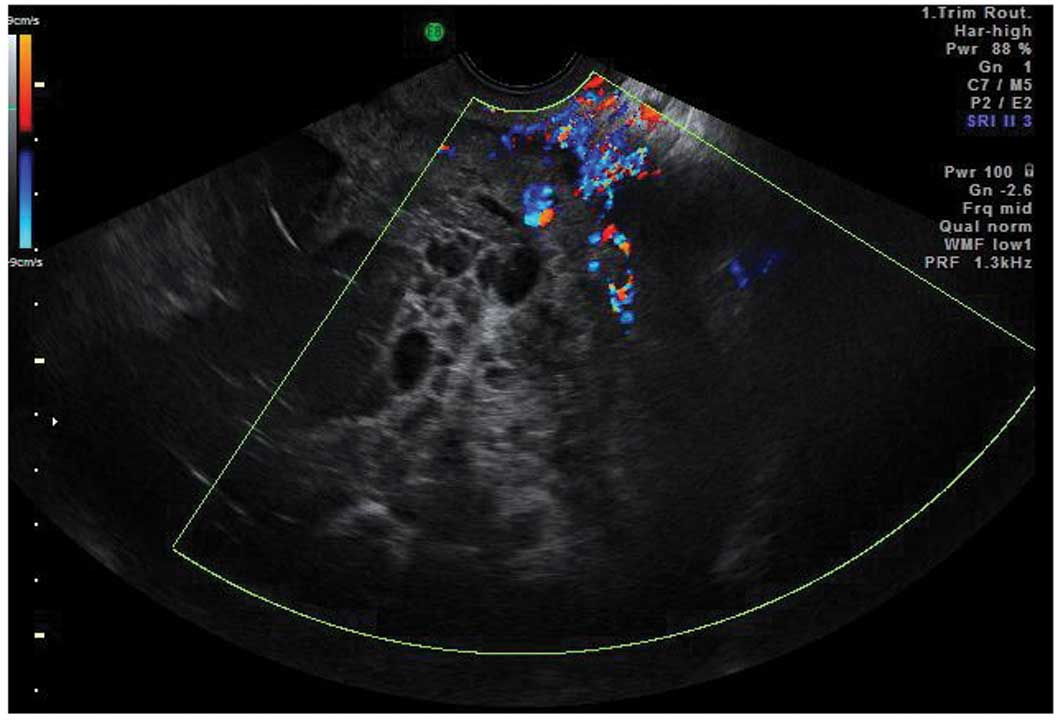
7 

Testa A, Kaijser J, Wynants L, et al: Strategies to diagnosie ovarian cancer: new evidence from phase 3 of the multicentre international IOTA study. Br J Cancer. 111:680–688. 2014. View Article : Google Scholar : PubMed/NCBI

8 

Valentin L, Ameye L, Savelli L, et al: Adnexal masses difficult to classify as benign or malignant using subjective assessment of gray-scale and Doppler ultrasound findings: logistic regression models do not help. Ultrasound Obstet Gynecol. 38:456–465. 2011. View Article : Google Scholar : PubMed/NCBI

9 

Timmerman D, Schwarzler P, Collins WP, et al: Subjective assessment of adnexal masses with the use of ultrasonography: an analysis of interobserver variability and experience. Ultrasound Obstet Gynecol. 13:11–16. 1999. View Article : Google Scholar : PubMed/NCBI

10 

Timmerman D: The use of mathematical models to evaluate pelvic masses; can they beat an expert operator? Best Pract Res Clin Obstet Gynaecol. 18:91–104. 2004. View Article : Google Scholar : PubMed/NCBI

11 

Valentin L, Hagen B, Tingulstad S and Eik-Nes S: Comparison of ‘pattern recognition’ and logistic regression models for discrimination between benign and malignant pelvic masses: a prospective cross validation. Ultrasound Obstet Gynecol. 18:357–365. 2001. View Article : Google Scholar

12 

Valentin L: Pattern recognition of pelvic masses by gray-scale ultrasound imaging: the contribution of Doppler ultrasound. Ultrasound Obstet Gynecol. 14:338–347. 1999. View Article : Google Scholar

13 

Sokalska A, Timmerman D, Testa AC, et al: Diagnostic accuracy of transvaginal ultrasound examination for assigning a specific diagnosis to adnexal masses. Ultrasound Obstet Gynecol. 34:462–470. 2009. View Article : Google Scholar : PubMed/NCBI

14 

Jeong YY, Outwater EK and Kang HK: Imaging evaluation of ovarian masses. Radiographics. 20:1445–1470. 2000. View Article : Google Scholar : PubMed/NCBI

15 

Valentin L: Use of morphology to characterize and manage common adnexal masses. Best Pract Res Clin Obstet Gynaecol. 18:71–89. 2004. View Article : Google Scholar : PubMed/NCBI

16 

Kurachi H, Murakami T, Nakamura H, et al: Imaging of peritoneal pseudocysts: value of MR imaging compared with sonography and CT. AJR Am J Roentgenol. 161:589–591. 1993. View Article : Google Scholar : PubMed/NCBI

17 

Jain KA: Imaging of peritoneal inclusion cysts. AJR Am J Roentgenol. 174:1559–1563. 2000. View Article : Google Scholar : PubMed/NCBI

18 

Savelli L, de Iaco P, Ghi T, Bovicelli L, Rosati F and Cacciatore B: Transvaginal sonographic appearance of peritoneal pseudocysts. Ultrasound Obstet Gynecol. 23:284–288. 2004. View Article : Google Scholar : PubMed/NCBI

19 

Dorum A, Blom GP, Ekerhovd E and Granberg S: Prevalence and histologic diagnosis of adnexal cysts in postmenopausal women: an autopsy study. Am J Obstet Gynecol. 192:48–54. 2005. View Article : Google Scholar : PubMed/NCBI

20 

Savelli L, Ghi T, De Iaco P, Ceccaroni M, Venturoli S and Cacciatore B: Paraovarian/paratubal cysts: comparison of transvaginal sonographic and pathological findings to establish diagnostic criteria. Ultrasound Obstetrics Gynecol. 28:330–334. 2006. View Article : Google Scholar

21 

Smorgick N, Herman A, Schneider D, Halperin R and Pansky M: Paraovarian cysts of neoplastic origin are underreported. JSLS. 13:22–26. 2009.PubMed/NCBI

22 

Timor-Tritsch IE, Lerner JP, Monteagudo A, Murphy KE and Heller DS: Transvaginal sonographic markers of tubal inflammatory disease. Ultrasound Obstet Gynecol. 12:56–66. 1998. View Article : Google Scholar : PubMed/NCBI

23 

Romosan G, Bjartling C, Skoog L and Valentin L: Ultrasound for diagnosing acute salpingitis: a prospective observational diagnostic study. Hum Reprod. 28:1569–1579. 2013. View Article : Google Scholar : PubMed/NCBI

24 

Guerriero S, Ajossa S, Lai MP, Mais V, Paoletti AM and Melis GB: Transvaginal ultrasonography associated 
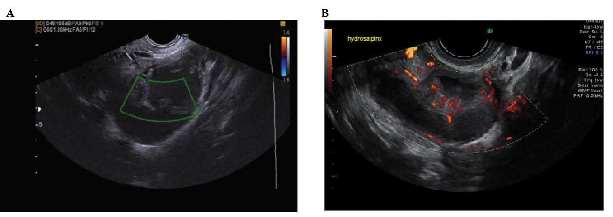
with colour Doppler energy in the diagnosis of hydrosalpinx. Hum Reprod. 15:1568–1572. 2000. View Article : Google Scholar : PubMed/NCBI

25 

Karlan BY, Bristow RE and Li AJ: Gynecologic Oncology: Clinical Practice & Surgical Atlas. McGraw-Hill Medical; New York, NY: 2012

26 

Caspi B, Hagay Z and Appelman Z: Variable echogenicity as a sonographic sign in the preoperative diagnosis of ovarian mucinous tumors. J Ultrasound Med. 25:1583–1585. 2006.PubMed/NCBI

27 

Alcazar JL, Errasti T, Minguez JA, Galan MJ, Garcia-Manero M and Ceamanos C: Sonographic features of ovarian cystadenofibromas: spectrum of findings. J Ultrasound Med. 20:915–919. 2001.PubMed/NCBI

28 

Ameye L, Timmerman D, Valentin L, et al: Clinically oriented three-step strategy for assessment of adnexal pathology. Ultrasound Obstet Gynecol. 40:582–591. 2012. View Article : Google Scholar : PubMed/NCBI

29 

Jermy K, Luise C and Bourne T: The characterization of common ovarian cysts in premenopausal women. Ultrasound Obstet Gynecol. 17:140–144. 2001. View Article : Google Scholar : PubMed/NCBI

30 

Cohen L and Sabbagha R: Echo patterns of benign cystic teratomas by transvaginal ultrasound. Ultrasound Obstet Gynecol. 3:120–123. 1993. View Article : Google Scholar : PubMed/NCBI

31 

Guerriero S, Ajossa S, Mais V, Risalvato A, Lai MP and Melis GB: The diagnosis of endometriomas using colour Doppler energy imaging. Hum Reprod. 13:1691–1695. 1998. View Article : Google Scholar : PubMed/NCBI

32 

Van Holsbeke C, Van Calster B, Guerriero S, et al: Endometriomas: their ultrasound characteristics. Ultrasound Obstet Gynecol. 35:730–740. 2010.PubMed/NCBI

33 

Asch E and Levine D: Variations in appearance of endometriomas. J Ultrasound Med. 26:993–1002. 2007.PubMed/NCBI

34 

Sayasneh A, Naji O, Abdallah Y, Stalder C and Bourne T: Changes seen in the ultrasound features of a presumed decidualised ovarian endometrioma mimicking malignancy. J Obstet Gynaecol. 32:807–811. 2012. View Article : Google Scholar : PubMed/NCBI

35 

Testa AC, Timmerman D, Van Holsbeke C, et al: Ovarian cancer arising in endometrioid cysts: ultrasound findings. Ultrasound Obstet Gynecol. 38:99–106. 2011. View Article : Google Scholar : PubMed/NCBI

36 

Yen P, Khong K, Lamba R, Corwin MT and Gerscovich EO: Ovarian fibromas and fibrothecomas: sonographic correlation with computed tomography and magnetic resonance imaging: a 5-year single-institution experience. J Ultrasound Med. 32:13–18. 2013.

37 

Paladini D, Testa A, Van Holsbeke C, Mancari R, Timmerman D and Valentin L: Imaging in gynecological disease (5): clinical and ultrasound characteristics in fibroma and fibrothecoma of the ovary. Ultrasound Obstet Gynecol. 34:188–195. 2009. View Article : Google Scholar : PubMed/NCBI

38 

Roth LM and Talerman A: The enigma of struma ovarii. Pathology. 39:139–146. 2007. View Article : Google Scholar : PubMed/NCBI

39 

Zalel Y, Seidman DS, Oren M, et al: Sonographic and clinical characteristics of struma ovarii. J Ultrasound Med. 19:857–861. 2000.PubMed/NCBI

40 

Savelli L, Testa AC, Timmerman D, Paladini D, Ljungberg O and Valentin L: Imaging of gynecological disease (4): clinical and ultrasound characteristics of struma ovarii. Ultrasound Obstet Gynecol. 32:210–219. 2008. View Article : Google Scholar : PubMed/NCBI

41 

Green GE, Mortele KJ, Glickman JN and Benson CB: Brenner tumors of the ovary: sonographic and computed tomographic imaging features. J Ultrasound Med. 25:1245–1254. 2006.PubMed/NCBI

42 

Sherer DM, Dalloul M, Salame G, et al: Color Doppler sonographic features of a Brenner tumor in pregnancy. J Ultrasound Med. 28:1405–1408. 2009.PubMed/NCBI

43 

Dierickx I, Valentin L, Van Holsbeke C, et al: Imaging in gynecological disease (7): clinical and ultrasound features of Brenner tumors of the ovary. Ultrasound Obstet Gynecol. 40:706–713. 2012. View Article : Google Scholar : PubMed/NCBI

44 

Valentin L, Ameye L, Testa A, et al: Ultrasound characteristics of different types of adnexal malignancies. Gynecol Oncol. 102:41–48. 2006. View Article : Google Scholar : PubMed/NCBI

45 

Exacoustos C, Romanini ME, Rinaldo D, et al: Preoperative sonographic features of borderline ovarian tumors. Ultrasound Obstet Gynecol. 25:50–59. 2005. View Article : Google Scholar

46 

Pascual MA, Tresserra F, Grases PJ, Labastida R and Dexeus S: Borderline cystic tumors of the ovary: gray-scale and color Doppler sonographic findings. J Clin Ultrasound. 30:76–82. 2002. View Article : Google Scholar : PubMed/NCBI

47 

Hassen K, Ghossain MA, Rousset P, et al: Characterization of papillary projections in benign versus borderline and malignant ovarian masses on conventional and color Doppler ultrasound. AJR Am J Roentgenol. 196:1444–1449. 2011. View Article : Google Scholar : PubMed/NCBI

48 

Fruscella E, Testa AC, Ferrandina G, et al: Ultrasound features of different histopathological subtypes of borderline ovarian tumors. Ultrasound Obstet Gynecol. 26:644–650. 2005. View Article : Google Scholar : PubMed/NCBI

49 

Darai E, Teboul J, Walker F, et al: Epithelial ovarian carcinoma of low malignant potential. Eur J Obstet Gynecol Reprod Biol. 66:141–145. 1996. View Article : Google Scholar : PubMed/NCBI

50 

Testa AC, Ferrandina G, Timmerman D, et al: Imaging in gynecological disease (1): ultrasound features of metastases in the ovaries differ depending on the origin of the primary tumor. Ultrasound Obstet Gynecol. 29:505–511. 2007. View Article : Google Scholar : PubMed/NCBI

51 

Testa AC, Mancari R, Di Legge A, et al: The ‘lead vessel’: a vascular ultrasound feature of metastasis in the ovaries. Ultrasound Obstet Gynecol. 31:218–221. 2008. View Article : Google Scholar : PubMed/NCBI

Related Articles

  • Abstract
  • View
  • Download
  • Twitter
Copy and paste a formatted citation
Spandidos Publications style
Sayasneh A, Ekechi C, Ferrara L, Kaijser J, Stalder C, Sur S, Timmerman D and Bourne T: The characteristic ultrasound features of specific types of ovarian pathology (Review). Int J Oncol 46: 445-458, 2015.
APA
Sayasneh, A., Ekechi, C., Ferrara, L., Kaijser, J., Stalder, C., Sur, S. ... Bourne, T. (2015). The characteristic ultrasound features of specific types of ovarian pathology (Review). International Journal of Oncology, 46, 445-458. https://doi.org/10.3892/ijo.2014.2764
MLA
Sayasneh, A., Ekechi, C., Ferrara, L., Kaijser, J., Stalder, C., Sur, S., Timmerman, D., Bourne, T."The characteristic ultrasound features of specific types of ovarian pathology (Review)". International Journal of Oncology 46.2 (2015): 445-458.
Chicago
Sayasneh, A., Ekechi, C., Ferrara, L., Kaijser, J., Stalder, C., Sur, S., Timmerman, D., Bourne, T."The characteristic ultrasound features of specific types of ovarian pathology (Review)". International Journal of Oncology 46, no. 2 (2015): 445-458. https://doi.org/10.3892/ijo.2014.2764
Copy and paste a formatted citation
x
Spandidos Publications style
Sayasneh A, Ekechi C, Ferrara L, Kaijser J, Stalder C, Sur S, Timmerman D and Bourne T: The characteristic ultrasound features of specific types of ovarian pathology (Review). Int J Oncol 46: 445-458, 2015.
APA
Sayasneh, A., Ekechi, C., Ferrara, L., Kaijser, J., Stalder, C., Sur, S. ... Bourne, T. (2015). The characteristic ultrasound features of specific types of ovarian pathology (Review). International Journal of Oncology, 46, 445-458. https://doi.org/10.3892/ijo.2014.2764
MLA
Sayasneh, A., Ekechi, C., Ferrara, L., Kaijser, J., Stalder, C., Sur, S., Timmerman, D., Bourne, T."The characteristic ultrasound features of specific types of ovarian pathology (Review)". International Journal of Oncology 46.2 (2015): 445-458.
Chicago
Sayasneh, A., Ekechi, C., Ferrara, L., Kaijser, J., Stalder, C., Sur, S., Timmerman, D., Bourne, T."The characteristic ultrasound features of specific types of ovarian pathology (Review)". International Journal of Oncology 46, no. 2 (2015): 445-458. https://doi.org/10.3892/ijo.2014.2764
Follow us
  • Twitter
  • LinkedIn
  • Facebook
About
  • Spandidos Publications
  • Careers
  • Cookie Policy
  • Privacy Policy
How can we help?
  • Help
  • Live Chat
  • Contact
  • Email to our Support Team