Introduction
Acute myeloid leukemia (AML) is the most common type
of leukemia in adults, and occurs in approximately one-third of
newly diagnosed patients, and remains one of the most difficult
hematological malignancies to treat (the 5-year overall survival
rate is 20–30% for adult primary AML) (1,2). AML
is characterized by the proliferation of clonal precursor myeloid
cells with arrested differentiation (3). In contrast to the poor prognosis of
most patients with AML, the use of differentiation therapy with
all-trans-retinoic acid (ATRA) for acute promyelocytic
leukemia (APL), a distinct type of AML, has revolutionized therapy
for this disease by converting it from fatal to curable (4). However, ATRA is not effective in
other AMLs. Furthermore, many APL patients treated with ATRA fail
to respond or invariably relapse. Therefore, alternative or
combination therapies are needed to improve the prognosis and
survival of patients.
Cotylenin A (CN-A), which is a fucicoccan-diterpene
glycoside with a complex sugar moiety, was originally isolated as a
plant growth regulator and has been shown to affect several
physiological processes in higher plants (5). We previously reported that CN-A
exhibited potent differentiation-inducing activity in several human
and murine myeloid leukemia cell lines and in leukemia cells that
were freshly isolated from patients with AML (6–9).
Furthermore, the administration of CN-A significantly prolonged the
survival of mice with severe combined immunodeficiency that had
been inoculated with the APL cells of the NB-4 cell line (10).
Previous studies reported that vitamin K2 (VK2)
effectively induced apoptosis in various types of primary cultured
leukemia cells and leukemia cell lines in vitro (11–13)
as well as in solid tumor cells (14–16).
On the other hand, in contrast to the induction of apoptosis in
leukemia cells, VK2 has been shown to exhibit
differentiation-inducing activity in AML cell lines, such as HL-60
and U937, in vitro (17–19).
Sada et al found that VK2 also had differentiation-promoting
effects on myeloid lineage progenitors (20).
Since c-MYC is aberrantly expressed in a wide
variety of human solid tumors (21) as well as in leukemia (22), it is an attractive target for
cancer therapy. The downregulation of c-MYC is known to play
a crucial role in ATRA-induced growth arrest and myeloid
differentiation of AML (23–27).
In addition, previous findings, including ours, indicated that the
expression of cyclin G2 was significantly upregulated during cell
cycle arrest responses to diverse growth-inhibitory signals and
strongly repressed by mitogens, suggesting the positive role of
cyclin G2 in the promotion or maintenance of cell cycle arrest
(28–30). In order to identify useful new
differentiation inducers and effective combination treatments for
various types of AML and APL, we searched for substances capable of
inducing cell differentiation and the expression of cyclin G2 as
well as strongly suppressing the expression of c-MYC in
HL-60 cells. In the present study, we demonstrated that the
combined treatment of VK2 and CN-A synergistically induced
monocytic differentiation in HL-60 cells and cooperatively
inhibited cell proliferation showing G1 arrest. Furthermore, we
showed that the combined treatment of VK2 and CN-A efficiently
suppressed the expression of c-MYC and cooperatively induced
the expression of cyclin G2.
Materials and methods
Reagents
VK2, nitroblue tetrazolium (NBT), all-trans
retinoic acid (ATRA), 1α,25-dihydroxyvitamin D3 (VD3), and
12-O-tetradecanoylphorbol-13-acetate (TPA) were purchased
from Sigma-Aldrich Inc. (St. Louis, MO, USA). CN-A was purified
from a stock ethyl acetate extract obtained from the culture
filtrate of Cladosporium fungus sp. 501-7 W by flash
chromatography on a silica gel with >99% purity (5).
Cells and cell culture
Human AML HL-60 cells were cultured in RPMI-1640
medium (Sigma-Aldrich Inc.) supplemented with 10% heat-inactivated
fetal bovine serum and 80 μg/ml gentamicin sulfate (MSD K.K, Tokyo,
Japan) at 37°C in a humidified atmosphere of 5% CO2 in
air.
Assay of cell growth
Cells were plated in multidishes (Falcon, Corning
Inc., Corning, NY, USA) at a density of 2.5×10⁴ cells/ml
and incubated with or without the test compounds. Cell numbers were
counted with a model Z1 Coulter Counter (Beckman Coulter Inc.,
Miami, FL, USA).
NBT reduction assay
The reduction of NBT was assayed colorimetrically as
previously described (31).
Briefly, cells were incubated in 1 ml of serum-free medium
containing 1 mg/ml NBT and 100 ng/ml TPA at 37°C for 60 min. The
reaction was stopped by adding HCl. Formazan solution at 560 nm was
measured in a spectrophotometer (DU730, Beckman Coulter Inc.).
Assessment of monocytic
differentiation
In order to assess monocytic differentiation,
non-specific esterase staining was performed using an Esterase
Staining kit (Muto Chemical Co., Tokyo, Japan).
Assessment of morphological
differentiation
Morphological changes were examined in cell smears
using light microscopy of cytospin preparations stained with
May-Grunwald-Giemsa solution (Merck, Darmstadt, Germany).
Cell cycle analysis
Cells were plated in 60-mm plastic dishes at a
density of 1×10⁵ cells/ml and incubated with VK2 in the
absence or presence of CN-A. After 96 h, the cells were washed with
phosphate-buffered saline (PBS) and fixed gently in 100% ethanol at
4°C for 30 min. Cells were suspended in propidium iodide (PI)-RNase
solution, which contained 50 μg/ml PI (MBL Co. Ltd., Nagoya, Japan)
and 0.1 mg/ml RNase (Sigma-Aldrich Inc.) in PBS for 30 min at room
temperature. The cell cycle analysis was performed by flow
cytometry (BD FACSCalibur, Becton Dickinson, East Rutherford, NJ,
USA).
RNA extraction and determination of mRNA
levels by reverse transcriptase (RT)-quantitative polymerase chain
reaction (qPCR)
RNA was extracted using an RNeasy Plus Mini kit
(Qiagen, Valencia, CA, USA) according to the manufacturer’s
instructions. Total RNA (1 μg) from leukemia cells was reverse
transcribed with the ReverTra Ace qPCR RT kit (Toyobo Co. Ltd.,
Osaka, Japan). qPCR using the SYBER Green method was carried out
with the Thunderbird SYBER qPCR Mix (Toyobo Co.) on a Thermal
Cycler Dice Real-time PCR instrument (Takara Bio, Shiga, Japan)
according to the manufacturer’s instructions. Real-time PCR results
were calculated according to the following protocol: Relative
expression level=2−ΔCt, where ΔCt=Ct (gene of interest)
- Ct (housekeeping gene). The c-MYC primers used for qPCR
were: forward, 5′-TTCGGGT AGTGGAAAACCAG-3′ and reverse,
5′-CAGCAGCTCGAA TTTCTTCC-3′. The GAPDH primers used for qPCR
were: forward, 5′-GACGCTGGGGCTGGCATTG-3′ and reverse,
5′-GCTGGTGGTCCAGGGGTC-3′ (32).
The cyclin G2 primers used for qPCR were: forward, 5′-ATCGTTTCAAG
GCGCACAG-3′ and reverse, 5′-CAACCCCCCTCAGGTA TCG-3′ (33). The P21/CIP1 primers used for qPCR
were: forward, 5′-CGATGCCAACCTCCTCAACGA-3′ and reverse,
5′-TCGCAGACCTCCAGCATCCA-3 (34).
Results
Effects of the combined treatment of
vitamin K2 (VK2) and cotylenin A (CN-A) on the cell proliferation
of HL-60 cells
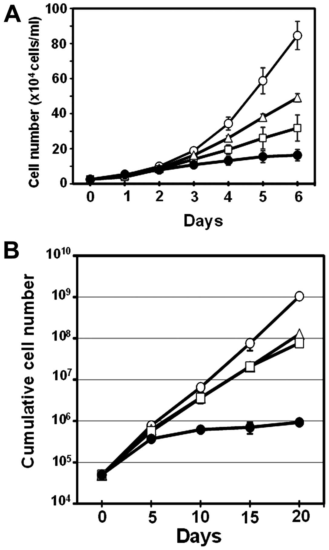
HL-60 cells (2.5×10⁴ cells/ml) were
cultured without or with VK2, CN-A, or VK2 plus CN-A for 6 days.
Fig. 1A shows the time course of
the combined effects of VK2 and CN-A on cell growth. The growth of
HL-60 cells was moderately inhibited by VK2 (5 μM) or CN-A (5
μg/ml) alone, but was still observed until at least 6 days;
however, no significant changes were observed in the cell number
after 4 days of the treatment with the combination of both VK2 and
CN-A (Fig. 1A). We also examined
the long-term effects of the combined treatment of VK2 and CN-A on
the proliferation of HL-60 cells. HL-60 cells (5×10⁴
cells/ml) were cultured without or with 10 μM VK2, 5 μg/ml CN-A, or
10 μM VK2 plus 5 μg/ml CN-A for 20 days (Fig. 1B). The culture medium was replaced
by fresh medium once every 5 days. Although the growth rate of VK2-
or CN-A-treated cells was significantly lower than that of control
cells under these culture conditions, the cell number markedly
increased (100-fold between days 5 and 20). On the other hand, cell
growth was greatly inhibited by the combined treatment of VK2 and
CN-A, and the cell number was almost the same as that at day 5
(Fig. 1B).
VK2 and CN-A synergistically induced
monocytic differentiation in HL-60 cells
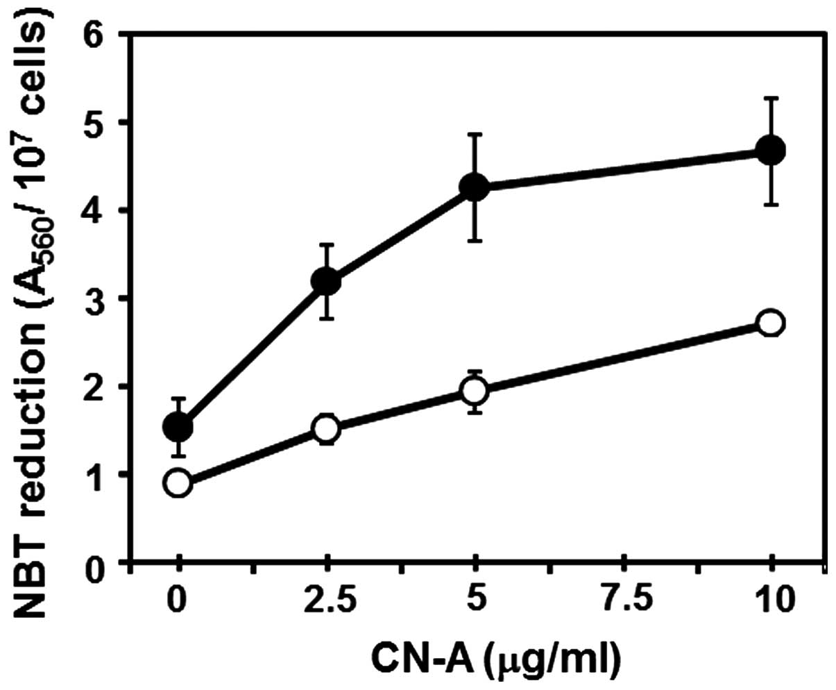
We examined the combined effects of VK2 and CN-A on
the induction of differentiation of HL-60 cells because VK2 or CN-A
alone are inducers of differentiation in HL-60 cells (6,7,17,18).
HL-60 cells (2.5×10⁴ cells/ml) were cultured with CN-A
in the presence or absence of VK2 for 6 days. CN-A and 10 μM VK2
synergistically induced the reduction of NBT (one of the typical
myelo/monocytic differentiation markers of human leukemia cells)
(Fig. 2). We then determined
whether the induction of differentiation induced with VK2 plus CN-A
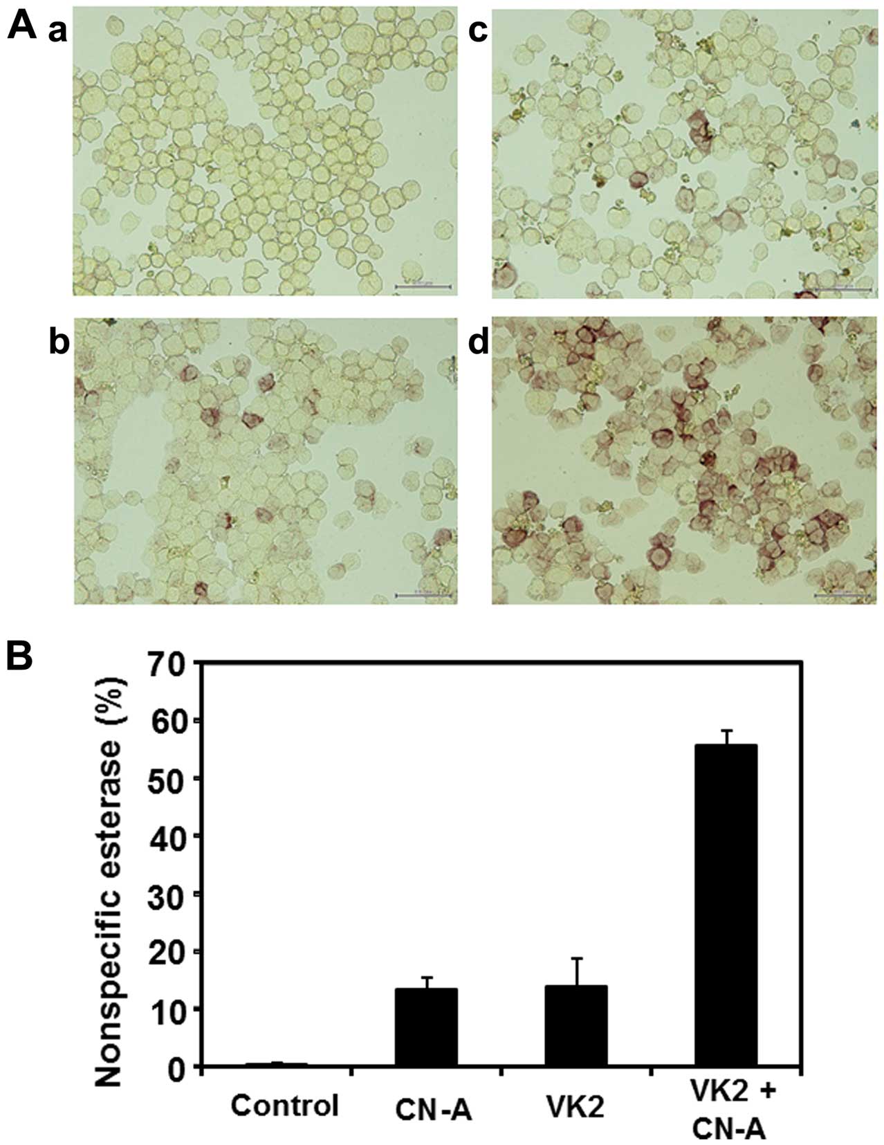
was a granulocytic or monocytic lineage. HL-60 cells
(2.5×10⁴ cells/ml) were cultured without or with 10
μg/ml CN-A, 10 μM VK2, or 10 μM VK2 plus 10 μg/ml CN-A for 5 days
(Fig. 3A). Nonspecific
esterase-positive cells were counted under a microscope (Fig. 3B). Cells treated with CN-A plus VK2
synergistically became positive for nonspecific esterase (Fig. 3A-d and B), whereas those treated
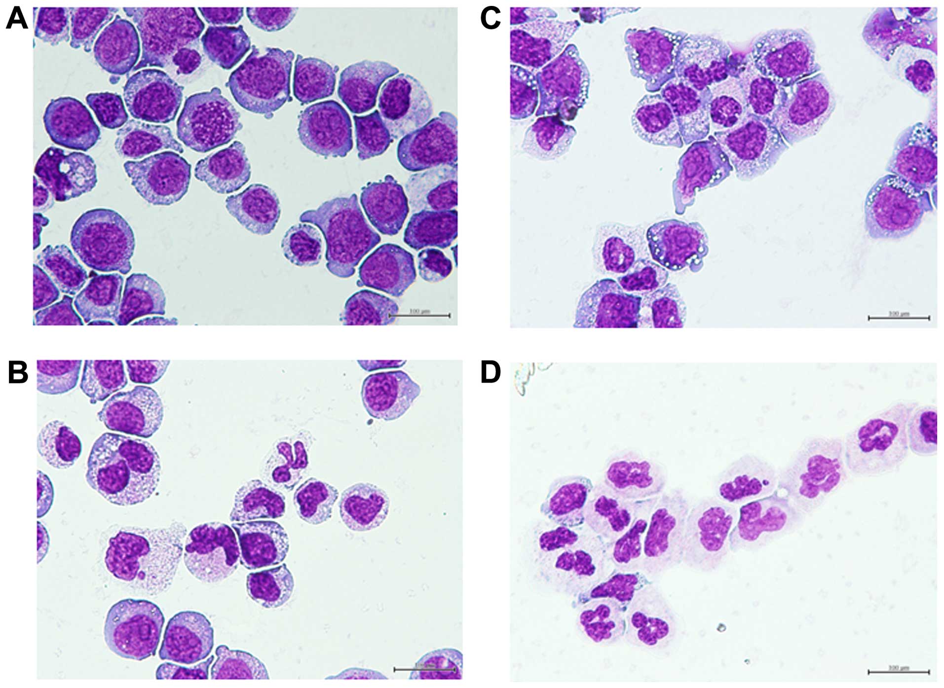
with CN-A or VK2 alone became weakly positive (Fig. 3A–b, A–c and B). The combined
treatment of VK2 and CN-A also induced the marked morphological
differentiation of HL-60 cells (Fig.
4D), whereas VK2 or CN-A alone induced the intermediate stage
of differentiation (Fig. 4B and
C). These results indicated that the treatment of HL-60 cells
with VK2 and CN-A effectively induced monocytic
differentiation.
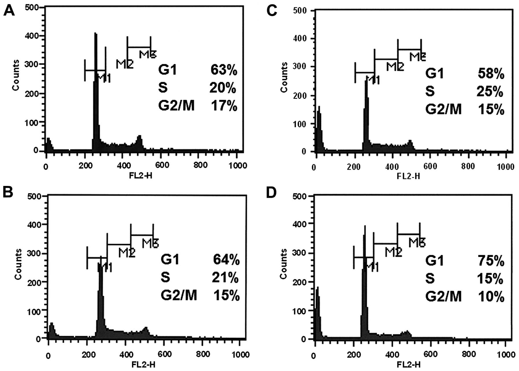
Induction of G1 arrest in HL-60 cells
with VK2 plus CN-A
In order to more clearly understand the combined
effects of VK2 and CN-A on cell growth, we exposed HL-60 cells
(1×10⁵ cells/ml) to 10 μM VK2 plus 10 μg/ml CN-A, and
then measured changes in cell cycle distribution after 4 days
(Fig. 5). Under these culture
conditions, VK2 or CN-A alone did not markedly affect the cell
cycle (Fig. 5B and C). On the
other hand, the percentage of cells in the G1 phase was
significantly increased from 63% to 75% (Fig. 5A and D). The percentages of cells
in the S phase and G2/M phase were inversely decreased in response
to the combined treatment of VK2 and CN-A (Fig. 5A and D).
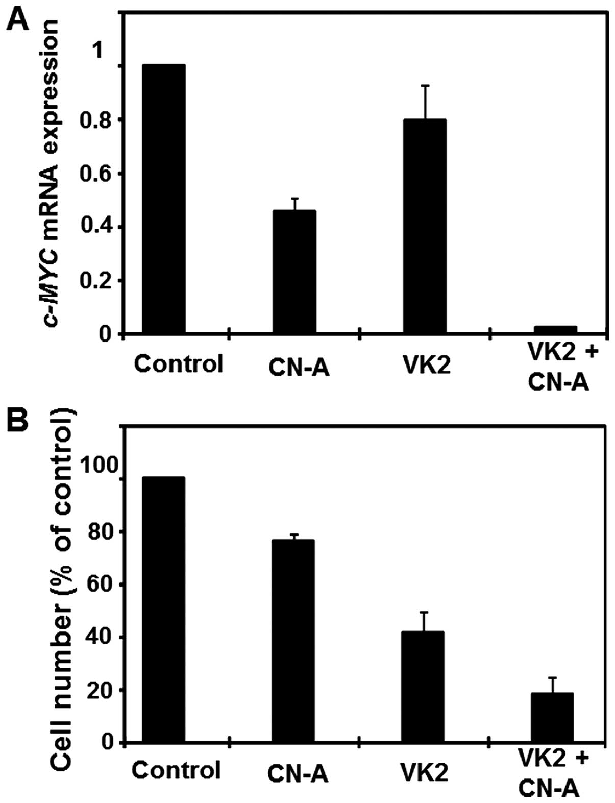
Combined treatment of VK2 and CN-A
synergistically inhibited c-MYC gene expression in HL-60 cells
Previous studies reported that the induction of
differentiation and growth arrest in HL-60 cells was associated
with the suppression of c-MYC gene expression (23–27);
therefore, we investigated whether the combined treatment of CN-A
and VK2 synergistically inhibited c-MYC gene expression in
HL-60 cells. HL-60 cells (2.5×10⁴ cells/ml) were
cultured without or with VK2 plus CN-A for 6 days. Although 5 μg/ml
CN-A or 5 μM VK2 alone inhibited c-MYC gene expression in
HL-60 cells to approximately 45 or 80% that of control cells,
respectively, the combined treatment almost completely suppressed
c-MYC gene expression (>95% inhibition) (Fig. 6A). This synergistic inhibition of
c-MYC gene expression in HL-60 cells was also observed when
HL-60 cells were treated with CN-A and VK2 for 4 days (data not
shown). As described above, the combined treatment of CN-A and VK2
more strongly inhibited cell growth than that of CN-A or VK2 alone
(Fig. 6B) and clearly induced
monocytic differentiation (Fig.
4).
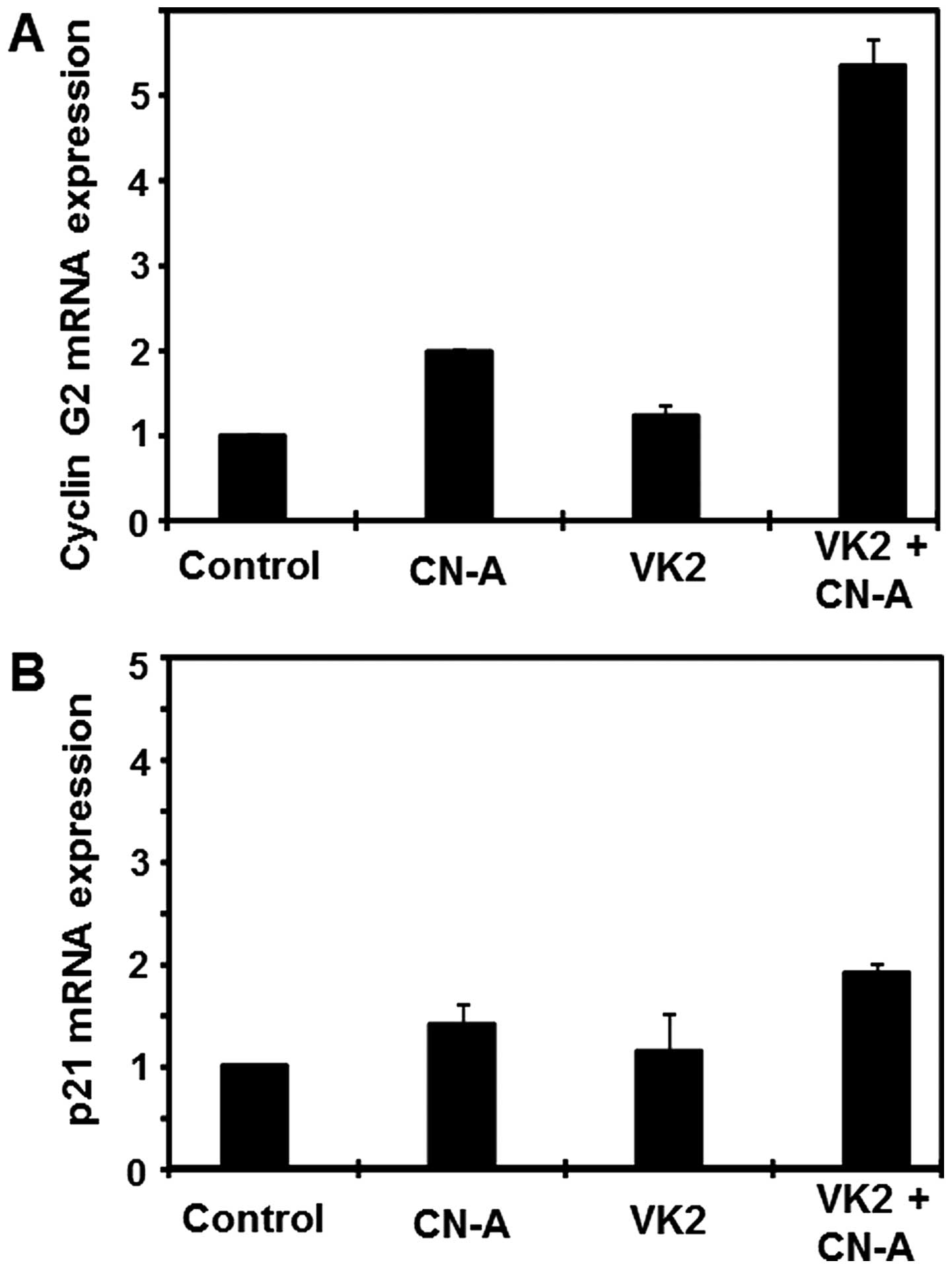
Combined treatment of VK2 and CN-A
synergistically induced cyclin G2 gene expression in HL-60
cells
Previous findings, including ours, indicated that
the expression of cyclin G2 was significantly upregulated during
cell cycle arrest responses to diverse growth-inhibitory signals
and strongly repressed by mitogens, suggesting a positive role for
cyclin G2 in the promotion or maintenance of cell cycle arrest
(28–30). Therefore, we determined whether the
differentiation of HL-60 cells induced with VK2 and CN-A was
accompanied by the induction of cyclin G2 expression. Cyclin G2
gene expression was markedly induced (>5-fold) in VK2 plus
CN-A-treated HL-60 cells (Fig.
7A). The expression of cyclin G2 was approximately 2-fold
higher in CN-A-treated HL-60 cells than in control cells, whereas
VK2-treated cells showed only a marginal increase (Fig. 7A). Similar results were obtained
when HL-60 cells were treated with VK2 plus CN-A for 4 days (data
not shown).
We also examined the gene expression levels of
several cell cycle regulators such as p21/CIP1, p27/KIP1, and
cyclin D1. We did not observe the marked induction (>2-fold) of
the expression of p21/CIP1 (Fig.
7B), p27/KIP1 (data not shown), or cyclin D1 (data not shown)
in VK2-, CN-A-, or VK2 plus CN-A-treated HL-60 cells.
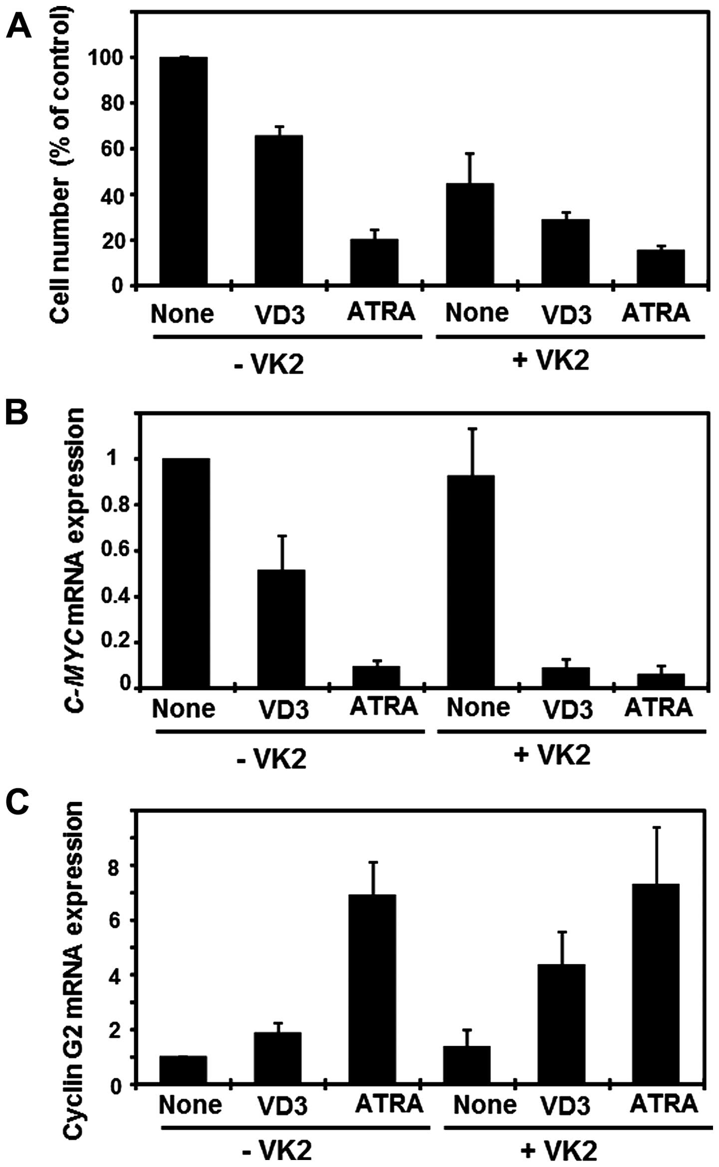
Effects of VK2 on the expression of c-MYC
and cyclin G2 in VD3- or ATRA-treated HL-60 cells
We examined the effects of VK2 on cell growth and
the expression of c-MYC and cyclin G2 in HL-60 cells treated
with two typical differentiation inducers. VD3 is one of the most
potent monocytic differentiation inducers identified to date
(35,36). Although VD3 or VK2 alone inhibited
cell growth to approximately 65 or 45% that of control cells and
also suppressed the expression of c-MYC to approximately 50
or 90% that of control cells, respectively, the combined treatment
of VD3 and VK2 inhibited cell growth to approximately 30% that of
control cells and suppressed the expression of c-MYC to
<10% that of control cells (Fig. 8A
and B). Furthermore, VD3 plus VK2 cooperatively induced cyclin
G2 gene expression more than that of the additive manner (Fig. 8C). On the other hand, ATRA alone,
which is a standard drug used for differentiation therapy in APL,
inhibited cell growth to approximately 20% that of control cells,
suppressed c-MYC expression to <10% that of control
cells, and markedly induced cyclin G2 gene expression (6.9-fold)
(Fig. 8). Under this treatment
condition using ATRA, VK2 marginally increased the effects of ATRA
on cell growth, c-MYC expression, and cyclin G2 expression
(Fig. 8).
Discussion
Vitamin Ks (VK) are known to act as co-factor for
the γ-carboxylation of prothrombin and other VK-dependent
coagulation factors (37). VK
promotes osteogenesis through the γ-carboxylation of glutamate
residues in osteocalcin. VK2 is a naturally-occurring and the main
form of vitamin K in the tissues. A synthetic VK2 analog has been
approved as an anti-osteoporotic medicine by the Ministry of
Health, Labor and Welfare in Japan. The safety of the long-term
administration of VK2 has been well established (38). Although the exact mechanism has not
yet been elucidated in detail, VK2 and their analogs have been
shown to inhibit the survival of various cancer cell lines
(14–16) and leukemia cells (11–13).
Furthermore, previous studies reported that VK2 exhibited some
differentiation-inducing activity in AML cell lines in vitro
(17–19). Only VK2 alone induced the
intermediate stage of differentiation in HL-60 cells in the present
study (Fig. 4D) and, even at
higher concentrations (>10 μM), VK2 could not induce mature
differentiation, but induced apoptosis (data not shown). As VK2 is
a naturally-occurring, safe, and clinically-utilized agent, we
searched for substances that could enhance the
differentiation-inducing activity of VK2. We found that CN-A, a
differentiation inducer, synergistically induced the
differentiation of HL-60 cells along with growth arrest, and
markedly suppressed the expression of c-MYC and induction of
cyclin G2 expression. This is the first study to examine the
effects of VK2 plus CN-A on the induction of differentiation and
expression of growth arrest-associated genes such as c-MYC
and cyclin G2.
The proto-oncogene c-MYC has been shown to
play an important role in cellular metabolism, apoptosis,
differentiation, cell cycle progress and tumorigenesis (36–43).
The expression of c-MYC in particular was found to
contribute to leukemogenesis and promote the progression of
leukemia (43). The downregulation
of c-MYC is critical for the ATRA-induced growth arrest and
myeloid differentiation of AML (23–27).
The inhibition of c-MYC was also shown to suppress the
proliferation and induce the differentiation of primary AML cells
(27). Furthermore, the
overexpression of c-MYC in an APL cell line inversely
inhibited ATRA-induced cell differentiation (27). These findings indicate that
c-MYC is an attractive target for differentiation
therapy.
In the present study, we found that VK2 markedly
enhanced the downregulation of c-MYC gene expression induced
by differentiation inducers, whereas VK2 alone at the doses used
weakly suppressed gene expression (Figs. 6A and 8B). The combined treatment of VK2 and
CN-A exhibited the most potent suppressive effects on c-MYC
gene expression among the inducers or their combinations tested.
This combined treatment reduced the expression of c-MYC to
approximately one fortieth that of control levels (Fig. 6A), and synergistically induced
differentiation (Figs. 3 and
4) and growth arrest (Figs. 1 and 5). Although VK2 also effectively enhanced
the suppressive effects of c-MYC expression induced by VD3
(Fig. 8B), VK2 plus VD3 reduced
the expression of c-MYC less than that of VK2 plus CN-A
(Figs. 6A and 8B). Furthermore, no derivative of active
vitamin D3 has so far been used clinically as an anticancer agent
because of the side effect of hypercalcemia (44). These results suggest that the
combination of VK2 and CN-A has therapeutic value in the treatment
of AML. Furthermore, since we previously found that CN-A was also
capable of stimulating the functional and morphological
differentiation of ATRA-resistant APL cells (10), the combined treatment of VK2 plus
CN-A may be useful for differentiation therapy in
retinoid-resistant leukemia.
The expression of cyclin G2 was shown to be
significantly upregulated during cell cycle arrest responses to
diverse growth-inhibitory signals and strongly repressed by
mitogens, suggesting the positive role of cyclin G2 in the
promotion or maintenance of cell cycle arrest (28–30).
We previously reported that the combination of differentiation
inducers including CN-A effectively inhibited the proliferation of
several human breast cancer cell lines as well as leukemia cells
(27,43). This treatment induced growth arrest
in cells at the G1 phase rather than apoptosis and also rapidly and
markedly induced cyclin G2 gene expression (29,45).
Cyclin G2 knockdown induced by cyclin G2 small
interfering RNA markedly reduced the potency of CN-A plus other
inducers to induce growth inhibition. Ectopically inducible cyclin
G2 expression was shown to potently inhibit the proliferation of
breast cancer cells (29).
Therefore, for more effective differentiation therapy in AML, it is
necessary to find new differentiation inducers or combination
therapies that can induce cell differentiation and growth arrest.
We have searched for substances that are capable of inducing cell
differentiation and the expression of cyclin G2, and that can also
strongly suppress the expression of c-MYC in HL-60 cells. We
showed that the treatment with VK2 plus CN-A induced functional and
morphological differentiation as well as growth arrest in HL-60 AML
cells. Furthermore, this treatment almost completely suppressed the
expression of c-MYC and markedly induced the expression of
cyclin G2. Therefore, our results suggest an attractive combination
for effective differentiation therapy in human myeloid leukemia.
More detailed studies on the mechanisms underlying this effective
combined treatment of VK2 plus CN-A are required.
Acknowledgements
This study was supported partly by a grant from the
Ministry of Education, Culture, Sports, Science and Technology of
Japan and by a grant from Shimane University ‘SUIGAN’ project.
References
|
1
|
Guerrouahen BS, Futami M, Vaklavas C,
Kanerva J, Whichard ZL, Nwawka K, Blanchard EG, Lee FY, Robinson
LJ, Arceci R, et al: Dasatinib inhibits the growth of molecularly
heterogeneous myeloid leukemias. Clin Cancer Res. 16:1149–1158.
2010. View Article : Google Scholar : PubMed/NCBI
|
|
2
|
Robak T and Wierzbowska A: Current and
emerging therapies for acute myeloid leukemia. Clin Ther.
31:2349–2370. 2009. View Article : Google Scholar
|
|
3
|
McCulloch EA: Stem cells in normal and
leukemic hemopoiesis (Henry Stratton Lecture, 1982). Blood.
62:1–13. 1983.PubMed/NCBI
|
|
4
|
Petrie K, Zelent A and Waxman S:
Differentiation therapy of acute myeloid leukemia: Past, present
and future. Curr Opin Hematol. 16:84–91. 2009. View Article : Google Scholar : PubMed/NCBI
|
|
5
|
Sassa T, Tojyo T and Munakata K: Isolation
of a new plant growth substance with cytokinin-like activity.
Nature. 227:3791970. View
Article : Google Scholar : PubMed/NCBI
|
|
6
|
Asahi K, Honma Y, Hazeki K, Sassa T,
Kubohara Y, Sakurai A and Takahashi N: Cotylenin A, a plant-growth
regulator, induces the differentiation in murine and human myeloid
leukemia cells. Biochem Biophys Res Commun. 238:758–763. 1997.
View Article : Google Scholar : PubMed/NCBI
|
|
7
|
Yamamoto-Yamaguchi Y, Yamada K, Ishii Y,
Asahi KI, Tomoyasu S and Honma Y: Induction of the monocytic
differentiation of myeloid leukaemia cells by cotylenin A, a plant
growth regulator. Br J Haematol. 112:697–705. 2001. View Article : Google Scholar : PubMed/NCBI
|
|
8
|
Yamada K, Honma Y, Asahi KI, Sassa T, Hino
KI and Tomoyasu S: Differentiation of human acute myeloid leukaemia
cells in primary culture in response to cotylenin A, a plant growth
regulator. Br J Haematol. 114:814–821. 2001. View Article : Google Scholar : PubMed/NCBI
|
|
9
|
Honma Y: Cotylenin A - a plant growth
regulator as a differentiation-inducing agent against myeloid
leukemia. Leuk Lymphoma. 43:1169–1178. 2002. View Article : Google Scholar : PubMed/NCBI
|
|
10
|
Honma Y, Ishii Y, Sassa T and Asahi K:
Treatment of human promyelocytic leukemia in the SCID mouse model
with cotylenin A, an inducer of myelomonocytic differentiation of
leukemia cells. Leuk Res. 27:1019–1025. 2003. View Article : Google Scholar : PubMed/NCBI
|
|
11
|
Yaguchi M, Miyazawa K, Katagiri T,
Nishimaki J, Kizaki M, Tohyama K and Toyama K: Vitamin K2 and its
derivatives induce apoptosis in leukemia cells and enhance the
effect of all-trans retinoic acid. Leukemia. 11:779–787. 1997.
View Article : Google Scholar : PubMed/NCBI
|
|
12
|
Yokoyama T, Miyazawa K, Naito M, Toyotake
J, Tauchi T, Itoh M, Yuo A, Hayashi Y, Georgescu MM, Kondo Y, et
al: Vitamin K2 induces autophagy and apoptosis simultaneously in
leukemia cells. Autophagy. 4:629–640. 2008. View Article : Google Scholar : PubMed/NCBI
|
|
13
|
Kitagawa J, Hara T, Tsurumi H, Ninomiya S,
Ogawa K, Adachi S, Kanemura N, Kasahara S, Shimizu M and Moriwaki
H: Synergistic growth inhibition in HL-60 cells by the combination
of acyclic retinoid and vitamin K2. J Cancer Res Clin Oncol.
137:779–787. 2011. View Article : Google Scholar
|
|
14
|
Mizuta T and Ozaki I: Hepatocellular
carcinoma and vitamin K. Vitam Horm. 78:435–442. 2008. View Article : Google Scholar : PubMed/NCBI
|
|
15
|
Matsumoto K, Okano J, Nagahara T and
Murawaki Y: Apoptosis of liver cancer cells by vitamin K2 and
enhancement by MEK inhibition. Int J Oncol. 29:1501–1508.
2006.PubMed/NCBI
|
|
16
|
Showalter SL, Wang Z, Costantino CL,
Witkiewicz AK, Yeo CJ, Brody JR and Carr BI: Naturally occurring K
vitamins inhibit pancreatic cancer cell survival through a
caspase-dependent pathway. J Gastroenterol Hepatol. 25:738–744.
2010. View Article : Google Scholar
|
|
17
|
Sakai I, Hashimoto S, Yoda M, Hida T,
Ohsawa S, Nakajo S and Nakaya K: Novel role of vitamin K2: A potent
inducer of differentiation of various human myeloid leukemia cell
lines. Biochem Biophys Res Commun. 205:1305–1310. 1994. View Article : Google Scholar : PubMed/NCBI
|
|
18
|
Miyazawa K, Yaguchi M, Funato K, Gotoh A,
Kawanishi Y, Nishizawa Y, Yuo A and Ohyashiki K:
Apoptosis/differentiation-inducing effects of vitamin K2 on HL-60
cells: Dichotomous nature of vitamin K2 in leukemia cells.
Leukemia. 15:1111–1117. 2001. View Article : Google Scholar : PubMed/NCBI
|
|
19
|
Funato K, Miyazawa K, Yaguchi M, Gotoh A
and Ohyashiki K: Combination of 22-oxa-1,25-dihydroxyvitamin D(3),
a vitamin D(3) derivative, with vitamin K(2) (VK2) synergistically
enhances cell differentiation but suppresses VK2-inducing apoptosis
in HL-60 cells. Leukemia. 16:1519–1527. 2002. View Article : Google Scholar : PubMed/NCBI
|
|
20
|
Sada E, Abe Y, Ohba R, Tachikawa Y,
Nagasawa E, Shiratsuchi M and Takayanagi R: Vitamin K2 modulates
differentiation and apoptosis of both myeloid and erythroid
lineages. Eur J Haematol. 85:538–548. 2010. View Article : Google Scholar : PubMed/NCBI
|
|
21
|
Field JK and Spandidos DA: The role of ras
and myc oncogenes in human solid tumours and their relevance in
diagnosis and prognosis (Review). Anticancer Res. 10:1–22.
1990.PubMed/NCBI
|
|
22
|
Delgado MD, Albajar M, Gomez-Casares MT,
Batlle A and León J: MYC oncogene in myeloid neoplasias. Clin
Transl Oncol. 15:87–94. 2013. View Article : Google Scholar
|
|
23
|
Kumakura S, Ishikura H, Tsumura H, Hayashi
H, Endo J and Tsunematsu T: c-myc protein expression during cell
cycle phases in differentiating HL-60 cells. Leuk Lymphoma.
14:171–180. 1994. View Article : Google Scholar : PubMed/NCBI
|
|
24
|
Kumakura S, Ishikura H, Tsumura H, Iwata
Y, Endo J and Kobayashi S: C-Myc and Bcl-2 protein expression
during the induction of apoptosis and differentiation in TNF
alpha-treated HL-60 cells. Leuk Lymphoma. 23:383–394. 1996.
View Article : Google Scholar : PubMed/NCBI
|
|
25
|
Jiang G, Albihn A, Tang T, Tian Z and
Henriksson M: Role of Myc in differentiation and apoptosis in HL60
cells after exposure to arsenic trioxide or all-trans retinoic
acid. Leuk Res. 32:297–307. 2008. View Article : Google Scholar
|
|
26
|
Cheng YC, Lin H, Huang MJ, Chow JM, Lin S
and Liu HE: Downregulation of c-Myc is critical for valproic
acid-induced growth arrest and myeloid differentiation of acute
myeloid leukemia. Leuk Res. 31:1403–1411. 2007. View Article : Google Scholar : PubMed/NCBI
|
|
27
|
Pan XN, Chen JJ, Wang LX, Xiao RZ, Liu LL,
Fang ZG, Liu Q, Long ZJ and Lin DJ: Inhibition of c-Myc overcomes
cytotoxic drug resistance in acute myeloid leukemia cells by
promoting differentiation. PLoS One. 9:e1053812014. View Article : Google Scholar : PubMed/NCBI
|
|
28
|
Horne MC, Donaldson KL, Goolsby GL, Tran
D, Mulheisen M, Hell JW and Wahl AF: Cyclin G2 is up-regulated
during growth inhibition and B cell antigen receptor-mediated cell
cycle arrest. J Biol Chem. 272:12650–12661. 1997. View Article : Google Scholar : PubMed/NCBI
|
|
29
|
Kasukabe T, Okabe-Kado J and Honma Y:
Cotylenin A, a new differentiation inducer, and rapamycin
cooperatively inhibit growth of cancer cells through induction of
cyclin G2. Cancer Sci. 99:1693–1698. 2008. View Article : Google Scholar : PubMed/NCBI
|
|
30
|
Zimmermann M, Arachchige-Don AS, Donaldson
MS, Dallapiazza RF, Cowan CE and Horne MC: Elevated cyclin G2
expression intersects with DNA damage checkpoint signaling and is
required for a potent G2/M checkpoint arrest response to
doxorubicin. J Biol Chem. 287:22838–22853. 2012. View Article : Google Scholar : PubMed/NCBI
|
|
31
|
Kasukabe T, Honma Y, Hozumi M and Nomura
H: Inhibition of proliferation and induction of differentiation of
human and mouse myeloid leukemia cells by new ethyleneglycol-type
nonphosphorus alkyl ether lipids. Jpn J Cancer Res. 81:807–812.
1990. View Article : Google Scholar : PubMed/NCBI
|
|
32
|
Marzi I, Cipolleschi MG, D’Amico M,
Stivarou T, Rovida E, Vinci MC, Pandolfi S, Dello Sbarba P, Stecca
B and Olivotto M: The involvement of a Nanog, Klf4 and c-Myc
transcriptional circuitry in the intertwining between neoplastic
progression and reprogramming. Cell Cycle. 12:353–364. 2013.
View Article : Google Scholar : PubMed/NCBI
|
|
33
|
Stossi F, Likhite VS, Katzenellenbogen JA
and Katzenellenbogen BS: Estrogen-occupied estrogen receptor
represses cyclin G2 gene expression and recruits a repressor
complex at the cyclin G2 promoter. J Biol Chem. 281:16272–16278.
2006. View Article : Google Scholar : PubMed/NCBI
|
|
34
|
Liang X-H, Li L-L, Wu G-G, Xie Y-C, Zhang
G-X, Chen W, Yang HF, Liu QL, Li WH, He WG, et al: Upregulation of
CPE promotes cell proliferation and tumorigenicity in colorectal
cancer. BMC Cancer. 13:4122013. View Article : Google Scholar : PubMed/NCBI
|
|
35
|
Tanaka H, Abe E, Miyaura C, Kuribayashi T,
Konno K, Nishii Y and Suda T: 1 alpha,25-Dihydroxycholecalciferol
and a human myeloid leukaemia cell line (HL-60). Biochem J.
204:713–719. 1982.PubMed/NCBI
|
|
36
|
Kasukabe T, Honma Y, Hozumi M, Suda T and
Nishii Y: Control of proliferating potential of myeloid leukemia
cells during long-term treatment with vitamin D3 analogues and
other differentiation inducers in combination with antileukemic
drugs: In vitro and in vivo studies. Cancer Res. 47:567–572.
1987.PubMed/NCBI
|
|
37
|
Vermeer C and Schurgers LJ: A
comprehensive review of vitamin K and vitamin K antagonists.
Hematol Oncol Clin North Am. 14:339–353. 2000. View Article : Google Scholar : PubMed/NCBI
|
|
38
|
Sasaki N, Kusano E, Takahashi H, Ando Y,
Yano K, Tsuda E and Asano Y: Vitamin K2 inhibits
glucocorticoid-induced bone loss partly by preventing the reduction
of osteoprotegerin (OPG). J Bone Miner Metab. 23:41–47. 2005.
View Article : Google Scholar
|
|
39
|
Masui K, Tanaka K, Akhavan D, Babic I,
Gini B, Matsutani T, Iwanami A, Liu F, Villa GR, Gu Y, et al: mTOR
complex 2 controls glycolytic metabolism in glioblastoma through
FoxO acetylation and upregulation of c-Myc. Cell Metab. 18:726–739.
2013. View Article : Google Scholar : PubMed/NCBI
|
|
40
|
Sheth A, Escobar-Alvarez S, Gardner J, Ran
L, Heaney ML and Scheinberg DA: Inhibition of human mitochondrial
peptide deformylase causes apoptosis in c-myc-overexpressing
hematopoietic cancers. Cell Death Dis. 5:e11522014. View Article : Google Scholar : PubMed/NCBI
|
|
41
|
Gómez-Casares MT, García-Alegria E,
López-Jorge CE, Ferrándiz N, Blanco R, Alvarez S, Vaqué JP,
Bretones G, Caraballo JM, Sánchez-Bailón P, et al: MYC antagonizes
the differentiation induced by imatinib in chronic myeloid leukemia
cells through downregulation of p27(KIP1). Oncogene. 32:2239–2246.
2013. View Article : Google Scholar
|
|
42
|
Singh AM and Dalton S: The cell cycle and
Myc intersect with mechanisms that regulate pluripotency and
reprogramming. Cell Stem Cell. 5:141–149. 2009. View Article : Google Scholar : PubMed/NCBI
|
|
43
|
Hoffman B, Amanullah A, Shafarenko M and
Liebermann DA: The proto-oncogene c-myc in hematopoietic
development and leukemogenesis. Oncogene. 21:3414–3421. 2002.
View Article : Google Scholar : PubMed/NCBI
|
|
44
|
DeLuca HF: Evolution of our understanding
of vitamin D. Nutr Rev. 66(Suppl 2): S73–S87. 2008. View Article : Google Scholar : PubMed/NCBI
|
|
45
|
Kasukabe T, Okabe-Kado J, Kato N, Sassa T
and Honma Y: Effects of combined treatment with rapamycin and
cotylenin A, a novel differentiation-inducing agent, on human
breast carcinoma MCF-7 cells and xenografts. Breast Cancer Res.
7:R1097–R1110. 2005. View Article : Google Scholar
|