Introduction
Antibody based molecular imaging provides a
non-invasive means of detecting biological processes and molecular
events in vivo. By combining the high sensitivity and
resolution of e.g. a PET (positron emission tomography)-scanner
with the tumor specificity of a tumor targeting molecule, this
technique is of increasing importance to visualize and characterize
tumor lesions. Additionally, it can be used for dosimetric
calculations, to monitor therapy response and to identify patients
who may benefit from a particular therapy (1). With advancements in instrumentation
and development of novel targeted probes, immuno-PET has firmly
established its role in drug development and in clinical
assessment.
Although intact monoclonal antibodies (mAbs) have
been considered candidates for molecular imaging due to their
specificity and high signal delivery to cell surface molecules,
their relatively large size (~150 kDa) and Fc-mediated half-life
extension tend to cause suboptimal imaging pharmacokinetics, poorer
tumor penetration and increased immunogenicity. Smaller targeting
moieties such as antibody fragments have emerged as an alternative
to mAbs to overcome the aforementioned limitations. Common antibody
fragments are smaller molecules that omit the Fc-region and consist
of one Fab (~50 kDa) or two F(ab′)2 (100 kDa) antigen
binding arms, respectively. Due to their smaller size, antibody
fragments are likely to exhibit better extravasation and diffusion
in the intra-cellular space and faster blood clearance compared to
their parental antibodies (2).
Consequently, normal tissue accumulation and immunologic reactions
is expected to be lower with a fragment than with the intact
antibody, thus enhancing tumor contrast and allowing imaging at
earlier time-points. For several targets, antibody fragments have
shown promising results as imaging agents in animals (3) and clinical studies (4). Generally, F(ab′)2
fragments are considered advantageous to Fab fragments in molecular
imaging due to their bivalent nature, which can result in an
increased functional affinity and tumor uptake compared to Fab.
Another possible disadvantage of Fab fragments is that their
molecular weight subjects them to glomerular filtration. The larger
F(ab′)2 format, with a size above the renal threshold,
is expected to exhibit a biodistribution shift from kidneys to
liver (5).
An alternative to antibody fragments derived from
full-length antibodies generated through immunization are
recombinant Fabs obtained in vitro by phage display,
generated from selections in phage libraries. Recombinant antibody
technology is a rapidly evolving field with a number of major
benefits over conventional antibody generation and production
methods (6). It enables the use of
in vitro selection steps that facilitate the isolation of
antibodies with desired characteristics, e.g. antibodies that
distinguish closely related antigens (7), and candidates can be engineered using
readily available DNA sequences. Furthermore, since recombinant
monoclonal antibody fragments can be produced in bacteria this
allows for an easier, faster, and less expensive production process
than using hybridoma or mammalian cell culture techniques.
Moreover, antibodies selected from in vitro libraries of
human antibody genes do not elicit the same immune responses in
patients that are seen with non-human antibodies. Therefore, such
antibodies can be used for therapeutic development. Thus, more and
more fully human antibodies obtained from antibody libraries are
entering clinical development and are reaching the market (8).
We have developed the CD44v6-targeting human
bivalent antibody fragment AbD19384, a recombinant human bivalent
Fab antibody, engineered from monovalent Fab AbD15179 by subcloning
of the Fab gene into an expression vector containing a
self-dimerizing helix-turn helix motif (dHLX) (9,10).
The Fab-dHLX constructs represent a novel, attractive platform for
cancer targeting. The size and format is functionally equivalent to
a F(ab′)2 fragment, but there are also important
differences besides the origin of the molecule. AbD19384 is a
non-covalent homodimer formed via C-terminal fusion of a small
homodimerization domain, whereas disulfide bridges are present in
F(ab′)2 fragments enzymatically derived from full length
antibodies. These non-covalent homodimers are stable under reducing
conditions, whereas F(ab′)2 fragments are not. CD44v6
was chosen as a target, since it is a surface antigen found to be
overexpressed in >90% of head and neck squamous cell carcinoma
(HNSCC), as well as in other cancers such as lung, esophagus and
breast, which makes it an attractive target for molecular imaging
and targeted therapy (11). To the
best of our knowledge, there are no previous studies of tumor
targeting abilities of the Fab-dHLX format in vivo, hence it
is not known how this construct functions in an in vivo
setting. Thus, the present study is not only relevant for
CD44v6-targeted molecular imaging, but also as the first study of a
tumor targeting Fab-dHLX construct in vivo.
Consequently, the aim of the present study was to
evaluate for the first time the in vitro and in vivo
binding properties of the Fab-dHLX construct, and to assess the
utility of radio-iodinated AbD19384 as a targeting agent for
molecular imaging of CD44v6-expressing tumors.
Materials and methods
AbD19384
AbD19384 is a bivalent antibody fragment, engineered
from two fully human AbD15179 Fab-fragments. This format is a
non-covalent homodimer of AbD15179 formed via C-terminal fusion of
a dHLX (synthetic double helix loop helix) dimerization motif
(9,10) to the heavy chain segment of
AbD15179. This format, Fab-dHLX, also referred to as ‘bivalent
mini-antibody’, has a molecular weight of ~110 kDa, and is
functionally equivalent to a F(ab′)2 fragment. The dHLX
helix-turn-helix motif is a de novo designed
self-associating peptide (9,12).
The dimerization occurs spontaneously, and thus the construct is in
equilibrium with the monomeric form and possibly also a small
fraction of tetramers or higher order aggregates, with a majority
of the molecules being in the dimeric form (9). The generation of the monovalent form
AbD15179 has been previously described (7). AbD19384 was supplied by Bio-Rad AbD
Serotec GmbH (Puchheim, Germany) in 3X PBS.
Surface plasmon resonance (SPR) analysis
of AbD19384
Recombinant CD44v3-10 (7) and two BSA-conjugated peptides (Elim
Biopharmaceuticals, Hayward, CA, USA) derived from human
(CGNRWHEGYRQTPREDS) and murine (CQNGWQGKNPPTPSEDS) CD44v6,
respectively, were immobilized by standard amine coupling on a
ProteOn XPR36 general layer medium sensor chip (Bio-Rad
Laboratories, Hercules, CA, USA). A v6-negative isoform of CD44
(CD44v3-10Δv6) was included as a negative control. Channels with
immobilization levels of 200 RU (BSA-conjugated peptides), 500 RU
(CD44v3-10) and 1000 RU (CD44v3-10 and CD44v3-10Δv6) were used.
Dilution series (3–50 nM) of the Fab-fragment AbD15179 and its
corresponding bivalent fragment AbD19384 in PBS 0.05% Tween-20 (pH
7.4) were injected at 50 μl/min at 25°C. Real-time signal from a
blank channel and a parallel buffer injection were subtracted and
binding data fitted to a 1:1 Langmuir isotherm using ProteOn
Manager software version 3.1.0.6 (Bio-Rad Laboratories). Data were
double referenced by subtraction of simultaneous responses from a
blank surface and a buffer injection. Experimental data were
plotted together with curves drawn from a fitted 1:1 Langmuir
isotherm. Kinetic constants were calculated from five separate
injections using two different immobilization levels of CD44v3-10.
Surfaces were regenerated with 10 mM HCl between injections.
Cell lines
The human SCC cell line A431 (obtained from the
American Type Culture Collection, Manassas, VA, USA), derived from
an epidermal carcinoma of the vulva, was cultured in Ham's F10,
supplemented with 10% fetal calf serum, 2 mM L-glutamine, and
antibiotics (100 IU penicillin and 100 μg/ml streptomycin). A431
has been shown to be a highly CD44v6-expressing cell line with
~3×10⁶ antigens/cell (13). The human SCC cell line H314
(obtained from the European Collection of Cell Cultures), derived
from floor of the mouth, was cultured in a one-to-one mixture of
Ham's F12 and Dulbecco's modified Eagle's medium (DMEM) with the
same supplements. H314 has demonstrated a medium antigen density of
~0.7×10⁶ CD44v6 antigens per cell (14). The human SCC cell line UM-SCC74B
derived from base of the tongue (kindly provided by Professor T.E.
Carey, University of Michigan, MI, USA) was cultured in DMEM with
the same supplements, as well as 1% non-essential amino acids.
UM-SCC74B has demonstrated low expression (~0.1×10⁵) of
CD44v6. As a negative control, the breast cancer cell line
MDA-MB-231 (obtained from the American Type Culture Collection) was
used. It was cultured in DMEM with the same supplements as
described above for A431. This cell line has demonstrated no
detectable CD44v6 expression (14). Cells were incubated at 37°C in an
atmosphere containing 5% CO2. For LigandTracer studies,
~1×10⁶ A431 cells, 2×10⁶ H314 cells,
1×10⁶ UM-SCC74B cells, or 2×10⁶ MDA-MB-231
cells were seeded 2–3 days before measurements in a local part on a
10-cm cell dish (#150350; Nunc) and allowed to adhere firmly to the
plastics prior to the addition of fresh medium.
Labeling
AbD19384 was labeled with 125I
(Perkin-Elmer, Waltham, MA, USA) using direct chloramine T labeling
(CAT) (15) (Sigma-Aldrich). CAT
and Na2SO5 (NBS) were dissolved in
MilliQ-water to 4 mg/ml. AbD19384 (100 μg) in PBS was added to 10
MBq of 125I and mixed before adding 30 μl of CAT. The
reaction mixture was incubated for 1 min on ice before ending the
reaction by adding 60 μl of NBS. The sample was purified on a
NAP5-size exclusion column (GE Healthcare Life Sciences Uppsala,
Sweden) equilibrated with PBS.
Labeling of AbD19384 with 124I
(Perkin-Elmer) using 1,3,4,6-Tetrachloro-3α,6α-diphenylglycouril
(Iodogen) was performed as following; three Iodogen buffers (A, B
and C) were prepared; A, 0.5 M sodium phosphate buffer, pH 7.4; B,
0.05 M sodium phosphate and 5 M NaCl, pH 7.4; C, 0.05 M sodium
phosphate, 5% potassium iodide and 0.5% BSA (w/v). All buffers were
prepared using MilliQ-water. Iodogen was dissolved in
dichloromethane to 0.2 mg/ml. 124I was incubated with
cold NaI in a 1:1 molar ratio with the added AbD19384 in order to
ensure that the nuclide was in iodide form prior to starting the
labeling procedure. 124I and AbD19384 (1 mg/ml in PBS)
were mixed in a tube previously coated with 50 μg Iodogen. Buffer A
was added in an equivalent volume, incubated at room temperature
for 7 min and agitated carefully every 30 sec using a vortex. The
reaction mixture was transferred to a new tube and buffer B (480
μl) was subsequently added. Following at least 10 min of rest,
buffer C (480 μl) was added, and the sample was mixed thoroughly.
Labeled F(ab′)2 was separated from non-reacted
radionuclide and low-molecular-weight reaction components by using
a NAP-5 or PD10 column pre-equilibrated with PBS. Iodination using
Iodogen was also performed using 125I in the same way,
in order to optimize and compare labelings.
To determine the yield, purity and stability of the
labeled conjugates, instant thin-layer chromatography (ITLC)
analyses were performed on labeled conjugates. Samples taken
immediately as well as 48 h after the labeling procedure were
analyzed. Serum stability tests were performed by 1 h incubation at
37°C in 42% murine serum in PBS, pH 7.4. Approximately 1 μl of the
conjugate was placed on an ITLC chromatography strip (Biodex) and
placed into a ‘running buffer’ (70% acetone), followed by
measurements on a Cyclone Storage Phosphor system. Data were
analyzed using OptiQuant image analysis software.
In vitro binding measurements on cultured
tumor cells
Real-time in vitro binding and retention
measurements of the radiolabeled conjugates were performed at room
temperature using LigandTracer instruments (Ridgeview Instruments
AB, Uppsala, Sweden) on CD44v6-positive A431, H314 and UM-SCC74B
cells. In vitro binding specificity measurements were
performed on LigandTracer instruments using MDA-MB-231 cells as
negative controls. LigandTracer Grey was used for
125I-AbD19384 and LigandTracer White was used for
124I-AbD19384. Binding traces using several subsequent
concentrations (0–90 nM) were obtained for at least 1 h per
concentration, followed by a dissociation measurement for at least
15 h. The shapes of the real-time binding curves produced in
LigandTracer were compared in the evaluation software TraceDrawer
1.6.1 (Ridgeview Instruments AB, Vänge, Sweden). Through kinetic
fitting using a 1:1 and 1:2 models, the dissociation equilibrium
constant KD (corresponding to the apparent affinity),
the association rate constant ka and the dissociation
rate kd were obtained.
Small animal studies
Female nu/nu Balb/c mice were housed under standard
laboratory conditions and fed ad libitum. All experiments
complied with the Swedish law and were performed with permission
from the Uppsala Committee of Animal Research Ethics, ethical
permission number C 410/12.
In vivo specificity of
125I-AbD19384
In the first group (n=5), aimed at analyzing in
vivo specific binding of 125I-AbD19384 to CD44v6,
approximately (8×10⁶) A431 cells (high CD44v6
expression) suspended in 150 μl 1:1 cell medium:Matrigel were
injected s.c. into the right posterior leg. In the same mice,
~8×10⁶ MDA-MB-231 cells (no CD44v6 expression) suspended
in 150 μl 1:1 cell medium:Matrigel were injected in the left
posterior leg. Two weeks after tumor cell injections, experiments
were performed. At the time of study, average animal weights were
19.9±0.9 g (SEM), A431 tumor weights 73±20 mg, and MDA-MB-231 tumor
weights 169±23 mg. The mice were injected with
125I-AbD19384 (11 μg in 200 μl PBS, 100 kBq) in the tail
vein. At 24 h post injection (p.i), animals were euthanized with a
mixture of ketamine and xylazine followed by heart puncture. Blood
and tumors were collected and weighed, and tracer uptake was
measured in a gamma well-counter (1480 Wizard; Wallace Oy, Turku,
Finland). Radioactivity uptake in the organ was calculated as
percent of injected activity per gram of tissue (%ID/g).
Tumor-to-blood ratio was calculated as activity/gtumor
divided by activity/gblood.
Ex vivo biodistribution of
125I-AbD19384
In a second group (n=20), used to analyze in
vivo binding of 125I-AbD19384 to xenografts with
varying CD44v6 receptor densities, approximately (8×10⁶)
A431 cells suspended in 150 μl 1:1 cell medium:Matrigel were
injected s.c. into the left posterior leg and 10×106
H314 cells (moderate CD44v6 expression) suspended in 150 μl 1:1
cell medium:Matrigel were injected in the right posterior leg.
Three weeks after tumor cell injections, experiments were
performed. At the time of study, average animal weights were
20.1±1.3 g, A431 tumor weights 219±54 mg and H314 tumor weights
23±3 mg. Twenty mice bearing dual A431 (high CD44v6 expression) and
H314 (moderate CD44v6 expression) xenografts received an
intravenous injection via the tail vein with
125I-AbD19384 (100 kBq), totally 11 μg
F(ab′)2 in 200 μl PBS per mouse. Animals were
sub-divided into four groups with five mice in each. At 6, 24, 48
and 72 h p.i. animals were euthanized with a mixture of ketamine
and xylazine followed by heart puncture. Blood was collected, and
tumors and organs of interest such as salivary glands, thyroid (en
bloc with larynx), tongue, heart, liver, kidneys, spleen, urinary
bladder, colon, upper gastrointestinal tract, skin, bone and muscle
were excised, weighed and measured in a gamma well-counter. The
tail and the rest of the body were also measured. Three injection
standards were measured for each group. Radioactivity uptake in the
organ was calculated as percent of injected activity per gram of
tissue (%ID/g). Thyroid was excised en bloc with larynx, and uptake
was calculated as percent of injected activity per organ. Tumor to
organ ratio was calculated as activity/gtumor divided by
activity/gorgan.
In vivo PET/CT and ex vivo
biodistribution studies with 124I-AbD19384
A third group was used to analyze in vivo
biodistribution of 124I-AbD19384 (n=12) or
18F-FDG (n=4) in mice with xenografts with varying
CD44v6 receptor densities and for PET/CT imaging. Approximately
(8×10⁶) A431 cells suspended in 150 μl 1:1 cell
medium:Matrigel were injected s.c. into the left posterior leg and
10×106 UM-SCC74B tumor cells (low CD44v6 expression)
suspended in 150 μl 1:1 cell medium:Matrigel were injected in the
right posterior leg. Tumors were allowed to grow for three weeks
before start of experiments. At the time of study, average animal
weights were 16.1±1.5 g. A day before conjugate injections, mice
were given potassium iodide (1%) in drinking water to block uptake
of free 124I in thyroid. Animals were then injected with
124I-AbD19384 (11 μg, 1.5 MBq, in 200 μl PBS per mouse)
in the tail vein as a single bolus. As verified with native gel
analysis, 17% of injected 124I-AbD19384 consisted of
monomers or smaller fragments.
Whole body PET/CT studies of
124I-AbD19384 were performed under general anesthesia
(isoflurane 1.0–2.5% in 50%/50% medical oxygen:air at 450 ml/min)
at 24, 48 and 72 h p.i. Four additional mice were imaged using
18F-FDG (2.7 Mbq) at 30 min p.i. under general
anesthesia. Sedated xenograft animals with pre-injected tracer were
placed in the gantry of the small animal PET/CT scanner (Triumph™
Trimodality System; TriFoil Imaging, Inc., Northridge, CA, USA).
Whole body PET scans were performed for 80 min in list mode
followed by a CT examination for 3 min (Field of View = 8.0 cm).
18F-FDG PET scan was acquired for 60 min. At indicated
times, animals were euthanized, and organs removed and measured as
described above. The breathing rate was monitored with a camera
under controlled anesthesia. Animals were placed on the heated bed
of the PET scanner to prevent hypothermia and fastened to prevent
large movements during study. 18F-FDG was kindly
provided by the Nuclear Medicine Department (Akademiska Sjukhuset,
Uppsala, Sweden).
The PET data were reconstructed into a static image
using an ordered subset expectation maximization 3-D algorithm (20
iterations). The CT raw files were reconstructed using Filter Back
Projection. PET data were reconstructed for attenuation and scatter
corrections with their respective CT data. PET and CT dicom files
were analyzed using PMOD v3.508 (PMOD Technologies Ltd., Zurich,
Switzerland). Volumes of interest were drawn manually on tumors,
muscle and left ventricle. The measurement from left ventricle was
accounted for blood. Tracer uptake in these organs from PET images
is expressed as tissue-to-reference tissue ratio. Blood was used as
reference tissue.
After PET/CT measurements, animals were euthanized;
blood, tumors and organs of interest were collected and treated as
described above for ex vivo biodistribution measurements.
For animals injected with 124I-AbD19384 (n=12), A431
tumor weights were 567±61 mg, and UM-SCC74B tumor weights 483±77
mg. For animals injected with 18F-FDG (n=4) A431 tumor
weights were 681±210 mg, and UM-SCC74B tumor weights 409±241 mg at
the time of experiments.
Statistical analyses
Statistical analyses were performed using GraphPad
Prism version 5.02 for Windows (GraphPad Software Inc., La Jolla,
CA, USA). For in vivo studies, the differences in uptake
between MDA-MB-231/UM-SCC74B tumors and A431 tumors were assessed
using a two-tailed t-test and were considered statistically
significant at P<0.05. The biodistribution data are presented as
mean ± standard deviation (SD). Significant differences between the
groups over time were tested with one-way analysis of variance
(ANOVA) with Newman-Keuls multiple comparison test. The differences
were considered statistically significant at P<0.05. The
differences in uptake of 125I-AbD19384 and
124I-AbD19384 between low/medium CD44v6 expressing
tumors and high CD44v6 expressing tumors were evaluated using
repeated measures ANOVA with Newman-Keuls multiple comparison test
and were considered statistically significant at P<0.05.
Results
SPR analysis
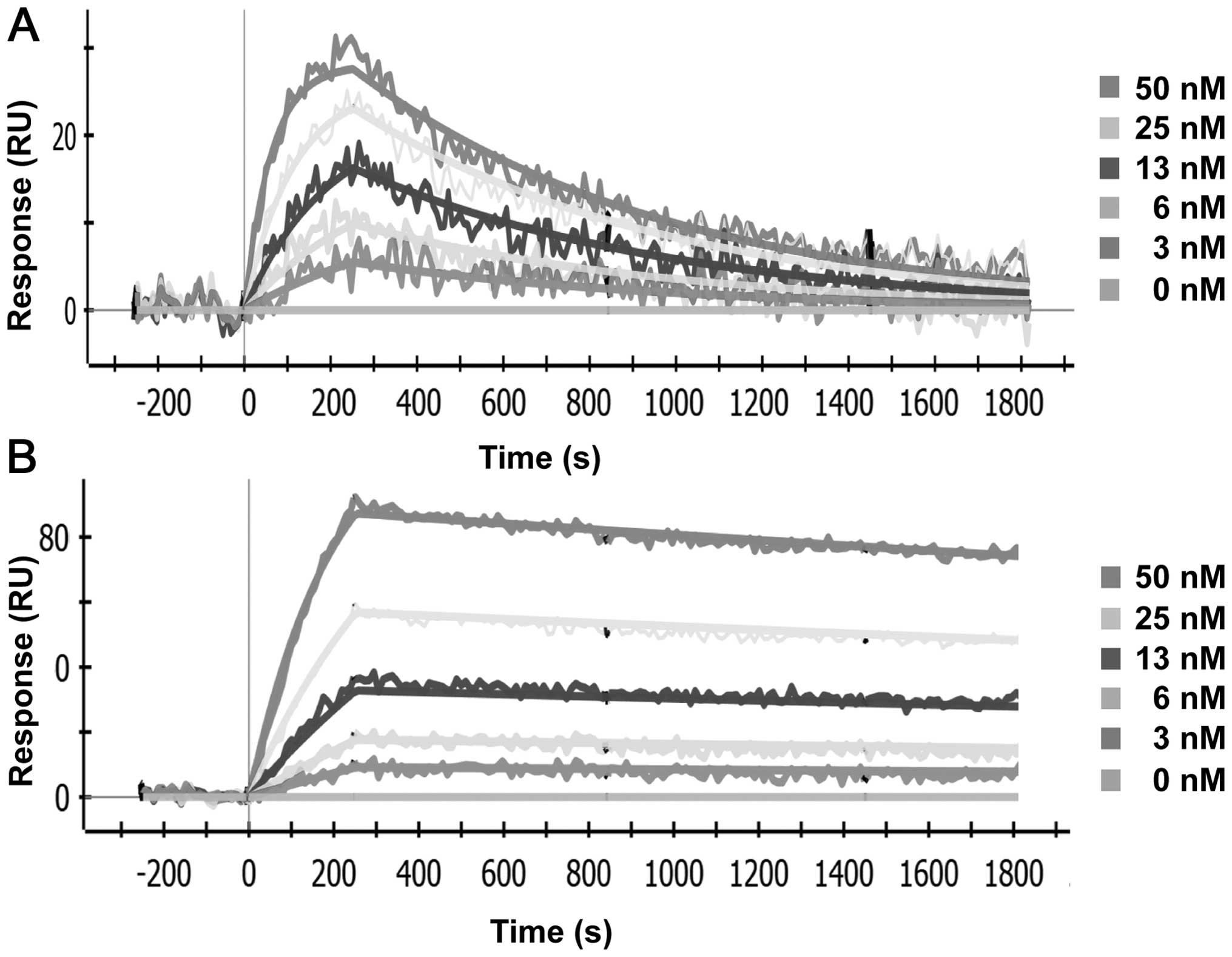
Both AbD19384 and AbD15179 bound CD44v3-10,
representative sensorgrams are shown in Fig. 1, and no binding was seen to the
negative control isoform. Reformatting Fab AbD15179 into the
bivalent AbD19384 improved the apparent affinity (KD)
almost 6-fold from 6 to 1 nM. This was a result of an improved
dissociation rate, which was decreased from
1.1(±0.3)×10−3 s−1 for AbD15179 to
1.6(±0.5)×10−4 s−1 for AbD19384. The
association rate constants were similar, 1.8(±0.5)×105
M−1s−1 for AbD15179 vs.
1.4(±0.8)×105 M−1s−1 for the
larger AbD19384. Both antibody fragments bound human CD44v6 peptide
and no signals were observed on surfaces immobilized with murine
CD44v6 peptide (data not shown).
Labeling
Labeling yields were 64–74% for
125I-AbD19384, and 64–73% for 124I-AbD19384,
respectively. Specific activity for 125I-AbD19384 was
adjusted before in vivo injections, using unlabeled
AbD19384, resulting in an injected activity dose of 100 kBq per
mouse (11 μg AbD19384 per mouse). The specific activity of the
injection solution for 124I-AbD19384 was 136 kBq/μg,
resulting in an injected activity dose of 1.5 MBq (11 μg) per
mouse. The radiochemical purity after size-exclusion chromatography
purification was >95% for all conjugates. Radiochemical purity
of labeled conjugates stored in PBS for 48 h, or in serum for 1 h,
was unchanged according to ITLC analysis for both conjugates.
Native gel analyses of radioiodinated conjugates demonstrated that
serum incubation did not influence either monomer/dimer
equilibrium, or aggregation of the conjugate (data not shown).
In vitro binding measurements of
iodinated AbD19384 on cultured tumor cells
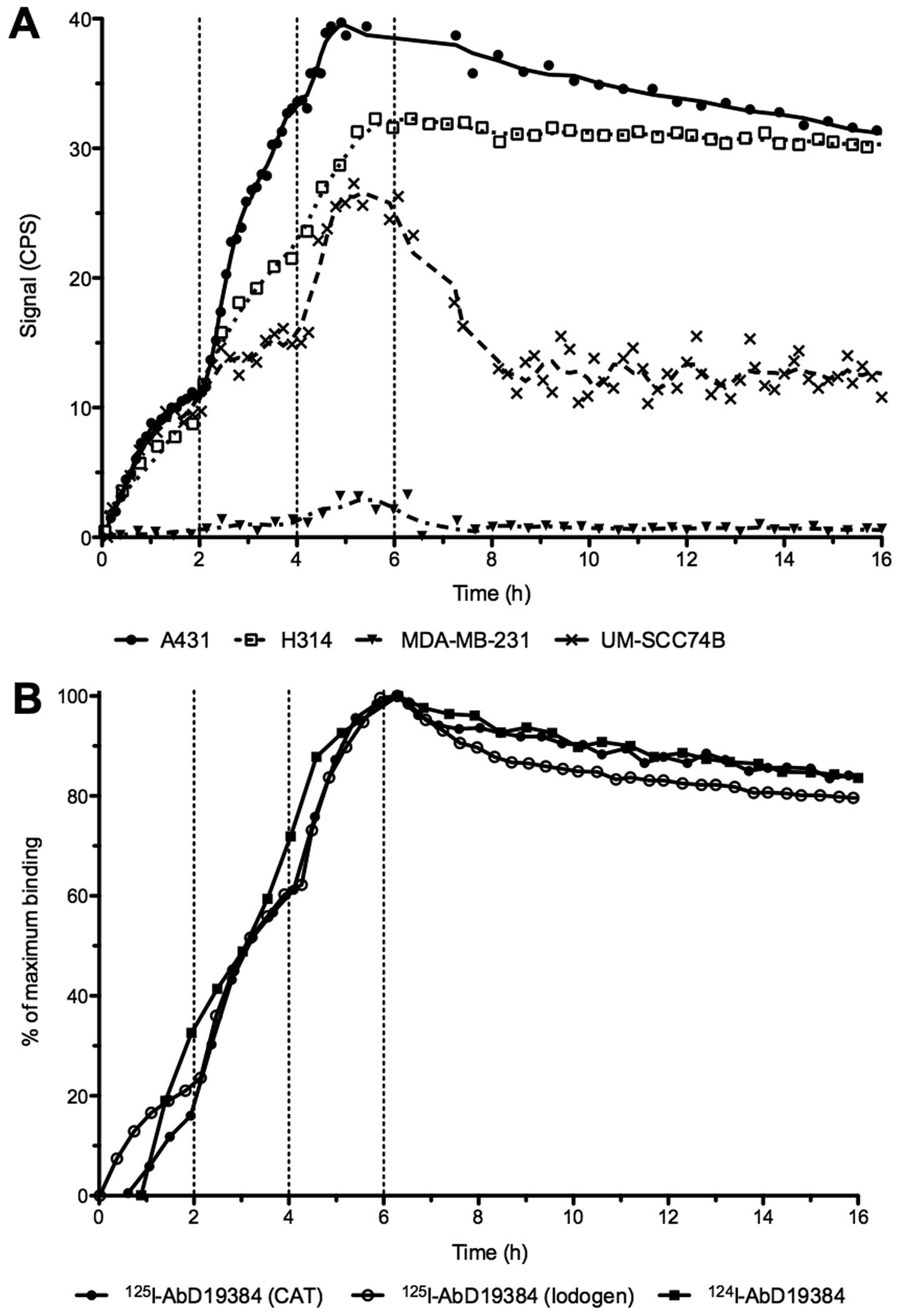
Real-time binding data from measurements of
radio-iodinated AbD19384 binding to cells can be seen in Fig. 2. A clear binding with slow off-rate
was seen on both A431 and H314 cells, whereas a lower, noisier
signal was seen for the low CD44v6-expressing UM-SCC74B cells and
no signal was detected on the negative control MDA-MB-231 cells
(Fig. 2A). All iodinated
conjugates behaved in a similar manner with comparable binding and
retention for conjugates regardless of labeling method (using CAT
or Iodogen) or nuclide (125I or 124I)
(Fig. 2B). Interactions were
mainly 1:1 and the apparent KD of the high-affinity
interaction was around 1 nM for all three conjugates (0.8±0.1,
0.7±0.12 and 1.3±0.2 nM for 125I-AbD19384 (CAT),
125I-AbD19384 (Iodogen), and 124I-AbD19384
(Iodogen), respectively).
Small animal studies
In vivo specificity of AbD19384
Tumor uptake in A431 tumors was 0.97±0.17%ID/g, and
uptake in MDA-MB-231-tumors was 0.24±0.04%ID/g, comparable to
amounts in blood (0.23±0.11%ID/g). The difference in uptake was
statistically significant (P<0.0001), with the CD44v6 positive
tumors displaying approximately four times higher uptake compared
to the CD44v6 negative tumors.
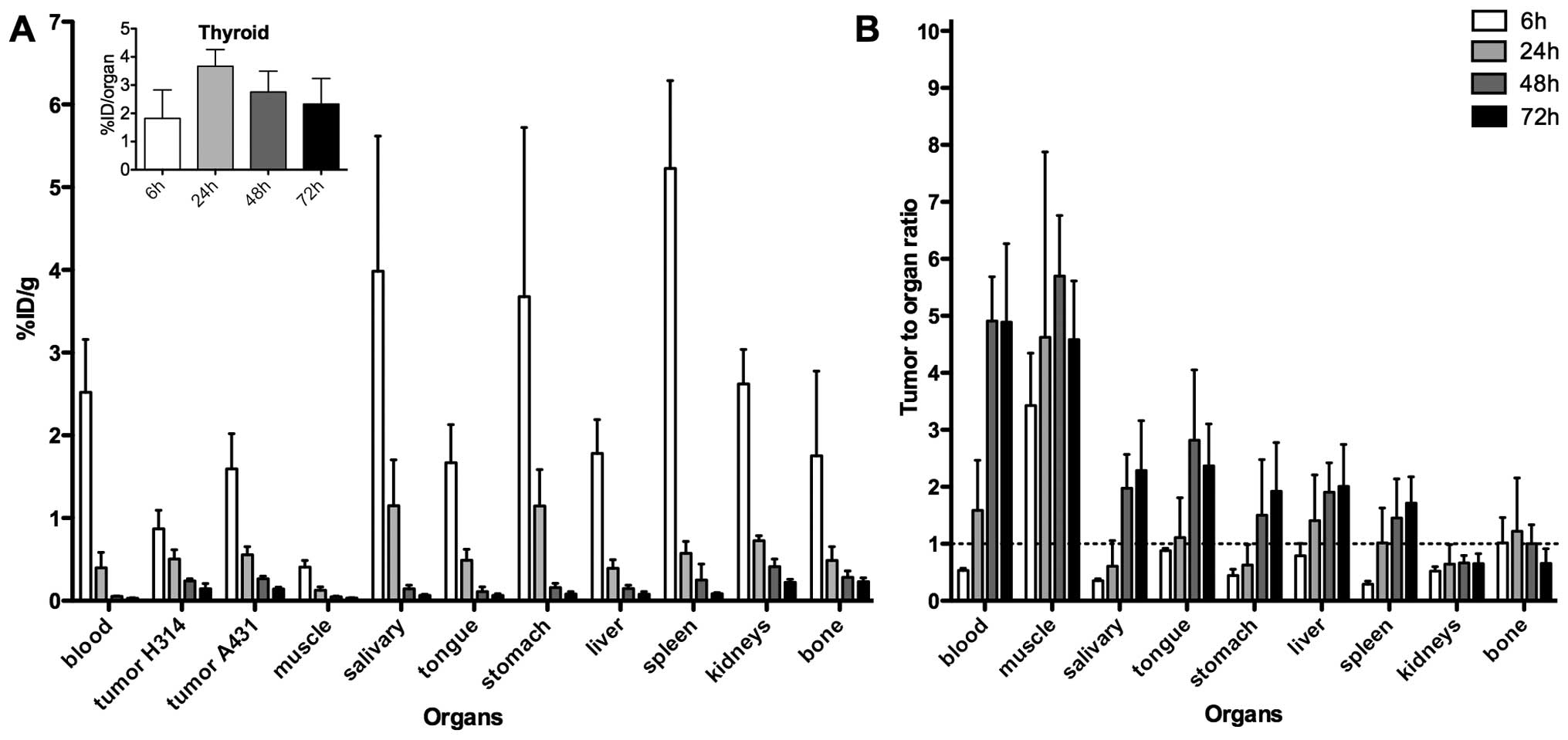
Ex vivo biodistribution study using
125I-AbD19384
Uptake of 125I-AbD19384 in tumors, blood,
and selected organs can be seen in Fig. 3A, and a comparison between
125I-AbD19384 and 125I-AbD15179 uptake in
liver and spleen is shown in Table
I. A pronounced clearance and decrease in activity over time
was seen for all organs, except thyroid, where activity remained
stable over time (inset, Fig 3A).
At 6 h p.i., a significant difference (P=0.02) in tumor uptake
between the high- and moderate-CD44v6-expressing tumors was
observed, with 1.60±0.42%ID/g in the high expressing A431 tumor,
compared to 0.92±0.24%ID/g in the low expressing H314 tumor. This
difference did, however, disappear over time. Activity in blood at
6 h p.i. was 2.52±0.64%ID/g, declining to 0.33±0.22%ID/g at 24 h
p.i. and 0.03±0.01%ID/g at the latest time-point. The organs with
the highest initial activity were the spleen, the stomach and the
salivary glands. Tumor-to-organ-ratios, calculated based on the
A431 tumors, can be seen in Fig.
3B. Tumor-to-organ ratios for 125I-AbD19384
increased over time for most organs of interest, as well as for
blood up to 48 h p.i. Tumor to blood ratio for
125I-AbD19384 was below one at 6 h p.i., however, at 72
h the ratio had increased to 4.90±1.38%ID/g. Tumor-to-organ ratios
above one were demonstrated in the organs in the head and neck
region at the later time-points.
 | Table IComparison of %ID/g of
125I-AbD19384 and 125I-AbD15179 (18) in liver and kidneys, as obtained by
ex vivo organ distribution. |
Table I
Comparison of %ID/g of
125I-AbD19384 and 125I-AbD15179 (18) in liver and kidneys, as obtained by
ex vivo organ distribution.
| | 6 h | 24 h | 48 h | 72 h |
|---|
| |
|
|
|
|
|---|
| Organ | Tracer | Mean | SD | Mean | SD | Mean | SD | Mean | SD |
|---|
| Blood | AbD15179 | 1.97 | 0.35 | 0.39 | 0.08 | 0.14 | 0.11 | 0.051 | 0.008 |
| AbD19384 | 2.52 | 0.64 | 0.40 | 0.19 | 0.054 | 0.003 | 0.030 | 0.006 |
| Tumor (A431) | AbD15179 | 1.58 | 0.12 | 0.60 | 0.09 | 0.44 | 0.27 | 0.22 | 0.04 |
| AbD19384 | 1.59 | 0.43 | 0.56 | 0.10 | 0.27 | 0.03 | 0.14 | 0.02 |
| Liver | AbD15179 | 1.09 | 0.19 | 0.30 | 0.06 | 0.12 | 0.06 | 0.067 | 0.012 |
| AbD19384 | 1.78 | 0.41 | 0.39 | 0.10 | 0.15 | 0.04 | 0.078 | 0.032 |
| Kidneys | AbD15179 | 6.46 | 2.18 | 1.37 | 0.25 | 0.78 | 0.31 | 0.57 | 0.09 |
| AbD19384 | 2.62 | 0.42 | 0.73 | 0.06 | 0.41 | 0.09 | 0.22 | 0.04 |
Ex vivo biodistribution and in vivo
small animal PET study with 124I-AbD19384
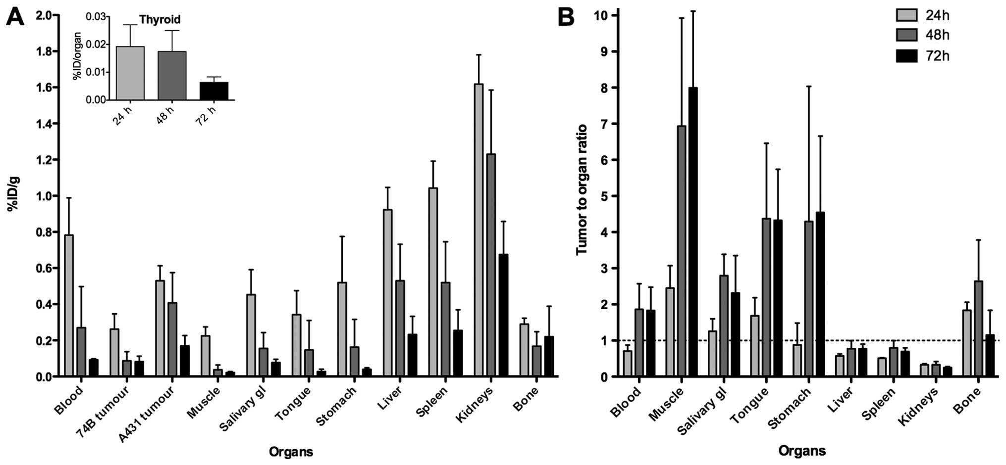
Uptake of 124I-AbD19384 in tumors, blood,
and selected organs, as assessed by ex vivo organ
distribution, are presented in Fig.
4A. A pronounced clearance and decrease in activity over time
was seen for all organs, except thyroid, where activity was very
low, and remained stable over time (inset, Fig 4A). At all three time-points, a
significant difference in tumor uptake between the high- and
low-CD44v6-expressing tumors was observed, with A431 tumors
displaying on average 2.3, 3.5 and 2.0 times higher uptake than
UM-SCC74B tumors at 24, 48 and 72 h p.i., respectively. Activity in
blood at 24 h p.i. was 0.78±0.21%ID/g, declining to 0.09±0.005%ID/g
at 72 h p.i. The organs with the highest initial activity were the
kidneys, the spleen and the liver. Tumor-to-organ ratios,
calculated based on the A431 tumors, can be seen in Fig. 4B, and tumor-to-blood ratios are
listed in Table II.
Tumor-to-organ ratios were below one for liver, spleen and kidneys
at all time-points, and above two for the late time-points in the
organs in the head and neck region. Biodistribution data verified
that animals imaged by PET were representative for each group.
| Table IIComparison of tumor uptake of
124I-AbD19384 relative to blood (heart), as obtained by
PET imaging and ex vivo organ distribution. |
Table II
Comparison of tumor uptake of
124I-AbD19384 relative to blood (heart), as obtained by
PET imaging and ex vivo organ distribution.
| | 24 h | 48 h | 72 h |
|---|
| |
|
|
|
|---|
| Tumor type | Mode | Mean | SD | N | Mean | SD | N | Mean | SD | N |
|---|
| A431 | PET | 0.83 | 0.18 | 4 | 1.32 | 0.32 | 3 | 1.44 | 0.58 | 3 |
| Ex vivo | 0.71 | 0.17 | 4 | 1.86 | 0.71 | 4 | 1.83 | 0.64 | 4 |
| UM-SCC74B | PET | 0.33 | 0.03 | 3 | 0.49 | 0.36 | 3 | 0.99 | 0.29 | 3 |
| Ex vivo | 0.34 | 0.04 | 4 | 0.52 | 0.38 | 4 | 0.90 | 0.29 | 4 |
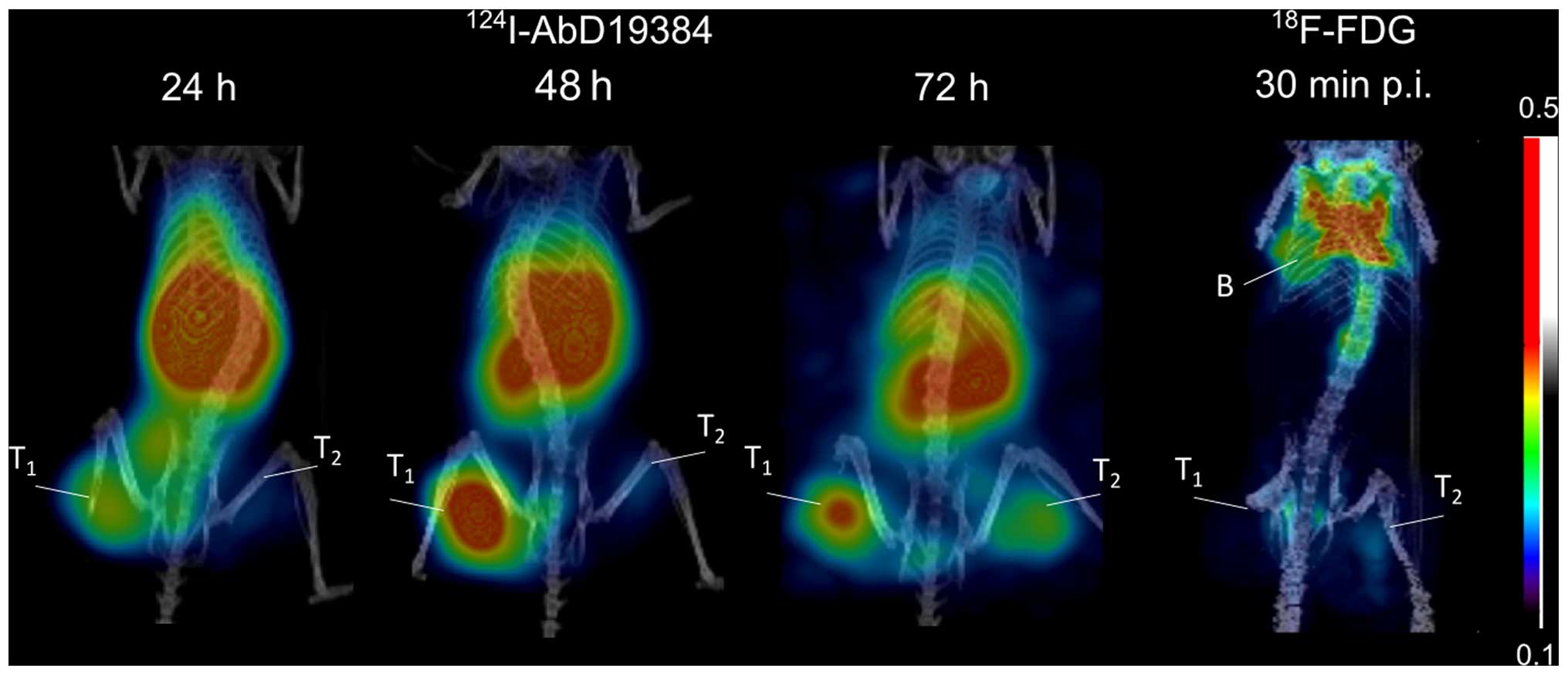
Representative PET images of
124I-AbD19384 at 24, 48 and 72 h p.i., as well as
18F-FDG 30 min p.i., can be seen in Fig. 5. The high CD44v6-expressing A431
tumors were visualized at all three time-points, with 48 h p.i.
providing best contrast. UM-SCC74B tumors were visualized to a
lower extent, in line with the biodistribution data of the
conjugate. Tumor to blood ratios were also calculated from the PET
examinations and compared to ratios generated from ex vivo
biodistribution measurements (Table
I). Results were in good agreement (no statistical differences
between any pair of measurements). 18F-FDG demonstrated
faint visualization of tumors, no discrepancy between the high- and
low-CD44v6 expressing tumor, and some uptake in brown adipose
tissue (Fig. 5).
Discussion
The objective of the present study was to evaluate
for the first time the in vitro and in vivo binding
properties of the radio-iodinated human bivalent fragment AbD19384
for targeting of CD44v6-expressing tumors, and to assess the
utility of Fab-dHLX constructs as targeting agents for molecular
imaging. As CD44v6 is overexpressed in a number of epithelial
tumors AbD19384 could prove to be valuable as a targeting molecule
for a number of cancers, for example HNSCC. To the best of our
knowledge, this is the first time a Fab-dHLX construct has been
used for in vivo targeting and molecular imaging. Previous
in vivo studies using a dHLX construct have, however,
assessed the possibility of antibody-mediated toxicity
neutralization in mice using Fab-dHLX (16), or scFv-dHLX (17) by pre-incubation of targeting
molecule and target with promising results.
In the present study, antigen specificity, binding
properties, and interaction analysis of AbD19384 were first
assessed in vitro. In vivo specificity and
biodistribution of 125I-AbD19384 was then evaluated in
tumor-bearing mice using a dual-tumor setup. Finally, AbD19384 was
labeled with 124I, and its imaging properties were
assessed in mice bearing tumors of both low and high CD44v6
expression using small animal PET/CT and compared to
18F-FDG.
SPR measurements clearly demonstrated in
vitro specificity of AbD19384, and an improved apparent
affinity compared to the monovalent AbD15179 fragment (Fig. 1). AbD19384 did not bind to the
murine v6-peptide. Binding of iodinated AbD19384 conjugates was
also followed in real-time in vitro on cultured tumor cells
with different CD44v6-expression (Fig.
2A). Results further confirmed the specific binding of AbD19384
to CD44v6. Binding and retention on CD44v6-positive cells was in
agreement with SPR measurements, with a slower off-rate for
AbD19384 (Fig. 2A) than previously
observed for the monovalent AbD15179 conjugate on cultured tumor
cells (7,14,18).
The impact of labeling method (using chloramine-T or Iodogen) and
radiohalogen (125I or 124I) was also assessed
in vitro (Fig. 2B). No
significant differences between the binding interactions of the
different conjugates could be seen, demonstrating that the in
vitro binding of the conjugates was not significantly
influenced by labeling method or radionuclide in the present
study.
In vivo specificity of
125I-AbD19384 was assessed by comparing uptake of
125I-AbD19384 in mice bearing both MDA-MB-231 tumors
(CD44v6 negative) and A431 tumors (high CD44v6 expression). Uptake
in A431 tumors was shown to be significantly higher (four times
higher) than the negative control tumors, confirming the specific
binding of AbD19384 to CD44v6 also in vivo. A full
biodistribution study of 125I-AbD19384 was performed in
mice bearing H314 tumors (medium CD44v6-expression) and high
CD44v6-expressing A431 tumors. Biodistribution data were favorable,
with a pronounced clearance and decrease in activity over time for
most organs, and tumor to blood ratios around five from 48 h p.i.
(Fig. 3). The high uptake in
thyroid, and initially high uptake in salivary glands and stomach,
where Na/I symporters are abundantly expressed, can probably be
attributed to radio-catabolites of 125I (19).
Biodistribution of 125I-AbD19384 was
similar to the previously observed biodistribution of the
monovalent 125I-AbD15179 fragment (18), with a maximum uptake in organs at
the first time-point and fast clearance of the conjugate. As seen
in Table I, kidney uptake was
reduced with ~50% for the bivalent fragment, whereas uptake in
liver was only slightly increased. The reduced kidney uptake was
expected, since AbD19384 displays a size above the renal threshold,
whereas AbD15179 does not (20).
However, since the bivalent Fab-dHLX construct is in equilibrium
with a smaller fraction of the monovalent form, some monovalent
tracers are expected to contribute to the biodistribution. It has
previously been hypothesized that this may be an advantage in a
tumor targeting setting, where monomers may penetrate tumors better
and create effective dimers once at the target cells, due to the
increased local concentration of the dimerization domains at the
target site (9). In the present
study, uptake in A431 tumors of monovalent 125I-AbD15179
(18) was comparable to that of
bivalent 125I-AbD19384 at all time-points with no
statistically significant difference (Table I). The bivalent fragment could have
been expected to exhibit higher tumor uptake due to slower off-rate
(21), as seen in the in
vitro measurements (Fig. 2),
but this was not confirmed in the in vivo studies. Thus, we
conclude that both radio-iodinated AbD15179 and AbD19384 are
suitable for CD44v6-specific tumor targeting in vivo, where
AbD19384 can provide a lower kidney uptake than AbD15179 at similar
tumor uptake levels. It is possible that tumor uptake for AbD19384
could be further increased by using a residualizing radiolabel,
since a bivalent fragment may trigger an increased level of
internalization, which could counteract a lower off-rate in the
case of non-residualizing radioiodine label (22).
Due to the favorable biodistribution properties seen
in the 125I-AbD19384 study, 124I was selected
for an imaging study and AbD19384 was subsequently labeled with
124I and assessed in PET/CT examinations, complemented
with ex vivo biodistribution measurements (Figs. 4 and 5). Biodistribution measurements
demonstrated more than two times higher uptake (calculated as
%ID/g) in high CD44v6-expressing A431 tumors compared to the low
CD44v6-expressing UM-SCC74B tumors at all time-points. This was
also shown by PET imaging, yielding higher contrast and higher
uptake in A431 tumors at all three time-points. Uptake and
elimination of 124I-AbD19384 was similar to
125I-AbD19384, with a maximum uptake in all organs at
the first time-point and fast clearance of the conjugate. There was
no significant difference in A431 tumor uptake between
124I-AbD19384 and 125I-AbD19384 conjugates.
However, activity in blood was approximately two times higher for
124I-AbD19384 than for 125I-AbD19384 at 24 h
p.i., resulting in a corresponding increase in the blood rich
organs such as liver, spleen and kidneys for
124I-AbD19384. Uptake of 124I in thyroid was
successfully blocked by adding potassium iodide in the drinking
water one day before measurements. Furthermore, radioactivity
uptake in salivary glands and stomach was reduced with ~2/3 at 24 h
p.i., most probably also a result of blocking. Even though the
higher blood activity of 124I-AbD19384 resulted in lower
tumor-to-blood ratios than obtained with 125I-AbD19384,
A431 tumors were clearly visualized at all time-points, with the
best contrast achieved at 48 h p.i. PET/CT images were in line with
the biodistribution measurements, and tumor-to-blood ratios
calculated from PET images were in very close agreement with the
ex vivo data (Table II),
further confirming the reliability of the non-invasive in
vivo imaging technique.
In order to compare 124I-AbD19384 with
standard imaging methods, 18F-FDG was also employed as
an imaging agent in PET/CT scans, and data were confirmed by ex
vivo biodistribution measurements. There was no clear
visualization of the tumors using 18F-FDG. Furthermore,
there was no clear discrimination between high CD44v6 expressing
A431 tumors and low CD44v6 expressing UM-SCC74B tumors, since
18F-FDG uptake is generally proportional to the
metabolic activity in tissues and is not specific to tumor cells.
High uptake of 18F-FDG was observed in brown adipose
tissue in the examinations (Fig.
5). This highlights one of the potential imaging pitfalls
sometimes found also in clinical 18F-FDG-PET during
tumor staging (23) and emphasizes
the need to complement this method with a more tumor-specific
imaging technique.
In conclusion, our results describe for the first
time a recombinant human Fab-dHLX fusion protein targeted to
CD44v6, that possess high affinity, target specificity and
potential for in vivo imaging of tumor biomarkers. This
bivalent Fab antibody, engineered from monovalent Fab AbD15179 by
subcloning of the Fab gene in fusion with a self-dimerizing
helix-turn helix motif, was successfully produced and
radioiodinated for molecular imaging of CD44v6 in vivo.
Biodistribution and small-animal PET studies demonstrated that
124I-AbD19384 is a promising PET probe for imaging
CD44v6 antigen expression in vivo. Furthermore, this
proof-of-concept research established the feasibility of using
recombinant Fab-dHLX constructs for in vivo imaging of tumor
biomarkers. Additionally, Fab-dHLX constructs could be useful in
the theranostic nanoplatform setting as a targeting ligand, and
with further development of the other components of a successful
theranostic agent it could also prove to be a valuable addition to
personalized cancer treatment.
Acknowledgements
The authors would like to thank Jonas Stenberg for
help with LigandTracer analysis and Veronika Asplund for help with
18F-FDG injections. The authors would like to
acknowledge the Swedish Research Council, the Swedish Cancer
Society, the Swedish Association for Medical Research and the
Sweden America Foundation for kind support.
References
|
1
|
Chames P, Van Regenmortel M, Weiss E and
Baty D: Therapeutic antibodies: Successes, limitations and hopes
for the future. Br J Pharmacol. 157:220–233. 2009. View Article : Google Scholar : PubMed/NCBI
|
|
2
|
Olafsen T and Wu AM: Antibody vectors for
imaging. Semin Nucl Med. 40:167–181. 2010. View Article : Google Scholar : PubMed/NCBI
|
|
3
|
Hoeben BA, Kaanders JH, Franssen GM,
Troost EG, Rijken PF, Oosterwijk E, van Dongen GA, Oyen WJ, Boerman
OC and Bussink J: PET of hypoxia with 89Zr-labeled
cG250-F(ab′)2 in head and neck tumors. J Nucl Med.
51:1076–1083. 2010. View Article : Google Scholar : PubMed/NCBI
|
|
4
|
Willkomm P, Bender H, Bangard M, Decker P,
Grünwald F and Biersack HJ: FDG PET and immunoscintigraphy with
99mTc-labeled antibody fragments for detection of the
recurrence of colorectal carcinoma. J Nucl Med. 41:1657–1663.
2000.PubMed/NCBI
|
|
5
|
Khawli LA, Alauddin MM, Hu P and Epstein
AL: Tumor targeting properties of indium-111 labeled genetically
engineered Fab' and F(ab')2 constructs of chimeric tumor
necrosis treatment (chTNT)-3 antibody. Cancer Biother Radiopharm.
18:931–940. 2003. View Article : Google Scholar
|
|
6
|
Bradbury AR, Sidhu S, Dübel S and
McCafferty J: Beyond natural antibodies: The power of in vitro
display technologies. Nat Biotechnol. 29:245–254. 2011. View Article : Google Scholar : PubMed/NCBI
|
|
7
|
Nilvebrant J, Kuku G, Björkelund H and
Nestor M: Selection and in vitro characterization of human
CD44v6-binding antibody fragments. Biotechnol Appl Biochem.
59:367–380. 2012. View Article : Google Scholar
|
|
8
|
Reichert JM and Dhimolea E: The future of
antibodies as cancer drugs. Drug Discov Today. 17:954–963. 2012.
View Article : Google Scholar : PubMed/NCBI
|
|
9
|
Pack P and Plückthun A: Miniantibodies:
Use of amphipathic helices to produce functional, flexibly linked
dimeric FV fragments with high avidity in Escherichia coli.
Biochemistry. 31:1579–1584. 1992. View Article : Google Scholar : PubMed/NCBI
|
|
10
|
Eisenberg D, Wilcox W, Eshita SM, Pryciak
PM, Ho SP and DeGrado WF: The design, synthesis, and
crystallization of an alpha-helical peptide. Proteins. 1:16–22.
1986. View Article : Google Scholar : PubMed/NCBI
|
|
11
|
Orian-Rousseau V: CD44, a therapeutic
target for metastasising tumours. Eur J Cancer. 46:1271–1277. 2010.
View Article : Google Scholar : PubMed/NCBI
|
|
12
|
Pluckthun A and Pack P: New protein
engineering approaches to multivalent and bispecific antibody
fragments. Immunotechnology. 3:83–105. 1997. View Article : Google Scholar : PubMed/NCBI
|
|
13
|
Nestor M, Sundström M, Anniko M and
Tolmachev V: Effect of cetuximab in combination with
alpha-radioimmunotherapy in cultured squamous cell carcinomas. Nucl
Med Biol. 38:103–112. 2011. View Article : Google Scholar : PubMed/NCBI
|
|
14
|
Stenberg J, Spiegelberg D, Karlsson H and
Nestor M: Choice of labeling and cell line influences interactions
between the Fab fragment AbD15179 and its target antigen CD44v6.
Nucl Med Biol. 41:140–147. 2014. View Article : Google Scholar
|
|
15
|
Hunter WM and Greenwood FC: Preparation of
iodine-131 labelled human growth hormone of high specific activity.
Nature. 194:495–496. 1962. View Article : Google Scholar : PubMed/NCBI
|
|
16
|
Larkin EA, Stiles BG and Ulrich RG:
Inhibition of toxic shock by human monoclonal antibodies against
staphylococcal entero-toxin B. PLoS One. 5:e132532010. View Article : Google Scholar
|
|
17
|
Kalinke U, Krebber A, Krebber C, Bucher E,
Plückthun A, Zinkernagel RM and Hengartner H: Monovalent
single-chain Fv fragments and bivalent miniantibodies bound to
vesicular stomatitis virus protect against lethal infection. Eur J
Immunol. 26:2801–2806. 1996. View Article : Google Scholar : PubMed/NCBI
|
|
18
|
Haylock AK, Spiegelberg D, Nilvebrant J,
Sandström K and Nestor M: In vivo characterization of the novel
CD44v6-targeting Fab fragment AbD15179 for molecular imaging of
squamous cell carcinoma: A dual-isotope study. EJNMMI Res.
4:112014. View Article : Google Scholar : PubMed/NCBI
|
|
19
|
Portulano C, Paroder-Belenitsky M and
Carrasco N: The Na+/I− symporter (NIS):
Mechanism and medical impact. Endocr Rev. 35:106–149. 2014.
View Article : Google Scholar :
|
|
20
|
Holechek MJ: Glomerular filtration: an
overview. Nephrol Nurs J. 30:285–290; quiz 291-282. 2003.PubMed/NCBI
|
|
21
|
Rudnick SI and Adams GP: Affinity and
avidity in antibody-based tumor targeting. Cancer Biother
Radiopharm. 24:155–161. 2009. View Article : Google Scholar : PubMed/NCBI
|
|
22
|
Schreiber AB, Libermann TA, Lax I, Yarden
Y and Schlessinger J: Biological role of epidermal growth
factor-receptor clustering. Investigation with monoclonal
anti-receptor antibodies. J Biol Chem. 258:846–853. 1983.PubMed/NCBI
|
|
23
|
Truong MT, Erasmus JJ, Munden RF, Marom
EM, Sabloff BS, Gladish GW, Podoloff DA and Macapinlac HA: Focal
FDG uptake in mediastinal brown fat mimicking malignancy: A
potential pitfall resolved on PET/CT. AJR Am J Roentgenol.
183:1127–1132. 2004. View Article : Google Scholar : PubMed/NCBI
|