Introduction
Metal complexes have important applications in many
aspects of life. In recent years, the successful development of
metal-based pharmaceuticals, including copper anticancer agents,
has attracted more attention on metal complexes in cancer treatment
(1–3). Studies have shown that the copper
complexes, such as dithiocarbamate (4,5),
8-hydroxyquinoline (6), clioquinol
(7) and some Schiff base copper
coordination compounds (8–10), exert promising anticancer activity
by targeting the ubiquitin-proteasome pathway (UPP) in preclinical
trials.
The UPP selectively mediates cellular protein
degradation which regulates cell cycle progression, signal
transduction, differentiation, proliferation and apoptosis
(11). The tagged proteins are
degraded by the 26S proteasome, a huge cylinder-like protein
complex, consists of one 20S core particle and two 19S regulatory
particles (12). The 20S core
particle comprises four packed rings, and each ring consists of 7
distinct subunits: the outer two rings are α subunits, acting like
a ‘gate’ and controlling which proteins may go through into the
cylinder; the inner two rings are β subunits, which form a
catalytically active protease and are responsible for protein
degradation (13). In proteasome
of mammal cells, all α and β subunits are distinct and only
subunits β1, β2 and β5 are catalytically active, which are
responsible for caspase or peptidyl-glutamyl peptide-hydrolyzing
(PGPH)-like activity, trypsin-like and chymotrypsin (CT)-like,
respectively (14–16).
Previous research has demonstrated that the
inhibition of proteasomal CT-like activity is closely associated
with tumor cell apoptosis (17–20).
Therefore, it is no surprise that selective inhibition of
proteasome in cancer cells could be a potential anticancer
strategy. To date, the full structure of human 20S proteasome has
not been reported yet. By BLAST sequence search against PDB
library, bovine 20S proteasome (PDB code: 1IRU) was discovered to
have very high sequence homology to human 20S proteasome,
especially for the catalytic subunits, β1, β2 and β5 (identity,
96.7, 98.5 and 98.0%, respectively) (21). Despite a few differences, for
example, β1 V6A/E205G and β2 C91Y, these 3 subunits are completely
identical between bovine and human. Thus, after some minor
modifications and energy optimization, this bovine 20S proteasome
structure could be directly used as receptor of docking
studies.
We previously reported that some metal complexes
possess the ability to inhibit the CT-like activity of proteasome
and induce cancer cell death in human cancer cells (22–25).
However, the involved mechanism is unknown. Hence, our results
prompted us to obtain further detailed understanding on such copper
complexes as anticancer agent candidates. In the present study, we
studied the effect of three ternary copper complexes with
3-indolecarboxylic and 1,10-phenanthroline in human breast cancer
cells. Similar to the previous finding in prostate cancer cells,
all these copper complexes could inhibit the CT-like activity of
proteasome and induce apoptosis. Furthermore, molecular docking
simulation was used to analyze the possible interaction between
these complexes and the catalytically active 20S proteasome. These
three complexes were found to have best binding with catalytic site
of subunit β5 and formed a few hydrogen bonds and hydrophobic
interactions with the receptor. Taken together, the information
from the present study provides a basis for developing promising
proteasome inhibitors in cancer treatment.
Materials and methods
Chemicals and reagents
3-indoleacetic acid (IAA) and 3-indolepropionic acid
(IPA), 3-indolebutyric acid (IBA) were purchased from J&K
Scientific Ltd. (Beijing, China). 1,10-Phenanthroline, DMSO and
copper acetate were obtained from Aladdin Co. The complexes
IAA-Cu-phen, IPA-Cu-phen and IBA-Cu-phen were synthesized in Jining
Medical University Pharmaceutical College (Rizhao, China) and were
dissolved by DMSO to 25 mM.
3-[4,5-dimethylthiazol2-yl]-2.5-diphenyl-tetrazolium bromide (MTT)
were obtained from Sigma-Aldrich (St. Louis, MO, USA). The
fluorogenic peptide substrate Suc-LLVY-AMC was obtained from
Calbiochem (San Diego, CA, USA). Mouse monoclonal antibody to human
poly(ADP-ribose) polymerase (PARP), mouse monoclonal antibody
against ubiquitin (Ub), goat polyclonal antibody against β-actin
(C-11) and all secondary antibodies were purchased from Cell
Signaling Technology, Inc. (Shanghai, China).
Instruments
Infrared spectra were performed on a Nicolet 170-SX
spectrophotometer using KBr pellets. 1H NMR spectra were
collected at a Bruker AVANCE III (600-MHz) spectrometer.
13C NMR spectra were carried out with a Bruker AV600
(600-MHz) spectrometer. Thermal analyses were recorded on a NETZSCH
thermal analyzer. Elemental chemical analysis (C, H and N) were
obtained on a Perkin-Elmer 2400 analyzer. Cellular changes were
observed using a Zeiss Axiovert 25 microscope.
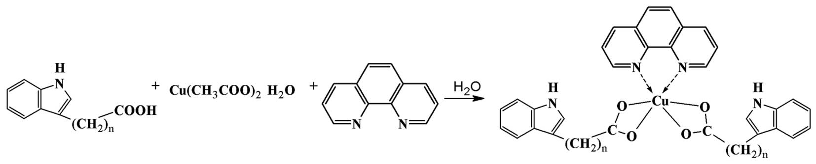
Synthesis of the complexes
The complexes were synthesized in two steps by
following a previously described procedure (22). In the first step, ligand IAA (0.351
mg, 2 mmol), IPA (0.378 mg, 2 mmol), or IBA (0.406 mg, 2 mmol) were
dissolved in ethyl alcohol to give a solution, respectively.
Cu(CH3COO)2. H2O (0.199 mg, 1
mmol), dissolved in ethyl alcohol, was added dropwise to the above
mentioned solution and reacted for 4 h to give a green solution. In
the second step, C12H8N2 (0.180
mg, 1 mmol) dissolved in ethyl alcohol, was added into the solution
of the first step and then reacted for 3 h to give a green
precipitate, which was filtered to give the product.
IAA-Cu-phen
[Cu(C10H8O2N)2(C12H8N2)],
FW=592.10 g·mol−1. Yield, 72%. IR (KBr,
cm−1): 3389.63 υ(-NH-); 1576.24
υas(COO−); 1408.06
υs(COO−); 528.27 υ(M-N); 476.85 υ(M-O);
1H NMR (DMSO, 600 MHz): d (ppm) 10.736 (s, -NH-, indole
ring); 8.931 (d, H-2, 9, 1,10-phenanthroline); 7.187 (s, H-5, 6,
1,10-phenanthroline); 7.352, 7.560 (d, H-4, 7, indole ring); 6.813,
6.975 (t, H-5, 6, indole ring); 7.113 (d, H-2, indole ring); 3.367
(s, -CH2-indole ring); 13C NMR (DMSO): d
(ppm) 184.35 (-COO); 155.28, 142.10, 133.14, 125.29
(1,10-phenanthroline carbons); 138.82, 127.41, 122.56, 121.27,
118.18, 117.93, 116.37 and 109.25 (indole ring carbons); 20.80
(-CH2). TG analysis: residue 14.05% (calculated 13.43%,
CuO). Anal. Calc. for Cu
(C10H8O2N)2
(C12H8N2): C, 64.91; H, 4.09; N,
9.46. Found (%): C, 64.84; H, 4.21; N, 9.32. Molar conductivity:
Λm (S•cm−1•mol−1): 13.22.
IP-Cu-phen
[Cu(C11H10O2N)2(C12H8N2)],
FW=620.16 g·mol−1. Yield, 69%. IR (KBr,
cm−1): 3405.72 υ(-NH-); 1586.96
υas(COO−); 1417.58
υs(COO−); 546.34 υ(M-N); 497.64 υ(M-O);
1H NMR (DMSO, 600 MHz): d (ppm) 10.473 (s, -NH-, indole
ring); 8.722 (d, H-2, 9, 1,10-phenanthroline); 7.262 (s, H-5, 6,
1,10-phenanthroline); 7.606, 7.398 (d, H-4, 7, indole ring); 6.877,
7.044 (t, H-5, 6, indole ring); 7.039 (d, H-2, indole ring); 4.372
(s, -CH2-indole ring); 3.420 (t, -CH2-indole
ring). 13C NMR (DMSO): d (ppm) 179.63(-COO); 157.32,
145.10, 140.81, 133.05 (1,10-phenanthroline carbons); 137.15,
129.38, 124.87, 119.04, 118.23, 117.65, 112.21 and 110.52 (indole
ring carbons); 25.19, 22.66 (-CH2). TG analysis: residue
13.26% (calculated 12.83%, CuO). Anal. Calc. for Cu
(C11H10O2N)2
(C12H8N2): C, 65.85; H, 4.55; N,
9.03. Found (%): C, 65.79; H, 4.60; N, 8.97. Molar conductivity:
Λm (S•cm−1•mol−1): 8.29.
IBA-Cu-phen
[Cu(C12H12O2N)2(C12H8N2)],
FW=648.21g·mol−1. Yield, 66%. IR (KBr, cm−1):
3417.19 υ(-NH-); 1592.77 υas(COO−); 1423.03
υs(COO−); 551.20 υ(M-N); 483.12 υ(M-O);
1H NMR (DMSO, 600 MHz): d (ppm) 10.559 (s, -NH-, indole
ring); 8.806 (d, H-2, 9, 1,10-phenanthroline); 7.224 (s, H-5, 6,
1,10-phenanthroline); 7.587, 7.390 (d, H-4, 7, indole ring); 6.846,
7.012 (t, H-5, 6, indole ring); 7.156 (d, H-2, indole ring); 4.437
(s, -CH2-indole ring); 3.382 (t, -CH2-indole
ring); 3.053 (t, -CH2-indole ring). 13C NMR
(DMSO): d (ppm) 180.76 (-COO); 153.27, 141.01, 139.84, 129.69
(1,10-phenanthroline carbons); 137.92, 127.58, 123.30, 120.16,
118.88, 117.43, 112.87 and 109.53 (indole ring carbons); 26.44,
24.60, 21.55 (-CH2). TG analysis: residue 12.80%
(calculated 12.27%, CuO). Anal. Calc. for Cu
(C12H12O2N)2
(C12H8N2): C, 66.70; H, 4.98; N,
8.64. Found (%): C, 66.64; H, 5.08; N, 8.55. Molar conductivity:
Λm (S•cm−1•mol−1): 11.04.
Cell culture and whole-cell extract
preparation
MDA-MB-231 and MCF-7 breast cancer cells were grown
at 37°C and 5% CO2 in DMEM/F-12 (1:1) or RPMI-1640
(Invitrogen), respectively, supplemented with 10% fetal bovine
serum (FBS). A whole cell extracts were prepared and applied to
assess the CT-like activity and western blot analysis (26).
Cell proliferation assay
MTT assay was applied to evaluate the
antiproliferative effect of each complex on breast cancer cells.
Cells were seeded in a 96-well plate and grown to 70–80%
confluency, then treated with the indicated concentration of each
complex at 37°C. After cultured for 24 h, cell proliferation was
assessed as previously described (26).
Proteasome activity assay
Purified 20S human proteasome (35 ng) or whole-cell
extract (10 μg) of human breast cancer cells were incubated with a
series of concentrations of three copper complexes in 100 μl of
assay buffer (20 mM Tris-HCl, pH 7.5) with 20 μM fluorogenic
peptide substrate Suc-LLVY-AMC. After 2-h incubation at 37°C, the
production of hydrolyzed AMC groups was measured as previously
described (26).
Western blot analysis
MAD-MB-231 human breast cancer cells were treated
with the complexes as already described (27). Proteins (40 μg) were separated by
SDS-PAGE and transferred to a nitrocellulose membrane. The
expression of ubiquitin, PARP and β-actin were examined by western
blot analysis.
Molecular docking
Sequence of human 20S proteasome β subunits were
retrieved from Protein KnowledgeBase (UniProtKB). For each subunit
sequence, a BLAST search was performed on PDB library and bovine
20S proteasome [PDB code 1IRU (21)] was found to be the most homologous
structure to human and was chosen for this docking study after some
minor modifications. The small molecules were sketched manually and
energy minimized and then docked to all three catalytic sites of
the proteasome. Molecular docking was performed with the CCDC Gold
5.2 (28). Scoring function was
set to ChemPLP, and other docking parameters were set to their
default values. Accelrys Discovery Studio Visualizer 4.0 was used
for result analysis and figure generation.
Statistical analysis
Statistical analysis was performed using Graphpad
Prism 5 software. Differences between groups were analyzed using
the Student's t-test.
Results
Synthesis and characterization of three
complexes
3-indolecarboxylic acid copper complexes could
exhibit potent anticancer activity through inhibiting proteasome
activity (22). Whereas the exact
mechanism is still unknown, there is a hypothesis that the copper
complexes interact with proteasome β5 catalytically active subunit.
At the same time, the length of linker between indole ring and
carboxylic acid group has a certain impact on the activity of these
complexes. In the present study, we synthesized three copper
complexes with similar structure: IAA-Cu-phen, IPA-Cu-phen and
IBA-Cu-phen (Fig. 1), among which
IBA-Cu-phen is novel, and then compared their anticancer activity
and investigated the potential mechanism of action. The obtained
complexes were characterized by IR, 1H NMR,
13C NMR, TG analysis and elemental analysis. Because
these compounds were not crystallized, X-ray diffraction was not
performed.
According to the IR spectra data of ligands and
complexes (IAA-Cu-phen, IPA-Cu-phen and IBA-Cu-phen), the strong
absorption peaks at 3389.63, 3405.81 and 3417.19 cm−1,
respectively, belong to the -NH- group. No significant shifts were
observed on the peaks among these ligands, suggesting that nitrogen
atom in the indole ring did not coordinate with copper. Compared
with the IR spectra of the ligands, two new peaks in the range of
1576.24 cm−1–1592.77 cm−1 and 1408.06
cm−1–1423.03 cm−1 were assigned to
υas(COO−) and υs(COO−),
respectively. In addition, the magnitude between
υas(COO−) and υs(COO−)
was <200 cm−1, indicating that the oxygen in
-COO− group formed a coordination bond with copper ion,
which is consistent with the literature (29).
The absorption vibration frequencies appeared at
476.85, 497.64 or 483.12 cm−1 in these complexes were
attributed to the formation of Cu-O bond. A new peak at 528.27,
546.34 or 551.20 cm−1, respectively, appeared in these
complexes, suggesting the formation of Cu-N bond.
The 1H NMR spectra of IAA-Cu-phen,
IPA-Cu-phen and IBA-Cu-phen showed that there were small shifts in
the signals among these ligands, suggesting the interaction between
ligands and Cu(II) ion. In addition, the signal for -NH- proton in
the spectra (10.736, 10.473 and 10.559 ppm) were still present,
while the signal for the -COOH proton of the ligands were absent in
these complexes, indicating the hydrogen atom of carboxyl group
were displaced by Cu(II) ion. In comparison with the 1H
NMR spectra of ligands, the signal of 1,10-phenanthroline did
appear in these complexes, indicating there was coordination of
1,10-phenanthroline and Cu (II) ion. The 1H NMR spectra
analysis results were further supported by 13C NMR
spectra data (1H NMR and 13C NMR data of the
ligands IAA, IPA and IBA were not shown).
Thermal analysis (TG) of complexes IAA-Cu-phen,
IPA-Cu-phen and IBA-Cu-phen were recorded in the range from 25 to
800°C. These copper-containing compounds were all decomposed
actually in one step and the residue rates of them were 14.05,
13.26 and 12.80%, respectively, which were consistent with the
calculated values (13.43, 12.83 and 12.27%).
Elemental analysis was applied to further confirm
the composition of these copper complexes. The contents of carbon,
hydrogen and nitrogen in each complex were consistent with the
calculated values, which supported the proposed structure of these
complexes.
The ligands and complexes were stable in air.
Furthermore, all the complexes were soluble in DMSO and their molar
conductivities (Λm) were 13.22, 8.2, and 11.04
S·cm2·mol−1, respectively, which were <35
S·cm2·mol−1 (30).
Consequently, the complexes IAA-Cu-phen, IPA-Cu-phen
and IBA-Cu-phen were considered as non-electrolytes and quite
stable in culture medium.
Antiproliferative effect of the complexes
in human breast cancer MDA-MB-231 and MCF-7 cell lines
We first studied effects of IAA-Cu-phen,
IPA-Cu-phen, IBA-Cu-phen and their ligands on cell growth (Fig. 2). Human breast cancer MDA-MB-231
and MCF-7 cells were treated with 1, 5, 10 and 20 μM (IAA-Cu-phen,
IPA-Cu-phen, IBA-Cu-phen) as well as their ligands for 24 h,
followed by an MTT assay. We found that these three complexes
possessed similar growth-inhibitory activity with >90%
inhibition at 20 μM in both cell lines in dose-dependent, compared
to a control treated with DMSO (Fig.
2). However, IAA-Cu-phen was most potent, IPA-Cu-phen came
next, followed by IBA-Cu-phen while none of the ligands were
active. The IC50 values for cell death induction by all
the ligands and copper complexes are given in Table I.
 | Table IIC50 values for cell death
induction by copper compounds. |
Table I
IC50 values for cell death
induction by copper compounds.
| Cell death
induction IC50 (μM) |
|---|
|
|
|---|
| Compound | MDA-MB-31 | MCF-7 |
|---|
|
Cu(CH3COO)2 | No activity | No activity |
| IAA | No activity | No activity |
| IPA | No activity | No activity |
| IBA | No activity | No activity |
| IAA-Cu-phen | 4.20 | 5.21 |
| IPA-Cu-phen | 4.71 | 6.29 |
| IBA-Cu-phen | 5.31 | 6.82 |
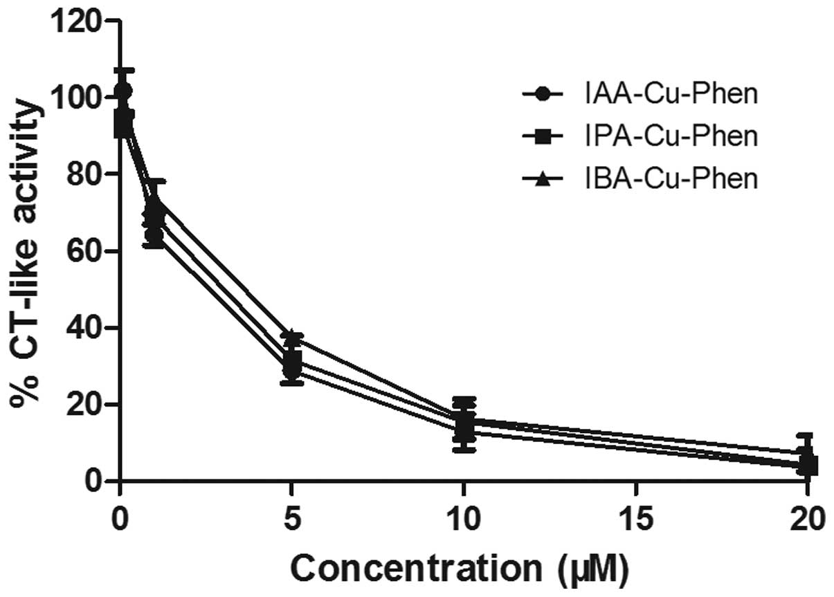
In vitro inhibition of purified 20S
proteasomal chymotryspin-like activity
In this part of the present study, we investigated
whether these three complexes could act as potent proteasome
inhibitors and possess similar effects of proteasome inhibition as
on cell proliferation. Purified human 20S proteasome were kept with
various concentrations of the three complexes for 2 h and DMSO was
used as a control. This result was consistent with MTT assays. The
three complexes inhibited the 20S proteasomal chymotrypsin-like
in vitro with rather similar IC50 (3.5–3.75 μM),
and the inhibition ability increased along the concentration of
these complexes (Fig. 3).
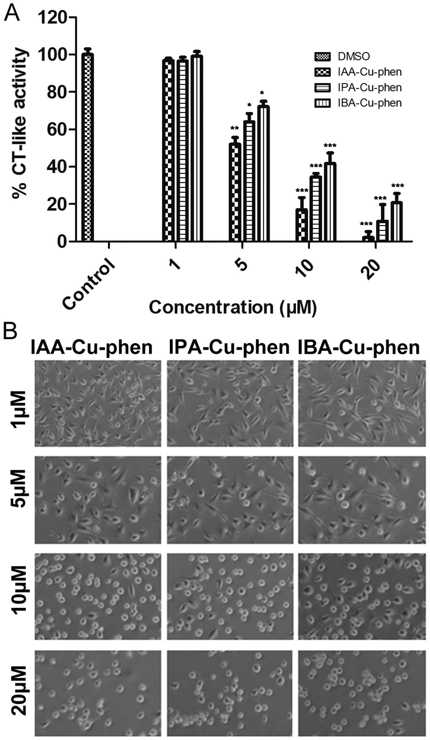
Inhibition of chymotrypsin-like activity
and induction of apoptosis by the complexes in MDA-MB-231 breast
cancer cells
Chemical structures of several complexes could play
a key role in inhibiting proteasome activity to some degree.
Therefore, we studied the effects on cellular proteasomal CT-like
inhibition and apoptosis induction of these three copper complexes
with similar structures. Human breast cancer MDA-MB-231 cells were
treated with three copper complexes at the concentrations of 1, 5,
10 and 20 μM for 24 h (DMSO as control), and cellular proteasomal
CT-like activity and morphological changes were measured. The
results indicated that all three complexes at 1 μM showed no less
than 5% inhibition of proteasomal CT-like activity (Fig. 4A) and no significant
apoptosis-associated cellular morphology changes were observed
(Fig. 4B). However, the abilities
on the proteasome inhibition of these three copper complexes were
obviously different at the concentrations of 5, 10 and 20 μM. We
found that IAA-Cu-phen was the most potent, inhibiting 48, 83 and
98% (Fig. 4A), followed by
IPA-Cu-phen, inhibiting 36%, 66 and 89 (Fig. 4A), and the last was IBA-Cu-phen,
with the degrees of inhibition being 28, 59 and 79% (Fig. 4A), respectively. Consistently,
cellular morphology changes (shrunken and rounded up) were found at
concentrations of 5, 10 and 20 μM (Fig. 4B). Our results suggested all three
copper complexes possessed the ability to inhibit the proteasome
activity and induce apoptosis in a dose-dependent manner with the
order IAA-Cu-phen >IPA-Cu-phen >IBA-Cu-phen.
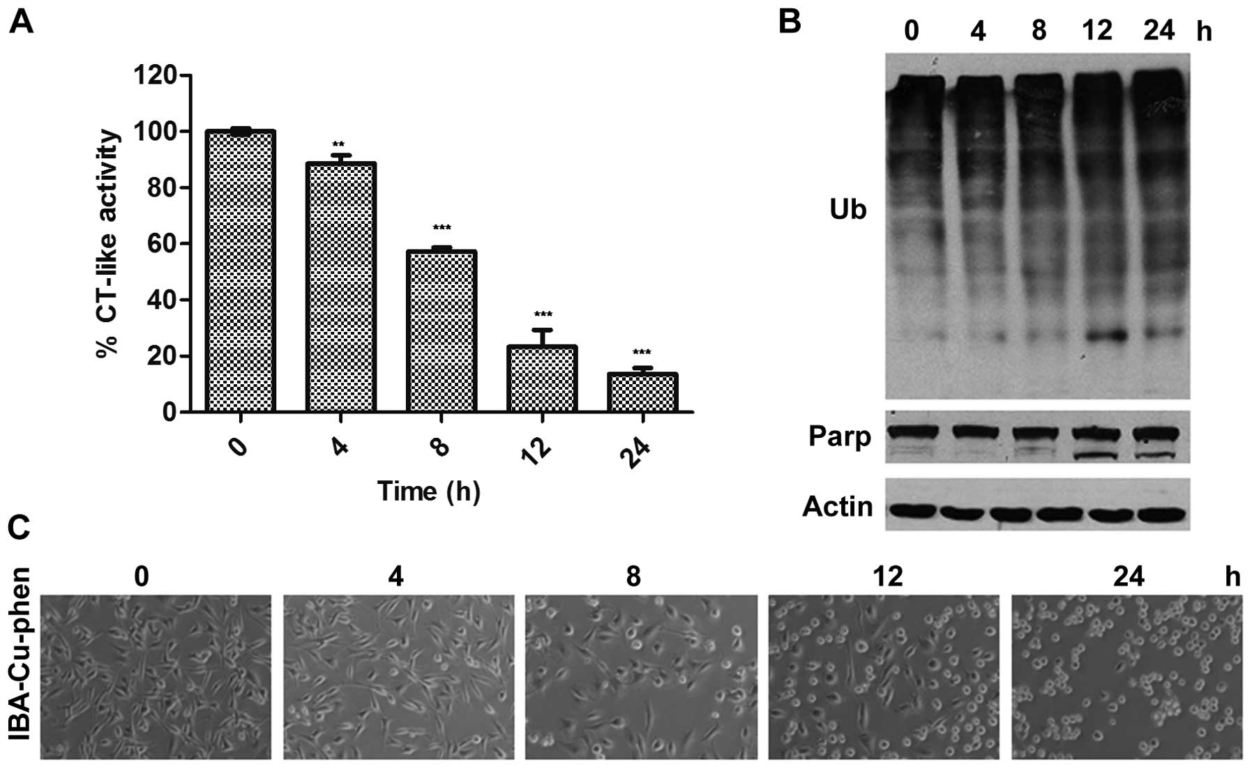
Kinetic effect of the complexes on
proteasome inhibition and apoptosis induction
Previous studies have shown that the decrease of
CT-like activity in proteasome is associated with induction of
tumor cell apoptosis (5,17–20).
Herein, we performed a kinetic experiment to investigate whether
the new complex IBA-Cu-phen possessed the same effect. MDA-MB-231
cells were treated with 20 μM compound for 0, 4, 8, 12 or 24 h,
followed by measurement of proteasomal inhibition and cell
apoptosis (Fig. 5). The result
showed that IBA-Cu-phen inhibited ~12% of proteasome activity after
4-h treatment (Fig. 5A). As time
went on, the chymotrypsin-like activity further decreased. The
inhibition ratios were 43, 77 and 86% at 8, 12 and 24 h,
respectively. Consistently, the accumulation of ubiquitinated
proteins were also detected at 4 h and the levels increased
gradually at later time-points (Fig.
5B). However, cellular apoptosis morphological changes
(Fig. 5C) and apoptosis-specific
PARP cleavage (Fig. 5B) both
appeared at 12 h and later time-point of treatment. These results
demonstrate that proteasome inhibition by IBA-Cu-phen triggers the
occurrence of tumor cell apoptosis.
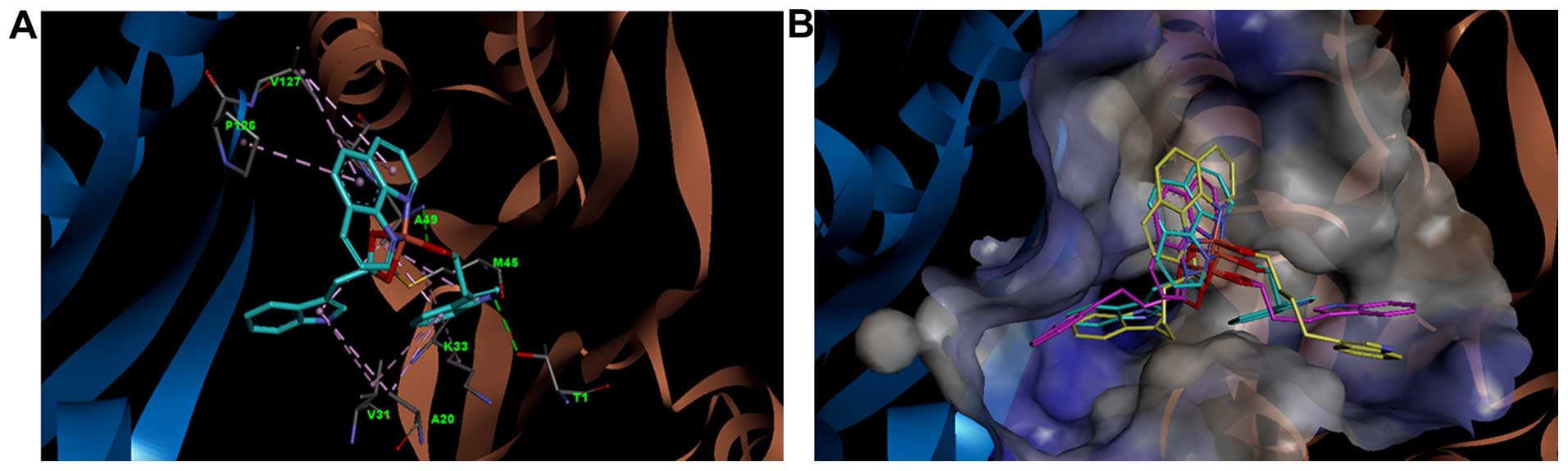
Molecular docking simulation
To further explain how and why these three copper
complexes could serve as proteasomal CT-like inhibitors, we
investigated the possible binding mode of the three compounds with
proteasome by molecular docking simulation. The small molecules
IAA-Cu-phen, IPA-Cu-phen and IBA-Cu-phen were docked to all the
three catalytic site of the above receptor structure using CCDC
GOLD 5. These three small molecules were found to have the best
binding with catalytic site of subunit β5, which locates near the
adjacent subunit β1 and also consists of a number of residues from
subunit β1, like SER123, ASP125, PRO126, VAL127 and GLN131
(Fig. 6).
The binding mode of IAA-Cu-phen, which has the
highest activity among all three complexes, was analyzed. As shown
in Fig. 6A, IAA-Cu-phen forms a
few hydrogen bonds and hydrophobic interactions with the receptor.
Briefly, it forms a hydrogen bond with THR1 sidechain by its indole
nitrogen atom; also, its carboxylic oxygen atom (which coordinates
with copper), forms a hydrogen bond with ALA49, which plays an
important role in IAA-Cu-phen binding. Residues ALA20, VAL31, LYS33
and MET45 form a large hydrophobic cavity with their sidechains, to
accommodate one indole ring of IAA-Cu-phen and bind it with
hydrophobic interactions. The naphthisodiazine group of IAA-Cu-phen
binds to ALA50 and two residues from subunit β1 (PRO126 and VAL127)
through hydrophobic interactions. Furthermore, binding modes of
three complexes were compared. As displayed in Fig. 6B, IAA-Cu-phen, IPA-Cu-phen and
IBA-Cu-phen in proteasome catalytic pocket have similar binding
conformations, but differ in the position and orientation of the
two terminal indole rings. IAA-Cu-phen binds to the pocket tightly,
while IBA-Cu-phen shows a little weaker binding to the pocket than
IAA-Cu-phen and IPA-Cu-phen. These results agree with the
biological study results.
Discussion
The ubiquitin-proteasome pathway is responsible for
selectively degradation of intracellular proteins, and has
attracted extensive interest in cancer therapeutics (1,31).
Some studies indicate that cancer cells are more dependent on the
UPP than normal cells (18,32).
Therefore, inhibition of proteasome activity could be a viable
option in treatment of human cancer. The first proteasome inhibitor
bortezomib (Velcade) was successfully applied clinically and used
in relapsed multiple myeloma treatment (33), and this raised concerns over the
development of proteasome inhibitor. The second-generation
proteasome inhibitors carfilzomib (Kyprolis®) (34) and Ninlaro (ixazomib) (35,36)
were developed and approved by the USFDA in 2012 and 2015,
respectively. But, unfortunately, the emergence of drug resistance
makes treatment less effective and limits their clinical use. Based
on the development of the antitumor metal-based drugs, designing
and developing novel metal-based complexes as a new generation
proteasome inhibitor could be a good choice (20).
Recent studies suggest that some copper complexes
could serve as potential proteasome inhibitors due to their potent
effect in the inhibition of proteasome and induction of cancer cell
apoptosis (22), although the
detailed mechanism of action remained unclear. Hence, in order to
further investigate the anticancer effect of this kind of copper
complexes and to understand the mechanisms by which these three
complexes inhibit the proteasomal activity and induce cell death,
in the current study we synthesized three Cu(II) complexes
IAA-Cu-phen, IPA-Cu-phen and IBA-Cu-phen using IAA, IPA and IBA as
ligands, and have shown that they exert very potent
growth-inhibitory activity in both ER-negative MDA-MB-231 and
ER-positive MCF-7 cells. IAA-Cu-phen was the most potent,
IPA-Cu-phen came next, followed by IBA-Cu-phen with none of the
ligands active (Fig. 2 and
Table I). In fact, these complexes
possess similar structures. Therefore, the length of linker between
indole ring and carboxylic acid group may have a certain impact in
the activity of these complexes.
We extended this knowledge and investigated the
potential proteasome-inhibitory activity of these copper complexes
in vitro and in vivo. We found that three complexes
were potent in their ability to inhibit the CT-like activity of
purified 20S proteasome and the cellular proteasome in
concentration-dependent effect (Figs.
3 and 4A). In addition,
cellular morphologic changes were observed at 10 and 20 μM
(Fig. 4B), indicating cellular
apoptosis induction. Correlating positively with the
anti-proliferation assay, these complexes inhibited proteasome
activity and induce apoptosis with the order IAA-Cu-phen
>IPA-Cu-phen >IBA-Cu-phen. Then, we performed a kinetic
experiment to test whether tumor cell apoptosis was caused by the
proteasome inhibition of IBA-Cu-phen. MDA-MB-231 cells were treated
with IBA-Cu-phen, the proteasomal CT-like activity was inhibited at
as early as 4 h (Fig. 5A), and
this was associated with accumulation levels of ubiquitinated
proteins (Fig. 5B). However,
cleavage fragment p85 of PARP was observed at 8-h treatment and
later time-points, indicating apoptosis occurrence, which also
complemented well with the morphologic changes (Fig. 5C).
Currently, the related mechanism of action for the
Cu complexes is still unclear. The first orally bioavailable and
reversible proteasome inhibitor, ixazomib, acts by binding to and
inhibiting the β5 subunit of the 20S proteasome (36,37).
The discovery encouraged us to further study whether the copper
complexes could bind to the β5 subunit of the 20S proteasome.
Therefore, we investigated the possible binding mode of the three
active compounds to proteasome by molecular docking simulation. The
bovine 20S proteasome, which was discovered to have very high
sequence homology to human 20S proteasome, especially for the
catalytic subunits, β1, β2 and β5, was used as receptor of docking
studies. We found that these three complexes have best binding with
catalytic site of subunit β5, which locates near the adjacent
subunit β1 (Fig. 6). These three
complexes could form a few hydrogen bonds and hydrophobic
interactions with the receptor and directly inhibit
chemotrypsin-like activity (Fig.
6A). The binding conformations of three copper complexes in
proteasome catalytic pocket are roughly similar, but differ in the
position and orientation of the two terminal indole rings (Fig. 6B). Furthermore, such differences
may be due to the different length of linker between indole ring
and carboxylic acid group. IAA-Cu-phen binds to the pocket tightly,
while IBA-Cu-phen shows a little weaker binding affinity to the
pocket than IAA-Cu-phen and IPA-Cu-phen, probably due to the larger
freedom of movement of the indole rings. This result may indicate
that the extra rotatable bond(s) of IPA-Cu-phen and IBA-Cu-phen is
unnecessary or detrimental. But in general, seen from the molecular
modeling results, binding ability of IPA-Cu-phen and IBA-Cu-phen do
not differ much to that of IAA-Cu-phen, which agree with the
biological study results.
As stated earlier, the ligands IAA, IPA and IBA have
no activity on inhibiting tumor cell proteasome or inducing
apoptosis. It is known that CuCl2 alone was not able to
induce any of these events (5) but
inhibited the CT-like activity of the purified 20S proteasome with
an IC50 value of 5.3 μM (38). However, the synthesized Cu
complexes exhibit cancer-specific proteasome-inhibitory and
apoptosis inducing activities. In combination with the results of
biological study and molecular docking simulation, we can draw the
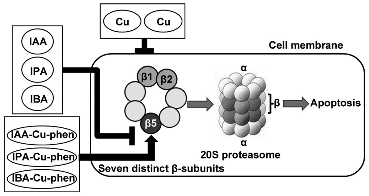
following conclusion: these three complexes, but not ligands or
CuCl2, could enter tumor cells and directly bind with
catalytic site of subunit β5 by forming a number of hydrogen bonds
and hydrophobic interactions and therefore inhibit
chemotrypsin-like activity and induce tumor cell death (Fig. 7). This study indicates that these
copper complexes and other similar complexes could act as potent
proteasome inhibitors and have great potential to be developed into
new metal-based anticancer agents.
Acknowledgements
The present study was supported by grants from the
Project of Shandong Province Higher Educational Science and
Technology Program to Zhen Zhang (no. J15LC22), the National
Science Foundation of China to Caifeng Bi (no. 21371161), the
Science and Technology Program of Jining to Zhen Zhang (no.
2015-92) and the Doctoral Foundation of Jining Medical University
to Zhen Zhang (no. JY14QD06).
Abbreviations:
|
IC50
|
half maximal (50%) inhibitory
concentration of a substance
|
|
CT
|
chymotrypsin
|
|
MTT
|
3-[4,5-dimethylthiazol-2-yl]-2.5-diphenyl-tetrazolium bromide
|
|
PARP
|
polyclonal antibody against human
poly(ADP-ribose) polymerase
|
References
|
1
|
Schmitt SM, Frezza M and Dou QP: New
applications of old metal-binding drugs in the treatment of human
cancer. Front Biosci (Schol Ed). 4:375–391. 2012. View Article : Google Scholar
|
|
2
|
Santini C, Pellei M, Gandin V, Porchia M,
Tisato F and Marzano C: Advances in copper complexes as anticancer
agents. Chem Rev. 114:815–862. 2014. View Article : Google Scholar
|
|
3
|
Krajčiová D, Melník M, Havránek E,
Forgácsová A and Mikuš P: Copper compounds in nuclear medicine and
oncology. J Coord Chem. 67:1493–1519. 2014. View Article : Google Scholar
|
|
4
|
Buac D, Schmitt S, Ventro G, Kona FR and
Dou QP: Dithiocarbamate-based coordination compounds as potent
proteasome inhibitors in human cancer cells. Mini Rev Med Chem.
12:1193–1201. 2012. View Article : Google Scholar : PubMed/NCBI
|
|
5
|
Chen D, Cui QC, Yang H and Dou QP:
Disulfiram, a clinically used anti-alcoholism drug and
copper-binding agent, induces apoptotic cell death in breast cancer
cultures and xenografts via inhibition of the proteasome activity.
Cancer Res. 66:10425–10433. 2006. View Article : Google Scholar : PubMed/NCBI
|
|
6
|
Zhai S, Yang L, Cui QC, Sun Y, Dou QP and
Yan B: Tumor cellular proteasome inhibition and growth suppression
by 8-hydroxyquinoline and clioquinol requires their capabilities to
bind copper and transport copper into cells. J Biol Inorg Chem.
15:259–269. 2010. View Article : Google Scholar :
|
|
7
|
Chen D, Cui QC, Yang H, Barrea RA, Sarkar
FH, Sheng S, Yan B, Reddy GP and Dou QP: Clioquinol, a therapeutic
agent for Alzheimer's disease, has proteasome-inhibitory, androgen
receptor-suppressing, apoptosis-inducing, and antitumor activities
in human prostate cancer cells and xenografts. Cancer Res.
67:1636–1644. 2007. View Article : Google Scholar : PubMed/NCBI
|
|
8
|
Adsule S, Barve V, Chen D, Ahmed F, Dou
QP, Padhye S and Sarkar FH: Novel Schiff base copper complexes of
quinoline-2 carboxaldehyde as proteasome inhibitors in human
prostate cancer cells. J Med Chem. 49:7242–7246. 2006. View Article : Google Scholar : PubMed/NCBI
|
|
9
|
Creaven BS, Czeglédi E, Devereux M, Enyedy
ÉA, Foltyn-Arfa Kia A, Karcz D, Kellett A, McClean S, Nagy NV,
Noble A, et al: Biological activity and coordination modes of
copper(II) complexes of Schiff base-derived coumarin ligands.
Dalton Trans. 39:10854–10865. 2010. View Article : Google Scholar : PubMed/NCBI
|
|
10
|
Zhang Z, Bi C, Fan Y, Zhang N, Deshmukh R,
Yan X, Lv X, Zhang P, Zhang X and Dou QP: L-Ornithine Schiff
base-copper and -cadmium complexes as new proteasome inhibitors and
apoptosis inducers in human cancer cells. J Biol Inorg Chem.
20:109–121. 2015. View Article : Google Scholar
|
|
11
|
Nalepa G, Rolfe M and Harper JW: Drug
discovery in the ubiquitin-proteasome system. Nat Rev Drug Discov.
5:596–613. 2006. View Article : Google Scholar : PubMed/NCBI
|
|
12
|
Peters JM, Cejka Z, Harris JR,
Kleinschmidt JA and Baumeister W: Structural features of the 26 S
proteasome complex. J Mol Biol. 234:932–937. 1993. View Article : Google Scholar : PubMed/NCBI
|
|
13
|
Groll M, Ditzel L, Löwe J, Stock D,
Bochtler M, Bartunik HD and Huber R: Structure of 20S proteasome
from yeast at 2.4 A resolution. Nature. 386:463–471. 1997.
View Article : Google Scholar : PubMed/NCBI
|
|
14
|
Groll M, Heinemeyer W, Jäger S, Ullrich T,
Bochtler M, Wolf DH and Huber R: The catalytic sites of 20S
proteasomes and their role in subunit maturation: A mutational and
crystallographic study. Proc Natl Acad Sci USA. 96:10976–10983.
1999. View Article : Google Scholar : PubMed/NCBI
|
|
15
|
Adams J: The proteasome: A suitable
antineoplastic target. Nat Rev Cancer. 4:349–360. 2004. View Article : Google Scholar : PubMed/NCBI
|
|
16
|
Frezza M, Schmitt S and Dou QP: Targeting
the ubiquitin-proteasome pathway: An emerging concept in cancer
therapy. Curr Top Med Chem. 11:2888–2905. 2011. View Article : Google Scholar : PubMed/NCBI
|
|
17
|
Lopes UG, Erhardt P, Yao R and Cooper GM:
p53-dependent induction of apoptosis by proteasome inhibitors. J
Biol Chem. 272:12893–12896. 1997. View Article : Google Scholar : PubMed/NCBI
|
|
18
|
An B, Goldfarb RH, Siman R and Dou QP:
Novel dipeptidyl proteasome inhibitors overcome Bcl-2 protective
function and selectively accumulate the cyclin-dependent kinase
inhibitor p27 and induce apoptosis in transformed, but not normal,
human fibroblasts. Cell Death Differ. 5:1062–1075. 1998. View Article : Google Scholar
|
|
19
|
Rajkumar SV, Richardson PG, Hideshima T
and Anderson KC: Proteasome inhibition as a novel therapeutic
target in human cancer. J Clin Oncol. 23:630–639. 2005. View Article : Google Scholar : PubMed/NCBI
|
|
20
|
Verani CN: Metal complexes as inhibitors
of the 26S proteasome in tumor cells. J Inorg Biochem. 106:59–67.
2012. View Article : Google Scholar
|
|
21
|
Unno M, Mizushima T, Morimoto Y, Tomisugi
Y, Tanaka K, Yasuoka N and Tsukihara T: The structure of the
mammalian 20S proteasome at 2.75 A resolution. Structure.
10:609–618. 2002. View Article : Google Scholar : PubMed/NCBI
|
|
22
|
Zhang Z, Bi C, Schmitt SM, Fan Y, Dong L,
Zuo J and Dou QP: 1,10-Phenanthroline promotes copper complexes
into tumor cells and induces apoptosis by inhibiting the proteasome
activity. J Biol Inorg Chem. 17:1257–1267. 2012. View Article : Google Scholar : PubMed/NCBI
|
|
23
|
Zhang Z, Bi C, Buac D, Fan Y, Zhang X, Zuo
J, Zhang P, Zhang N, Dong L and Dou QP: Organic cadmium complexes
as proteasome inhibitors and apoptosis inducers in human breast
cancer cells. J Inorg Biochem. 123:1–10. 2013. View Article : Google Scholar : PubMed/NCBI
|
|
24
|
Zhang Z, Bi C, Fan Y, Wang H and Bao Y:
Cefepime, a fourth-generation cephalosporin, in complex with
manganese, inhibits proteasome activity and induces the apoptosis
of human breast cancer cells. Int J Mol Med. 36:1143–1150.
2015.PubMed/NCBI
|
|
25
|
Zhang N, Fan YH, Zhang Z, Zuo J, Zhang PF,
Wang Q, Liu SB and Bi CF: Syntheses, crystal structures and
anticancer activities of three novel transition metal complexes
with Schiff base derived from 2-acetylpyridine and l-tryptophan.
Inorg Chem Commun. 22:68–72. 2012. View Article : Google Scholar
|
|
26
|
Daniel KG, Chen D, Orlu S, Cui QC, Miller
FR and Dou QP: Clioquinol and pyrrolidine dithiocarbamate complex
with copper to form proteasome inhibitors and apoptosis inducers in
human breast cancer cells. Breast Cancer Res. 7:R897–R908. 2005.
View Article : Google Scholar : PubMed/NCBI
|
|
27
|
Chen D, Peng F, Cui QC, Daniel KG, Orlu S,
Liu J and Dou QP: Inhibition of prostate cancer cellular proteasome
activity by a pyrrolidine dithiocarbamate-copper complex is
associated with suppression of proliferation and induction of
apoptosis. Front Biosci. 10:2932–2939. 2005. View Article : Google Scholar : PubMed/NCBI
|
|
28
|
Jones G, Willett P and Glen RC: Molecular
recognition of receptor sites using a genetic algorithm with a
description of desolvation. J Mol Biol. 245:43–53. 1995. View Article : Google Scholar : PubMed/NCBI
|
|
29
|
Kazuo N, Huang DR and Wang QR: Infrared
and Raman spectra of inorganic and coordination compounds. Chemical
Industry Press; Beijing: 1988
|
|
30
|
Gong QJ, Jin WJ, Dong C and Liu CS:
Synthesis of new fluorescence reagent: 4-Aminoantipyrine aromatic
Schiff bases. Appl Chem. 17:227–229. 2000.
|
|
31
|
Li B and Dou QP: Bax degradation by the
ubiquitin/proteasome-dependent pathway: Involvement in tumor
survival and progression. Proc Natl Acad Sci USA. 97:3850–3855.
2000. View Article : Google Scholar : PubMed/NCBI
|
|
32
|
Orlowski RZ and Kuhn DJ: Proteasome
inhibitors in cancer therapy: Lessons from the first decade. Clin
Cancer Res. 14:1649–1657. 2008. View Article : Google Scholar : PubMed/NCBI
|
|
33
|
Kane RC, Bross PF, Farrell AT and Pazdur
R: Velcade: U.S. FDA approval for the treatment of multiple myeloma
progressing on prior therapy. Oncologist. 8:508–513. 2003.
View Article : Google Scholar : PubMed/NCBI
|
|
34
|
Dou QP and Zonder JA: Overview and
perspective of proteasome inhibitor-based anti-cancer therapies:
Bortezomib and second generation proteasome inhibitors versus
future generation inhibitors of ubiquitin-proteasome system. Curr
Cancer Drug Targets. 14:517–536. 2014. View Article : Google Scholar
|
|
35
|
Lee EC, Fitzgerald M, Bannerman B, Donelan
J, Bano K, Terkelsen J, Bradley DP, Subakan O, Silva MD, Liu R, et
al: Antitumor activity of the investigational proteasome inhibitor
MLN9708 in mouse models of B-cell and plasma cell malignancies.
Clin Cancer Res. 17:7313–7323. 2011. View Article : Google Scholar : PubMed/NCBI
|
|
36
|
Shirley M: Ixazomib: First Global
Approval. Drugs. 76:405–411. 2016. View Article : Google Scholar : PubMed/NCBI
|
|
37
|
Offidani M, Corvatta L, Gentili S, Maracci
L and Leoni P: Oral ixazomib maintenance therapy in multiple
myeloma. Expert Rev Anticancer Ther. 16:21–32. 2016. View Article : Google Scholar
|
|
38
|
Milacic V, Chen D, Giovagnini L, Diez A,
Fregona D and Dou QP: Pyrrolidine dithiocarbamate-zinc(II) and
-copper(II) complexes induce apoptosis in tumor cells by inhibiting
the proteasomal activity. Toxicol Appl Pharmacol. 231:24–33. 2008.
View Article : Google Scholar : PubMed/NCBI
|