Introduction
Osteosarcoma (OS) is the most common primary tumor
of bone with high mortality and poor prognosis (1). In recent years, the adjuvant
chemotherapy and radiotherapy are common treatments for OS.
However, owing to the recurrence and metastasis, the overall 5-year
survival rate for osteosarcoma patients remains unsatisfactory
(2-4). Therefore, the development of new
diagnosis and treatment strategies is required to reduce recurrence
and improve the survival rate.
Long non-coding RNAs (lncRNAs) are a class of
transcripts longer than 200 nucleotides in length with little
functional protein-coding ability. Emerging data have reported that
lncRNAs are involved in a wide range of biological processes, such
as proliferation, apoptosis, and differentiation (5,6).
Several lncRNAs, such as MEG3 (maternally expressed gene 3)
(7) and TUG1 (taurine upregulated
gene 1) (8) have been reported to
play crucial roles during the development and progression of OS.
lncRNA XIST (X-inactive specific transcript) is a product of the
XIST gene which has been found to be dysregulated in a variety of
human cancers, such as hepatocellular carcinoma (HCC), gastric
cancer (GC) and human nasopharyngeal carcinoma (NPC) (9-11).
Furthermore, increasing evidence demonstrated that XIST can act as
tumor suppressors, and play important roles in carcinogenesis and
cancer development (12-15). Silence of XIST reduced cell
proliferation, migration and invasion as well as inducing apoptosis
in human glioblastoma stem cells (15). XIST was reported to inhibit HCC
cell proliferation and metastasis (14). Recently, a study from Huang et
al showed that XIST expression was decreased in breast tumor
samples and breast cancer cell lines and functioned as a tumor
suppressor through inhibition of AKT activation (13). However, it is unknown whether XIST
plays a tumor suppressive role in OS.
Abundant evidence indicates that lncRNAs suppress
the expression and biological functions of miRNAs by acting as a
competitive endogenous RNA (16,17).
For example, HOTAIR is specifically upregulated in gastric cancer
and functions as a ceRNA to regulate human epithelial growth factor
receptor 2 (HER2) expression by competitively binding to miR-331-3p
(18). lncRNA H19 functions as a
miRNA sponge to restrain the activity of many miRNAs, such as
miR-138 and miR-200a, leading to the suppression of its target
gene, vimentin, ZEB1, and ZEB2, thereby promoting EMT progression
in colorectal cancer (CRC) (19).
A recent study from Chen et al showed that knockdown of
lncRNA XIST exerted its tumor-suppressive effect though
downregulating the expression of EZH2 via miR-101 in gastric
pathogenesis (20). However,
whether lncRNA-XIST affects the biological behavior of OS cells by
regulating miRNAs has not yet been reported.
In the present study, we found that XIST was
significantly downregulated in both OS tissues and cell lines, and
over-expression of XIST inhibited cell proliferation and metastasis
in vitro as well as tumorigenesis in vivo. Then, we
investigated the molecular mechanism of XIST in the progression of
OS and the reciprocal regulation between XIST and miR-21-5p, a
potent oncogenic microRNA. Our findings provide new insights into
the molecular function of XIST/miR-21-5p/PDCD4 signaling pathway in
OS, and will give a novel strategy for the treatment of OS.
Materials and methods
Cell culture and tissue samples
Osteosarcoma cell lines MG63, U2OS, Saos2, HOS,
SOSP-9697 and SV40 immortalized human fetal osteoblastic cell line
hFOB 1.19 were cultured in were grown in RPMI-1640 medium
supplemented with 10% fetal bovine serum (Gibco, Beijing, China).
Cultures were maintained at 37°C in a humidified atmosphere with 5%
CO2. A total of 50 pairs of tumor tissues and adjacent
normal tissues were collected from routine therapeutic surgery at
our department. All samples were obtained with informed consent and
approved by the hospital institutional review board.
Quantitative reverse transcription-PCR
(qRT-PCR)
Total RNAs from frozen OS and paired non-cancerous
tissues or cell lines were extracted using TRIzol (Invitrogen,
Carlsbad, CA, USA) according to the instructions provided by the
manufacturer. cDNA was synthesized by reverse transcribing the
total RNA using PrimeScript™ RT Master Mix (Takara Biotechnology,
Dalian, China). The expression level of XIST and miR-21-5p was
detected by qRT-PCR using the Ultra SYBR Mixture with ROX (CWBio
Co., Ltd.) and ABI7100 system (Applied Biosystems, Darmstadt,
Germany). GAPDH and U6 were used as internal controls for XIST,
PDCD4 and miR-21-5p. All qRT-PCR reactions were performed in
triplicate. Relative quantification of gene expression was
calculated by the 2−ΔΔCt method. The primers used in
this study were: XIST forward 5′-CTAGCTAGCTTTTGTAGTGAGCTT GCTCCT-3′
and reverse 5′-GCTCTAGAATGTCTCCATCTCCATTTTGC-3′; PDCD4 forward
5′-CGACAGTGGGAGTGACGCCCTTA-3′; reverse 5′-CAGACACCTTTGCCTCC
TGCACC-3′; GAPDH forward 5′-GCACCGTCAAGGCTG AGAAC-3′ and reverse
5′-TGGTGAAGACGCCAGTGGA-3′; miR-21-5p forward
5′-GTGCAGGGTCCGAGGT-3′, reverse 5′-GCCGCTAGCTTATCAGACTGATGT-3′; U6
forward 5′-TGCGGGTGCTCGCTTCGGCAGC-3′ and reverse 5′-CCA
GTGCAGGGTCCGAGGT-3′.
Cell transfection
XIST overexpression lentivirus (Lv-XIST) vector as
well as the NC lentivirus vector was obtained from GenePharma
(Shanghai, China). The HEK293 cells were cotransfected with
Lenti-Pac HIV Expression Packaging Mix and the lentiviral vectors
(or the control lentivirus vectors) using Lipofectamine 2000 (Life
Technologies Corp., Carlsbad, CA, USA). After 48 h, lentiviral
particles in the supernatant were harvested and filtered by
centrifugation at 600 × g for 10 min. The packaged lentiviruses
were named Lv-XIST and Lv-control. The MG63 and Saos2 cells were
then infected with Lv-XIST or Lv-control at a MOI of 20.
miR-21-5p mimics, miR-21-5p inhibitor and miR-21-5p
negative control (NC), small interfering RNA (siRNAs) against PDCD4
(si-PDCD4) and si-NC were also employed and synthesized by
GenePharma, and transfected into MG63 and Saos2 cells using
Lipofectamine 2000 (Invitrogen) according to the manufacturer's
protocol. All the transfections were repeated more than three times
independently.
Immunohistochemical staining
The tumor tissues were paraffin-embedded and cut
into 5-µm-thick slides for immuno-histochemical analysis.
After incubating in 0.15% Triton X-100 at room temperature and
blocking with 1% goat serum albumin in modified D-PBS Tween-20 for
1 h, the sections were incubated overnight with PDCD4 antibody
(1:1500, Cell Signaling Technology, Danvers, MA, USA). After
washing with PBS three times, the sections were incubated with
secondary antibodies (anti-rabbit IgG antibodies) at 37°C for 30
min, and visualized with diaminobenzidine (DAB). Finally, the
slides were counterstained with 10% hematoxylin, and analyzed under
a light microscope with a digital camera. Image analysis was
performed by Image-Pro Plus software.
CCK-8 assay
MG63 and Saos2 cells were seeded into a 96-well
plate (5×10³ cells/well) and cultured at 37°C and cell
proliferation was measured by the Cell Counting Kit-8 (CCK-8;
Dojindo, Kumamoto, Japan) according to the manufacturer's protocol.
At different time points, 10 µl CCK-8 solution was added
into each well and incubated for an additional 2 h at 37°C. The
absorbance at 450 nm was measured using a microplate reader
(ELx800; BioTek Instuments, Inc., Winooski, VT, USA).
Apoptosis assay
Cell apoptosis was measured by flow cytometry
following the instructions of the Annexin V-FITC/PI apoptosis
detection kit (KeyGEN, Nanjing, China). Cultured cells were
harvested and washed twice in PBS, re-suspended in binding buffer,
and then incubated with Annexin V-FITC and propidium iodide (PI)
for 15 min at room temperature. Afterwards, flow cytometry was
performed to determine rate of apoptosis on a FACSAria flow
cytometer (BD Biosciences, Franklin Lakes, NJ, USA).
In vitro invasion and migration
assay
At 24 h after transfection, cell invasion of MG63
and Saos2 cells was assessed using Transwell chambers (8 µm,
24-well format; Corning Inc., Corning, NY, USA) as previously
described in detail (14). The
number of invaded cells was calculated by counting five random
views under the microscope. The experiment was performed in
triplicate and repeated three times.
At 24 h after transfection, MG63 and Saos2 cells
were seeded into 96-well plates (3×10³ cells/well), and
wound healing assays was monitored to measure cell migration as
previously described (14). The
wound closure was observed and imaged under a microscope. Then, the
wound area was measured and the percentage of the wound healing was
calculated by ImageJ software (NIH, Bethesda, MD, USA).
Western blotting
Total cellular proteins were extracted using RIPA
lysis buffer containing proteinase inhibitor (Sigma, St. Louis, MO,
USA). Concentrations of total cellular protein were determined
using a BCA assay kit (Pierce, Rockford, IL, USA). Total protein
samples (25 µg) were analyzed by 8% SDS-PAGE gel. The
protein was transferred to polyvinylidene difluoride (PVDF)
membranes by a wet blotting procedure (100 V, 120 min, 4°C). After
blocked with 5% blocking buffer, the membranes were incubated with
primary antibodies at 4°C overnight using the following
concentration: cleaved caspase-3, cleaved caspase-9, Bax, Bcl-2
(1:1000, Cell Signaling Technology), PDCD4, ZEB1, vimentin,
N-cadherin, E-cadherin (1:2000; Cell Signaling Technology) and
anti-β-actin (1:500; Santa Cruz Biotechnology, Santa Cruz, CA,
USA), followed by horseradish peroxidase-conjugated secondary
antibody (anti-rabbit, 1:2000, Cell Signaling Technology).
Anti-β-actin antibody was used as an internal control. The protein
bands were visualized by enhanced chemiluminescence detection
reagents (Applygen Technologies, Inc., Beijing, China) according to
the manufacturer's instructions. Relative band intensities were
determined by densitometry using Scion image software (version
4.0).
Luciferase reporter assay
The fragment from XIST containing the predicted
miR-21-5p binding site was amplified by PCR and cloned into a
pmirGLO Dual-luciferase Target Expression Vector (Promega, Madison,
WI, USA) to form the reporter vector XIST-wild-type (XIST-Wt). To
test the binding specificity, the corresponding mutant was created
by mutating the miR-21-5p seed region binding site (seed sequence
binding fragment 5′-TATTCGA-3′ changed to 5′-CGTGAGC-3′), which
were named as pmirGLO-XIST-Mt (XIST-MUT). pmirGLO-XIST-Mt or
pmirGLO-XIST-WT was co-transfected with miR-21-5p mimics, miR-21-5p
inhibitor or miR-21-5p NC into HEK 293T cells using Lipofectamine
2000. Luciferase reporter assay was performed using the
Dual-Luciferase Reporter Assay System (Promega) 48 h later, the
firefly luciferase activity was measured and normalized by
Renilla luciferase activity.
OS xenograft mouse model
After infection with Lv-XIST (1.2×10⁷ TU)
or Lv-control, MG63 and Saos2 cells (5×10⁶) were
suspended in 100 µl PBS and injected subcutaneously to the
right flank of the BALB/c nude mice purchased from the Shanghai
Institute of Materia Medica (Shanghai, China). The tumor volume was
measured every week and calculated as follows: tumor volume
(mm3) = (length x width2)/2. On day 35 after
cell implantation, the mice were sacrificed and the tumor specimens
were removed and weighed. All animal studies were conducted in the
Animal Institute of Shanghai Jiaotong University according to the
protocols approved by the Medical Experimental Animal Care
Commission of Shanghai Jiaotong University.
Statistical analysis
Statistical analyses were performed with SPSS 13.0
software. The results were evaluated by χ2 test and the
other data were evaluated by Student's t-test and expressed as the
mean ± SD from three independent experiments. A P-value of <0.05
was considered to indicate a statistically significant
difference.
Results
Downregulation of XIST is correlated with
poor outcome of OS patients
To elucidate the expression of XIST in OS, qRT-PCR
was performed to detect the expression of XIST in 41 pairs of human
osteosarcoma and adjacent normal tissues (among which 26 metastases
and 15 non-metastases osteosarcoma tissues, 23 recurrent and 18
non-recurrent osteosarcoma tissues). The transcript level of XIST
was lower in OS tissues when compared with normal tissues (Fig. 1A). In addition, the expression
level of XIST was also significantly lower in metastatic group
compared with non-metastatic group (Fig. 1B). It was noteworthy that XIST
low-expression was significantly associated with tumor recurrence
(Fig. 1C). Furthermore, the
Kaplan-Meier method and log-rank test revealed that patients with
high XIST expression in OS had significantly longer overall
survival than those with low XIST expression (Fig. 1D). Taken together, these data
suggest decreased XIST expression might be critically involved in
OS progression.
Overexpression of XIST inhibits OS cell
growth in vitro as well as tumorigenesis in vivo
It has been reported that XIST exerted
tumor-suppressive effects in many cancers (12,20).
However, it is not clear whether XIST functions as a tumor
suppressor in OS. To explore the biological functions of XIST in
OS, we first measured the expression levels of XIST in five OS cell
lines including mG63, U2OS, Saos2, HOS and SOSP-9697. Consistent
with expression levels in OS tissues, the expression levels of XIST
markedly decreased in OS cell lines, especially in MG63 and Saos2
cells compared with the human osteoblast cell line hFOB 1.19
(Fig. 2A). Then, the lentivirus
containing XIST (MOI=20) was infected into both mG63 and Saos2
cells and its overexpression efficiency was remarkable compared
with Lv-control (Fig. 2B). CCK-8
assays were performed to detect the impact of XIST over expression
on cell viability of OS cell lines. As shown in Fig. 2C and D, overexpression of XIST
significantly inhibited the cell proliferation compared with
Lv-control group. Subsequently, flow cytometry showed that
overexpression of XIST significantly increased the percentage of
apoptotic cells in both MG63 and Saos2 cells (Fig. 2E). In addition, we detected the
protein levels of classic apoptotic markers in MG63 and Saos2
cells. As expected, western blot analysis showed that XIST
overexpression markedly increased the expression levels of
cleaved-caspase-3, cleaved-caspase-9 and Bax, while that of
anti-apoptotic protein Bcl-2 was decreased (Fig. 2F).
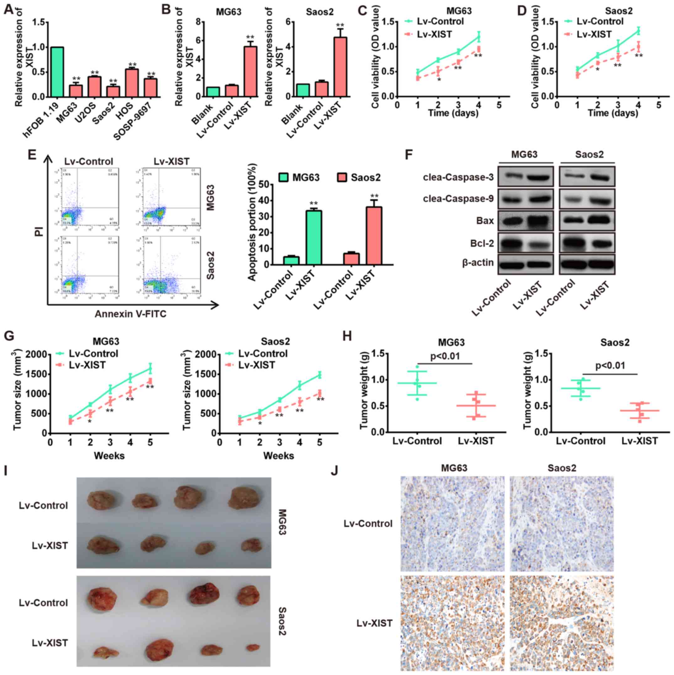 | Figure 2Overexpression of XIST inhibits the
proliferation, invasion and migration as well as promotes apoptosis
of OS cells. (A) qRT-PCR analysis of XIST expression levels in five
OS cell lines (mG63, U2OS, Saos2, HOS and SOSP-9697).
**P<0.01 vs. hFOB 1.19. (B) qRT-PCR analysis of XIST
in MG63 and Saos2 cells infected with lentivirus overexpressing
XIST (Lv-XIST). **P<0.01 vs. Lv-control group. (C and
D) CCK8 assay was performed to evaluate the OS cell growth, and the
relative cell viability was determined at different times. Lv-XIST
inhibited cell growth in MG63 and Saos2 cells.
*P<0.05, **P<0.01 vs. Lv-control. (E)
Flow cytometer was performed to evaluate the OS cell apoptosis, and
the cell apoptosis was determined after transfection for 48 h.
Lv-XIST promoted cell apoptosis in MG63 and Saos2 cells.
**P<0.01 vs. Lv-control. (F) Apoptosis related
proteins (cleaved caspase-3, cleaved caspase-9, Bax and Bcl-2) were
detected by western blotting in MG63 and Saos2 cells, and Lv-XIST
promoted the expression levels of cleaved caspase-3, cleaved
caspase-9 and Bax, while inhibited the expression of Bcl-2. (G and
H) Lv-control or Lv-XIST (MOI=20) was infected into MG63 and Saos2
cells, which were injected into nude mice, respectively. Tumor
sizes were calculated every 7 days after injection (G).
*P<0.05, **P<0.01 vs. Lv-control. Tumor
weights were measured 35 days after injection (H). P<0.01 vs.
Lv-control. Data are presented as mean ± SD from three independent
experiments. (I) Representative images of tumors from xenografts
with MG63 and Saos2 cells infected with Lv-XIST or Lv-control. (J)
The expression of PDCD4 was detected by immunohistochemistry
staining in cell-derived xenograft tumor model. |
To explore whether overexpression of XIST affects
tumor growth in vivo, MG63 and Saos2 cells infected with
Lv-XIST or Lv-control were subcutaneously injected into the flank
of nude mice. Tumor sizes were obviously smaller in the Lv-XIST
group compared with the Lv-control group (Fig. 2G and I). In addition, tumor weights
in the Lv-XIST group were significantly lower than in the
Lv-control group (Fig. 2H). These
results indicate that XIST plays a tumor suppressor role in the
progression of OS.
Overexpression of XIST inhibits OS cell
migration and invasion in vitro
Next, we investigated the association of XIST and OS
metastasis. Two OS cell lines, MG63 and Saos2, were injected with
Lv-NC or Lv-XIST (MOI=20), and then Transwell and wound healing
assays were carried out to explore the effects of XIST on OS cell
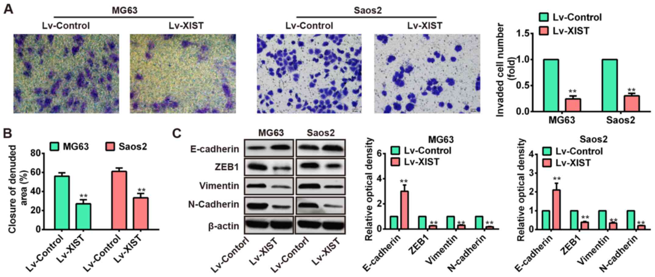
metastasis. The results of Transwell assay showed that numbers of
invaded cells were obviously attenuated in the Lv-XIST groups
compared with Lv-control groups (Fig.
3A). In addition, wound healing assay showed that wound closure
of both MG63 and Saos2 cells with ectopic expression of XIST was
slower than that in Lv-control groups (Fig. 3B). It is well known that EMT is a
crucial event in the invasion and migration of tumor cells
(21). Therefore, we used western
blotting to explore whether XIST can affect EMT in OS cells. Of
interest, overexpression of XIST increased the level of epithelial
marker E-cadherin while reduced the level of mesenchymal markers
such as ZEB1, vimentin and N-cadherin (Fig. 3C). These data suggested that over
expression of XIST could inhibit OS cell metastasis in
vitro.
XIST binds to miR-21-5p and represses its
expression
Recent studies have suggested lncRNA could
communicate with miRNAs via shared common miRNA binding sites and
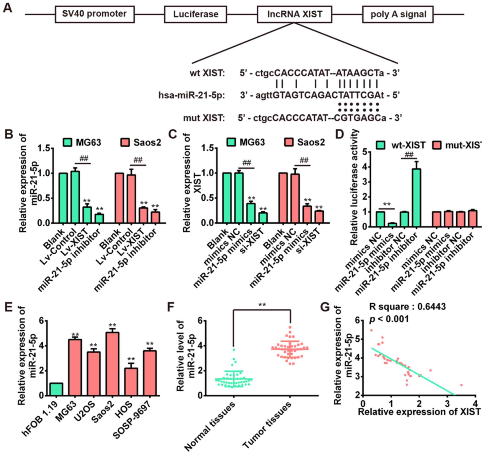
regulate its expression and activity (22). To determine whether XIST could
serve as a ceRNA, we used two bioinformatic databases (starBase and
TargetScanS) to search for the potential miRNAs that can be
regulated by XIST. Interestingly, miR-21-5p, a well-known oncogene
in osteosarcoma (23), could bind
to XIST. Fig. 4A revealed the
presence of XIST binding sites in the 3′-UTR of miR-21-5p. We
subsequently investigated the correlation between XIST and
miR-21-5p in MG63 and Saos2 cell lines. The results of qRT-PCR
indicated that miR-21-5p expression was downregulated after
over-expression of XIST in both MG63 and Saos2 cells (Fig. 4B), whereas the XIST level could
also be repressed by miR-21-5p overexpression (Fig. 4C). These results suggested that
there was reciprocal repression between XIST and miR-21-5p.
To confirm the direct binding relationship between
XIST and miR-21-5p, a luciferase activity assay was conducted. The
predicted miR-21-5p binding site (XIST-wt) and its mutant type
(XIST-mt) were amplified and directly fused to the downstream of
the luciferase reporter gene in the pmirGLO-basic vector.
Co-transfection of miR-21-5p mimic and pmirGLO-XIST-wt
significantly decreased the luciferase activity, whereas
co-transfection of miR-21-5p inhibitor and pmirGLO-XIST-wt
increased the luciferace activity (Fig. 4D). Likewise, cells cotransfected
with miR-21-5p and pmirGLO-XIST-mut showed no obvious change in
luciferase activity (Fig. 4D).
These data indicated that the miR-21-5p binding site within XIST
was functional.
Next, we investigated the expression of miR-21-5p in
OS cell lines and 41 pairs of OS and correspondingly adjacent
tissues. As shown in Fig. 4E and
F, the expression of miR-21-5p was significantly increased
compared with the human osteoblast cell line hFOB 1.19 and adjacent
normal tissues. This result is consistent with a previous study
(24). Moreover, we examined the
potential correlation among the expression levels of XIST and
miR-21-5p, and an inverse correlation between XIST and miR-21-5p
expression levels was observed (Fig.
4G). These results demonstrated that there exist a negative
regulation between XIST and miR-21-5p.
Knockdown of miR-21-5p inhibits cell
proliferation and metastasis by targeting PDCD4
Previous studies demonstrated that miR-21-5p could
play oncogenic roles in several types of cancer by regulating cell
growth, EMT and metastasis (25,26).
Moreover, a recent study showed that miR-21 promoted cell
proliferation through the downregulation of PDCD4 expression in
head and neck squamous carcinoma (27). However, it is unknown whether or
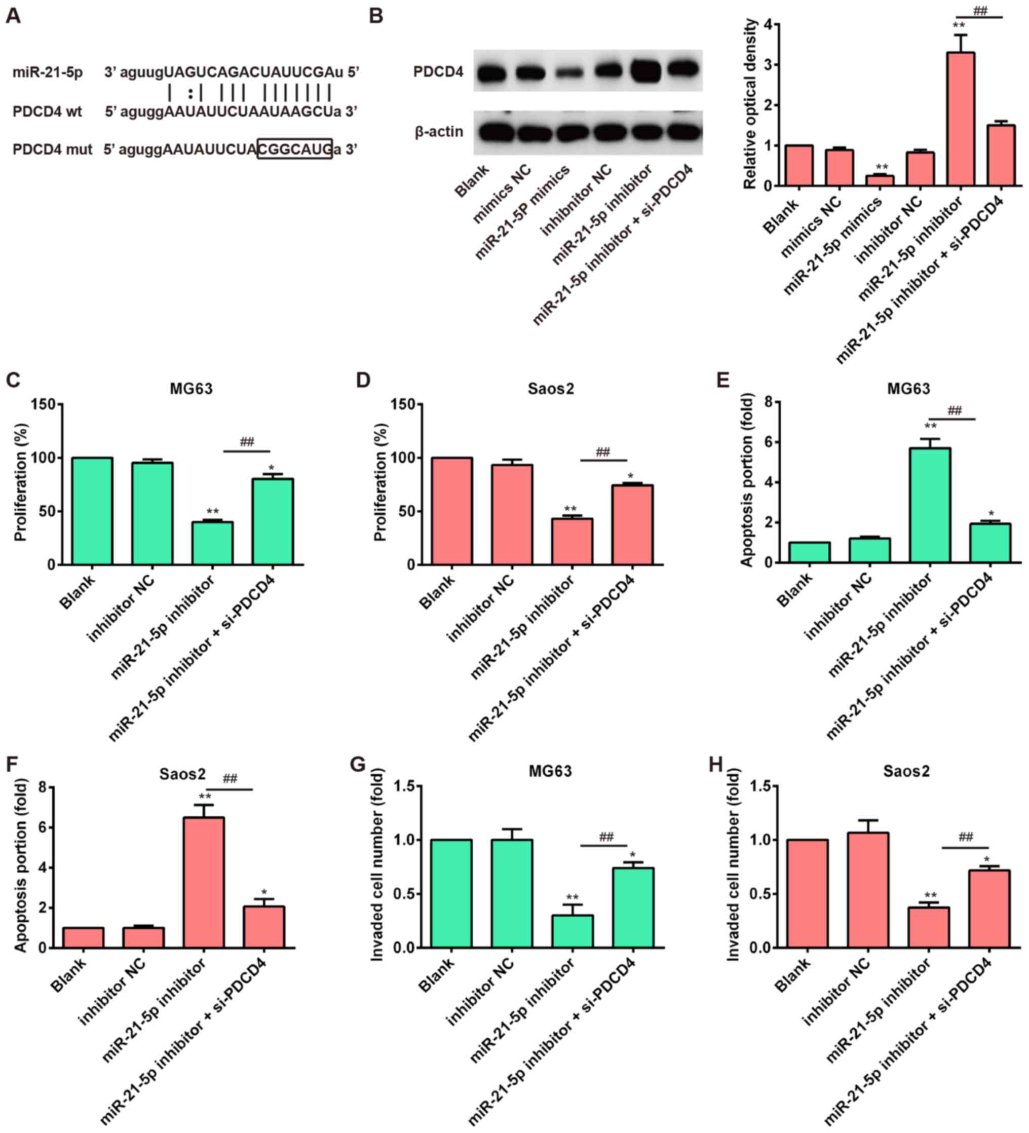
not PDCD4 is a function target of miR 21-5p in OS. Analysis using
targeting algorithms (TargetScan and microRNA.org)
indicated that PDCD4 could be a potential target gene of miR-21-5p.
Fig. 5A illustrates the predicted
miR-21-5p binding site in the 3′-UTR of PDCD4. Western blot
analysis showed that overexpression of miR-21-5p markedly reduced
the protein levels of PDCD4 in the OS cell lines, whereas it was
enhanced by knockdown of miR-21-5p (Fig. 5B). Consistent with previous
research, we also demonstrated that knockdown of miR-21-5p
inhibited cell proliferation and invasion as well as increased cell
apoptosis (Fig. 5C–H). To
determine whether PDCD4 acts as a functional target of miR-21-5p,
miR 21-5p inhibitor together with si-PDCD4 was transfected into
MG63 and Saos2 cells. Knockdown of PDCD4 by siRNA significantly
restored reduced cell proliferation, migration, and invasion as
well as increased apoptosis caused by inhibition of miR-21-5p in OS
cells (Fig. 5C–H). These results
indicate that inhibition of miR-21-5p suppresses the cell
proliferation, invasion and promoted apoptosis of OS cells, at
least in part by targeting PDCD4.
miR-21-5p mediates the expression of
PDCD4 involved in XIST-regulated antitumor effects of OS cells
Increasing evidence suggests that PDCD4 functions as
a tumor suppressor in several cancers including OS (28,29).
Our finding that miR-21-5p exerted an oncogenic role through
negatively regulating PDCD4 in OS cells led us to examine whether
XIST inhibited OS cell growth and metastasis via suppression of
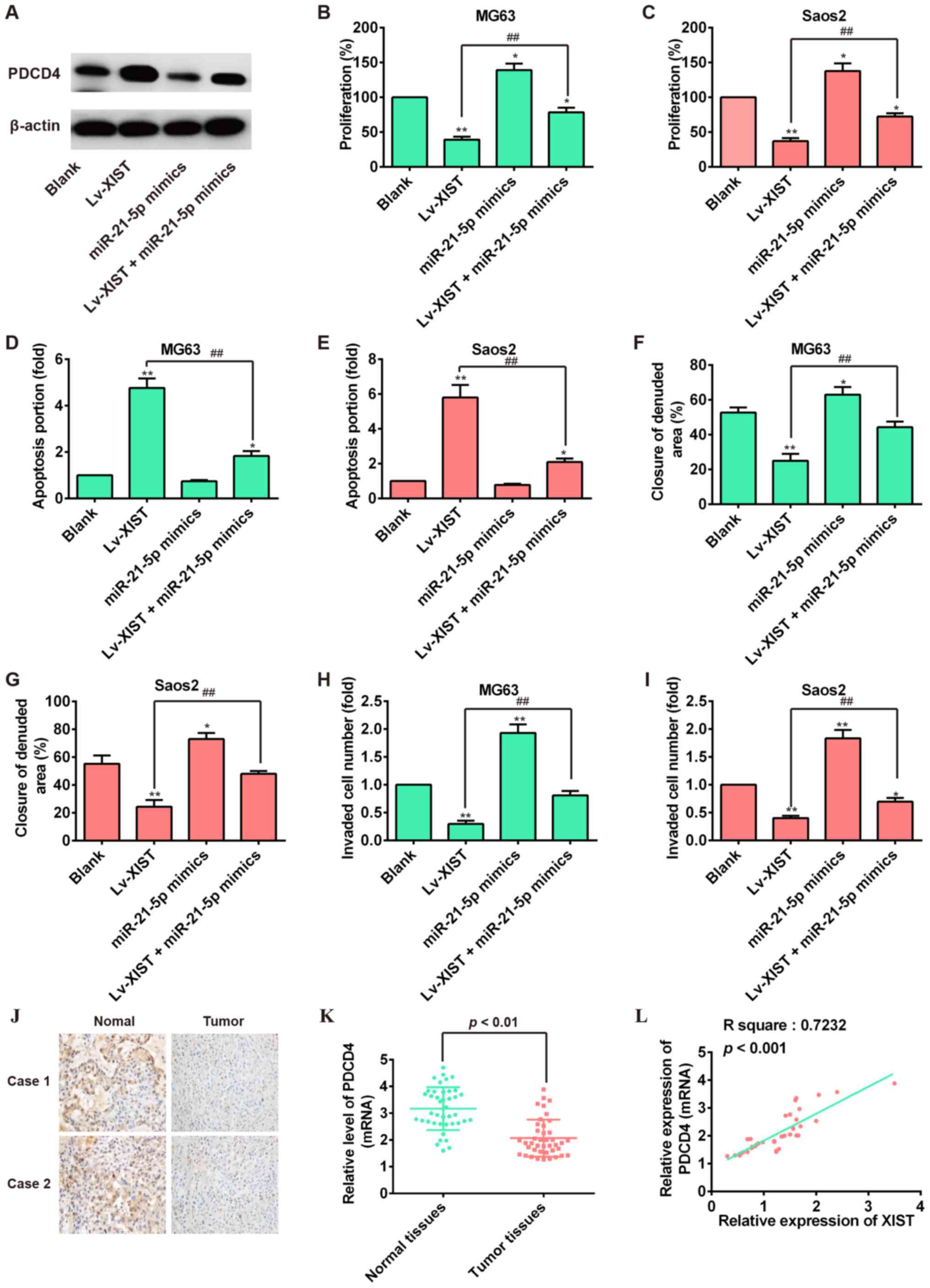
miR-21-5p activity and promotion of PDCD4 expression. First, we
explore whether miR-21-5p was involved in the effect of XIST in
regulating the PDCD4 expression in OS cells. As shown in Fig. 6A, western blot analysis showed that
overexpression of XIST could increase the expression of PDCD4, but
miR-21-5p mimics reversed the promoting effect of XIST
overexpression on PDCD4 expression. Subsequently, the cell function
assay showed that miR-21-5p overexpression could restore the
reduction of cell proliferation and reverse the enhancement of cell
apoptosis induced by XIST overexpression (Fig. 6B–E). Importantly, the Transwell
invasion assay and would healing assay revealed that the
suppression of lv-XIST-regulated cell migration and invasion was
attenuated by miR-21-5p over expression (Fig. 6F–I). We also examine the expression
of PDCD4 in OS tissue samples by immunohistochemical staining and
qRT-PCR. As expected, PDCD4 was down regulated in OS tissue samples
(Fig. 6J and K). Additionally, the
correlation of XIST and PDCD4 in OS tissue samples was investigated
and a positive relationship was observed between XIST and PDCD4
(Fig. 6L). In cell-derived
xenograft tumors model, we also found that overexpression of XIST
positively regulated the expression of PDCD4 by immunohistochemical
staining (Fig. 2J). Collectively,
we concluded that XIST functions as a ceRNA to enhance the
expression of PDCD4 by competitively binding miR-21-5p, leading to
the inhibition of OS cell growth and metastasis.
Discussion
In the present study, we demonstrated that XIST
expression was downregulated and associated with a poor clinical
outcome in OS patients. Overexpression of XIST inhibited the cell
proliferation, migration and invasion, and promoted apoptosis in
vitro and reduced tumor growth in vivo. Furthermore,
this study provided evidence that XIST exerted tumor-suppressive
functions by downregulating miR-21-5p, thereby enhancing the
expression of PDCD4, a well-known tumor suppressor. Our study not
only revealed the important role of XIST/miR-21-5p/PDCD4 axis in OS
pathogenesis but also implicated the potential role of XIST in the
clinical diagnosis and treatment of OS.
Recently, more studies have reported that lncRNAs
play considerable functional roles in the initiation and
progression of multiple cancers, including OS (30,31).
lncRNA XIST was a product of the XIST gene which was located in the
X inactivation center (32).
Previous studies have found that XIST was upregulated in glioma
tissues and human glioblastoma stem cells (GSCs), and knockdown of
XIST exerted tumor-suppressive roles by reducing cell
proliferation, migration and invasion as well as inducing apoptosis
(15). Song et al
discovered that XIST was upregulated in NPC tissues and XIST
overexpression enhanced, while XIST silencing hampered the cell
growth in NPC (11). However,
other researchers reported that XIST expression was lost in several
cancers, including ovarian, cervical cancer and breast cell lines
(13,33,34).
These results demonstrated that XIST have different roles either as
an oncogene or a tumor suppressor role in different tumors. In the
present study, our results confirmed that XIST was markedly
downregulated in OS, and low XIST expression was associated with a
poor clinical outcome in OS patients. Moreover, we found that
overexpression of XIST led to repressed cell proliferation,
migration and invasion as well as increased apoptosis, which was
consistent with the suppressive roles of XIST revealed by previous
studies. In addition, the in vivo studies also confirmed
that overexpression of XIST suppressed tumor growth in nude mice.
These findings suggest that XIST may function as a tumor suppressor
in OS progression.
It is worth noting that XIST, an lncRNA from the
inactive X-chromosome, is required for the silencing of one X
chromosome in female mammalian cells to achieve equivalent X-linked
gene dosage between females and males (35-37).
Previous studies showed that XIST was expressed mainly in female
cells, and loss of XIST contributes to cancer progression in breast
cancer (13), hematologic cancer
(12) and hepatocellular carcinoma
(38). A tumor suppressor role of
XIST in breast cancer has been widely suggested (13,39-41),
but still remains controversial (42,43).
Similarly, we observed that a recent paper has claimed that the
role of XIST as tumor progressor contributed to osteosarcoma cell
proliferation and invasion and XIST exerted its function through
the miR-320b/RAP2B axis (44).
However, there are no animal models such as nude mice used in that
study.
In our study, we confirmed that XIST remarkably
inhibited the growth of OS cells in vitro as well as the
xenograft tumor formation in vivo. Moreover, both cell
invasion and migration were inhibited by XIST overexpression via
suppressing the EMT process. Most importantly, our data show that
loss of XIST associated with recurrence and short overall survival
in OS patients. It was reported that multiple demographics
including gender were related to the incidence rate and outcome of
osteosarcoma (45,46). Since XIST was proved expressed
mainly in female cells in other tumors, we suspect this may be
because of gender differences of samples lead to different results.
In the future, our work will focus the correlation between gender
disparity of OS incidence and different expression patterns of XIST
in a large number of samples.
It has been reported that EMT is associated with the
acquisition of metastasis in cancer (47). During EMT, the epithelial protein
level, such as E-cadherin, is downregu-lated, while mesenchymal
protein such as N-cadherin and vimentin are upregulated (48). Zhuang et al found that high
level of XIST seems to be associated with distant metastasis and
poor prognosis in patients with hepatocellular carcinoma (HCC)
(38). A recent study from Chen
et al showed that XIST could affect the metastasis of human
gastric cancer cell via inducing EMT (20). In this study, we found that
overexpression of XIST promoted E-cadherin protein expression but
inhibited the expression of ZEB1, vimentin and N-cadherin in OS
cells in vitro, suggesting that XIST overexpression
inhibited cell invasion and migration via suppressing the EMT
process.
As a newly described regulatory mechanism, lncRNAs
can antagonize miRNA function by sponging miRNAs via a competing
endogenous mechanism (22). For
example, Ma et al have shown that lncRNA-ATB acts as a ceRNA
to regulate the expression of TGF-β by competing for miR-200a in
glioma (49). Several recent
studies have demonstrated that XIST functions as a ceRNA in many
types of cancer, such as human nasopharyngeal carcinoma (NPC)
(11), hepato cellular carcinoma
(HCC) (9), and gastric cancer (GC)
(10). XIST upregulated the
expression of miR-34a-5p targeted gene E2F3 through acting as a
ceRNA of miR-34a-5p in NPC (11).
XIST upregulated EZH2 by competitively binding the miR-101 and then
induced cell proliferation and invasion in GC (10). Based on these studies, we
hypothesized that XIST may act as a ceRNA in OS and so we searched
for potential interactions with miRNAs. In support of this notion,
bioinformatics analysis and luciferase assays were performed to
verify the direct binding ability of the predicted miRNA on the
XIST transcript. As expected, we discovered that XIST is directly
bound to miR-21-5p and there was reciprocal repression between XIST
and miR-21-5p in OS cells. In addition, qRT-PCR analysis showed
that expression of miR-21-5p was inversely correlated with XIST in
OS tissues. Moreover, knockdown of miR-21-5p expression could
arrest cell proliferation and invasion, which was consistent with
results of overexpression of XIST expression in OS cells. Taken
together, these data are consistent with our hypothesis and
indicate that XIST affects the biological characteristic of OS
cells by modulating the miR-21-5p function.
PDCD4 is a novel tumor suppressor that frequently
exhibits downregulated expression in a number of cancers (50-52).
Downregulation of PDCD4 was reported to be significantly associated
with short overall survival of patients with head and neck,
digestive system, and urinary system cancers (53-55).
Lin et al previously reported that PDCD4 was identified as a
target of miR-202 and involved in the inhibitory effect of miR-202
on cell apoptosis and drug resistance of osteosarcoma cells
(28). Other research demonstrates
that PDCD4 inhibition may not be affected by gene amplification
alone, but is also likely to be influenced by transcriptional
activation and/or post-transcriptional mechanisms in carcinomas
(56). In previous studies, PDCD4
mRNA and protein over expression have been directly affected by
miRNA-mediated post-transcriptional mechanisms in cancers (57-59).
Our study also confirms PDCD4 as a direct target of miR-21-5p.
Considering the interaction of XIST/miR-21-5p, we therefore
hypothesize that XIST may also regulate PDCD4 expression in OS
which signifies the role of XIST in the tumorigenesis-regulating
network. As expected, we demonstrated that PDCD4 expression was
positively associated with XIST in OS cells, and miR-21-5p reversed
the enhancement of PDCD4 mediated by XIST overexpression. More
importantly, we demonstrated overexpression of miR-21-5p could
attenuate the inhibitory effects on cell proliferation, invasion
and migration, as well as inhibit cell apoptosis induced by
overexpression of XIST. These results support our hypothesis that
XIST can antagonize miRNA-21-5p, thereby protecting PDCD4 from
repression, finally inhibiting OS tumorigenesis.
In conclusion, our data demonstrated that XIST was
downregulated in OS and its expression was associated with overall
survival of OS patients. Furthermore, we determined that XIST
suppressed OS cell proliferation and metastasis via downregulation
of miR-21-5p and activation of PDCD4 function. Thus,
XIST/miR-21-5p/PDCD4 axis may represent a novel prognostic
biomarker and therapeutic target in OS.
References
|
1
|
Ottaviani G and Jaffe N: The epidemiology
of osteosarcoma. Cancer Treat Res. 152:3–13. 2009. View Article : Google Scholar
|
|
2
|
Meazza C, Luksch R, Daolio P, Podda M,
Luzzati A, Gronchi A, Parafioriti A, Gandola L, Collini P, Ferrari
A, et al: Axial skeletal osteosarcoma: A 25-year monoinstitutional
experience in children and adolescents. Med Oncol. 31:8752014.
View Article : Google Scholar : PubMed/NCBI
|
|
3
|
Ando K, Heymann MF, Stresing V, Mori K,
Rédini F and Heymann D: Current therapeutic strategies and novel
approaches in osteosarcoma. Cancers (Basel). 5:591–616. 2013.
View Article : Google Scholar
|
|
4
|
Wittig JC, Bickels J, Priebat D, Jelinek
J, Kellar-Graney K, Shmookler B and Malawer MM: Osteosarcoma: A
multidisciplinary approach to diagnosis and treatment. Am Fam
Physician. 65:1123–1132. 2002.PubMed/NCBI
|
|
5
|
Shi X, Sun M, Liu H, Yao Y and Song Y:
Long non-coding RNAs: A new frontier in the study of human
diseases. Cancer Lett. 339:159–166. 2013. View Article : Google Scholar : PubMed/NCBI
|
|
6
|
Wapinski O and Chang HY: Long noncoding
RNAs and human disease. Trends Cell Biol. 21:354–361. 2011.
View Article : Google Scholar : PubMed/NCBI
|
|
7
|
Tian ZZ, Guo XJ, Zhao YM and Fang Y:
Decreased expression of long non-coding RNA MEG3 acts as a
potential predictor biomarker in progression and poor prognosis of
osteosarcoma. Int J Clin Exp Pathol. 8:15138–15142. 2015.
|
|
8
|
Zhang Q, Geng PL, Yin P, Wang XL, Jia JP
and Yao J: Down-regulation of long non-coding RNA TUG1 inhibits
osteosarcoma cell proliferation and promotes apoptosis. Asian Pac J
Cancer Prev. 14:2311–2315. 2013. View Article : Google Scholar : PubMed/NCBI
|
|
9
|
Mo Y, Lu Y, Wang P, Huang S, He L, Li D,
Li F, Huang J, Lin X, Li X, et al: Long non-coding RNA XIST
promotes cell growth by regulating miR-139-5p/PDK1/AKT axis in
hepatocellular carcinoma. Tumour Biol. 39:10104283176909992017.
View Article : Google Scholar : PubMed/NCBI
|
|
10
|
Ma L, Zhou Y, Luo X, Gao H, Deng X and
Jiang Y: Long non-coding RNA XIST promotes cell growth and invasion
through regulating miR-497/mACC1 axis in gastric cancer.
Oncotarget. 8:4125–4135. 2017.
|
|
11
|
Song P, Ye LF, Zhang C, Peng T and Zhou
XH: Long non-coding RNA XIST exerts oncogenic functions in human
nasopharyngeal carcinoma by targeting miR-34a-5p. Gene. 592:8–14.
2016. View Article : Google Scholar : PubMed/NCBI
|
|
12
|
Yildirim E, Kirby JE, Brown DE, Mercier
FE, Sadreyev RI, Scadden DT and Lee JT: Xist RNA is a potent
suppressor of hematologic cancer in mice. Cell. 152:727–742. 2013.
View Article : Google Scholar : PubMed/NCBI
|
|
13
|
Huang YS, Chang CC, Lee SS, Jou YS and
Shih HM: Xist reduction in breast cancer upregulates AKT
phosphorylation via HDAC3-mediated repression of PHLPP1 expression.
Oncotarget. 7:43256–43266. 2016. View Article : Google Scholar : PubMed/NCBI
|
|
14
|
Chang S, Chen B, Wang X, Wu K and Sun Y:
Long non-coding RNA XIST regulates PTEN expression by sponging
miR-181a and promotes hepatocellular carcinoma progression. BMC
Cancer. 17:2482017. View Article : Google Scholar : PubMed/NCBI
|
|
15
|
Yao Y, Ma J, Xue Y, Wang P, Li Z, Liu J,
Chen L, Xi Z, Teng H, Wang Z, et al: Knockdown of long non-coding
RNA XIST exerts tumor-suppressive functions in human glioblastoma
stem cells by up-regulating miR-152. Cancer Lett. 359:75–86. 2015.
View Article : Google Scholar : PubMed/NCBI
|
|
16
|
John-Aryankalayil M, Palayoor ST, Makinde
AY, Cerna D, Simone CB II, Falduto MT, Magnuson SR and Coleman CN:
Fractionated radiation alters oncomir and tumor suppressor miRNAs
in human prostate cancer cells. Radiat Res. 178:105–117. 2012.
View Article : Google Scholar : PubMed/NCBI
|
|
17
|
Cesana M, Cacchiarelli D, Legnini I,
Santini T, Sthandier O, Chinappi M, Tramontano A and Bozzoni I: A
long noncoding RNA controls muscle differentiation by functioning
as a competing endogenous RNA. Cell. 147:358–369. 2011. View Article : Google Scholar : PubMed/NCBI
|
|
18
|
Liu XH, Sun M, Nie FQ, Ge YB, Zhang EB,
Yin DD, Kong R, Xia R, Lu KH, Li JH, et al: Lnc RNA HOTAIR
functions as a competing endogenous RNA to regulate HER2 expression
by sponging miR-331-3p in gastric cancer. Mol Cancer. 13:922014.
View Article : Google Scholar : PubMed/NCBI
|
|
19
|
Liang WC, Fu WM, Wong CW, Wang Y, Wang WM,
Hu GX, Zhang L, Xiao LJ, Wan DC, Zhang JF, et al: The lncRNA H19
promotes epithelial to mesenchymal transition by functioning as
miRNA sponges in colorectal cancer. Oncotarget. 6:22513–22525.
2015. View Article : Google Scholar : PubMed/NCBI
|
|
20
|
Chen DL, Ju HQ, Lu YX, Chen LZ, Zeng ZL,
Zhang DS, Luo HY, Wang F, Qiu MZ, Wang DS, et al: Long non-coding
RNA XIST regulates gastric cancer progression by acting as a
molecular sponge of miR-101 to modulate EZH2 expression. J Exp Clin
Cancer Res. 35:1422016. View Article : Google Scholar : PubMed/NCBI
|
|
21
|
Bonnomet A, Brysse A, Tachsidis A, Waltham
M, Thompson EW, Polette M and Gilles C: Epithelial-to-mesenchymal
transitions and circulating tumor cells. J Mammary Gland Biol
Neoplasia. 15:261–273. 2010. View Article : Google Scholar : PubMed/NCBI
|
|
22
|
Salmena L, Poliseno L, Tay Y, Kats L and
Pandolfi PP: A ceRNA hypothesis: The Rosetta Stone of a hidden RNA
language? Cell. 146:353–358. 2011. View Article : Google Scholar : PubMed/NCBI
|
|
23
|
Lv C, Hao Y and Tu G: MicroRNA-21 promotes
proliferation, invasion and suppresses apoptosis in human
osteosarcoma line mG63 through PTeN/Akt pathway. Tumour Biol.
37:9333–9342. 2016. View Article : Google Scholar : PubMed/NCBI
|
|
24
|
Xu B, Xia H, Cao J, Wang Z, Yang Y and Lin
Y: MicroRNA-21 inhibits the apoptosis of osteosarcoma cell line
SAOS-2 via targeting caspase 8. Oncol Res. 25:1161–1168. 2017.
View Article : Google Scholar : PubMed/NCBI
|
|
25
|
Selcuklu SD, Donoghue MT and Spillane C:
miR-21 as a key regulator of oncogenic processes. Biochem Soc
Trans. 37:918–925. 2009. View Article : Google Scholar : PubMed/NCBI
|
|
26
|
Dillhoff M, Liu J, Frankel W, Croce C and
Bloomston M: MicroRNA-21 is overexpressed in pancreatic cancer and
a potential predictor of survival. J Gastrointest Surg.
12:2171–2176. 2008. View Article : Google Scholar : PubMed/NCBI
|
|
27
|
Sun Z, Li S, Kaufmann AM and Albers AE:
miR-21 increases the programmed cell death 4 gene-regulated cell
proliferation in head and neck squamous carcinoma cell lines. Oncol
Rep. 32:2283–2289. 2014. View Article : Google Scholar : PubMed/NCBI
|
|
28
|
Lin Z, Song D, Wei H, Yang X, Liu T, Yan W
and Xiao J: TGF-β1-induced miR-202 mediates drug resistance by
inhibiting apoptosis in human osteosarcoma. J Cancer Res Clin
Oncol. 142:239–246. 2016. View Article : Google Scholar
|
|
29
|
Asangani IA, Rasheed SA, Nikolova DA,
Leupold JH, Colburn NH, Post S and Allgayer H: MicroRNA-21 (miR-21)
post-transcriptionally downregulates tumor suppressor Pdcd4 and
stimulates invasion, intravasation and metastasis in colorectal
cancer. Oncogene. 27:2128–2136. 2008. View Article : Google Scholar
|
|
30
|
Dong Y, Liang G, Yuan B, Yang C, Gao R and
Zhou X: MALAT1 promotes the proliferation and metastasis of
osteosarcoma cells by activating the PI3K/Akt pathway. Tumour Biol.
36:1477–1486. 2015. View Article : Google Scholar
|
|
31
|
Wang B, Su Y, Yang Q, Lv D, Zhang W, Tang
K, Wang H, Zhang R and Liu Y: Overexpression of long non-coding RNA
HOTAIR promotes tumor growth and metastasis in human osteosarcoma.
Mol Cells. 38:432–440. 2015. View Article : Google Scholar : PubMed/NCBI
|
|
32
|
Escamilla-Del-Arenal M, da Rocha ST and
Heard E: Evolutionary diversity and developmental regulation of
X-chromosome inactivation. Hum Genet. 130:307–327. 2011. View Article : Google Scholar : PubMed/NCBI
|
|
33
|
Benoît MH, Hudson TJ, Maire G, Squire JA,
Arcand SL, Provencher D, Mes-Masson AM and Tonin PN: Global
analysis of chromosome X gene expression in primary cultures of
normal ovarian surface epithelial cells and epithelial ovarian
cancer cell lines. Int J Oncol. 30:5–17. 2007.
|
|
34
|
Kawakami T, Zhang C, Taniguchi T, Kim CJ,
Okada Y, Sugihara H, Hattori T, Reeve AE, Ogawa O and Okamoto K:
Characterization of loss-of-inactive X in Klinefelter syndrome and
female-derived cancer cells. Oncogene. 23:6163–6169. 2004.
View Article : Google Scholar : PubMed/NCBI
|
|
35
|
Xiao C, Sharp JA, Kawahara M, Davalos AR,
Difilippantonio MJ, Hu Y, Li W, Cao L, Buetow K, Ried T, et al: The
XIST noncoding RNA functions independently of BRCA1 in X
inactivation. Cell. 128:977–989. 2007. View Article : Google Scholar : PubMed/NCBI
|
|
36
|
Marahrens Y, Panning B, Dausman J, Strauss
W and Jaenisch R: Xist-deficient mice are defective in dosage
compensation but not spermatogenesis. Genes Dev. 11:156–166. 1997.
View Article : Google Scholar : PubMed/NCBI
|
|
37
|
Penny GD, Kay GF, Sheardown SA, Rastan S
and Brockdorff N: Requirement for Xist in X chromosome
inactivation. Nature. 379:131–137. 1996. View Article : Google Scholar : PubMed/NCBI
|
|
38
|
Zhuang LK, Yang YT, Ma X, Han B, Wang ZS,
Zhao QY, Wu LQ and Qu ZQ: MicroRNA-92b promotes hepatocellular
carcinoma progression by targeting Smad7 and is mediated by long
non-coding RNA XIST. Cell Death Dis. 7:e22032016. View Article : Google Scholar : PubMed/NCBI
|
|
39
|
Soudyab M, Iranpour M and Ghafouri-Fard S:
The role of long non-coding RNAs in breast cancer. Arch Iran Med.
19:508–517. 2016.PubMed/NCBI
|
|
40
|
Salvador MA, Wicinski J, Cabaud O, Toiron
Y, Finetti P, Josselin E, Lelièvre H, Kraus-Berthier L, Depil S,
Bertucci F, et al: The histone deacetylase inhibitor abexinostat
induces cancer stem cells differentiation in breast cancer with low
Xist expression. Clin Cancer Res. 19:6520–6531. 2013. View Article : Google Scholar : PubMed/NCBI
|
|
41
|
Vincent-Salomon A, Ganem-Elbaz C, Manié E,
Raynal V, Sastre-Garau X, Stoppa-Lyonnet D, Stern MH and Heard E: X
inactive-specific transcript RNA coating and genetic instability of
the X chromosome in BRCA1 breast tumors. Cancer Res. 67:5134–5140.
2007. View Article : Google Scholar : PubMed/NCBI
|
|
42
|
Schouten PC, Vollebergh MA, Opdam M,
Jonkers M, Loden M, Wesseling J, Hauptmann M and Linn SC: High XIST
and low 53BP1 expression predict poor outcome after high-dose
alkylating chemotherapy in patients with a BRCA1-like breast
cancer. Mol Cancer Ther. 15:190–198. 2016. View Article : Google Scholar
|
|
43
|
Sirchia SM, Tabano S, Monti L, Recalcati
MP, Gariboldi M, Grati FR, Porta G, Finelli P, Radice P and Miozzo
M: Misbehaviour of XIST RNA in breast cancer cells. PLoS One.
4:e55592009. View Article : Google Scholar : PubMed/NCBI
|
|
44
|
Lv GY, Miao J and Zhang XL: Long
non-coding RNA: Long non-coding RNA XIST promotes osteosarcoma
progression by targeting Ras-related protein RAP2B via miR-320b.
Oncol Res. Apr 12–2017.Epub ahead of print. View Article : Google Scholar
|
|
45
|
Lindsey BA, Markel JE and Kleinerman ES:
Osteosarcoma overview. Rheumatol Ther. 4:25–43. 2017. View Article : Google Scholar :
|
|
46
|
Smeland S, Müller C, Alvegard TA, Wiklund
T, Wiebe T, Björk O, Stenwig AE, Willén H, Holmström T, Follerås G,
et al: Scandinavian Sarcoma Group Osteosarcoma Study SSG VIII:
Prognostic factors for outcome and the role of replacement salvage
chemotherapy for poor histological responders. Eur J Cancer.
39:488–494. 2003. View Article : Google Scholar : PubMed/NCBI
|
|
47
|
Kalluri R and Weinberg RA: The basics of
epithelial-mesenchymal transition. J Clin Invest. 119:1420–1428.
2009. View Article : Google Scholar : PubMed/NCBI
|
|
48
|
Yang J, Mani SA, Donaher JL, Ramaswamy S,
Itzykson RA, Come C, Savagner P, Gitelman I, Richardson A and
Weinberg RA: Twist, a master regulator of morphogenesis, plays an
essential role in tumor metastasis. Cell. 117:927–939. 2004.
View Article : Google Scholar : PubMed/NCBI
|
|
49
|
Ma CC, Xiong Z, Zhu GN, Wang C, Zong G,
Wang HL, Bian EB and Zhao B: Long non-coding RNA ATB promotes
glioma malignancy by negatively regulating miR-200a. J Exp Clin
Cancer Res. 35:902016. View Article : Google Scholar : PubMed/NCBI
|
|
50
|
Yang HS, Jansen AP, Nair R, Shibahara K,
Verma AK, Cmarik JL and Colburn NH: A novel transformation
suppressor, Pdcd4, inhibits AP-1 transactivation but not NF-kappaB
or ODC transactivation. Oncogene. 20:669–676. 2001. View Article : Google Scholar : PubMed/NCBI
|
|
51
|
Mudduluru G, Medved F, Grobholz R, Jost C,
Gruber A, Leupold JH, Post S, Jansen A, Colburn NH and Allgayer H:
Loss of programmed cell death 4 expression marks adenoma-carcinoma
transition, correlates inversely with phosphorylated protein kinase
B, and is an independent prognostic factor in resected colorectal
cancer. Cancer. 110:1697–1707. 2007. View Article : Google Scholar : PubMed/NCBI
|
|
52
|
Yang HS, Knies JL, Stark C and Colburn NH:
Pdcd4 suppresses tumor phenotype in JB6 cells by inhibiting AP-1
transactivation. Oncogene. 22:3712–3720. 2003. View Article : Google Scholar : PubMed/NCBI
|
|
53
|
Feng G, Li P, You H, Liu W, Zhang X, Xu X,
Sun G and Li F: The expression and clinical pathological
significance of PDCD in laryngocarcinoma. Lin Chung Er Bi Yan Hou
Tou Jing Wai Ke Za Zhi. 25:16–19. 2011.In Chinese. PubMed/NCBI
|
|
54
|
Zhen Y, Liu Z, Yang H, Yu X, Wu Q, Hua S,
Long X, Jiang Q, Song Y, Cheng C, et al: Tumor suppressor PDCD4
modulates miR-184-mediated direct suppression of C-MYC and BCL2
blocking cell growth and survival in nasopharyngeal carcinoma. Cell
Death Dis. 4:e8722013. View Article : Google Scholar : PubMed/NCBI
|
|
55
|
Li X, Xin S, Yang D, Li X, He Z, Che X,
Wang J, Chen F, Wang X and Song X: Down-regulation of PDCD4
expression is an independent predictor of poor prognosis in human
renal cell carcinoma patients. J Cancer Res Clin Oncol.
138:529–535. 2012. View Article : Google Scholar
|
|
56
|
Leupold JH, Asangani IA, Mudduluru G and
Allgayer H: Promoter cloning and characterization of the human
programmed cell death protein 4 (pdcd4) gene: Evidence for ZBP-89
and Sp-binding motifs as essential Pdcd4 regulators. Biosci Rep.
32:281–297. 2012. View Article : Google Scholar
|
|
57
|
Li JZ, Gao W, Lei WB, Zhao J, Chan JY, Wei
WI, Ho WK and Wong TS: MicroRNA 744-3p promotes MMP-9-mediated
metastasis by simultaneously suppressing PDCD4 and PTEN in
laryngeal squamous cell carcinoma. Oncotarget. 7:58218–58233. 2016.
View Article : Google Scholar : PubMed/NCBI
|
|
58
|
Li C, Deng L, Zhi Q, Meng Q, Qian A, Sang
H, Li X and Xia J: MicroRNA-183 functions as an oncogene by
regulating PDCD4 in gastric cancer. Anticancer Agents Med Chem.
16:447–455. 2016. View Article : Google Scholar : PubMed/NCBI
|
|
59
|
Ma QQ, Huang JT, Xiong YG, Yang XY, Han R
and Zhu WW: MicroRNA-96 regulates apoptosis by targeting PDCD4 in
human glioma cells. Technol Cancer Res Treat. 16:92–98. 2017.
View Article : Google Scholar :
|


