Introduction
Renal cell carcinoma (RCC) is a major cause of
cancer-associated mortality and one of the most common types of
urological cancer worldwide alongside prostate and bladder cancer
(1). An estimated 65,240 newly
diagnosed RCC cases and >14,970 cases of RCC-associated
mortality were reported in the USA in 2018 (2). Although the diagnosis and treatment
strategies for RCC are continuously improving, the proportion of
patients who present with metastases at initial diagnosis is 20-30%
(3-5). Additionally, one-third of patients
with localized RCC may experience recurrence or progression to
metastatic disease following curative tumour resection (6,7). For
metastatic RCC, the therapeutic efficacy of radiotherapy and
chemotherapy is low, leading to a poor prognosis and a 5-year
survival rate of <10% (8,9). At
present, several well-known risk factors, including
patient-specific (smoking, obesity, hypertension, etc.) and
tumour-specific (TNM stage, Eastern Cooperative Oncology Group
performance status, etc.) factors, are widely used for RCC
prognosis (7,10,11).
However, the complex and heterogeneous characteristics of RCC may
affect the value and accuracy of these predictors. Therefore, it is
important to identify and understand the alterations in the cancer
in order to determine the clinical significance of biological
markers and develop targeted therapeutics (10).
Previous molecular studies have revealed that
abnormal expression of microRNAs (miRNAs) is involved in
tumorigenesis (12-14). miRNAs are endogenous non-coding
molecules 18-25 nucleotides in length, which regulate the processes
of cellular homeostasis and tumorigenesis, including cell
proliferation and apoptosis, through specific binding to the
3′-untranslated region (UTR) of target mRNAs (14,15).
Abnormal expression of miRNAs has been reported in most tumours; in
breast cancer, miR-30 has been identified to directly target
multiple bone metastasis-associated genes, including cadherin 11,
which affects tumour cell osteomimicry, and integrin subunit a 5,
which affects invasiveness, thereby inhibiting cancer cell
invasion, osteomimicry, and bone destruction (16). Similarly, miR-125b may act as a
tumour suppressor by inducing cellular senescence and apoptosis
during hepatocellular carcinogenesis by directly targeting the
3′-UTR of sirtuin 6 (17).
However, in previous study, microRNA (miR)-21 was identified to
promote cellular hyperplasia in numerous types of cancers including
thyroid (18), colorectal
(19) and pancreatic cancer
(20) and contribute to malignant
cell transformation. Additionally, it downregulated the expression
of the tumour suppressor programmed cell death protein 4 (PDCD4)
protein, which was consistent with the findings of fluorescence
microscopic evaluation (21). The
aim of the present study was to investigate the potential role of
miR-21 in the angiogenesis, invasiveness and progression of RCC
cells, and to elucidate the underlying mechanism.
Materials and methods
Search strategy
A search was performed in the PubMed (ncbi.nlm.nih.gov/pubmed/), Medline (ovid.com/) and Cochrane electronic databases
(cochranelibrary.com) and the reference
lists of the identified articles, in order to identify relevant
studies in these databases that were published between 1994 and
2018. In addition to electronic searches of original papers, we
also reviewed the abstracts that were published in major academic
conferences (European Society of Urology, American Urological
Association, Asian Society of Urology, American Society of Clinical
Oncology, European Society for Medical Oncology and others). The
search terms included 'microRNA', 'miR-21', 'renal cell carcinoma',
'kidney cancer', 'prognosis', 'mortality', 'recurrence',
'progression' and 'relapse'. There were no language restrictions.
Studies were considered eligible if: i) They reported an effect
measure [i.e., hazard ratios (HRs), Kaplan-Meier survival curves or
log-rank P-values] of miR-21 expression on overall survival (OS),
cancer-specific survival (CSS), recurrence free survival,
progression-free survival (PFS) or metastasis-free survival; ii)
miR-21 expression was evaluated in primary kidney cancer exhibiting
histological homogeneity; iii) the search was limited to human
subjects; and iv) the study was a cohort or case-control study.
Reviews, abstracts, non-clinical studies and duplicate publications
were excluded in the present study.
Data extraction
The following data were extracted from each study
(22-28) where available: Last name of first
author, publication year, origin of study population, patient
number, RCC stage, miR-21 detection method, cut-off value, patient
outcomes, follow-up, effect assessments [such as relative risk
(RR), HR or odds ratio (OR) with a corresponding 95% confidence
interval (CI)] of mortality and overall assessment of potential
bias. When studies reported only Kaplan-Meier survival curves or a
log-rank P-value, HR and 95% CI were calculated using the method
described by Tierney et al (29), with any discrepancies regarding
evaluation of RCC stage, miR-21 detection method, cut-off value and
patient outcomes resolved through discussion based on evaluation of
inclusion and exclusion criteria.
Cell culture and transfection
Human renal carcinoma 786-O and A498 cell lines, and
human micro vessel endothelial (HMEC-1) cells were obtained from
the American Tissue Culture Collection and grown in DMEM (Thermo
Fisher Scientific, Inc.) supplemented with 15% FBS (Gibco; Thermo
Fisher Scientific, Inc.), 100 U/ml penicillin and 100 mg/ml
streptomycin at 37°C with 5% CO2. HMEC-1 cells were
grown in the same culture medium, which was additionally
supplemented with 10 ng/l vascular endothelial growth factor (VEGF;
Beyotime Institute of Biotechnology).
For the cell transfection assay, the cells were
first seeded into a 6-well plate, and when they had grown to ~50%
confluence, Lipofectamine® 2000 (Invitrogen; Thermo
Fisher Scientific, Inc.) was used to transfect the cells with a
final concentration of 100 nM miR-21 inhibitor
(5′-UCAACAUCAGUCUGAUAAGCUA-3′) or miR-21 mimics (sense,
5′-UAGCUUAUCAGACUGAUGUUGA-3′; antisense,
5′-AACAUCAGUCUGAUAAGCUAUU-3′) to specifically inhibit or upregulate
miR-21 expression, miR-21 inhibitor negative control
(5′-CAGUACUUUUGUGUAGUACAA-3′) and miR-21 mimics negative control
(sense, 5′-UUCUCCGAACGUGUCACGUTT-3′; antisense,
5′-ACGUGACACGUUCGGAGAATT-3′), PDCD4 small interfering RNA (siRNA)
(sense, 5′-GUGCAUCCGUACUCCCAAA-3′; antisense,
5′-UUUGGGAGUACGGAUGCAC-3′), c-Jun siRNA (sense,
5′-GAAAGUCAUGAACCACGUUTT-3′; antisense,
5′-UAGUAAGAGAGGCUAUCCCTT-3′) and scrambled siRNA negative control
(NC) (sense, 5′-UUCUCCGAACGUGUCACGUTT-3′; antisense,
5′-ACGUGACACGYYCGGAGAATT-3′), which were purchased from Shanghai
GenePharma Co., Ltd. Total RNA or protein was extracted after 36 or
48 h and used for further experiments.
Cell migration assay
Transwell chambers with 8-µm pore filters
were used to assess the migratory ability of 786-O and A498 cells.
After 24 or 36 h of transfection, cells with different transfection
treatments were trypsinized, and ~5×10⁴ cells were
seeded in the upper chamber in serum-free DMEM. DMEM supplemented
with 15% FBS as a chemoattractant was added to the lower chamber.
Following incubation for 24 h at 37°C, cotton swabs were used to
remove the non-migratory cells. Subsequently, the cells that had
migrated to the bottom of the membrane were fixed with 95% ethanol
for 20 min at room temperature, and 1% eosin was used for staining
for 15 min at room temperature, followed by washing with PBS. The
number of the stained cells were counted by Image-Pro Plus v6.0
software (National Institutes of Health) using an inverted light
microscope at a ×200 magnification.
Cell invasion assay
For the cell invasion assay, 5×10⁴
transfected cells were added to the top chamber using the same
procedure as the cell migration assay. However, the upper chambers
were pre-coated with 40 µg Matrigel (BD Biosciences) at 37°C
for 30 min at a dilution of 1:8. Subsequently, the cells were
cultured for 48 h in a humidified incubator at 37°C, and cotton
swabs were used to remove non-invading cells. The invading cells
were then fixed, stained and visualized as aforementioned in cell
migration assay section.
Tube formation assay
Serum-free DMEM and pre-cooled melted Matrigel were
mixed at a ratio of 1:1. The Matrigel mixture (50 µl) was
coated onto a 96-well plate, and polymerized for 1 h at 37°C.
Subsequently, 2×10³ HMEC-1 cells were seeded on the
surface of the Matrigel into each well and incubated at 37°C. Tube
formation was observed using an inverted light microscope at a
magnification of ×100 after 1, 2, 6, 12 and 24 h. For
quantification, the extent of tube formation was assessed by
determining the number and branching points of the tube
formations.
Western blot analysis
Cell Extraction buffer (Thermo Fisher Scientific,
Inc.) was used to lyse the cells to extract protein, and the
bicinchoninic acid assay kit (Thermo Fisher Scientific, Inc.) was
used to quantify the protein. Protein (30 µg) was then
separated using 10% SDS-PAGE and transferred to PVDF membranes (EMD
Millipore). The membranes were blocked with 5% non-fat milk at room
temperature for 2 h, followed by incubation overnight at 4°C with
the relevant primary antibodies against PDCD4 (1:1,000; Abcam; cat.
no. ab51495), c-Jun (1:1,000; Abcam; cat. no. ab31419),
phosphorylated (p-)c-Jun (1:1,000; Abcam; cat. no. ab32385), matrix
metalloproteinase (MMP)2 (1:1,000; ProteinTech Group, Inc.; cat.
no. 10373-2-AP), MMP9 (1:800; ProteinTech Group, Inc.; cat. no.
10375-2-AP), angiotensin (ANG)-1 (1:1,000; ProteinTech Group, Inc.;
cat. no. 23302-1-AP), vascular endothelial growth factor (VEGF)A
(1:2,000; ProteinTech Group, Inc.; cat. no. 66828-1-lg) and GAPDH
(1:4,000; Santa Cruz Biotechnology, Inc.; cat. no. sc-47724). The
PVDF membranes were washed with TBS with 0.1% Tween-20 and
incubated for 1 h at room temperature with a horseradish peroxidase
conjugated anti-mouse or rabbit secondary antibody (1:3,000;
ProteinTech Group, Inc.; cat. nos. SA00001-1 and SA00001-2). An ECL
kit (Advansta, Inc.) and Image Lab software 4.0 (Bio-Rad
Laboratories, Inc.) were used to detect the protein levels and band
intensities.
Reverse transcription-quantitative
polymerase chain reaction (RT-qPCR) analysis
TRIzol® reagent (Invitrogen; Thermo
Fisher Scientific, Inc.) was used to extract total RNA from A498
and 786-O cells according to the manufacturer's protocol. miR-21
expression was detected with the NCode EXPRESS SYBR-GreenER miRNA
RT-qPCR kit (Invitrogen; Thermo Fisher Scientific, Inc.) and
normalized to the expression of U6 snRNA. c-Jun mRNA expression was
detected with the SYBR PrimeScript RT-PCR kit II (Takara Bio, Inc.)
and normalized to the expression levels of GAPDH. The reverse
transcription temperature protocol was as follows: 42°C for 30 min
and 85°C for 5 sec. The thermocycling conditions for qPCR were as
follows: 95°C for 10 min; followed by 40 cycles of 95°C for 10 sec
and 58°C for 45 sec. (Invitrogen; Thermo Fisher Scientific, Inc.).
The relative expression level was calculated using the
2−ΔΔCq method (30).
The PCR primers were as follows: miR-21 forward,
5′-TAGCTTATCAGACTGATG-3′ and reverse, 5′-TGGTGTCGTGGAGTCG-3′; U6
forward, 5′-CTCGCTTCGGCAGCACA-3′ and reverse,
5′-AACGCTTCACGAATTTGCGT-3′; c-Jun forward,
5′-ATCCTGAAACAGAGCATGAC-3′ and reverse, 5′-TTGCTGGACTGGATTATCA-3′;
and GAPDH forward, 5′-TCAACGACCACTTTGTCAAGCTCA-3′ and reverse,
5′-GCTGGTGGTCCAGGGGTCTTACT-3′.
Chromatin immunoprecipitation (ChIP)
assay
To examine whether c-Jun or p-c-Jun [containing
activator protein 1 (AP-1) binding site] directly interacted with
the promoter region of miR-21, ChIP assays were performed using
A498 and 786-O cells. In brief, the cells were crosslinked with 1%
formaldehyde for 10 min at room temperature. Subsequently, the
cells were lysed with cell lysis buffer and then the cell lysates
were sonicated 10-15 times for 10 sec each time. The sonicated
lysates were then incubated with normal immunoglobulin G (1:1,000;
Abcam; cat. no. ab171870), c-Jun (1:1,000; Abcam; cat. no. ab31419)
or p-c-Jun (1:1,000; Abcam; cat. no. ab32385) overnight at 4°C,
followed by immunoprecipitation with protein A/G agarose beads. The
samples were reverse-cross-linked at 65°C for 4 h, and the
immunoprecipitated DNA fragments were extracted with a QiAquick PCR
Purification Kit (Qiagen, Inc.; cat. no. 28104). DNA was quantified
by PCR, using the following primers: Forward,
5′-GCCTCCCAAGTTTGCTAATG-3′ and reverse,
5′-TGTACTCTGGTATGGCACAAAGA-3′, in a 1% agarose gel with the DNA
fluorescent dye GelStain (1:10,000; cat. no. GS101; Beijing
Transgen Biotech Co., Ltd.). The DNA bands were observed using a
TGel Image System (OSE-470P; Tiangen Biotech Co., Ltd.) The
thermocycling conditions were: 10 min at 95°C; followed by 40
cycles of 30 sec at 95°C, 30 sec at 60°C and 30 sec at 72°C.
Data analysis
Stata v13.0 software (StataCorp LP) was used to
perform the complete data meta-analysis. The effects of the factors
of interest were evaluated with HR/RR/OR estimates and 95% CIs. To
test heterogeneity, the χ2 and I2 tests were
used. In each analysis, the potential bias was assessed by a funnel
plot and Egger's test. SPSS version 20.0 (IBM, Corp.) was used to
perform the statistical analyses. Values are presented as the mean
± standard error of the mean of three independent experiments.
Mann-Whitney U test or Student's t-test were used to analyse the
differences between two groups, and a one-way ANOVA with a post-hoc
Tukey's test was used to compare multiple groups. P<0.05 was
considered to indicate a statistically significant difference.
Results
Identification and eligibility of studies
included in the meta-analysis
Table I summarizes
the main characteristics of the seven relevant cohort studies
(22-28). These studies were published between
2012 and 2018. Of the included studies, three were performed in
North America, three in Central Europe and one in East Asia. The
sample size of the studies ranged between 36 and 121 patients
(total, n=473). Patients with pT1-pT3 RCC and T3 RCC were enrolled
respectively in three studies and one study included patients with
all stages of RCC. The stage of patients RCC patients was unknown
in two of the studies. The detection method in six of the studies
was RT-qPCR and in one study, it was qPCR.
 | Table IMain characteristics of the seven
included cohort studies. |
Table I
Main characteristics of the seven
included cohort studies.
| Author, year | Origin of
populations | Number of
patients | Stage | Sample type | Detection
method | Cut-off value | Outcome | Follow up
(Month) | (Refs.) |
|---|
| Lokeshwar et
al, 2018 | USA | 75 | pT0-pT4 | Frozen tissue | qPCR | ROC curve | OS MFS | 23 | (22) |
| Kowalczyk et
al, 2016 | Poland | 56 | pT1-pT3 | Frozen tissue | RT-qPCR
(TaqMan) | - | OS | 28.8 | (23) |
| Tang and Hu,
2015 | China | 45 | - | Frozen tissue | RT-qPCR (SYBR) | X-tile
algorithm | CSS | 58.4 | (24) |
| Vergho et
al, 2014 | Germany | 103 | pT1-pT3 | Frozen tissue | RT-qPCR
(TaqMan) | ROC curve | CSS | 68 | (25) |
| Vergho et
al, 2014 | Germany | 37 | T3 | FFPE | RT-qPCR
(TaqMan) | ROC curve | CSS | 152 | (26) |
| Faragalla et
al, 2012 | Canada | 121 | pT1-pT3 | FFPE | RT-qPCR
(TaqMan) | X-tile
algorithm | OS DFS | 50 | (27) |
| Zaman et al,
2012 | USA | 36 | - | FFPE | RT-qPCR
(TaqMan) | - | OS | - | (28) |
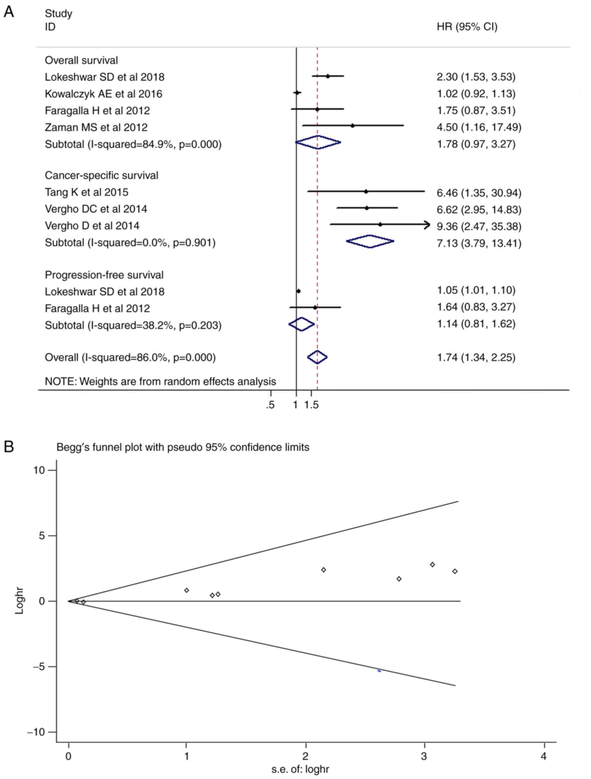
Meta-analysis of the effect of miR-21
expression on RCC prognosis and patient survival
Among the seven included studies, which contained a
total of 473 patients with survival data, three studies reported an
association between miR-21 expression and CSS in RCC, and the
remaining studies investigated OS. Due to heterogeneity, a
random-effects model was used to calculate the pooled HR value for
the survival and progression data (Fig. 1). The combined HR was calculated as
1.74 (95% CI, 1.34-2.25; P<0.001) in Fig. 2A, suggesting that increased
expression levels of miR-21 were significantly associated with
adverse prognosis in the pooled patient group. In the subgroup
analysis, the expression levels of miR-21 were significantly
associated with CSS, with a pooled HR estimate of 7.13 (95% CI,
3.79-13.41; P<0.001). However, non-significant decreases in OS
and PFS were observed in patients with RCC with high miR-21
expression (P=0.063 and P=0.443, respec tively). The Begg's funnel
plot revealed asymmetry (Fig. 2B),
which is typically associated with publication bias. For the
Egger's regression asymmetry test, the P-value was 0.002,
indicating publication bias.
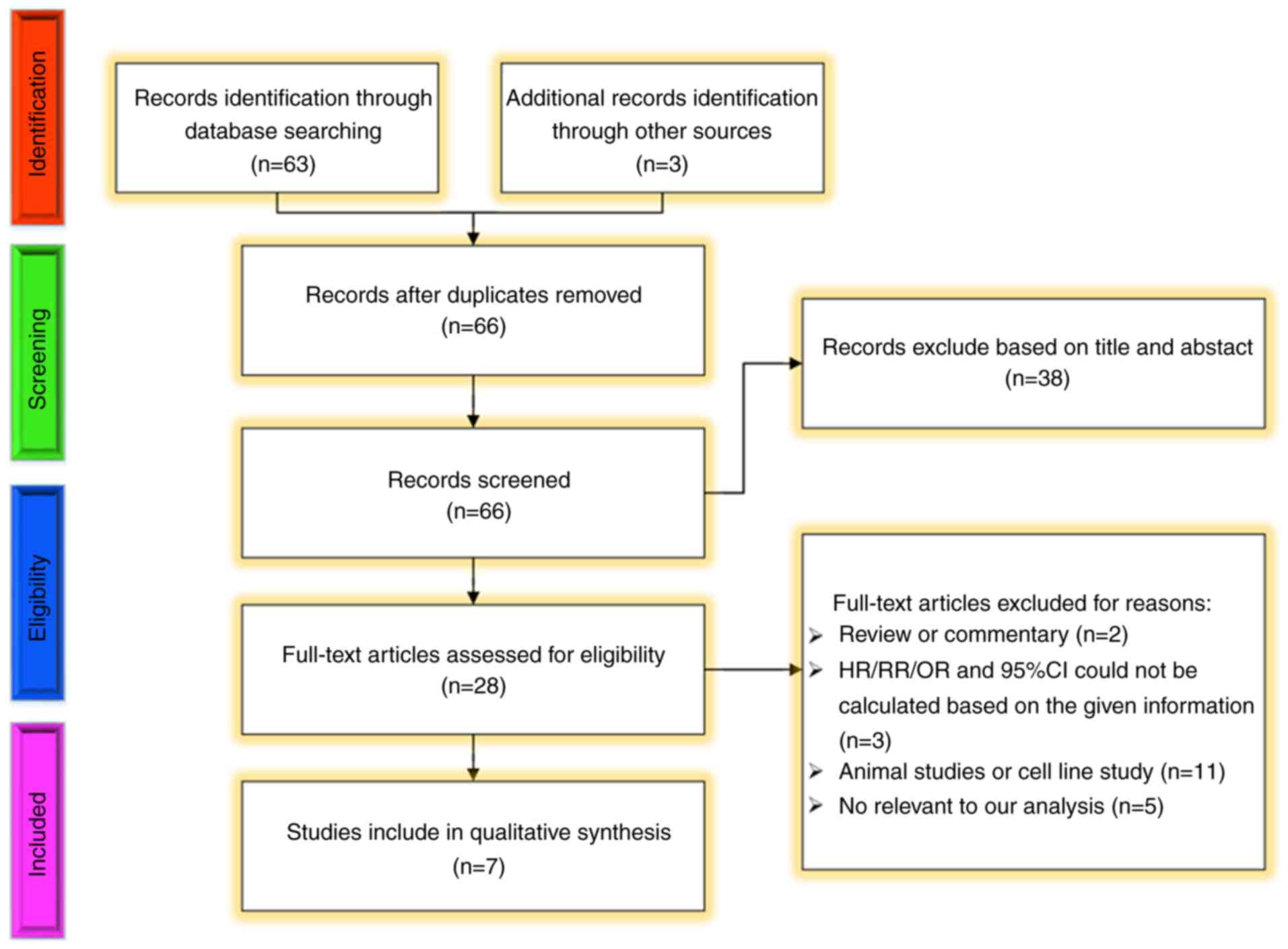 | Figure 1Methodological flow diagram of the
systematic review process. Among these, other sources included
potentially eligible articles or abstracts which were published in
major academic conferences (European Society of Urology, American
Urological Association, Asian Society of Urology, American Society
of Clinical Oncology, European Society for Medical Oncology amongst
others). HR, hazard ratio; RR, risk ratio, OR, odds ratio; CI,
confidence interval. |
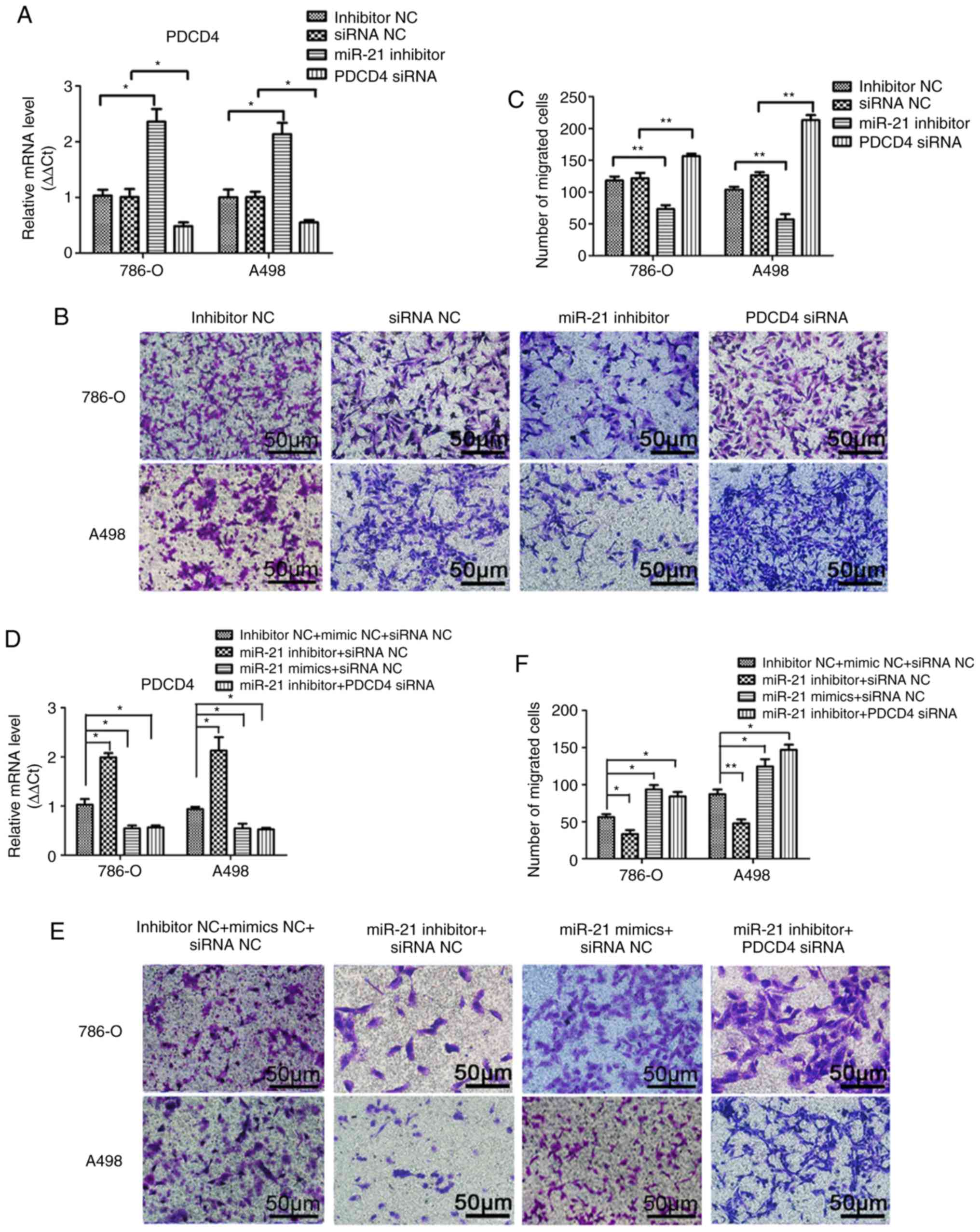
miR-21 regulates the migration, invasion
and angiogenic abilities of A498 and 786-O cells by targeting
PDCD4
A Transwell assay was performed to investigate the
effects of miR-21 on RCC cell migration and invasion. As shown in
Figs. 3B and C, and 4B and C, notable differences were
observed in the migration and invasion abilities among the
different groups of A498 or 786-O cells following transfection.
Transfection efficiency was verified by qPCR (Figs. 3A and 4A). Compared with the miRNA inhibitor NC
and siRNA NC groups, the migration and invasion abilities were
significantly decreased in the miR-21 inhibitor group, and
significantly increased in the group transfected with PDCD4 siRNA.
To elucidate the effect of the supernatant of A498 or 786-O cells
on the endothelial cell line HMEC-1, a tube formation assay in
Matrigel was performed, and the results are presented in Fig. 5. The transfection efficiency was
verified by qPCR (Fig. 5A).
Similarly, by inhibiting miR-21 expression, the number of tubes and
tube junctions in the miR-21 inhibitor group was significantly
decreased, whereas downregulating PDCD4 increased the angiogenic
ability (Fig. 5B and C).
Additionally, the migration, invasion and tube formation abilities
in the miR-21 inhibitor + siRNA NC group were lower compared with
those in the inhibitor NC + siRNA NC and the miR-21 inhibitor +
PDCD4 siRNA groups, whereas the decrease in these abilities was
reversed in the miR-21 mimics + siRNA NC group (Figs. 3E and F; 4E and F and 5E and F). These findings indicated that
miR-21 may regulate the migration, invasion and angiogenic
abilities of RCC cells through PDCD4.
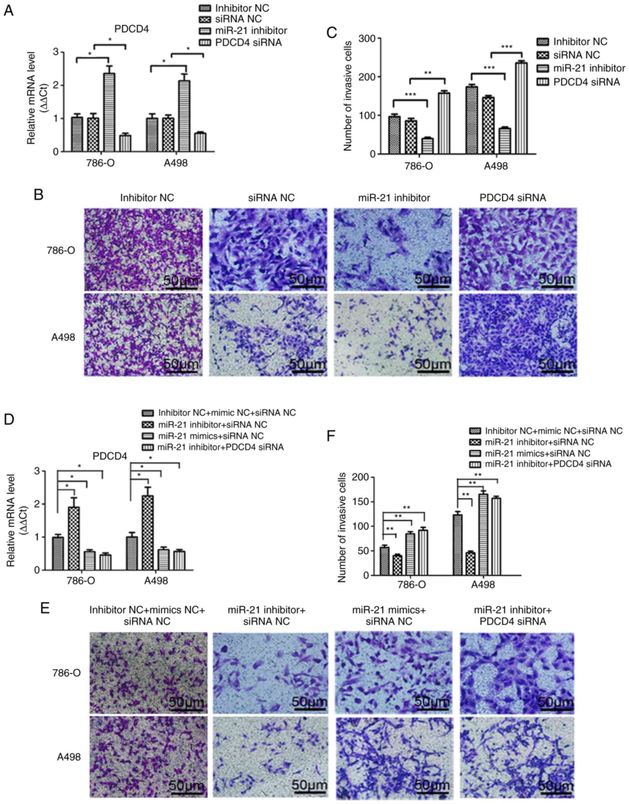 | Figure 3miR-21 promotes the migration of A498
and 786-O cells through PDCD4 regulation. (A) Transfection
efficiency was verified by quantitative PCR after transfection with
miR-21 inhibitor, PDCD4 siRNA, miRNA inhibitor NC or siRNA NC for
48 h. (B) Migratory ability was observed using a Transwell assay
after transfection with miR-21 inhibitor, PDCD4 siRNA, miRNA
inhibitor NC or siRNA NC. (C) The number of migrating cells were
calculated. (D) Transfection efficiency was verified by
quantitative PCR after transfection with combined miR-21 inhibitor,
miR-21 mimics, PDCD4 siRNA and the respective NCs. (E) Migratory
ability was observed using a Transwell assay after transfection
with combined miR-21 inhibitor, miR-21 mimics, PDCD4 siRNA and
respective NCs. (F) The number of migrating cells were calculated.
Data are presented as the mean ± standard error of the mean.
*P<0.05; **P<0.01; ***P<0.001.
PDCD4, programmed cell death protein 4; miRNA, microRNA; siRNA,
small interfering RNA; NC, negative control. |
 | Figure 4Effect on the invasion of A498 and
786-O cells by miR-21 expression through PDCD4. (A) Transfection
efficiency was verified by quantitative PCR after transfected with
miR-21 inhibitor, PDCD4 siRNA, miRNA inhibitor NC or siRNA NC for
48 h. (B) The invasion ability was observed by Transwell assay
after transfected with miR-21 inhibitor, PDCD4 siRNA, miRNA
inhibitor NC and siRNA NC. (C) Number of invading cells were
calculated. (D) Transfection efficiency was verified by
quantitative PCR after transfected with combined miR-21 inhibitor,
miR-21 mimics, PDCD4 siRNA and respective NC. (E) The invasion
ability was observed by Transwell assay after transfected with
combined miR-21 inhibitor, miR-21 mimics, PDCD4 siRNA and
respective NC. (F) Number of invading cells were calculated. Data
are presented as the mean ± standard error of the mean.
*P<0.05; **P<0.01. PDCD4, programmed
cell death protein 4; miRNA, microRNA; siRNA, small interfering
RNA; NC, negative control. |
 | Figure 5miR-21 promotes the angiogenic
ability of A498 and 786-O cells through targeting PDCD4. (A)
Transfection efficiency was determined by quantitative PCR after
transfection with miR-21 inhibitor, PDCD4 siRNA, miRNA inhibitor NC
or siRNA NC. (B) Conditioned medium from 786-O and A498 cells
transfected with miR-21 inhibitor, PDCD4 siRNA, miRNA inhibitor NC
and siRNA NC was used to overlay HMEC-1 cells seeded on a bed of
Matrigel for 6 h in an endothelial cell tube formation assays. (C)
Number of tubes and tube junctions were calculated. (D)
Transfection efficiency was verified by quantitative PCR after
transfected with combined miR-21 inhibitor, miR-21 mimics, PDCD4
siRNA and respective NC. (E) Conditioned medium from 786-O and A498
cells transfected with miR-21 inhibitor, miR-21 mimics, PDCD4 siRNA
and respective NC was used to overlay HMEC-1 cells seeded on a bed
of Matrigel for 6 h in endothelial cell tube formation assays. (F)
Number of tubes and tube junctions were calculated. Data are
presented as the mean ± standard error of the mean.
*P<0.05. PDCD4, programmed cell death protein 4;
miRNA, microRNA; siRNA, small interfering RNA; NC, negative
control. |
miR-21 regulates AP-1 signalling in RCC
cells via PDCD4
To elucidate the mechanism underlying the effect of
miR-21 on the migration, invasion and angiogenesis of RCC cells by
regulating the PDCD4/AP-1 signalling pathway, A498 and 786-O cells
were temporarily transfected with miR-21 inhibitor, PDCD4 siRNA,
inhibitor NC or siRNA NC. The results demonstrated that miR-21
inhibition decreased the protein levels of p-c-Jun and total c-Jun
in A498 and 786-O cells, and inhibited the expression of downstream
molecules, including MMP2, MMP9, ANG-1 and VEGFA (Fig. 6A D). Furthermore, the present study
investigated the ability of PDCD4 to regulate the miR-21-mediated
inhibition of MMP2, MMP9, ANG-1 and VEGFA protein expression.
miR-21 inhibition induced downregulation of MMP2, MMP9, ANG-1,
VEGFA and p-c-Jun levels in A498 and 786-O cells was rescued by
PDCD4 siRNA (Fig. 6E G), and
miR-21 mimics exhibited the same effects as PDCD4 siRNA.
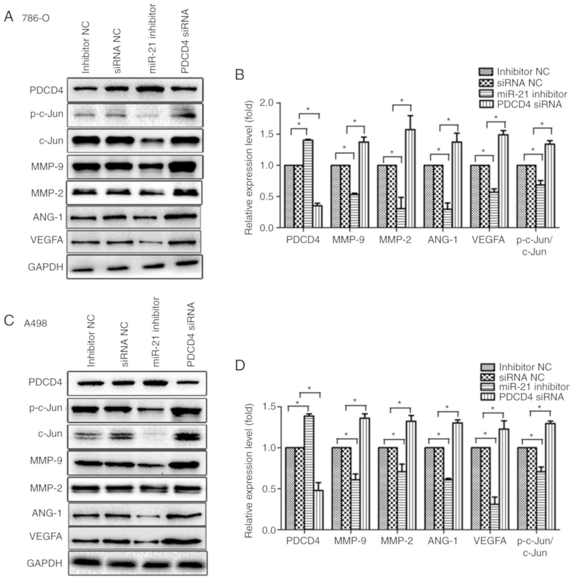 | Figure 6Suppression of PDCD4 expression,
activation of c-Jun and expression of downstream signalling
molecules, including MMP2, MMP9, ANG-1 and VEGFA, with increased
miR-21 expression. (A) Transfection of 786-O cells with miRNA
inhibitor NC, siRNA NC, miR-21 inhibitor or PDCD4 siRNA. Proteins
were extracted and analysed by western blotting. (B) Relative
protein expression by scanning densitometry was calculated. (C)
Transfection of A498 cells with miRNA inhibitor NC, siRNA NC,
miR-21 inhibitor or PDCD4 siRNA. Proteins were extracted and
analysed by western blotting. (D) Relative protein expression by
scanning densitometry was calculated. (E) Transfection of 786-O
cells with a combination of miR-21 inhibitor, miR-21 mimics, PDCD4
siRNA and respective NC. Total protein was extracted and analysed
by western blotting. (F) Relative protein expression by scanning
densitometry was calculated. Suppression of PDCD4 expression,
activation of c-Jun and expression of downstream signalling
molecules, including MMP2, MMP9, ANG-1 and VEGFA, with increased
miR-21 expression. (G) Transfection of A498 cells with a
combination of miR-21 inhibitor, miR-21 mimics, PDCD4 siRNA and
respective NC. Total protein was extracted and analysed by western
blotting. (H) Relative protein expression by scanning densitometry
was calculated. Data are presented as the mean ± standard error of
the mean. *P<0.05. PDCD4, programmed cell death
protein 4; miRNA, microRNA; siRNA, small interfering RNA; NC,
negative control; VEGF, vascular endothelial growth factor; MMP,
matrix metalloproteinase. |
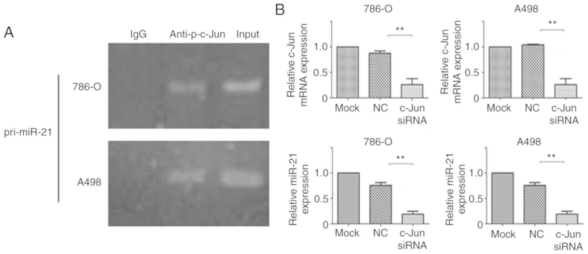
Transcription of miR-21 is activated by
c-Jun
It has been previously demonstrated that c-Jun
motivates gene transcription by attaching to its binding sites
within the promoter regions of genes (31,32).
To examine whether c-Jun directly interacts with the promoter
region of miR-21, the pri-miR-21 promoter regions associated with
p-c-Jun were assessed by ChIP assay. The results (Fig. 7A) demonstrated that p-c-Jun binds
specifically to the pri-miR-21 promoter region in A498 and 786-O
cells. To assess the control of miR-21 expression by c-Jun in RCC,
A498 and 786-O cells were transfected with c-Jun siRNA and the
expression levels of miR-21 were subsequently determined. As shown
in Fig. 7B, the expression levels
of miR-21 were significantly decreased in cells following
transfection with c-Jun siRNA compared with in control cells,
indicating that c-Jun may activate miR-21 transcription.
Discussion
Several studies have indicated that high miR-21
expression in patients with RCC is associated with an unfavourable
prognosis, including poorer metastasis-free survival, OS and
disease-specific mortality (33,34).
However, Kowalczyk et al (23) identified dissimilar outcomes
regarding miR 2l expression and its prognostic value in RCC. A
survey of 56 patients with RCC undergoing radical nephrectomy
revealed that high levels of miR-21 expression were not an
independent predictor of OS (23).
Therefore, the prognostic implications of miR-21 in patients with
RCC are inconsistent. On one hand, specific race/sex/age-associated
factors may be responsible for these differences (35). Delfino et al (36) reported that four miRNAs, including
ebv-miR-bhrf1-1, hsa-miR-565, hsa-miR-137 and hsa-miR-512-3p, are
associated with OS and PFS in glioblastoma. On the other hand,
different sample types and lack of a unified cut-off value for
miR-21 may affect the results and produce statistical heterogeneity
(37,38). Frozen or formalin-fixed tissues and
paraffin-embedded tissues are the sources for total RNA extraction.
However, RNA degradation caused by formalin fixation may affect
subsequent quantitative analyses (37,38).
Kakimoto et al (39)
revealed that the mean read length of RNAs from formalin-fixed and
paraffin-embedded (FFPE) tissue is shorter compared with that from
the matched refrigerated sample, demonstrating that longer RNA is
segmented into smaller RNA, resulting in an increase in total
reading count in FFPE samples. Finally, certain quantitative
methods for miRNAs are based on RT-qPCR, including TaqMan and SYBR.
TaqMan's advanced miRNA assays can translate all miRNAs into cDNA
in the same tube. As TaqMan analysis is occasionally restricted by
the efficiency of the additional enzymatic steps required,
exceptive reagents, including enzymatic stem loop probes and locked
nucleic acid modified primers, which may reduce nonspecific
ligation of probes and interference of precursor miRNA, are
required (40,41). Androvic et al (42) used a two-tailed RT-qPCR approach,
which uses SYBR Green to achieve the efficiency of a
poly-tail-based approach.
miR-21 is frequently overexpressed in cancer, acting
as an oncogene and tumour prognostic marker (43,44).
In patients with pancreatic cancer, overexpression of miR-21 is
associated with a low OS rate and a HR of 2.01 (45). In gastric cancer, Ren et al
(46) reported the association
between miR-21 and lymph node metastasis and suggested that the
expression of miR-21 may be applied to predict lymph node
metastasis. Additionally, miR-21 exerts an effect on the molecular
and cellular biology of multiple types of tumours, including the
following aspects: i) Promoting malignant biological behaviour. In
hepatocellular carcinoma, the overexpression of miR-21 can enhance
the liver cancer stem cell phenotype and promote invasion,
migration and tumorigenesis (47).
Similarly, Qi et al (48)
reported that miR-21 expression promotes the growth of gastric
cancer cells by targeting prostaglandin E2 to control the PTEN/AKT
signalling pathway. ii) Regulating the drug resistance of tumours.
By targeting HMG box transcription factor 1, a transcriptional
repressor 513 amino acid residues in length, miR-21 markedly
affects drug sensitivity and invasion of drug-resistant lung
adenocarcinoma cells (49). In
epithelial ovarian cancer, miR-21 may enhance resistance of
epithelial cancer cells, and chemoresistance to cisplatin may be
improved through downregulation of PTEN (50). iii) Participating in intercellular
communication through vesicular miRNAs. As mediators of
carcinogenesis, extracellular vesicles are responsible for the
communication between the cells of the tumour microenvironment
(51). Samsonov et al
(52) reported that miR-21 and
miR-18Ia-Sp are expressed in the exosomes of patients with thyroid
cancer (TC). This comparative assessment may contribute to
distinguishing between papillary and follicular types of TC with
100% sensitivity and 77% specificity (52). Finally, in gastric cancer,
proliferation of BGC-823 cells may be caused by the release of a
small RNA-21 inhibitor from macrophages (53).
It has been widely reported that miR-21 is involved
in the regulation of the aggressiveness of several diseases, such
as hepatic fibrosis in chronic hepatitis C virus (54), myeloid leukaemia (55) and hypertensive kidney injury
(56). As miR-21 is expressed
aberrantly in rheumatoid arthritis (RA), an inhibitor targeting
miR-21 may reduce the invasive ability of RA-derived
fibroblast-like synoviocytes (FLSs) by inhibiting the transforming
growth factor β1/Smad4/7 signalling pathway and altering the
expression of MMPs, thereby suppressing the invasiveness of FLSs
(57). In addi tion to benign
diseases, miR-21 is overexpressed in a variety of diverse
malignancies and is associated with metastasis of tumour cells as
follows: i) As an upstream promoter, miR-21 affects the expression
or biological function of downstream genes associated with tumour
suppression, including PTEN, which negatively regulates the
PI3K/AKT signalling pathway in the oral squamous cell carcinoma
SCC15 and SCC25 cell lines (58);
Snail1, which is implicated in epithelial-mesenchymal transition
(EMT) by directly suppressing the level of E-cadherin in salivary
adenoid cystic carcinoma (59);
and PDCD4, which activates the expression of AKT, IKKβ and mTORC1,
which are necessary for the migration and inva sion of RCC cells
(60). Additionally, Bera et
al (60) reported that there
is a positive feedback loop among miR-21 level, phosphorylated IKKβ
and NF κB activation, as PDCD4 has been demonstrated to negatively
affect the phosphorylation and activation of IKKβ and NF κB.
Furthermore, the present study revealed that miR-21 resulted in
variations in the expression levels of MMPs by targeting the
PDCD4/c-Jun signalling pathway, which is involved in the metastasis
of renal cancer. ii) As a downstream tumour promoting miRNA, the
levels or target genes of miR-21 are regulated by upstream
oncogenes, including Sox2, which not only positively regulates
miR21 associated migration/invasion signalling in glioma cells, but
also induces EMT of laryngeal cancer by activation of Wnt/β catenin
signalling (61). As an upstream
regulator of miR-21, p-STAT3 may regulate the metastatic capacity
of hepatocellular carcinoma cells by targeting miR-21, which
increases expression of cysteine rich proteins with kazal motifs
and PDCD4 (62). iii) As
endocytosis of exosomes of target cancer cells contributes to the
intracellular release of vesicular contents, exosomal miR-21 can
increase the ability of invasion potential and aggressive phenotype
of ovarian cancer cells through upregulation of MMP1, which is
transferred by cell-cell communication (63).
Angiogenesis is not only a key developmental
process, such as ocular neovascularization, it is also essential in
the pathological processes of various diseases including
cardiovascular diseases (64),
osteoarthritis (65) and diabetic
peripheral neuropathy (66,67).
Therefore, research regarding the role of miR-21 in angiogenesis is
currently focused mainly on haematological tumours. In diffuse
large B-cell lymphoma, Zheng et al (68) demonstrated a direct link between
miR-21 and tumour angiogenesis in lymphoma. miR-21 can increase the
interaction of endothelial cells with Treg cells. Subsequently,
after enhancing the expression of inducible T cell costimulator
(ICOS) on Treg cells, miR-21 prompts tumour angiogenesis via
ICOS/inducible T cell costimulator ligand pathway signalling, which
promotes disease progression and chemoresistance of B-cell lymphoma
(68). In acute monocytic
leukaemia (AML), the expression levels of miR-21 and VEGF in the
peripheral blood monocytes of the patients was higher compared with
that in healthy controls. Being the direct target of miR-21, the
level of interleukin 12 (IL-12) in the supernatant of THP-1 cells
is increased following transfection with miR-21 mimic (69). Additionally, IL-12 may induce VEGF
expression and angiogenic ability in human umbilical vein
endothelial cells, which suggests that miR-21 may possess
pro-angiogenic properties in human AML (69). At present, there is little research
on the role of miR-21 in the process of solid tumour angiogenesis.
In colorectal cancer, miR-21 is overexpressed and affects cell
cycle progression, apoptosis and viability of colon cancer cells
(70). Song and Rossi (70) identified an anti-miR-21 that
targets miR-21 to inhibit genes by post transcriptional or
transcriptional gene silencing. Since anti-miR-21 and pri-miR30
exhibit homology between anti-miR-21 and the 3′ end of pri-miR30,
anti-miR-21 may reduce the expression of miR30, which affects
vessel number and length, by inhibition of angiogenic pathways
(70). Therefore, anti-miR-21 may
be a useful curative strategy by regulating the process of
angiogenesis in colon cancer. In the present study, the effects of
the supernatant from A498 or 786-O cells on the endothelial HMEC-1
cell line were investigated, and it was observed that the quantity
of formed tubes and tube junctions in the miR-21 inhibitor group
was significantly decreased, whereas that in the PDCD4 siRNA group
was significantly increased, compared with the respective control
groups. To the best of our knowledge, this constitutes the first
evidence that miR-21 expression may promoted the ability of HMEC-1
cells to become organised into tubular networks by directly
targeting the PDCD4/c-Jun signalling pathway and regulating the
level of ANG-1 and VEGFA, indicating a direct association between
miR-21 and tumour angiogenesis and progression in RCC.
There are several miR-21 target genes. PDCD4 has
been recognized as a protein expressed during the processes of
apoptosis and tumour suppression. However, only few studies have
specifically demonstrated that there is an interaction between
miR-21 and PDCD4 in RCC (71). As
AP-1 is a transcription activating heterodimer composed of c-Jun
and c-Fos, studies have demonstrated (72-74)
that AP-1 is associated with invasion and metastasis of tumours by
regulating MMP2 or MMP9, and with angiogenesis by controlling ANG-1
or VEGFA. Under the influence of miR-21, the protein levels of
downstream molecules of the PDCD4 AP-1 signalling pathway,
including MMP2, MMP9, ANG-1 and VEGFA, were decreased.
Additionally, in A498 and 786-O cells, the miR-21 inhibition
induced downregulation of MMP2, MMP9, ANG-1, VEGFA and p-c-Jun
expression was reversed by PDCD4 siRNA. Among the targets (Fig. 8), ANG-1 usually promotes the
interaction between endothelial and perivascular cells to obtain a
stable vasculature (75,76). VEGFA serves a key role in the
growth of new vessels and has become a promising target for
anti-angiogenesis based tumour therapy (77,78).
As MMPs have been initially identified as proteases that act on the
extracellular matrix, the overexpression of MMP2 and MMP9 has been
associated with aggressive behaviour of tumours and metastasis
(79-81).
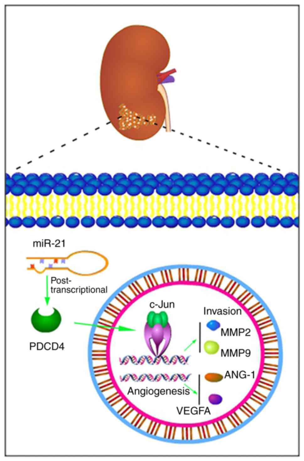 | Figure 8Schematic diagram of the roles of
miR-21 and the PDCD4/c-Jun signalling pathway in renal cell
carcinoma. The overexpression of miR-21 and the reduced PDCD4
expression, leading to the activation of c-Jun, further increased
the levels of MMP2, MMP9, ANG-1 and VEGFA, which promoted tumour
cell migration, invasion and angiogenesis. PDCD4, programmed cell
death protein 4; miR, microRNA; VEGF, vascular endothelial growth
factor; MMP, matrix metalloproteinase. |
In summary, the present study attempted to combine
evidence-based medicine and molecular biology and demonstrated that
increased miR-21 levels were significantly associated with adverse
prognosis in patients with RCC. Additionally, the migration,
invasion and angiogenic abilities of RCC cells were markedly
affected by the expression of miR-21 through direct targeting of
the PDCD4/c-Jun signalling pathway, indicating that miR-21 may be
of value as a therapeutic target for RCC.
Funding
The present study was supported by grants from the
National Natural Science Foundation of China (grant no. 31800787,
grant no. 81572505 and grant no. 81972831), the Natural Science
Foundation of Liaoning Province (grant no. LQ2017025), the Doctoral
Research Startup Foundation of Liaoning Province (grant no.
20180540020) and the Medical Scientific Research Project of Dalian
City (grant no. 1812038).
Availability of data and materials
The datasets used and/or analysed during the current
study are available from the corresponding author on reasonable
request.
Authors' contributions
BF, YYJ, HSZ, TJL and XCL conceived and designed the
study and wrote the manuscript. BF, YYJ and HSZ performed the
experiments. BF, RZ, MS and MFS collected the data. YYJ, WW and XGW
analysed the data. HSZ, WKL, NY and QW interpreted the data. BF,
HSZ, TJL and XCL reviewed the manuscript. All authors have read and
approved the final version of this manuscript for publication.
Ethics approval and consent to
participate
Not applicable.
Patient consent for publication
Not applicable.
Competing interests
The authors declare that they have no competing
interests.
Acknowledgments
Not applicable.
References
|
1
|
Bhatt JR and Finelli A: Landmarks in the
diagnosis and treatment of renal cell carcinoma. Nat Rev Urol.
11:517–525. 2014. View Article : Google Scholar : PubMed/NCBI
|
|
2
|
Siegel RL, Miller KD and Jemal A: Cancer
statistics, 2018. CA Cancer J Clin. 68:7–30. 2018. View Article : Google Scholar : PubMed/NCBI
|
|
3
|
Stahl M, Wilke H, Schmoll HJ, Schöber C,
Diedrich H, Casper J, Freund M and Poliwoda H: A phase II study of
high dose tamoxifen in progressive, metastatic renal cell
carcinoma. Ann Oncol. 3:167–168. 1992. View Article : Google Scholar : PubMed/NCBI
|
|
4
|
Choueiri TK and Motzer RJ: Systemic
therapy for metastatic renal cell carcinoma. N Engl J Med.
376:354–366. 2017. View Article : Google Scholar : PubMed/NCBI
|
|
5
|
Dabestani S, Marconi L, Hofmann F, Stewart
F, Lam TB, Canfield SE, Staehler M, Powles T, Ljungberg B and Bex
A: Local treatments for metastases of renal cell carcinoma: A
systematic review. Lancet Oncol. 15:e549-e5612014. View Article : Google Scholar
|
|
6
|
Qu L, Wang ZL, Chen Q, Li YM, He HW, Hsieh
JJ, Xue S, Wu ZJ, Liu B, Tang H, et al: Prognostic value of a long
non-coding RNA signature in localized clear cell renal cell
carcinoma. Eur Urol. 74:756–763. 2018. View Article : Google Scholar : PubMed/NCBI
|
|
7
|
Sekar RR, Patil D, Baum Y, Pearl J, Bausum
A, Bilen MA, Kucuk O, Harris WB, Carthon BC, Alemozaffar M, et al:
A novel preoperative inflammatory marker prognostic score in
patients with localized and metastatic renal cell carcinoma. Asian
J Urol. 4:230–238. 2017. View Article : Google Scholar
|
|
8
|
Escudier B, Motzer RJ, Sharma P, Wagstaff
J, Plimack ER, Hammers HJ, Donskov F, Gurney H, Sosman JA, Zalewski
PG, et al: Treatment beyond progression in patients with advanced
renal cell carcinoma treated with nivolumab in checkmate 025. Eur
Urol. 72:368–376. 2017. View Article : Google Scholar : PubMed/NCBI
|
|
9
|
Kroeger N, Stenzl A, Burchardt M and Bedke
J: Adjuvant treatment of high-risk renal cell carcinoma: Leaving
the det. Eur Urol. 71:695–696. 2017. View Article : Google Scholar : PubMed/NCBI
|
|
10
|
Leibovich BC, Lohse CM, Cheville JC, Zaid
HB, Boorjian SA, Frank I, Thompson RH and Parker WP: Predicting
oncologic outcomes in renal cell carcinoma after surgery. Eur Urol.
73:772–780. 2018. View Article : Google Scholar : PubMed/NCBI
|
|
11
|
Verbiest A, Couchy G, Job S, Caruana L,
Lerut E, Oyen R, de Reyniès A, Tosco L, Joniau S, Van Poppel H, et
al: Molecular subtypes of clear-cell renal cell carcinoma are
prognostic for outcome after complete metastasectomy. Eur Urol.
74:474–480. 2018. View Article : Google Scholar : PubMed/NCBI
|
|
12
|
Hydbring P, Wang Y, Fassl A, Li X, Matia
V, Otto T, Choi YJ, Sweeney KE, Suski JM, Yin H, et al:
Cell-cycle-targeting MicroRNAs as therapeutic tools against
refractory cancers. Cancer Cell. 31:576–590.e8. 2017. View Article : Google Scholar : PubMed/NCBI
|
|
13
|
Yang HY, Barbi J, Wu CY, Zheng Y, Vignali
PD, Wu X, Tao JH, Park BV, Bandara S, Novack L, et al: MicroRNA-17
modulates regulatory T cell function by targeting Co-regulators of
the Foxp3 transcription factor. Immunity. 45:83–93. 2016.
View Article : Google Scholar : PubMed/NCBI
|
|
14
|
Lin XJ, Chong Y, Guo ZW, Xie C, Yang XJ,
Zhang Q, Li SP, Xiong Y, Yuan Y, Min J, et al: A serum microRNA
classifier for early detection of hepatocellular carcinoma: A
multicentre, retrospective, longitudinal biomarker identification
study with a nested case-control study. Lancet Oncol. 16:804–815.
2015. View Article : Google Scholar : PubMed/NCBI
|
|
15
|
Iqbal MA, Arora S, Prakasam G, Calin GA
and Syed MA: MicroRNA in lung cancer: Role, mechanisms, pathways
and therapeutic relevance. Mol Aspects Med. August 18–2018.Epub
ahead of print. PubMed/NCBI
|
|
16
|
Croset M, Pantano F, Kan CWS, Bonnelye E,
Descotes F, Alix-Panabières C, Lecellier CH, Bachelier R, Allioli
N, Hong SS, et al: miRNA-30 family members inhibit breast cancer
invasion, osteomimicry, and bone destruction by directly targeting
multiple bone metastasis-associated genes. Cancer Res.
78:5259–5273. 2018. View Article : Google Scholar : PubMed/NCBI
|
|
17
|
Song S, Yang Y, Liu M, Liu B, Yang X, Yu
M, Qi H, Ren M, Wang Z, Zou J, et al: miR-125b attenuates human
hepatocellular carcinoma malignancy through targeting SIRT6. Am J
Cancer Res. 8:993–1007. 2018.PubMed/NCBI
|
|
18
|
Li Q, Zhang S, Wang M, Dong S, Wang Y, Liu
S, Lu T, Fu Y, Wang X and Chen G: Downregulated miR-21 mediates
matrine-induced apoptosis via the PTEN/Akt signaling pathway in FTC
133 human follicular thyroid cancer cells. Oncol Lett.
18:3553–3560. 2019.PubMed/NCBI
|
|
19
|
Dino P, D'Anna C, Sangiorgi C, Di Sano C,
Di Vincenzo S, Ferraro M and Pace E: Cigarette smoke extract
modulates E-Cadherin, Claudin-1 and miR-21 and promotes cancer
invasiveness in human colorectal adenocarcinoma cells. Toxicol
Lett. 317:102–109. 2019. View Article : Google Scholar : PubMed/NCBI
|
|
20
|
Sun J, Jiang Z, Li Y, Wang K, Chen X and
Liu G: Downregulation of miR-21 inhibits the malignant phenotype of
pancreatic cancer cells by targeting VHL. Onco Targets Ther.
12:7215–7226. 2019. View Article : Google Scholar : PubMed/NCBI
|
|
21
|
Li X, Xin S, He Z, Che X, Wang J, Xiao X,
Chen J and Song X: microRNA-21 (miR-21) post-transcriptionally
downregulates tumor suppressor PDCD4 and promotes cell
transformation, proliferation, and metastasis in renal cell
carcinoma. Cell Physiol Biochem. 33:1631–1642. 2014. View Article : Google Scholar : PubMed/NCBI
|
|
22
|
Lokeshwar SD, Talukder A, Yates TJ, Hennig
MJP, Garcia Roig M, Lahorewala SS, Mullani NN, Klaassen Z, Kava BR,
Manoharan M, et al: Molecular characterization of renal cell
carcinoma: A potential three MicroRNA prognostic signature. Cancer
Epidemiol Biomarkers Prev. 27:464–472. 2018. View Article : Google Scholar : PubMed/NCBI
|
|
23
|
Kowalczyk AE, Krazinski BE, Godlewski J,
Grzegrzolka J, Kiewisz J, Kwiatkowski P, Sliwinska-Jewsiewicka A,
Dziegiel P and Kmiec Z: SATB1 is down-regulated in clear cell renal
cell carcinoma and correlates with miR-21-5p overexpression and
poor prognosis. Cancer Genomics Proteomics. 13:209–217.
2016.PubMed/NCBI
|
|
24
|
Tang K and Xu H: Prognostic value of
meta-signature miRNAs in renal cell carcinoma: An integrated miRNA
expression profiling analysis. Sci Rep. 5:102722015. View Article : Google Scholar : PubMed/NCBI
|
|
25
|
Vergho D, Kneitz S, Rosenwald A, Scherer
C, Spahn M, Burger M, Riedmiller H and Kneitz B: Combination of
expression levels of miR-21 and miR-126 is associated with
cancer-specific survival in clear cell renal cell carcinoma. BMC
Cancer. 14:252014. View Article : Google Scholar
|
|
26
|
Vergho DC, Kneitz S, Kalogirou C, Burger
M, Krebs M, Rosenwald A, Spahn M, Löser A, Kocot A, Riedmiller H
and Kneitz B: Impact of miR-21, miR-126 and miR-221 as prognostic
factors of clear cell renal cell carcinoma with tumor thrombus of
the inferior vena cava. PLoS One. 9:e1098772014. View Article : Google Scholar : PubMed/NCBI
|
|
27
|
Faragalla H, Youssef YM, Scorilas A,
Khalil B, White NM, Mejia-Guerrero S, Khella H, Jewett MA, Evans A,
Lichner Z, et al: The clinical utility of miR-21 as a diagnostic
and prognostic marker for renal cell carcinoma. J Mol Diagn.
14:385–392. 2012. View Article : Google Scholar : PubMed/NCBI
|
|
28
|
Zaman MS, Shahryari V, Deng G, Thamminana
S, Saini S, Majid S, Chang I, Hirata H, Ueno K, Yamamura S, et al:
Up regulation of microRNA-21 correlates with lower kidney cancer
survival. PLoS One. 7:e310602012. View Article : Google Scholar
|
|
29
|
Tierney JF, Stewart LA, Ghersi D, Burdett
S and Sydes MR: Practical methods for incorporating summary
time-to-event data into meta-analysis. Trials. 8:162007. View Article : Google Scholar : PubMed/NCBI
|
|
30
|
Livak KJ and Schmittgen TD: Analysis of
relative gene expression data using real-time quantitative PCR and
the 2(−Delta Delta C(T)) method. Methods. 25:402–408. 2001.
View Article : Google Scholar
|
|
31
|
Zhou J, Wang KC, Wu W, Subramaniam S, Shyy
JY, Chiu JJ, Li JY and Chien S: microRNA-21 targets peroxisome
proliferators-activated receptor-alpha in an autoregulatory loop to
modulate flow-induced endothelial inflammation. Proc Natl Acad Sci
USA. 108:10355–10360. 2011. View Article : Google Scholar : PubMed/NCBI
|
|
32
|
Echevarría-Vargas IM, Valiyeva F and
Vivas-Mejía PE: Upregulation of miR-21 in cisplatin resistant
ovarian cancer via JNK-1/c-Jun pathway. PLoS One. 9:e970942014.
View Article : Google Scholar : PubMed/NCBI
|
|
33
|
Carlsson J, Christiansen J, Davidsson S,
Giunchi F, Fiorentino M and Sundqvist P: The potential role of miR
126, miR-21 and miR 10b as prognostic biomarkers in renal cell
carcinoma. Oncol Lett. 17:4566–4574. 2019.PubMed/NCBI
|
|
34
|
Chen J, Gu Y and Shen W: microRNA-21
functions as an oncogene and promotes cell proliferation and
invasion via TIMP3 in renal cancer. Eur Rev Med Pharmacol Sci.
21:4566–4576. 2017.PubMed/NCBI
|
|
35
|
Fan B, Zhang H, Jin H, Gai Y, Wang H, Zong
H, Jin M, Yang H, Wan S, Zhu J, et al: Is overexpression of Ki-67 a
prognostic biomarker of upper tract urinary carcinoma? A
retrospective cohort study and meta-analysis. Cell Physiol Biochem.
40:1613–1625. 2016. View Article : Google Scholar : PubMed/NCBI
|
|
36
|
Delfino KR, Serão NV, Southey BR and
Rodriguez-Zas SL: Therapy-, gender- and race-specific microRNA
markers, target genes and networks related to glioblastoma
recurrence and survival. Cancer Genomics Proteomics. 8:173–183.
2011.PubMed/NCBI
|
|
37
|
Czachorowski MJ, Amaral AF, Montes Moreno
S, Lloreta J, Carrato A, Tardón A, Morente MM, Kogevinas M, Real FX
and Malats N; SBC/EPICURO investigators: Cyclooxygenase-2
expression in bladder cancer and patient prognosis: Results from a
large clinical cohort and meta-analysis. PLoS One. 7:e450252012.
View Article : Google Scholar : PubMed/NCBI
|
|
38
|
Ku JH, Byun SS, Jeong H, Kwak C, Kim HH
and Lee SE: The role of p53 on survival of upper urinary tract
urothelial carcinoma: A systematic review and meta-analysis. Clin
Genitourin Cancer. 11:221–228. 2013. View Article : Google Scholar : PubMed/NCBI
|
|
39
|
Kakimoto Y, Tanaka M, Kamiguchi H, Ochiai
E and Osawa M: MicroRNA stability in FFPE tissue samples:
Dependence on GC content. PLoS One. 11:e01631252016. View Article : Google Scholar : PubMed/NCBI
|
|
40
|
Wyman SK, Parkin RK, Mitchell PS, Fritz
BR, O'Briant K, Godwin AK, Urban N, Drescher CW, Knudsen BS and
Tewari M: Repertoire of microRNAs in epithelial ovarian cancer as
determined by next generation sequencing of small RNA cDNA
libraries. PLoS One. 4:e53112009. View Article : Google Scholar : PubMed/NCBI
|
|
41
|
Stratmann J, Wang CJ, Gnosa S, Wallin A,
Hinselwood D, Sun XF and Zhang H: Dicer and miRNA in relation to
clinicopathological variables in colorectal cancer patients. BMC
Cancer. 11:3452011. View Article : Google Scholar : PubMed/NCBI
|
|
42
|
Androvic P, Valihrach L, Elling J, Sjoback
R and Kubista M: Two-tailed RT-qPCR: A novel method for highly
accurate miRNA quantification. Nucleic Acids Res. 45:e1442017.
View Article : Google Scholar : PubMed/NCBI
|
|
43
|
Macfarlane LA and Murphy PR: MicroRNA:
Biogenesis, function and role in cancer. Curr Genomics. 11:537–561.
2010. View Article : Google Scholar
|
|
44
|
Nassar FJ, Nasr R and Talhouk R: MicroRNAs
as biomarkers for early breast cancer diagnosis, prognosis and
therapy prediction. Pharmacol Ther. 172:34–49. 2017. View Article : Google Scholar
|
|
45
|
Negoi I, Hostiuc S, Sartelli M, Negoi RI
and Beuran M: microRNA-21 as a prognostic biomarker in patients
with pancreatic cancer - A systematic review and meta-analysis. Am
J Surg. 214:515–524. 2017. View Article : Google Scholar : PubMed/NCBI
|
|
46
|
Ren J, Kuang TH, Chen J, Yang JW and Liu
YX: The diagnostic and prognostic values of microRNA-21 in patients
with gastric cancer: A meta-analysis. Eur Rev Med Pharmacol Sci.
21:120–130. 2017.PubMed/NCBI
|
|
47
|
Jiang J, Yang P, Guo Z, Yang R, Yang H,
Yang F, Li L and Xiang B: Overexpression of microRNA-21 strengthens
stem cell-like characteristics in a hepatocellular carcinoma cell
line. World J Surg Oncol. 14:2782016. View Article : Google Scholar : PubMed/NCBI
|
|
48
|
Qi R, Wang DT, Xing LF and Wu ZJ: miRNA-21
promotes gastric cancer growth by adjusting prostaglandin E2. Eur
Rev Med Pharmacol Sci. 22:1929–1936. 2018.PubMed/NCBI
|
|
49
|
Su C, Cheng X, Li Y, Han Y, Song X, Yu D,
Cao X and Liu Z: miR-21 improves invasion and migration of drug
resistant lung adenocarcinoma cancer cell and transformation of EMT
through targeting HBP1. Cancer Med. 7:2485–2503. 2018. View Article : Google Scholar : PubMed/NCBI
|
|
50
|
Yu X, Chen Y, Tian R, Li J, Li H, Lv T and
Yao Q: miRNA-21 enhances chemoresistance to cisplatin in epithelial
ovarian cancer by negatively regulating PTEN. Oncol Lett.
14:1807–1810. 2017. View Article : Google Scholar : PubMed/NCBI
|
|
51
|
Peinado H, Alečković M, Lavotshkin S,
Matei I, Costa-Silva B, Moreno Bueno G, Hergueta Redondo M,
Williams C, García-Santos G, Ghajar C, et al: Melanoma exosomes
educate bone marrow progenitor cells toward a pro-metastatic
phenotype through MET. Nat Med. 18:883–891. 2012. View Article : Google Scholar : PubMed/NCBI
|
|
52
|
Samsonov R, Burdakov V, Shtam T,
Radzhabovа Z, Vasilyev D, Tsyrlina E, Titov S, Ivanov M, Berstein
L, Filatov M, et al: Plasma exosomal miR-21 and miR-181a
differentiates follicular from papillary thyroid cancer. Tumour
Biol. 37:12011–12021. 2016. View Article : Google Scholar : PubMed/NCBI
|
|
53
|
Wang JJ, Wang ZY, Chen R, Xiong J, Yao YL,
Wu JH and Li GX: Macrophage-secreted exosomes delivering miRNA-21
inhibitor can regulate BGC-823 cell proliferation. Asian Pac J
Cancer Prev. 16:4203–4209. 2015. View Article : Google Scholar : PubMed/NCBI
|
|
54
|
Besheer T, Elalfy H, Abd El-Maksoud M, Abd
El-Razek A, Taman S, Zalata K, Elkashef W, Zaghloul H, Elshahawy H,
Raafat D, et al: Diffusion weighted magnetic resonance imaging and
micro-RNA in the diagnosis of hepatic fibrosis in chronic hepatitis
C virus. World J Gastroenterol. 25:1366–1377. 2019. View Article : Google Scholar : PubMed/NCBI
|
|
55
|
Panagal M, S R SK, P S, M B, M K,
Gopinathe V, Sivakumare P and Sekar D: MicroRNA21 and the various
types of myeloid leukemia. Cancer Gene Ther. 25:161–166. 2018.
View Article : Google Scholar : PubMed/NCBI
|
|
56
|
Chen C, Lu C, Qian Y, Li H, Tan Y, Cai L
and Weng H: Urinary miR-21 as a potential biomarker of hypertensive
kidney injury and fibrosis. Sci Rep. 7:177372017. View Article : Google Scholar : PubMed/NCBI
|
|
57
|
Xiong G, Huang Z, Jiang H, Pan Z, Xie J
and Wang S: Inhibition of microRNA-21 decreases the invasiveness of
fibroblast-like synoviocytes in rheumatoid arthritis via TGFβ/Smads
signaling pathway. Iran J Basic Med Sci. 19:787–793.
2016.PubMed/NCBI
|
|
58
|
Zheng Y, Xie J, Jiang F, Li Y, Chang G and
Ma H: Inhibition of miR-21 promotes cell apoptosis in oral squamous
cell carcinoma by upregulating PTEN. Oncol Rep. 40:2798–2805.
2018.PubMed/NCBI
|
|
59
|
Yan F, Wang C, Li T, Cai W and Sun J: Role
of miR-21 in the growth and metastasis of human salivary adenoid
cystic carcinoma. Mol Med Rep. 17:4237–4244. 2018.PubMed/NCBI
|
|
60
|
Bera A, Das F, Ghosh-Choudhury N, Kasinath
BS, Abboud HE and Choudhury GG: microRNA-21 induced dissociation of
PDCD4 from rictor contributes to Akt-IKKβ-mTORC1 axis to regulate
renal cancer cell invasion. Exp Cell Res. 328:99–117. 2014.
View Article : Google Scholar : PubMed/NCBI
|
|
61
|
Luo G, Luo W, Sun X, Lin J, Wang M and
Zhang Y, Luo W and Zhang Y: microRNA-21 promotes migration and
invasion of glioma cells via activation of Sox2 and β-catenin
signaling. Mol Med Rep. 15:187–193. 2017. View Article : Google Scholar
|
|
62
|
Zhang N, Duan WD, Leng JJ, Zhou L, Wang X,
Xu YZ, Wang XD, Zhang AQ and Dong JH: STAT3 regulates the migration
and invasion of a stem-like subpopulation through microRNA-21 and
multiple targets in hepatocellular carcinoma. Oncol Rep.
33:1493–1498. 2015. View Article : Google Scholar : PubMed/NCBI
|
|
63
|
Au Yeung CL, Co NN, Tsuruga T, Yeung TL,
Kwan SY, Leung CS, Li Y, Lu ES, Kwan K, Wong KK, et al: Exosomal
transfer of stroma-derived miR21 confers paclitaxel resistance in
ovarian cancer cells through targeting APAF1. Nat Commun.
7:111502016. View Article : Google Scholar : PubMed/NCBI
|
|
64
|
Wang L, Jia Q, Xinnong C, Xie Y, Yang Y,
Zhang A, Liu R, Zhuo Y and Zhang J: Role of cardiac progenitor
cell-derived exosome-mediated microRNA-210 in cardiovascular
disease. J Cell Mol Med. 23:7124–7131. 2019. View Article : Google Scholar : PubMed/NCBI
|
|
65
|
Xie W, Su W, Xia H, Wang Z, Su C and Su B:
Synovial Fluid microRNA-210 as a potential biomarker for early
prediction of osteoarthritis. Biomed Res Int. 2019:71654062019.
View Article : Google Scholar : PubMed/NCBI
|
|
66
|
Zhuang Y, Peng H, Mastej V and Chen W:
MicroRNA regulation of endothelial junction proteins and clinical
consequence. Mediators Inflamm. 2016:50786272016. View Article : Google Scholar : PubMed/NCBI
|
|
67
|
Merrigan SL and Kennedy BN: Vitamin D
receptor agonists regulate ocular developmental angiogenesis and
modulate expression of dre-miR-21 and VEGF. Br J Pharmacol.
174:2636–2651. 2017. View Article : Google Scholar : PubMed/NCBI
|
|
68
|
Zheng Z, Xu PP, Wang L, Zhao HJ, Weng XQ,
Zhong HJ, Qu B, Xiong J, Zhao Y, Wang XF, et al: MiR21 sensitized
B-lymphoma cells to ABT-199 via ICOS/ICOSL-mediated interaction of
Treg cells with endothelial cells. J Exp Clin Cancer Res.
36:822017. View Article : Google Scholar : PubMed/NCBI
|
|
69
|
He XP, Chen P, Yang K, Liu B, Zhang Y,
Wang F, Guo Z, Liu XD, Lou JX and Chen HR: Overexpression of miR-21
is involved in acute monocytic leukemia-associated angiogenesis by
targeting IL-12. Mol Med Rep. 18:4122–4128. 2018.PubMed/NCBI
|
|
70
|
Song MS and Rossi JJ: The anti-miR21
antagomir, a therapeutic tool for colorectal cancer, has a
potential synergistic effect by perturbing an angiogenesis
associated miR30. Front Genet. 4:3012014. View Article : Google Scholar
|
|
71
|
Yuan H, Xin S, Huang Y, Bao Y, Jiang H,
Zhou L, Ren X, Li L, Wang Q and Zhang J: Downregulation of PDCD4 by
miR-21 suppresses tumor transformation and proliferation in a nude
mouse renal cancer model. Oncol Lett. 14:3371–3378. 2017.
View Article : Google Scholar : PubMed/NCBI
|
|
72
|
Suman P, Godbole G, Thakur R,
Morales-Prieto DM, Modi DN, Markert UR and Gupta SK: AP-1
transcription factors, mucin-type molecules and MMPs regulate the
IL-11 mediated invasiveness of JEG 3 and HTR 8/SVneo trophoblastic
cells. PLoS One. 7:e297452012. View Article : Google Scholar
|
|
73
|
Hwang YP, Yun HJ, Choi JH, Han EH, Kim HG,
Song GY, Kwon KI, Jeong TC and Jeong HG: Suppression of EGF-induced
tumor cell migration and matrix metalloproteinase-9 expression by
capsaicin via the inhibition of EGFR-mediated FAK/Akt, PKC/Raf/ERK,
p38 MAPK, and AP-1 signaling. Mol Nutr Food Res. 55:594–605. 2011.
View Article : Google Scholar : PubMed/NCBI
|
|
74
|
Chen Q, Chen P, Pang X, Hu Y and Zhang Y:
Adrenomedullin up regulates the expression of vascular endothelial
growth factor in epithelial ovarian carcinoma cells via JNK/AP-1
pathway. Int J Gynecol Cancer. 25:953–960. 2015. View Article : Google Scholar : PubMed/NCBI
|
|
75
|
Cossutta M, Darche M, Carpentier G, Houppe
C, Ponzo M, Raineri F, Vallée B, Gilles ME, Villain D, Picard E, et
al: Weibel-palade bodies orchestrate pericytes during angiogenesis.
Arterioscler Thromb Vasc Biol. 39:1843–1858. 2019. View Article : Google Scholar : PubMed/NCBI
|
|
76
|
Pellegrinelli V, Rouault C, Veyrie N,
Clément K and Lacasa D: Endothelial cells from visceral adipose
tissue disrupt adipocyte functions in a three-dimensional setting:
Partial rescue by angiopoietin-1. Diabetes. 63:535–549. 2014.
View Article : Google Scholar
|
|
77
|
Brudno Y, Ennett Shepard AB, Chen RR,
Aizenberg M and Mooney DJ: Enhancing microvascular formation and
vessel maturation through temporal control over multiple
pro-angiogenic and pro maturation factors. Biomaterials.
34:9201–9209. 2013. View Article : Google Scholar : PubMed/NCBI
|
|
78
|
Khan JA, Maki RG and Ravi V: Pathologic
angiogenesis of malignant vascular sarcomas: Implications for
treatment. J Clin Oncol. 36:194–201. 2018. View Article : Google Scholar
|
|
79
|
Liu Z, Ivanoff A and Klominek J:
Expression and activity of matrix metalloproteases in human
malignant mesothelioma cell lines. Int J Cancer. 91:638–643. 2001.
View Article : Google Scholar : PubMed/NCBI
|
|
80
|
Hirano H, Tsuji M, Kizaki T, Sashikata T,
Yoshi Y, Okada Y and Mori H: Expression of matrix
metalloproteinases, tissue inhibitors of metalloproteinase,
collagens, and Ki67 antigen in pleural malignant mesothelioma: An
immunohistochemical and electron microscopic study. Med Electron
Microsc. 35:16–23. 2002. View Article : Google Scholar : PubMed/NCBI
|
|
81
|
Zhu L, Kate P and Torchilin VP: Matrix
metalloprotease 2-responsive multifunctional liposomal nanocarrier
for enhanced tumor targeting. ACS Nano. 6:3491–3498. 2012.
View Article : Google Scholar : PubMed/NCBI
|