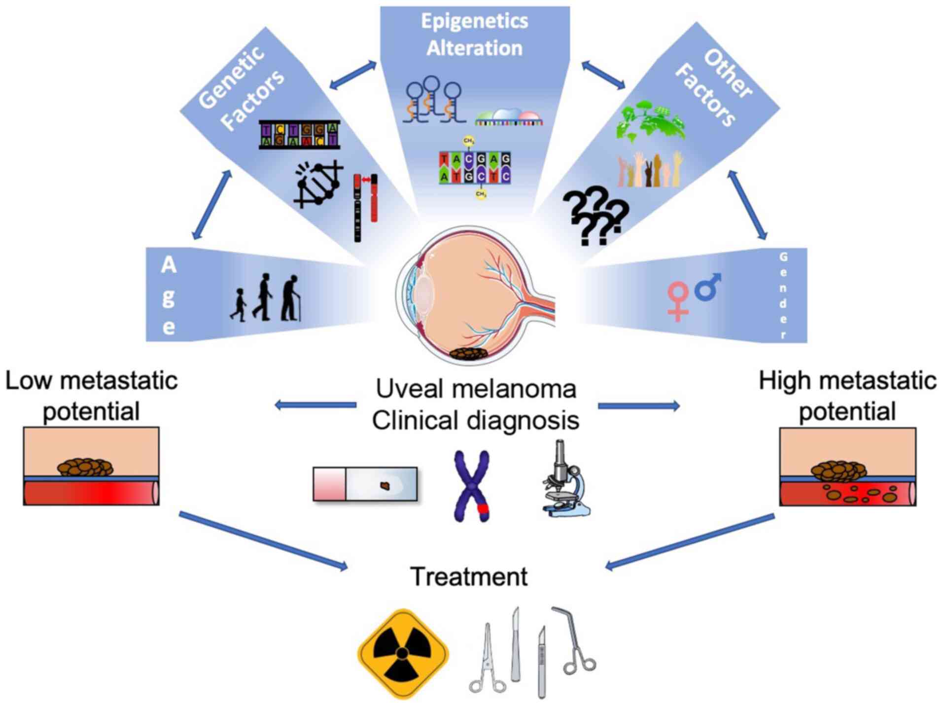
1. Introduction
Uveal melanoma (UM) represents the most prominent
primary eye cancer in adults. Although it could be considered a
relatively rare tumor, UM remains a disease of primary interest due
to its high mortality rate.
Notable efforts have been made to improve the
management of this malignancy. The introduction of globe-sparing
treatments was the most significant breakthrough of the last
century in this field. However, the overall survival of affected
patients has remained unaltered, and there is still no effective
treatment for metastatic disease.
Recently, increasing attention has focused on the
molecular mechanisms involved in UM carcinogenesis and progression,
which could allow for the identification of valuable diagnostic and
prognostic biomarkers, as well as novel therapeutic targets.
Herein, the available evidence on epidemiological,
clinical and molecular aspects of UM is discussed and reviewed,
with an aim of providing an updated and comprehensive tool, which
may be useful for both clinicians and researchers.
2. Epidemiology
UM represents 3-5% of all melanomas (1,2).
The largest proportion of cases of UM, approximately 85-90%, arises
from the choroid, while the 5-8% arises from the ciliary body and
the 3-5% from the iris (2,3).
The incidence of UM in the United States, between 1973 and 2008,
amounted to 5.1 cases per million annually (4). In Europe, a cancer registry-based
study demonstrated a north-to-south decreasing gradient in the
incidence of UM, with an incidence of >8 cases per million in
Norway and Denmark compared with approximately 2 cases per million
in Spain and Southern Italy (5).
Likewise, in Africa and Asia, the incidence is low, amounting to
0.2-0.3 cases per million per year (6). This latitude-related decreasing
trend in the incidence of UM has been associated with the
protective role of ocular pigmentation, which is higher in southern
countries compared to northern ones (5). Similarly, dark skin pigmentation may
play a role in protecting populations of African origin: The ratio
of UM among populations of African origin vs. Caucasian populations
ranges from 1:15 to 1:50 (7-9). A
population-based study investigated the relative risk of UM in
several racial cohorts, revealing a 5-fold higher risk among
Hispanic populations and 19-fold higher risk among non-Hispanic
Caucasian populations compared populations of African origin
(9).
UM is typically an adult malignancy, affecting older
age groups (10). The median age
at diagnosis has been reported to be approximately 62 years
(2), with an incidence rate that
tends to increase progressively up to 70 years of age, and then
levels off after 75 years of age (4,5,10,11). The mean age at diagnosis for UM
seems to decrease from 59-62 years of age in Caucasians, (4,12)
to 55 years in Japanese, 51 years in Taiwanese and 45 years in
Chinese populations (13-15). UM is uncommon in children and
extremely rare in newborns; congenital melanoma is also rare
(11,16,17). Shields et al investigated
the incidence rate of UM in children and teenagers, and
demonstrated that 50% of cases were >15 years of age, 35% were
between 10 and 15 years, 11% between 5 and 10 years, and only 3% of
cases were between 0 and 5 years of age at the time of diagnosis
(17).
The incidence of UM appears to be related to sex as
well (2,10). Population-based studies have
demonstrated a higher age-adjusted incidence in the male sex
compared to females, with a 20-30% greater rate in males (1,4).
An Australian population-based study found this difference to be
more prominent in the population which was ≥65 years of age, whilst
there was no significant difference in sex as regards the incidence
of UM when considering the population <65 years of age (18).
3. Risk factors
Several risk factors have been associated with the
development of UM. Host susceptibility variables, such as fair skin
color, inability to tan and light eye color have been significantly
associated with UM, with a risk ratio of 1.80, 1.64 and 1.75,
respectively (19). This
association is likely to be related to the poor amount of melanin
in the skin and eyes. It has been assumed that a poor amount of
melanin is present in the choroid and retinal pigment epithelium,
leading to an increased susceptibility to ultraviolet light and a
higher risk of developing UM (19). Oculodermal melanocytosis, also
known as Nevus of Ota, represents a relevant risk factor for
developing UM (20,21). This condition is characterized by
an abnormal congenital hyper-pigmentation within the V1/V2
trigeminal nerve area, and can involve periocular skin, orbit,
uvea, sclera and conjunctiva, as well as the palate, meninges and
tympanic membrane (20,21). Usually this condition is
unilateral and, is mostly confined to the eye. Oculodermal
melanocytosis is 35-fold more common in patients with UM compared
to the healthy population: The incidence rate among the Caucasian
population is 0.04 vs. 1.4% to 3% in patients with UM (20,21). A Caucasian patient affected by
oculodermal melanocytosis presents a lifetime risk for developing
UM equal to 1:400 (22). The
presence of atypical cutaneous nevi and intraocular nevi has been
also associated with the development of UM. In particular, the risk
of developing UM is 4-10-fold higher in patients affected by
atypical cutaneous nevi than in the healthy population (23,24). Intraocular nevi, such as iris nevi
and choroidal nevi, are considered risk factors for UM. Iris nevi
have been reported to have a potential risk of malignant
transformation, although the rate of this transformation has not
been clearly established, ranging from 2-5% (25,26). Predictive factors for an iris
nevus to transform into an iris melanoma have been summarized in
the ABCDEF acronym: A stands for young age; B stands for blood; C
stands for clock-hour (inferior location); D stands for diffuse
flat shape; E stands for ectropion uveae; F stands for feathery
margins (26). Choroidal nevus is
a common finding in the healthy population, with an incidence rate
of approximately 5% in the United States (27). Based on the concept that all
melanomas originate from a nevus, the rate of transformation of a
choroidal nevus into melanoma has been reported as 1:8845,
increasing to 1:3664 in the older aged cohort (80-84 years old)
(28). Predictive factors for a
choroidal melanoma to become malignant are a thickness of >2 mm,
the presence of subretinal fluid, presence of orange pigment,
proximity to optic disc, the absence of drusen or halo and
ultrasonographic hollowness (29,30).
A further relevant risk factor for the development
of UM is the mutation of the onco-suppressor gene, BRCA1 associated
protein 1 (BAP1) (10,31). BAP1 is located on chromosome 3.
The mutation of this gene has been associated with a hereditary
cancer syndrome. Tumors, such as malignant mesothelioma, basal cell
carcinoma, cutaneous melanomas, UMs and renal cell carcinoma, can
be developed following either the somatic or germline mutation of
BAP1 (31). In the case of
germline mutation, the tumors seem to be less aggressive than those
without this mutation (31). BAP1
has been found mutated in up to 47% of UM cases (32). Of note, patients affected by UM
present a higher risk compared to the general population
(approximately ≥11%) of a developing a secondary cancer, including
renal cell carcinoma and cutaneous melanoma, which could be related
to germline BAP1 mutations (2).
Sunlight ultraviolet exposure has been clearly
identified as a risk factor for skin melanomas (33), although there is still debate as
to whether this could represent a risk factor for UM: Some authors
support this hypothesis (34),
while others refuse this (35). A
previous meta-analysis revealed that chronic occupational natural
ultraviolet light exposure was a borderline non-significant risk
factor for UM, whereas geographic latitude and outdoor leisure UV
exposure were not significant (35). Conversely, welding was found to be
a significant variable associated with the development of UM
(35). As regards other
artificial lights, blue light exposure has been hypothesized to
play a role in the oncogenetic process and progression of UM
(36). In addition, occupational
cooking seems to be associated with a higher risk of developing UM
(37).
4. Clinical characteristics
Patients affected by UM can be asymptomatic (up to
30% of cases), with this malignancy being an incidental finding at
the time of diagnosis (38).
Symptoms, when preset, are related to the location of the tumor.
Iris melanomas are relatively uncommon (3-5% of UMs) and diagnosis
is mostly secondary to heterochromia, i.e., changes in iris color,
and corectopia, i.e., abnormality in pupil shape, which is present
in approximately 45% of cases (10,39). Usually, the tumor is located in
the inferior quadrant of the iris (45% of cases) and can cause
secondary glaucoma (direct or indirect obstruction to trabecular
outflow), ectropion uveae, angle seeding and bleeding with hyphema
(10). In some cases, it can be
complicated by extraocular extension (3%) (39). Extraocular extension, as well as
high intraocular pressure have been shown to be variables
associated with metastatic disease (39). Clinically, an iris melanoma can
present several types of configuration and levels of pigmentation
(from amelanotic to pigmented). In the majority of cases, iris
melanoma is circumscribed, while in a few cases, approximately 10%,
it can be diffuse (39,40). Diffuse iris melanoma is an
infiltrative form, undefined and flat, which can prove difficult to
diagnose: The presence of ipsilateral ocular hypertension and
acquired heterochromia of the iris are typically associated with
this condition (40). A rare
variant of diffuse iris melanoma is the ring melanoma of the
anterior chamber, a tumor that arises from the angle and tends to
infiltrate the angle structures >360 degrees, with a ring
pattern of growth (minimal extension towards iris or ciliary body)
(41). The main symptom is
unilateral glaucoma and diagnosis is very difficult (depends on
careful gonioscopy and ultrasound biomicroscopy) (41).
When it comes to choroidal and ciliary body
melanomas, also known as posterior UMs, the main symptoms are
blurriness (38% of cases), and floaters and flashing lights (7 and
9%, respectively). Less commonly, visual field loss, metamorphopsia
and pain have been reported (38). The diagnosis of ciliary body
melanomas is usually delayed due to their location and as symptoms
tend to appear only when the tumors are large (10). A study including 492 ciliary body
melanomas found at baseline, a mean tumor base of 11.7 mm and a
mean thickness of 6.6 mm, proving that the size of these tumors at
the time of diagnosis was relatively large (42). However, the same study also
included 7,256 choroidal melanomas, presenting a baseline mean base
of 11.3 mm and a mean thickness of 5.5 mm (42). The average choroidal melanoma
thickness at diagnosis has exhibited a decreasing trend from 5.5 mm
in the 1970s, to 4.5 mm in the 1990s, to 4 mm in more recent times
(43) This demonstrates the
efforts in improving the early diagnosis of the tumor. Choroidal
melanoma appears as a pigmented lesion in 55% of cases; in 15% of
cases is non-pigmented and in 30% of cases has mixed pigmented and
non-pigmented features (43). The
most common configuration of choroidal melanoma is dome-shaped, in
75% of cases. When the tumor grows, breaking through Bruch's
membrane, it acquires a typical mushroom-shaped configuration (20%
of cases) (43). Less commonly,
in approximately 5% of cases, choroidal melanoma presents a diffuse
configuration, which can make diagnosis more challenging (43). Orange pigment and subretinal fluid
are typically associated with choroidal melanomas. The tumor can
cause bleeding with subsequent vitreous hemorrhage, which can
obscure the view of the fundus. Neovascular glaucoma can develop in
advanced cases (43).
5. Diagnosis
The diagnosis of UM is based on a clinical
examination and ancillary tests (Table I). The diagnosis of iris melanoma
relies on slit lamp biomicroscopy, anterior segment-optical
coherence tomography (as-OCT) and ultrasound biomicroscopy (UBM).
In particular, UBM and as-OCT are helpful tools for the assessment
of the posterior extension of the tumor (10). Gonioscopy is important to evaluate
possible angle involvement (10).
Transillumination may help to evaluate ciliary body involvement
(10). A thorough fundus
examination is also required to assess retinal and choroidal
condition. The diagnosis of iris melanoma can be challenging, in
particular in cases of small/circumscribed lesions and diffuse
melanomas. Differential diagnosis includes most commonly, iris
nevus, as well as less common lesions, such as cysts, metastasis,
leiomyoma, melanocytosis and inflammatory conditions (granulomas)
(44,45). The differential diagnosis of
diffuse melanomas includes diffuse iris nevus, congenital
heterochromia, congenital, ectropion iridis, melanocytomalytic
glaucoma, pigmentary glaucoma, siderosis and iridocorneal
endothelial syndrome (40).
Photographic documentation plays a relevant role in case of small
lesion with a basal diameter <3 mm, providing information on
tumor growth during the follow-up. As reported above, the ABCDEF
rule is useful in differentiating an iris nevus from a melanoma. In
doubtful cases, such as small lesions, fine-needle biopsy could be
very helpful in the diagnostic process, with a low risk of
complications and a good rate of adequate sampling (46).
 | Table IKey points of primary tumor diagnosis
and treatment. |
Table I
Key points of primary tumor diagnosis
and treatment.
Diagnosis
| Treatment
|
|---|
| Tumor location | Clinical
examination (Refs.) | Tests and imaging
(Refs.) | Relevant studies
(Refs.) | Type (Refs.) | Relevant studies
(Refs.) |
|---|
| Iris melanoma | -Type:
Circumscribed tumor (most cases); diffuse iris melanoma (10% of
cases, undefined, flat, infiltrative form) (39,40); ring melanoma (rare, angle location
with a ring pattern of growth, glaucoma) (41); color: Different levels of
pigmentation (from amelanotic to pigmented); diameters; location
(inferior quadrant in 45% of cases) (10); associated findings: Heterochromia,
corectopia, bleeding, ectropion uveae, extraocular extension,
glaucoma (10);
-Gonioscopy: Angle involvement (10);
-Transillumination: Ciliary body involvement (10).
-Anterior segment-optical coherence tomography (size and posterior
and posterior extension) (10); | -Ultrasound
biomicroscopy; Anterior (size and posterior extension) (10);
-Photographic documentation (tumor growth) (10);
-fine-needle biopsy (doubtful cases, genetic profile) (58). | ABCDEF acronym
(26) (predictive factors for
differentiating an iris melanoma from a nevus): A: Young age; B:
Blood; C: Clock-hour (inferior location); D: Diffuse flat shape; E:
Ectropion uveae; F: Feathery margins. | -Resection (small
melanoma) iridectomy, iridotrabeculectomy,
iridocyclectomy;
-Radiotherapy (non-resectable lesion; seeding): Proton beam and
plaque radiotherapy, good local tumor control (102,103);
-Enucleation (large tumors, poor visual function, recurrent tumors,
multifocal melanoma and diffuse melanoma). | Shields et
al, 2013 (104): 144 iris
melanomas treated with iodine-125 plaque radiotherapy: 15% local
recurrence at 7 years, 1% metastasis rate at 7 years. |
| Ciliary body
melanoma | -Slit lamp
examination and dilated fundoscopy with scleral
indentation;
-Transillumination (10). | -Ultrasound
biomicroscopy: Useful for small melanomas (<4 mm) (47). | -In most cases
tumors are diagnosed when are large: Baseline mean tumor base of
11.7 mm and mean thickness of 6.6 mm (47). | Posterior UM
(including both ciliary body melanoma and choroidal
melanoma):
-Brachytherapy: Tumors <10 mm thickness and | Posterior UM
(including both ciliary body melanoma and choroidal
melanoma):
-COMS trial: Medium |
| Choroidal
melanoma | -Dilated fundus
examination: Configuration dome-shaped (75% of cases),
mushroom-shaped (20% of cases), diffuse (5% of cases) (43); color pigmented (55% of cases),
mixed (30% of cases), non-pigmented (15% of cases) (43); associated features subretinal
fluid; orange pigment; bleeding. | -Ocular
ultrasonography: (B-scan, A-scan) low-medium reflectivity/
ultrasonographic hollowness (10,49);
-Fluorescein and indocyanine green angiography: Progressive
hyperfluorescence, 'double circulation' pattern (51); | | <18 mm maximum
basal diameter (62); apex dose
70-100 Gy (105); Ruthenium-106
lower penetration depth (tumors <6 mm thickness) (106); local recurrence rate: 3% for
palladium-103, 7-10% for iodine-125, 14.7% for ruthenium-106
(2); | choroidal melanoma
(2.5-10 mm apical height and maximum basal tumor diameter ≤16 mm)
randomized to iodine-125 brachytherapy (85 Gy apex dose) or
enucleation. No survival differences: 5-, 10- and 12-year all-cause
mortality rate was 19, 35 and 43% in the |
-Optical coherence
tomography: Posterior location; accurate for detecting subretinal
fluid; useful for small lesion (50);
-Photographic documentation: Tumor growth, follow-up;
-Fine-needle biopsy (mainly for genetic profile) (58). | -TFSOM UHHD acronym
(30) (predictive factors for
differentiating a small melanoma from a nevus): Thickness (>2
mm); fluid (subretinal fluid); symptoms; orange pigment
(lipofuscin); margin (≤3 mm from optic disc); tumor
ultrasonographic hollowness; halo (absent); drusen
(absent);
-TFSOM DIM (54): Thickness
(>2 mm); fluid (subretinal fluid on optical coherence
tomography); symptoms; orange pigment (on autofluorescence),
ultrasonographic hollowness, diameter >5 mm (photography). | -Proton beam
radiotherapy: Tumor control and prognosis comparable to
brachytherapy (113); tumor
control over 90%, and 5-year overall survival of 70-85% (112); preferred to brachytherapy for
posterior pole location (112);
in large melanomas (>10 mm thickness or >16 mm largest
diameter) good tumor control but risk of ischemic and inflammatory
complications (115);
-Stereotactic radiotherapy (comparable to proton beam) (117);
-Enucleation: Thickness >10 or 12 mm and/or a basal diameter
>18 mm (62,119);
-Orbital exenteration: Extensive extraocular growth or orbital
invasion (10,106);
-Local resection (exoresection, endoresection): Selected
cases. | brachytherapy arm,
and 19, 35 and 41% in the enucleation arm, respectively; 5-, 10-
and 12-year metastasis-related mortality rate (histopathologically
confirmed) was 10, 18 and 21% in the brachytherapy arm, and 11, 17
and 17% in the enucleation arm, respectively (72). Brachytherapy 'as safe as
enucleation' (108);
-Papakostas et al, 2017 (116): More than 300 patients affected
by large choroidal melanoma (>10 mm thickness or >16 mm
largest basal diameter) treated with proton beam radiotherapy: 70%
Eye retention at 10 years; 60% 10-year mortality (comparable with
enucleation); 87% 10-year local tumor control; poor visual outcome:
20/200 or better in 8.7% at 10 years; 25% neovascular
glaucoma. |
The diagnosis of ciliary body melanomas can prove
difficult when the lesion is small, as the location does not allow
for a good visualization (10). A
good scleral indentation during fundus examination could be useful
to bring into the view these tumors. However, in the majority of
cases, tumors are diagnosed when are large with choroidal or iris
invasion (10). Transillumination
may help to visualize large lesions. A valuable examination for
detecting small ciliary body melanomas (<4 mm) is UBM, which is
useful in follow-up as well (47).
The diagnosis of choroidal melanoma depends mostly
on fundus examination with indirect ophthalmoscopy. The most
important test in the diagnostic process is represented by ocular
ultrasonography (10,48). In particular, B-scan
ultrasonography provides information on tumor size and extension.
A-scan ultrasonography provides valuable information on the
reflectivity of the lesion. The presence of acoustic hollowing is a
typical characteristic of UM (10). Ossoinig considered the presence of
low-medium reflectivity as one of the cardinal hallmark of a
melanoma lesion (49). Other
A-scan characteristics may be a quite regular internal structure
with spikes showing a similar height or a decreasing height, solid
consistency and sign of vascularization such as a spike showing
fast, vertical motion with flickering (49). Of note, A-scan ultrasonography has
a limited use in the case of very shallow lesions, with a thickness
<1.5/2 mm. Clinical examination and information provided by
ultrasonography, when carried out by an ocular oncology expert,
ensure a high level of accuracy, minimizing the necessity for
biopsy (48). Other useful tests
are OCT imaging, fluorescein angiography and indocyanine green
angiography. Enhanced deep imaging OCT is helpful for studying
small lesion (<3 mm in diameter) which are difficult to study
with other methods (50).
Furthermore, it has a high accuracy in detecting subretinal fluid
and may help to differentiate small choroidal malanomas from other
lesion, including nevi (10).
However, its use may be limited when it comes to lesions with a
thickness >3 mm (10).
Fluorescein angiography may be characterized by a progressive
hyperfluorescence which may last for >30-40 min. In the case of
Bruch's membrane break, the examination can reveal a typical
'double circulation' pattern, due to the presence of tumor vessels
underneath the retinal vasculature (51). Indocyanine green angiography is
more useful in showing intra-lesion vasculature, with the average
peak of hyperfluorescence at 18 min (52). The use of computed tomography and
magnetic resonance imaging with the purpose of studying choridal
melanomas has been investigated (10); however, their application in
clinical practice is very limited. The most common differential
diagnosis of choroidal melanomas is the choroidal nevus. Others can
be congenital hypertrophy of the retinal pigment epithelium
(CHRPE), peripheral eccentric choroidal neovascular membrane,
choroidal hemangioma, hemorrhagic detachment of pigment
epithelium/retina (53).
Differentiating a small choroidal melanoma from a choroidal nevus
may prove very difficult in some cases. Shields et al
provided a mnemonic acronym which can be useful in daily practice:
'To Find Small Ocular Melanoma Using Helpful Hints Daily (TFSOM
UHHD), which stands also for thickness (>2 mm), fluid
(subretinal fluid), symptoms, orange pigment (lipofuscin), margin
(≤3 mm from optic disc), ultrasonographic hollowness, halo
(absent), drusen (absent) (30).
Additionally, it has to be taken into account that a few number of
choroidal nevi may transform into choroidal melanomas (<1:8,000)
(28). When one of the TFSOM UHHD
factor is present, there is a 38% risk for the lesion to transform
into melanoma at 5 years, increasing to 50% when at least two
factors are present (29,30). If the lesion has the following 3
TFSOM UHHD factors, such as a thickness of >2 mm, a location
close to the disc and symptoms, the risk for transformation into
melanoma at 5 years increases to 69% (29,30). A choroidal nevus with drusen
(signs of chronicity), a thickness <2 mm and no other TFSOM UHHD
factor, can be considered 'low-risk'. The presence of one or more
TFSOM UHHD factors indicates a 'high-risk' nevus (10). Lesions with ≥2 TFSOM UHHD factors
are likely to represent small choroidal melanomas and treatment
should be indicated (29,30). Recently, TFSOM UHHD has been
updated, introducing the use of multimodal imaging. The new acronym
'To Find Small Ocular Melanoma Doing Imaging (TFSOM DIM) stands for
thickness (>2 mm on ultrasonography), fluid (subretinal fluid on
spectral domain-OCT), symptoms, orange pigment (on autofluorescence
imaging), melanoma ultrasonographic hollowness, diameter >5 mm
(photography). The 5-year risk for transformation of a nevus into
melanoma was 22% with two factors, 34% with three factors and 51%
with four factors (54).
Therefore, documenting with fundus photograph a choroidal
nevus/indeterminate lesion, which appears suspicious, plays a
relevant role for detecting lesion growth during the follow-up.
Data from the Collaborative Ocular Melanoma Study (COMS)
demonstrated a misdiagnosis rate of approximately 0.5% (55). This finding suggests that the
diagnosis of UM can be based on clinical examination and tests.
However, the COMS applied strict eligibility criteria, which could
have had an influence on the rate of misdiagnosis. Indeed, other
studies have found diagnostic fine-needle biopsy necessary in 1-9%
of cases (56,57). The biopsy of intraocular tumors is
debated due to the risk of tumor dissemination, as well as the risk
of ocular complications and inadequate sampling. However,
currently, tumor sampling has become more diffuse, usually not for
confirming the diagnosis, but with the purpose of analyzing the
genetic profile for assessing metastatic risk and prognosis
(58). For choroidal melanomas,
fine needle aspiration biopsy (FNAB) is performed using particular
precautions to prevent tumor seeding as well as subsequent
application of radiotherapy, which can help to sterilize seeded
cells (58).
6. Staging
The 8th edition of the American Joint Committee on
Cancer (AJCC) classification was published in 2016 and provides the
classification of UM as well (59). This is an updated version of the
7th edition (60). However, the
differences between the two editions are minimal. The widespread T
(tumor), N (node), M (metastasis) staging has been used also for
UM. Two classifications have been developed, one for iris melanoma,
and one for choroidal and ciliary body melanoma due to different
primary tumor staging (T). In either case, T0 refers to cases with
no evidence of primary tumors and Tx to cases where primary tumor
cannot be evaluated. Iris melanoma primary tumor (T) classification
is presented in Table II.
| Table IIIris melanoma primary tumor (T)
classification according to the American Joint Cancer Committee
(AJCC 8th edition) (59). |
Table II
Iris melanoma primary tumor (T)
classification according to the American Joint Cancer Committee
(AJCC 8th edition) (59).
| Primary tumor (T)
classification | Explanation | Sub-stages |
|---|
| T1 | Tumor limited to
the iris | T1a: not >3
clock hours in size |
| T1b: >3 clock
hours in size |
| T1c: T1 with
secondary glaucoma |
| T2 | Tumor confluent
with or extending into the ciliary body, choroid, or both | T2a: Confluent with
or extending into the ciliary body, without secondary glaucoma |
| T2b: Confluent with
or extending into the ciliary body and choroid without secondary
glaucoma |
| T2c: Confluent with
or extending into the ciliary body, choroid, or both, with
secondary glaucoma |
| T3 | Tumor confluent
with or extending into the ciliary body, choroid, or both, with
scleral extension | |
| T4 | Tumor with
extrascleral extension | T4a: Extrascleral
extension ≤5 mm in diameter |
| T4b: Extrascleral
extension >5 mm in diameter |
Primary tumor (T) classification for choroidal and
ciliary body melanomas depends on tumor size (thickness and largest
basal diameter), as well as ciliary body involvement and
extraocular extension (59).
Primary tumor classification according to tumor size is displayed
in Table III.
| Table IIIPrimary tumor (T) classification for
choroidal and ciliary body melanoma based on thickness and largest
diameter (59). |
Table III
Primary tumor (T) classification for
choroidal and ciliary body melanoma based on thickness and largest
diameter (59).
| Thickness | Largest basal
diameter, mm
|
|---|
| ≤3 | 3.1-6 | 6.1-9 | 9.1-12 | 12.1-15 | 15.1-18 | >18 |
|---|
| ≤3 mm | 1 | 1 | 1 | 1 | 2 | 2 | 4 |
| 3.1-6 mm | 1 | 1 | 1 | 2 | 2 | 3 | 4 |
| 6.1-9 mm | 2 | 2 | 2 | 2 | 3 | 3 | 4 |
| 9.1-12 mm | 3 | 3 | 3 | 3 | 3 | 3 | 4 |
| 12.1-15 mm | 3 | 3 | 3 | 3 | 3 | 4 | 4 |
| >15 mm | 4 | 4 | 4 | 4 | 4 | 4 | 4 |
All T values can be featured by a letter from 'a' to
'd', where 'a' indicates nor ciliary body involvement neither
extraocular extension, and 'b' indicates ciliary body involvement
without extraocular extension; 'c' indicates no ciliary body
involvement, but a ≤5 mm extraocular extension; and 'd' indicates
both ciliary body involvement and a ≤5 mm extraocular extension
(59). Additionally, the T4e
category includes any tumor size with an extraocular extension of
>5 mm (59). Regional lymph
nodes (N) include preauricular, submandibular and cervical sites.
Node assessment applies to tumors with extrascleral growth and
conjunctival involvement. Nx includes cases where nodes cannot be
evaluated; N0 indicates absence of node metastasis; and N1
indicates the presence of node metastasis or discrete tumor
deposits in the orbit; the N1 stage is classified into N1a
(metastasis in one or more regional lymph nodes) and N1b (no
positive regional lymph nodes, but the presence of discrete tumor
deposits in the orbit that are not contiguous to the eye). Distant
metastases are evaluated in the 'M' category: M0 indicates no
metastasis; M1 indicates the presence of distant metastasis ('a' ≤3
cm metastasis; 'b' 3.1-8 cm metastasis; 'c' >8.1 cm metastasis)
(59). The AJCC anatomic stage is
presented in Table IV.
| Table IVAnatomic stage according to AJCC
cancer staging manual, 8th edition (59). |
Table IV
Anatomic stage according to AJCC
cancer staging manual, 8th edition (59).
| Stage | T | N | M |
|---|
| I | T1a | N0 | M0 |
| IIA | T1b-d | N0 | M0 |
| T2a | N0 | M0 |
| IIB | T2b | N0 | M0 |
| T3a | N0 | M0 |
| IIIA | T2c-d | N0 | M0 |
| T3b-c | N0 | M0 |
| T4a | N0 | M0 |
| IIIB | T3d | N0 | M0 |
| T4b-c | N0 | M0 |
| IIIC | T4d-e | N0 | M0 |
| IV | Any | N1 | M0 |
| Any | N1 | M1a-c |
A further classification depends on the histological
grade (G) of the tumor. Basically, there are three histopathologic
types of UMs, according to their cytological composition: Spindle
cell UM (>90% spindle cells); epithelioid cell UM (>90%
epithelioid cells); mixed UM, consisting of <90% spindle cells
and >10% epithelioid cells (59). Spindle cells feature ovoid nuclei
and their growth exhibits a compact and cohesive pattern.
Epithelioid cells are pleomorphic, with larger and irregular shape
compared to spindle ones. The growth pattern of their nuclei and
nucleoli is less cohesive compared to spindle type; their cytoplasm
is acidophilic (59). Gx
indicates cases where the grade cannot be evaluated. G1 includes
spindle cell UM; G2 mixed cell UM; G3 epithelioid cell UM (59).
At the time of diagnosis, it is mandatory to carry
out imaging tests to identify systemic metastases, as the presence
of metastases has a relevant effect on the management plan. In the
past, baseline imaging consisted of abdominal ultrasonography and
chest radiography. Given the low sensitivity of those tests
(61), baseline modern imaging
work-up for ruling out metastases includes usually one of the
following protocols: Computed tomography (CT) of chest and abdomen;
chest CT and liver magnetic resonance imaging (MRI); whole body
positron emission tomography-CT (62).
7. Prognosis
UM presents a high mortality rate and up to 50% of
cases metastasize (2,63). A large study including both iris
and posterior melanomas revealed a metastatic rate of 15% at 5
years and 25% at 10 years (42).
The most common metastasis site is the liver (60-89%), followed by
the lungs (24-29%), skin and soft tissue (11-12%), bone (8-17%) and
lymph nodes (11%) (63,64). The prognosis of UM has been
related to several variables. First, location has been shown to
have an influence on prognosis. Iris melanoma has a mortality rate
5-10-fold lower than posterior UM. In a review article of >8,000
cases of UM, the 10-year metastasis disease was shown to be 33.4%
for ciliary body melanomas, 25% for choroidal melanomas and 6.9%
for iris melanomas (42). The
better prognosis of iris melanoma can be explained by factors
including a lower biologic activity, younger age and smaller size
(65,66). The cumulative proportion of
metastatic disease and mortality at 5 years has been found to be
5.2 and 2.2%, respectively, increasing to 8.8 and 3.3% at 10 years,
respectively (39). In the case
of iris melanoma, factors predicting melanoma-related metastasis
are an older age, increased thickness, secondary glaucoma, angle
involvement and extraocular extension (39,42,67). A study investigating the very
long-term prognosis of patients with posterior UM reported a
melanoma-related mortality rate of 31% at 5 years, 45% at 15 years
and 49% at 25 years (68). The
COMS study, which included choroidal melanomas and featured strict
eligibility criteria excluding peripapillary tumors and
predominately ciliary body tumors, reported a cumulative metastasis
rate of 25% at 5 years and 34% at 10 years (64). Following the development of
metastasis, the mortality rate was 80% within one year and 92%
within two years (64).
Several factors have been investigated with the
purpose of assessing their possible influence on the prognosis of
UMs, and the role of some is still debated (10). For instance, whether age and sex
may have an influence on the prognosis is not yet completely clear.
It seems that a younger age may exert a protective effect against
metastatic disease as the immune response is more robust, lesions
tend to be smaller and genetic mutations are less common compared
to older aged patients (17,66). In a study including >8,000
patients with melanoma, Shields et al (69) reported a cumulative rate of
metastasis at 10 year of 9% in patients <20 years of age, 23% in
patients 21-60 years of age and 28% in patients >60 years of
age; the cumulative 10-year mortality rate was 5% in patients
<20 years of age, 11% in patients 21-60 years of age and 16% in
patients >60 years of age; at 10 years, metastases were found in
0% of patients 0-10 years of age, 10% of patients 11-20 years of
age, 21% of patients 41-50 years of age, and in 30% of patients
71-80 years of age. The authors concluded that young patients had a
lower rate of metastatic disease. However, the proportion of iris
melanoma was 21% in young (≤20 years old), 4% in mid adult (21-60
years old) and 2% in older adult (>60 years old) patients
(69). Whether the female sex
could have a better prognosis compared to the male sex remains
controversial: In one study, the mortality rate at 10 years was
found to be 2-fold greater in males compared to females, and time
to develop metastatic disease was shorter in males compared to
females (metastatic disease at 5 years from diagnosis of UM in 84%
of males compared to 50% of females) (70); however, no sex-related differences
in survival analysis were found in the COMS study (71).
Tumor size has been demonstrated to have a
significant effect on the development of metastases. In a large
study including both iris and posterior UM, the 10-year metastasis
rate was 6% for a thickness of 0-1 mm, 12% for a thickness of 2.1-3
mm, 16% for a thickness of 3.1-4 mm, 27% for a thickness of 4.1-5
mm, 41% for a thickness of 7.1-8 mm, and 51% for a thickness of
>10 mm (42). A hazard ratio
of 1.06 was found for a 1 mm increase in thickness (42). The COMS report on the mortality
outcome of medium choroidal melanomas (2.5-10 mm thickness and ≤16
mm largest basal diameter) revealed a similar rate of melanoma
metastasis-related mortality at 10 years in both brachytherapy and
enucleation arms (18 and 17%, respectively), and depicted a larger
maximum basal tumor diameter as a primary predictor of melanoma
metastasis-related death (72).
Likewise, the COMS report on large choroidal melanomas (>10 mm
thickness and >16 mm diameter) revealed a melanoma
metastasis-related mortality of 40% in the enucleation arm at 10
years, and depicted a larger maximum basal tumor diameter as a
primary predictor of melanoma metastasis-related mortality
(71). A previous meta-analysis
on choroidal melanomas treated with enucleation reported a 5-year
mortality rate of 16% in the case of tumors with a thickness of
<2 or 3 mm and a basal diameter <10 or 11 mm, 32% in case of
tumors with a thickness of 3-8 mm and a basal diameter <15 or 16
mm, and 53% in case of tumors with a thickness of >8 mm and a
>16 mm basal diameter (73).
Not surprisingly, the AJCC staging has been
demonstrated to play a prognostic role. At the 10-year follow-up,
tumors with a T1 stage had a 15% metastatic rate, increasing to 25%
for T2 tumors and to 49% for T3 tumors. Melanomas with a T4 stage
presented a 63% metastatic rate (74). As reported above, the location of
the melanoma has a noticeable relevance in terms of metastatic
risk. Overall, ciliary body melanoma could be considered the most
aggressive. Ciliary body melanomas present a 2 to 4-fold higher
risk of metastasis than choroidal ones (75). The possible reasons for this
higher tendency to metastasize may be related to the delay in
diagnosis (ciliary body tumors are less symptomatic and, usually,
are large at time of diagnosis), the relevant vascularization of
ciliary body, and the higher incidence of chromosomal predisposing
alterations (10,76). The presence of oculodermal
melanocytosis represents a risk factor for the development of UM,
as well as for metastasis development in patients affected by UM
(76).
Other tumor-related features that may affect
prognosis are 'diffuse' configuration and extraocular extension.
The risk of melanoma-related metastasis has been demonstrated to be
higher in diffuse iris melanoma and diffuse choroidal melanoma
compared to non-diffuse iris and choroidal melanomas, respectively
(76). Extraocular extension has
a significant negative effect on prognosis when >5 mm: The
mortality rate at 5 years has been shown to be 37, 24 and 78% for
patients with microscopic, small (1-4 mm) and large extrascleral
extension, respectively (77).
Histopathology has been shown to play a relevant role in
prognostication. As regards cell type, the optimal prognosis has
been associated with spindle cell melanoma, the worst with
epithelioid cell type, while mixed type has an intermediate
prognosis (76). UM cases with a
high mitotic activity have a worse prognosis compared to those with
a low mitotic activity (78).
Nucleoli size is another histopathologic variable which affects
prognosis. The mean diameter of the 10 nucleoli with the largest
size (MLN) is used for prognostication. A large MLN predicts a poor
prognosis (76). Of note,
epithelioid cells are characterized by larger MLN, but MLN has been
shown to be an independent factor as well (79). Tumor vascularity also has an
influence on prognosis. A high microvascular density, as well as
specific microvascular patterns, such as the presence of networks
or loops, have been identified as predictors of a worse prognosis
(76). An unfavorable prognosis
has also been associated with the presence of numerous
tumor-infiltrating macrophages, high insulin-like growth factor-1
receptor expression, and a high expression of human leukocyte
antigen (HLA) class I and II (76).
Recently, when it comes to prognostication, further
attention has been paid to cytogenetic characteristics. Usually,
genetic tests are carried out on samples obtained from FNAB or
enucleation specimens. Most relevant cytogenetic alterations
include chromosome 3, 1, 8 and 6 (76). Chromosome 3 loss represents a
predictor of a poor prognosis. In particular, the complete loss of
one chromosome 3, known as monosomy 3, has been identified as the
most relevant prognostic factor, being associated with an increased
risk of metastatic disease (76).
In a series of 54 UMs, monosomy 3 was found in 56% of cases. Those
with monosomy 3 presented a 3-year mortality rate of 50 vs. 0% of
those without monosomy 3 (80).
Monosomy 3 was found in association with other unfavorable
prognostic factors, such as epithelioid type, vascular loops, high
mitotic activity, extrascleral extension, ciliary body location and
a large diameter (76).
Furthermore, BAP1 has been located on the short arm of this
chromosome (3p21.1) and BAP1 mutation has been found to be a
prognostic factor for metastatic disease (32). The partial or complete loss of
chromosome 1p predicts a negative prognosis. It is usually
associated with monosomy 3, but can also occur alone (76). Concomitant monosomy 3 and
chromosome 1p loss is by far a stronger predicting factor for
metastatic disease compared to the loss of either chromosome 3 or
chromosome 1p (81). The most
common alteration affecting chromosome 8 is a gain in chromosome
number. Indeed, chromosome 8q gain has been found in 41-53% of UM
cases, whereas a loss of chromosome 8q is rare (76). Similar to chromosome 1p loss,
chromosome 8q gain can occur alone or in combination with monosomy
3. Chromosome 8q gain in combination with monosomy 3 has been
associated with a poorer prognosis compared to each alteration
alone: The 5-year mortality rate has been reported to be 31% in
cases of 8q gain, 40% in cases with monosomy 3, 66% in cases of
concomitant 8q gain and monosomy 3 (82). Conversely to chromosome 8 gain,
chromosome 6 gain is a predictor of a good prognosis and tends to
be mutually exclusive with monosomy 3 (76). The occurrence of both monosomy 3
and chromosome 6 gain has been reported only in 4% of UM (83). On the contrary, chromosome 6 loss
is a predictor of an unfavorable prognosis: The loss of 6q has been
found in 40% of tumors with metastatic disease vs. 7% of
metastasis-free melanomas (84).
Over the past years, considerable efforts have been
made to improve epigenetic and transcriptomic analyses. Gene
expression profiling has provided a prognostic classification of
UM. This classification consists of two main classes: Class I
melanomas associated with a low risk of metastasis development and
class II melanomas associated with a high risk of metastatic
development (85). These results
were based on the analysis of the mRNA expression of 15 genes (12
target genes and 3 controls) and have been validated in a clinical
setting (86). A test analyzing
these 15 genes is available and can be used in clinical practice
with ease, on samples obtained from enucleation, tumor resection
and FNAB (87). Class I can be
divided into class IA with a 2% metastatic risk at 5 years and
class IB with a 20% metastatic risk at 5 years. Class II presents a
72% metastatic risk at 5 years (88). These data allow patients to be
offered a personalized management based on risk stratification
(88). A further point that needs
to be mentioned with regards to prognostication is the concept of
micrometastasis. Eskelin et al investigated the metastasis
doubling time and postulated that micrometastases could begin up to
5 years prior to primary tumor treatment (89). Taking into account all these
considerations, the early diagnosis and treatment of UM, including
small melanomas, may represent a key strategy for a positive
long-term prognosis (10).
8. Primary tumor treatment
The management of UM represents a multi-disciplinary
challenge, involving a variety of physicians specialized in ocular
oncology, such as ophthalmologists, radiologists, medical and
radiation oncologists (90). It
is important to note that despite improvements being made in
primary tumor treatment, the metastasis rate and overall survival
has remained unaltered over the past decades (4,91).
Once metastatic disease has been diagnosed, the overall survival is
as low as roughly one year, as shown by a recent meta-analysis
(91). In fact, patients who
present with metastasis at the diagnosis of the primary tumor often
do not undergo the aggressive treatment of the primary tumor
(62). Primary UM treatment can
be divided into two types: Globe-preserving treatment and
enucleation (Table I). The former
one includes radiation therapy, laser and surgical therapy. For
many years the only available treatment for UM was enucleation. In
the 1970s, efforts were made to develop globe-preserving
alternatives (92). Subsequently,
with the introduction of radiation therapy, there has been a shift
towards a globe-sparing approach rather than enucleation surgery,
in particular since the COMS study revealed comparable survival
rate between enucleation and plaque radiotherapy in patients with
medium choroidal melanomas (72).
Radiotherapy tends to be the preferred treatment for small and
medium UMs, whilst enucleation is usually performed for larger and
more advanced melanomas (10).
Tumor characteristics, as well as patient characteristics must be
taken into account when selecting the appropriate treatment.
Iris melanoma treatment depends on the size of the
lesion, as well as on its characteristics. A small lesion with a
basal diameter <3 mm, with no other sign and no symptoms, that
may be a nevus or a small melanoma (indeterminate lesions), can be
monitored periodically with photographic documentation for
evaluating possible growth (10).
Small circumscribed lesions with documented growth can be treated
with sector iridectomy (93). If
there is an involvement of the anterior chamber, a portion of the
trabecular meshwork needs to be removed as well; this type of
resection is termed iridotrabeculectomy. If there is an involvement
of the ciliary body, an iridocyclectomy can be performed, resecting
a portion of the iris and ciliary body (10). Intraocular surgery can be
associated with complications, such as hypotony, retinal
detachment, lens subluxation, phthisis, endophthalmitis and
sympathetic ophthalmia (93-95). Larger melanomas are usually
non-resectable and treatment is based on radiotherapy or
enucleation (10,96-98). In 1955, Lloyd and Ellis described
the use of radioactive wires (tantalum), inserted into the eye, for
the treatment of small iris melanoma (99). Subsequently, external beam and
plaque radiotherapy became available for the treatment of iris
melanomas. Anterior segment irradiation can be beneficial for
non-resectable lesions and for extensive seeding, as treatment
margins are larger compared to simple resection (100,101). In a small series of patients
with iris melanoma, it has been shown that proton beam and plaque
radiotherapy can achieve local tumor control in up to 93% (102) and 97% (103) of cases, respectively. A larger
study on 144 patients reported local recurrence in approximately
15% of cases at 7 years, showing an adequate local tumor control;
metastasis rate at 7 years was 1% (104). Even if radiation treatment is a
globe-spearing approach, complications may be severe and
sight-threatening, including corneal opacities, cataract and iris
neovascularization, culminating in vision loss (10,101). Enucleation surgery is usually
reserved for large tumors, poor visual function, recurrent tumors,
multifocal iris melanoma and diffuse iris melanoma (10). However, radiation therapy has
recently exhibited good local tumor control also for both diffuse
and multifocal iris melanomas (100,101).
The treatment of posterior UM can be surgery,
radiation therapy or laser. In general, the most common treatments
are radiotherapy, including plaque brachytherapy or external beam
radiation therapy, mostly used for small/medium melanomas, and
enucleation surgery, mostly used for large melanomas and poor
visual function (10). Other
possible treatment options include surgical resection and laser
treatments, such as transpupillary thermotherapy and photodynamic
therapy (PDT) (2). Importantly,
in the case of indeterminate lesions, which can be either a
choroidal nevus or a small melanoma, an observation can represent
the first approach: The patient is monitored for documented growth
or TFSOM UHHD (30) risk factors
(as reported above). If there is evidence of documented growth or
the presence of TFSOM UHHD (30)
factors, treatment should be considered (62). In some selected patients affected
by small choroidal melanomas (thickness of <3 mm and largest
basal diameter <10 mm), usually presenting with low-grade tumor
(stable or growing slowly), an advanced age, multiple comorbidities
and limited life expectancy, observation can represent an
alternative to the treatment. However, patients must be informed
about both the risks of treatment (visual loss) and the risk of
metastasis (unquantified albeit small) for observation (62,105).
Radiotherapy for UM includes plaque brachytherapy,
proton beam radiotherapy and photo beam radiation therapy
(stereotactic radiotherapy). Radiotherapy has gained increasing
popularity for the treatment of UM and has replaced enucleation
surgery for melanoma of suitable size and location (62). It is a globe-preserving treatment,
which ensures excellent local tumor control (2). Following the introduction of
radiotherapy for the treatment of UM, the main concern of
physicians was whether there was a difference in survival between
radiotherapy and enucleation (106). Therefore, from 1986 to 2003, the
COMS group conducted two large multicenter clinical trials
comparing survival between radiotherapy and enucleation in patients
affected by medium and large choroidal melanoma (72). Patients affected by large
choroidal melanoma (apical height >10 mm and maximum basal tumor
diameter >16 mm) were randomized to enucleation alone or
external beam irradiation (20 G) preceding enucleation surgery;
patients affected by medium choroidal melanoma (2.5-10 mm apical
height and maximum basal tumor diameter ≤16 mm) were randomized to
iodine-125 brachytherapy or enucleation (107). The COMS was the largest
randomized controlled trial (RCT) performed in ocular oncology,
with >2,000 patients enrolled (107). The results revealed no survival
differences at 5, 10 and 12 years between plaque brachytherapy and
enucleation in patients with medium choroidal melanoma: The 5-, 10-
and 12-year all-cause mortality rate was 19, 35 and 43% in the
brachytherapy arm, and 19, 35 and 41% in the enucleation arm,
respectively; the 5-, 10- and 12-year metastasis-related mortality
rate (histopathologically confirmed) was 10, 18 and 21% in the
brachytherapy arm and 11, 17 and 17% in the enucleation arm,
respectively (72). This
reassured that brachytherapy is 'as safe as enucleation' (108). However, in a number of cases,
metastases developed in a very short amount of time, suggesting
that systemic spread was present at the time of primary lesion
treatment; this could have led to a lack of statistical power
(108). Nonetheless, the
conclusion of comparable efficacy in terms of survival between
brachytherapy and enucleation could be considered correct (106). As regards the large choroidal
melanoma arms, no differences in 5- and 10-year tumor-related
mortality were found between enucleation alone and enucleation with
preoperative irradiation (71,107); this finding confirmed that
primary enucleation alone does not increase mortality from
metastatic disease as was hypothesized by Zimmerman et al
(109).
Brachytherapy is one of the most largely used
conservative treatments for UM (92). Following the publication of COMS
reports, brachytherapy has become the treatment of choice for
suitable tumors (62).
Brachytherapy is used for posterior UMs with a thickness of <10
mm and a maximum basal diameter<18 mm. Selected iris melanomas
and ciliary body melanomas (<10 mm thickness and no extensive
circumferential growth) can be considered for brachytherapy as well
(62). The radiation dose
delivered to the tumor apex is 80-100 Gy (10). According to the 2014 consensus
guidelines from the American Brachytherapy Society, the apex dose
can range from 70-100 Gy (105).
The apex dose in the COMS trial was 85 Gy (110). The plaque features a saucer
shape and contains the radioisotope. The plaque is sutured to the
sclera (positioned corresponding to the tumor) until the dose has
been delivered. Plaque size has to physically exceed tumor margin
by at least 2 mm (free-margin) (106). During the surgery, the plaque
has to be positioned adequately, in relation to tumor location:
Intraoperative US or transillumination are used to ensure a proper
positioning; a notched plaque is used in case of juxta-papillary
lesions (106). The most largely
used radioisotopes are iodine-125, ruthenium-106 and palladium-103
(62). Ruthenium-106 emits beta
radiation, while iodine-125 gamma radiation. Beta radiation has a
lower depth of penetration compared to gamma radiation. As a
consequence, ruthenium-106 can be used for tumor with a thickness
<6 mm (2,106). The advantage of this limited
depth of penetration should be a reduced damage to eye structures
(92) A previous study on 400
eyes treated with palladium-103 plaque, revealed a favorable visual
outcome and local tumor control compared with ruthenium-106 and
iodine-125; the mean apex dose was 73.3 Gy (for an equivalent dose,
more radiation was delivered in palladium-treated tissue compared
to iodine-treated) (111). The
local recurrence rate has been reported as 3% for palladium-103,
7-10% for iodine-125 and 14.7% for ruthenium-106 (2). Local recurrence can be either
re-treated with brachytherapy or treated with enucleation. A
further therapeutic option suitable for minimal margin recurrence
can be transpupillary thermotherapy (TTT) (62). Proton beam radiotherapy delivers
high dose radiation by using charged particles and relatively
sparing superficial tissues (106). Proton beam radiotherapy can be
used for the treatment of both posterior UMs and iris melanomas
(96,112). Tantalum markers are sutured to
the sclera and their distance from tumor margins, limbus and from
each other, is measured for proper localization and treatment
planning. Usually, a 2-mm safety margin is used. Following a
simulation phase, treatment is delivered in 4 consecutive
day-sessions, with a total dose of 56 Gy (106). In the past, the total dose
amounted to 60-70 Gy, whereas more recent studies have used a dose
ranging between 50 and 60 Gy (112). Proton beam radiotherapy for
posterior UM presents comparable outcomes in terms of tumor
control, systemic prognosis and visual result compared to
brachytherapy (113). Proton
beam radiotherapy is considered as an effective and safe treatment
for UM, with a rate of local tumor control >90%, and a 5-year
overall survival of 70-85% (112). Charged-particle radiotherapy
could be preferred to brachytherapy for tumors with a location that
may challenge plaque positioning, with also a risk of suboptimal
immobilization of the plaque (for instance, posterior pole)
(112,114). Proton beam radiotherapy can also
be used for the treatment of large tumors; however, it could be
challenged by a high rate of local recurrence and a high risk of
radiation-induced complications that can lead to vision loss and/or
secondary enucleation. Additionally, a 'toxic tumor syndrome' has
been described following radiotherapy for large tumors as a result
of severe intraocular inflammation, which causes exudative and
ischemic complications (115). A
previous study on >300 patients affected by large choroidal
melanoma (>10 mm thickness or >16 mm largest basal diameter;
>8 mm thickness in case of optic nerve involvement) demonstrated
that proton beam radiotherapy allowed patients to retain the eye in
70% of cases at 10 years; the 10-year mortality (60%) was
comparable with enucleation; and the 10-year local tumor control
was 87% (116). However, visual
outcome was poor, with only 8.7% of cases with a visual acuity of
20/200 or better at 10 years; additionally, 25% of cases developed
neovascular glaucoma; this rate increased to roughly 35% by 5 years
following treatment (116).
Stereotactic photon beam radiotherapy with gamma knife, cyber knife
or linear accelerator, delivers high doses from multiple
directions, trying to spare surrounding tissues (106). Tumor control, survival outcome
and visual outcome have been reported comparable with those of
proton beam radiotherapy (117).
Common sight-threatening complications from
radiotherapy are radiation retinopathy, known as radiation
maculopathy when affecting the macula, and radiation papillopathy,
when affecting the optic disc. Radiation maculopathy and optic
nerve atrophy can lead to visual loss (118). These complications are related
to the radiation-induced damage to the retina and optic nerve.
Tumor size and location, as well as dosimetric parameters have an
influence on their development (112,118). For instance, the cumulative
incidence rate of radiation maculopathy has been reported as high
as 64% at 5 years for tumors located within 4 disc diameters to the
macula (118). Other
complications of radiotherapy include glaucoma, neovascular
glaucoma, cataract, vitreous bleeding, ocular surface problems,
radiation-induced dry eye, keratitis, diplopia/strabismus and
scleral necrosis (10,106,112).
Enucleation represented the mainstay treatment for
UM prior to the advent of radiotherapy (92). Thereafter, enucleation has been
the second most common treatment for UM (119). In the 1970s, a concern as to
whether enucleation could increase the risk of metastasis was
raised due to the diffusion of the 'Zimmerman hypothesis', which
based this assumption on the potential dissemination of tumor cells
into the blood system at the moment of optic nerve cutting
(109). As reported above, this
hypothesis was disproved following the publication of the COMS
findings. Indications for enucleation include the presence of a
large tumor, poor visual potential and extraocular growth (10,114). Enucleation is the preferred
treatment for tumors with a thickness >10 or 12 mm and/or a
basal diameter >18 mm (62,119). For these lesions,
charged-particle radiotherapy can still be offered, although
high-dose irradiation carries a high risk of complications that can
lead to vision loss and, possibly, to eye loss; furthermore, these
patients have to cope with the anxiety related to possible
recurrence (62,119). In a series of 1,632 patients
treated for UMs from 1993 to 2002, 35% underwent primary
enucleation, 31% plaque brachytherapy and 17% proton beam
radiotherapy. Factors associated with primary enucleation were
tumor size, proximity to the optic disc, extensive involvement of
iris, angle or ciliary body (120). Orbital exenteration can be
required in the case of extensive extraocular growth or orbital
invasion (10,106).
The local resection of the tumor represents a
globe-sparing surgical treatment which may be suitable for selected
patients affected by posterior UMs. Tumor resection can be based
either on an external approach, known as exoresection, or on an
ab-interno approach, known as endoresection. The
exoresection of a posterior UM needs to be performed under
hypotensive general anesthesia due to the high hemorrhagic risk
(106). A lamellar, partial
thickness, scleral flap is created around the lesion, which is
'en-bloc' excided together with the inner scleral layer. The
superficial scleral flap is, then, used to close the deep opening
(121-124). This surgery is not commonly
performed and can be associated with sight-threating complications,
such as bleeding, retinal detachment, cataract and tumor
recurrence. Shields et al reported outcomes of 95 posterior
UMs treated with exoresection: Retinal hemorrhage, retinal
detachment and tumor recurrence/residual occurred in 35, 28 and 15%
of cases, respectively (121).
Adjuvant plaque brachytherapy can be associated with exoresection
to reduce the risk of recurrence (125). Recently, Caminal et al
(126) described the outcomes of
transcleral resection performed without hypotensive anesthesia and
combined with vitrectomy with silicone oil and plaque
brachytherapy. The authors concluded that this demanding procedure,
when successful, could provide a better visual outcome compared to
plaque brachytherapy. However, submacular hemorrhage, retinal
detachment and ocular hypertension occurred in 16, 21 and 21% of
cases, respectively (126).
Endoresection involves the piecemeal removal of the tumor by using
a vitreous cutter during a pars plana vitrectomy (127,128). Tumor residual can be destroyed
using endolaser photocoagulation. Silicone oil is used as an
endotamponade. The timing of silicone oil removal is variable,
ranging from 3 to 8.8 months (127,128). Due to concerns regarding tumor
seeding during surgical manipulation, adjuvant radiotherapy has
been associated with endoresection surgery (128,129). Endoresection has been proposed
to treat posterior UMs with a juxta-papillary location, as
radiotherapy is likely to cause radiation-induced optic neuropathy
(127). Konstantinidis et
al (127) reported on 71
patients with juxta-papillary UM treated with endoresection. Over a
median follow-up of 4 years, local recurrence occurred in 3% of
cases, and retinal detachment in 22% of cases. All-cause mortality
was 9 and 20% at 5 and 10 years, respectively. The authors
concluded that the procedure could be a useful alternative to
irradiation for juxta-papillary melanomas (127). Endoresection has been used also
in the treatment of large UMs, in combination with preoperative
stereotactic radiotherapy and adjuvant plaque brachytherapy
(128). Over a mean follow-up of
32 months, 15% of cases required a further vitrectomy, mainly due
to retinal detachment, 5% of cases had a local recurrence, and
15.5% of cases died from metastatic disease (128).
Laser treatment for posterior UMs includes TTT and
PDT. TTT delivers an infrared laser light (810 nm) to the tumor
surface through a dilated pupil. The laser causes an increase in
tumor temperature, heating its cells to 45-60°. As a result, tumor
abnormal vessels are obliterated, leading to a necrotic process
(130). This treatment can be
used for small choroidal melanomas (thickness ≤3 mm), due to
limited laser penetration (maximum penetration of 4 mm) (130). Tumor pigmentation may also have
an influence on treatment outcome because amelanotic lesions
feature poor heat absorption (131). Therefore, small pigmented
lesions may be suitable for TTT. However, the use of primary TTT as
a sole treatment of small choroidal melanomas has been questioned
due to high rate of local recurrence, reported up to 29% of cases
(132). As a consequence, TTT
should be preferably used in combination with radiotherapy
(106). A study including 143
patients with choroidal melanoma compared brachytherapy alone vs.
brachytherapy combined with TTT: Combined treatment provided a
lower recurrence rate, while the metastasis rate and overall
survival were comparable (133).
However, a larger study including 449 patients with choroidal
melanoma revealed no difference in tumor control and vital
prognosis between brachytherapy and brachytherapy combined with
TTT; of note, brachytherapy alone provided a better visual outcome
(134). The treatment of large
UMs with proton beam radiotherapy combined with TTT could reduce
the number of secondary enucleations (135). It is worth mentioning a large
study on 391 patients with choroidal melanoma treated with primary
TTT: Tumor recurrence occurred in 28% of patients and its
predictive factors were the presence of ocular symptoms, proximity
to the optic disc, subretinal fluid, greater thickness and
elevation of post-treatment tumor scar (130). Common complications following
TTT for UM were retinal vein occlusion (26%), macular epiretinal
membrane (23%), macular edema (9%) and vitreous hemorrhage (10%)
(130). In general, TTT or PDT
are used only if the lesion is very small and there is a high risk
of visual loss from radiotherapy (106). Of note, TTT represents a
treatment option in case of tumor recurrence after brachytherapy
(136).
PDT is a non-thermal laser treatment which involves
the administration of a photosensitizer activated with laser light
(137). This minimally invasive
therapy has also been described for the treatment of ocular tumors,
including choroidal melanoma (137). Following the intravenous
administration of the photosensitizer and its accumulation into the
tumor tissue, laser light is delivered to activate the
photosensitizer. This, once activated, it exerts a direct cytotoxic
effect on the tumor, causing peritumoral vasculature destruction
and local inflammation, with subsequent autophagy (137). It is important to highlight that
the presence of pigmented tumor is a contraindication for PDT
(137,138). Pigmentation seems to prevent
light penetration into the lesion (137). Thus, PDT could be used for the
treatment of small amelanotic melanomas (<4 mm thickness)
(10). Most commonly, PDT is
performed using verteporfin as a photosensitizer. Verteporfin PDT
for choroidal melanoma treatment can be with either standard
fluence (50 j/cm2 in 83 sec) or a double fluence (100
j/cm2 in 166 sec) (137). A previous review article of 6
reports including a total of 38 choroidal melanoma cases primarily
treated with verteporfin PDT showed 80% tumor control over 31
months (137). A more recent
study on 12 eyes with choroidal melanoma reported tumor control in
67% of cases, while 33% failed to regress (138). PDT with indocyanine green has
been used for the treatment of choroidal melanoma, exhibiting a
high rate of tumor control (137). Notably, a previous study on 25
patients demonstrated good local tumor control combining both TTT
and indocyanine green PDT for the treatment of small and medium
choroidal melanomas (139).
9. Surveillance and metastatic disease
treatment
Following primary tumor treatment, patients with UM
need to receive periodical ocular and systemic surveillance. The
aim of ocular follow-up is the early detection and management of
possible local tumor recurrence and treatment-related
complications. In general, close follow-up visits are scheduled in
the early post-operative period, which, then, are extended to a 3-
to 6-month interval for a few years; thereafter, if the clinical
condition is stable, follow-up can be arranged each year (106). Local tumor recurrence is usually
managed in the same manner as the primary tumor (106). The treatment of secondary
orbital involvement is challenging and includes radiotherapy and
surgery (excision, debulking, or exenteration) (140). The early detection of
radiation-induced complications can allow their early treatment.
Anti-vascular endothelium growth factor agents have been used for
the treatment of radiation retinopathy, radiation maculopathy,
radiation-induced optic neuropathy and neovascular glaucoma, and
can help to stabilize or, in some cases, to improve clinical
conditions (141-144).
As regards systemic surveillance, an ideal
surveillance protocol which would define timing, duration and the
type of examinations according to patient characteristics has not
yet been developed (62,106). Systemic monitoring is aimed at
the early detection of metastasis, which could have some clinical
relevance as highly selected cases of hepatic metastasis could be
managed with surgical resection, resulting in an improved survival
(145). In addition, no adjuvant
therapy has been demonstrated to be effective in reducing the risk
of metastasis (146). In this
scenario, risk stratification for metastasis development may play a
key role and may allow for the planning of a surveillance protocol
which could be suited to individual risk (146,147). As aforementioned, prognosis is
dependent on multiple factors, including clinical variables and
genetic prolife (GEP class). According to the GEP class, patients
can be classified as low-risk (class I) and high-risk (class II)
(88). Surveillance imaging tends
to be focused on hepatic monitoring due to the tendency of UM to
metastasize to the liver (146).
Low-risk patients can be monitored with hepatic ultrasound at
6-month intervals (146).
Hepatic ultrasound features a good specificity (100%), but poor
sensitivity (14%) (61).
Therefore, high-risk patients are recommended to undergo a more
frequent (<6-month interval) and a more intensive hepatic
monitoring, which would include more sensitive and specific imaging
tests, such as liver CT/MRI (146). However, other authors recommend
annual liver ultrasound and physical examination for low-risk
patients, and 6-monthly liver imaging (ultrasound alternated with
liver/abdomen MRI) plus annual physical examination for high-risk
patients (62). Low-risk and
high-risk patients can be transitioned to the GP at 5 and 10 years,
respectively (62).
To date, no therapy has been demonstrated to be
effective for the treatment of metastatic disease in patients with
UM. Metastatic UM is associated with a poor prognosis. Several
chemotherapeutic drugs, including dacarbazine, cisplatin,
trosulfan, temozolomide and fotemustine, have been investigated,
exhibiting a low response rate and disappointing outcomes (114,147). While immunotherapy has
noticeably improved outcomes in metastatic cutaneous melanoma, this
has not been the case for metastatic UM (148). A possible reason for such a
different response to immunotherapy could be related to the
different biological characteristics between cutaneous and UMs, as
well as their different immunogenicity (149,150). Ipilimumab has shown a response
rate of approximately 5-10%, with an overall survival ranging from
6 to 9.7 months (147).
Nivolumab has shown in a prospective trial a 6% response rate and
an overall survival of 11 months (151). A previous retrospective study
reported outcomes of 89 patients treated with ipilimumab plus
nivolumab, showing a 11.6% response rate and an overall survival of
15 months (152). The
understanding of the molecular mechanisms involved in UM
carcinogenesis and progression will contribute to the development
of targeted therapy for the treatment of metastatic UM. While BRAF
inhibitors, such as dabrafenib and vemurafenib, have been used in
cutaneous melanoma, which typically harbors BRAF and NRAS
mutations, there is no rationale for the use of these agents in UM
due to the different molecular profile compared to cutaneous ones
(153). Given the commonly
harbored GNAQ/GNA11 mutations in UM, agents targeting downstream
effectors of biological pathways GNAQ/GNA11-related, such as MEK
and protein kinase C (PKC), have been investigated. However,
similar to other therapeutic approaches, disappointing results have
been reported and response rates, in general, are <10-15%
(114,147). MEK inhibitors include
selumetinib and trametinib. Initially, the use of selumetinib in
metastatic UM seemed to provide promising results: A randomized
clinical trial enrolling 101 patients compared selumetinib with
traditional chemotherapy and reported longer progression-free
survival (PFS) and a higher response rate (14 vs. 0%) (154). However, these quite positive
outcomes failed to be achieved in a subsequent phase III randomized
trial: The SUMIT trial compared selumetinib plus dacarbazine with
dacarbazine alone in 129 patients with metastatic UM and revealed
no significant difference in PFS between the two interventions
(median PFS, 2.8 months in the selumetinib plus dacarbazine group
vs. 1.8 months in the dacarbazine alone group), and no difference
in response rate (3% in selumetinib plus dacarbazine group vs. 0%
in dacarbazine group) (155).
Trametinib was investigated in a phase I trial enrolling 16
patients with metastatic UM and 81 patients with cutaneous or
unknown primary melanoma. In patients affected by UM, trametinib
revealed no objective response (0% response rate) and a median PFS
of 1.8 months (156). Trametinib
was also used in combination with Akt inhibitor: A randomized trial
compared trametinib alone (18 patients receiving ≥1 study drug
dose) vs. trametinib combined with the Akt inhibitor GSK2141795 (21
patients receiving ≥1 study drug dose), revealing no difference in
median PFS (15.7 weeks vs. 15.6 weeks) and only one partial
response in each group (157).
Another biological pathway which has been studied as a possible
target in UM is the MAPK pathway, through PKC inhibition.
Sotrastaurin, a PKC inhibitor, has shown a median PFS of 15.4
weeks, a partial response in one patient and stable disease in 55
patients (47%) (158).
Furthermore, growth factor receptors which have been found
overexpressed in UM, have been studied as possible targets.
Sunitinib, a C-kit inhibitor, was used in a pilot study on 20
patients with C-kit expressing metastatic UM and demonstrawted a
partial response in one patient and 12 stable disease. The median
PFS was 4.2 months (159). A
retrospective study evaluated the use of sunitinib as an adjuvant
therapy in high-risk UM and reported a longer overall survival in
those receiving adjuvant sunitinib compared to historical controls
(160). Cabozantinib is a
multiple tyrosine kinase receptor inhibitor, including c-MET, Axl
and VEGF. This has been used in the treatment of metastatic UM.
Very recently, the results of a RCT comparing cabozantinib vs.
dacarbazine or temozolomide, have been published and have
demonstrated no improvement in PFS in patients treated with
cabozantinib (161). Sorafenib,
multi-kinase inhibitor, was used in a trial enrolling 152 patients
with metastatic UM: Of the 118 patients evaluable for response,
32.2% exhibited progression, 1.7% had a partial response, and 66.1%
had stable disease. Patients with stable disease were randomized to
sorafenib continuation or placebo, with a median PFS significantly
longer in sorafenib arm (5.5 vs. 1.9 months) (162).
Since the liver represents the most common site for
UM metastasis, several liver-directed treatments have been
investigated. A recent meta-analysis of trials (phase Ib-III) in
metastatic UM revealed that liver directed treatments were
associated with a longer overall survival and PFS compared to other
therapies (chemotherapy, immunotherapy and targeted therapy)
(163). Liver directed
treatments include surgical resection, stereotactic radiotherapy,
radiofrequency ablation, regional chemotherapy and embolization
(114,164). Surgical resection could be a
therapeutic option in very selected cases. A study including 155
patients with liver metastases from UM reported that 11% of
patients underwent liver resection, with a better survival compared
with those who did not receive surgical resection (145). Regional chemotherapy includes
hepatic intra-arterial infusion and isolated hepatic perfusion
(IHP). Fotemustine has been shown to have a better response rate
(10.5 vs. 2.5%) and longer PFS (4.5 vs. 3.5 months) when
administered as intra-arterial perfusion compared as intravenously,
but with no difference in overall survival (165). Isolated hepatic perfusion is
based on the isolation of liver blood supply from systemic
circulation with the purpose of delivering high-dose chemotherapy
and reducing at the same time systemic exposure. A retrospective
study on 18 patients treated with percutaneous IHP with melphalan
reported a median overall survival of 9.6 months and a median PFS
of 12.4 months (166).
Embolization treatment modalities include chemoembolization,
radioembolization and immunoembolization (164). A recent prospective trial
investigated radioembolizaton in two groups of patients:
Treatment-naïve patients and patients who exhibited progression
following immunoembolization. In the treatment-naïve group, median
PFS was 8.1 months and median overall survival 18.5 months; no case
of complete response was found, but 20 out of 23 patients had a
partial response or stable disease. In the group resistant to
immunoembolization, the median PFS was 5.2 months, and the median
overall survival was 19.2 months; there was no case of compete
response, but 14 out of 24 patients had partial response or stable
disease (167).
10. Genetic and epigenetic features in uveal
melanoma
Genetic and epigenetic characteristics of tumors
have been given ever-increasing attention over the past years, not
only due to their relevant role in the carcinogenesis process, but
also as they can provide new insight into tumor behavior (168). This can potentially allow the
development of reliable biomarkers and novel therapeutic targets,
leading to new breakthroughs in the management of UM. Genetic
alterations affect directly and permanently the DNA sequence and
include the following: Chromosomal aberration, copy number
variation (CNV), mutations either somatic or germline and single
nucleotide polymorphism (SNP). Conversely, epigenetic alterations
modulate gene activity and expression without involving any changes
in the DNA sequence; these include the alteration of microRNA
(miRNA/miR) expression levels, DNA methylation and histone
modifications (168). The
interaction between genetic, epigenetic and other possible factors
involved in UM carcinogenesis is illustrated in Fig. 1.
UM is a sporadic tumor, rarely exhibiting a
familial inheritance (168).
This is the case of tumor syndromes, such as Lynch syndrome
(169) and BAP1-tumor
predisposition syndrome (170).
As reported above, chromosomal alterations in UM mainly affect
chromosomes 1,3,6 and 8, and present a meaningful prognostic
relevance. As regards somatic mutations, the most commonly mutated
genes that have been identified in UM patients are GNAQ, GNA11
mainly affected by specific point mutations (p.Q209P and p.Q209L,
respectively) and BAP1 that is subjected to several mutations
occurring in the whole gene sequence (88,171). Unlike cutaneous melanoma, UM
does not harbor typical mutations in BRAF and NRAS genes (153,172). The GNAQ and GNA11 genes encode
for the Gα11 subunits and the Gαq subunits of G proteins,
respectively (171). G proteins
are involved in signal transduction, controlling gene transcription
and, subsequently, cell survival, growth and mortality (171). GNAQ and GNA11 are located on
chromosome 9 (q21.2) and chromosome 19 (p13.3), respectively
(168). These genes have been
considered as driver oncogenes, representing early or initiating
mutations in UM (168,171). Oncogenic mutations of GNAQ and
GNA11 genes are usually mutually exclusive and were found in up to
83% of Ums (173). Oncogenic
mutations of these genes determine a constitutive activation of G
proteins, which, in turn, affect downstream signaling (171). Several intracellular pathways
are regulated by GNAQ and GNA11 genes, including the
RAF/mitogen-activated protein kinase kinase (MEK)/extracellular
signal-regulated kinase (ERK) pathway (174). The activation of the RAF/MEK/ERK
pathway causes an overexpression of cell-cycle regulatory protein
cyclin D1 (CCND1), leading to the inactivation of the
retinoblastoma tumor suppressor gene (174). Mutations in this pathway have
been assumed to be early or initiating events in UM carcinogenesis
(174). The BAP1 gene is an
onco-suppressor gene located on chromosome 3 (p21.1). Inactivating
somatic mutations of BAP1 have been associated with metastatic
disease: Up to 84% of metastasizing UMs harbor inactivating
mutations of this onco-suppressor gene (32). As regards BAP1 function, this is a
deubiquitinating enzyme that regulates specific proteins. In
particular, BAP1 regulates genes involved in melanocyte function
and differentiation: Inactivating mutations can lead to melanocytic
de-differentiation, promoting a pro-metastatic behavior. The loss
of the tumor suppressor activity of BAP1 has been identified in
breast and lung cancers as well (175). Of note, BAP1 mutations can also
occur in germline featuring the BAP1 familial cancer syndrome,
which predisposes to several cancers, including UM (176).
As regards epigenetic alterations, biological
mechanisms related to miRNAs and DNA methylation have been
identified in UM, whereas modest evidence on histone modification
is available. The role of histone modification has been associated
with BAP1 loss. It has been shown that BAP1 depletion determines a
loss of differentiation in cancer cells through the
hyperubiquitination of histone H2A (175). The use of histone deacetylases
inhibitors proved to reverse H2A hyperubiquitination, inducing cell
differentiation and inhibiting tumor growth (175). Indeed, the nuclear expression of
several histone deacetylases has been found in UMs, confirming the
role of this epigenetic alteration as a potential therapeutic
target (177).
DNA methylation represents an important epigenetic
mechanism which regulates the expression of several genes involved
in UM carcinogenesis. This regulation is based on
methylation/demethylation mechanisms, that is the addition/removal
of a methyl group (CH3) to/from a DNA sequence mediated
by DNMT3A and TET1. It has been widely demonstrated that certain
methylated DNA regions can be used as stable biomarkers,
considering that DNA methylation is maintained almost unchanged
during the cell replication process by DNA methyltransferase 1
(DNMT1) (178,179). The functional alteration of this
enzyme, together with that of other methyltransferases (DNMT3A and
DNMT3B) that catalyze the de novo DNA methylation, is
responsible for the global hypomethylation and hypermethylation of
widespread regions of the tumor cell genome (180).
Identifying the gene regions affected by
methylation phenomena is important to establish the effect that
this modification determines at the transcriptional level. It is
currently known that promoter methylation determines gene
suppression by blocking the access of transcription factors to
binding sites on the promoter (181,182), while it has not yet been fully
clarified the functional role of intragenic and intergenic
methylation in the control of gene expression (183). On this matter, several studies
have described how DNA methylation can modulate the expression of
non-coding RNA, the alternative splicing, the recruitment of
enhancers, and the increase of RNA polymerase activity that
influences the expression levels of the genes involved (184-186). As regards UM, the
hypomethylation of sites close to the preferentially expressed
antigen in melanoma (PRAME) promoter has been shown to promote
PRAME activation with subsequent increase in metastatic risk
(187). The hypomethylation of
the deleted in split hand/split foot 1 (DSS1) promoter has been
found to be a frequent event in UM (188). Hypermethylation of the following
oncosuppressor gene promoters has been demonstrated in UM: p16,
RASSF1A, RASEF, TIMP3 and EFS (189-194). This event leads to the
inactivation of these genes. The methylation of human telomerase
reverse transcriptase (TERT) promoter has been described in UM.
Human TERT is an oncogene which has been found to be upregulated in
UM (190). TRAIL receptors DcR1
and DcR2 were found hypermethylated in both UMs and cutaneous
melanomas (195). Of note, the
hypermethylation of a site on chromosome 3 at BAP1 locus determines
BAP1 downregulation, showing the epigenetic regulation of this gene
(196).
miRNA-based epigenetic mechanisms have been also
investigated in UM (197).
miRNAs are non-coding RNA molecules, consisting of a
single-stranded sequence of 18-22 nucleotides. miRNAs regulate gene
expression, playing important roles in both physiological and
pathological processes, such as cell proliferation,
differentiation, apoptosis, organ formation, angiogenesis,
extracellular matrix remodeling, etc. (198,199). The dysregulation of specific
miRNAs has been associated with the onset and progression of a
number of types of cancer, including UM (200-203). Research in this field has
exhibited marked progress. Venza et al found 96 miRNAs
dysregulated in both UM and cutaneous melanoma cell lines (204). Radhakrishnan et al found
specific miRNAs associated with metastatic UM, which were in
association with chromosome 1,3 and 8 aberrations (205). However, Larsen et al
failed to demonstrate the role of this association between
chromosome alterations and miRNAs expression in predicting
metastatic disease in UM (206).
Worley et al demonstrated that the expression of 6 miRNAs
(let-7b, miR-199a*, miR-199a, miR-193b, miR-143 and miR-652) could
be used to distinguish class1 UM (low metastatic risk) from class 2
ones (high metastatic risk) with maximum sensitivity and
specificity (207). Among the
miRNAs involved in cell proliferation and apoptosis, which have
been found abnormally expressed in UM, noteworthy are the
following: miR-137 (downregulated); miR-144 (downregulated);
miR-145 (downregulated); miR-92a-3p (upregulated); and miR-181b
(upregulated) (208-213). Among the miRNAs involved in cell
migration and invasion, which have been found abnormally expressed
in UM, noteworthy are the following: miR-20a (upregulated); miR-155
(upregulated); miR-296-3p (downregulated); miR-454 (upregulated);
miR-367 (upregulated); miR-21 (upregulated); miR-23a
(downregulated); and miR-224-5p (downregulated) (214-221). Additionally, miR-204 and miR-145
have been found to be downregulated, whereas miR-20a, miR-17,
miR-106a, miR-34a and miR-21 have been found to be upregulated in
UM samples (222). Circulating
levels of the following miRNAs have been found to be dysregulated
in metastatic UM: miR-125b, miR-20a, miR-146a, miR-181a, miR-155
and miR-223 (223). miRNAs are
also involved in the regulation of immune mediators, which can
modulate UM behavior. For instance, interleukin (IL)-10 may play a
role in promoting cancer as can suppress immune response against
the tumor. Specific miRNAs seem to be involved in IL-10 modulation
(224). miRNAs may also play a
promising role in the development of therapeutic targets. For
instance, genistein, an antitumor drug, has been shown to inhibit
miR-27a expression and, as a result, to increase ZBTB10 gene
expression (miR-27a target gene); the antitumor action may be
related to the miR-27a regulatory mechanism (225). Overall, all these studies
provide important insight into the involvement of miRNAs in UM
development and progression, as well as in the prognosis of UM
patient. However, some data are still conflicting; therefore, there
is an urgent need to clarify which miRNAs are effectively involved
in UM development or in specific clinical-pathological features of
patients.
Recent studies have proposed the evaluation of all
these genetic and epigenetic biomarkers for the early diagnosis of
tumors. In particular, a previous study demonstrated how the
analysis of liquid biopsy samples and circulating DNA may be useful
for the early detection of genetic aberrations and epigenetic
markers of precancerous and cancerous lesions (226). In this context, novel
high-sensitive techniques, such as next generation sequencing and
droplet digital PCR (ddPCR), have been used for the analysis of
liquid biopsy samples to detect low amounts of miRNAs, circulating
mutations, microbial nucleic acids, etc. (227-229).
Therefore, the analysis of both genetic and
epigenetic factors in liquid biopsy and tissue samples obtained
from patients with UM by using these high-sensitive techniques
would contribute to the discovery of novel effective diagnostic and
prognostic biomarkers for the management of UM.
11. Conclusions
The present review article focused on UM and
collected an updated evidence on epidemiological, clinical and
molecular aspects of this malignancy. It is suggested that even if
UM is a relatively rare type of cancer, the prognosis remains
unfavorable in a consistent percentage of cases. The treatment of
the primary tumor has made significant improvements thanks to the
introduction of globe-preserving approaches in the previous
century. However, metastatic disease remains a critical issue for
clinicians due to the lack of effective therapeutic strategies.
Molecular studies have provided new insight into the genetic and
epigenetic mechanisms that regulate UM biological activity.
However, it seems that the available knowledge has only scratched
the surface and there is hope for the identification of effective
biomarkers and new therapy which can make the difference in the
management of metastatic disease. This could be the breakthrough of
the current century.
Availability of data and materials
Not applicable.
Authors' contributions
MF, LF and TA conceived the study. MF and GR wrote
the original draft of the manuscript. AL, MR, VB, AR, GG and LF
performed the bibliography research and collected the data. RC
collected data on the pathological staging of uveal melanoma. MF,
GG and LF prepared the figure and tables. All authors contributed
to the critical revisions, editing and reviewing of the final
version of the manuscript.
Ethics approval and consent to
participate
Not applicable.
Patient consent for publication
Not applicable.
Competing interests
The authors declare that they have no competing
interests.
Acknowledgments
Not applicable.
Funding
The present study was partially supported by the Italian League
Against Cancer (LILT).
References
|
1
|
McLaughlin CC, Wu XC, Jemal A, Martin HJ,
Roche LM and Chen VW: Incidence of noncutaneous melanomas in the
U.S. Cancer. 103:1000–1007. 2005. View Article : Google Scholar : PubMed/NCBI
|
|
2
|
Krantz BA, Dave N, Komatsubara KM, Marr BP
and Carvajal RD: Uveal melanoma: Epidemiology, etiology, and
treatment of primary disease. Clin Ophthalmol. 11:279–289. 2017.
View Article : Google Scholar : PubMed/NCBI
|
|
3
|
Damato B: Progress in the management of
patients with uveal melanoma. the 2012 Ashton Lecture. Eye (Lond).
26:1157–1172. 2012. View Article : Google Scholar
|
|
4
|
Singh AD, Turell ME and Topham AK: Uveal
melanoma: Trends in incidence, treatment, and survival.
Ophthalmology. 118:1881–1885. 2011. View Article : Google Scholar : PubMed/NCBI
|
|
5
|
Virgili G, Gatta G, Ciccolallo L,
Capocaccia R, Biggeri A, Crocetti E, Lutz JM and Paci E; EUROCARE
Working Group: Incidence of uveal melanoma in Europe.
Ophthalmology. 114:2309–2315. 2007. View Article : Google Scholar : PubMed/NCBI
|
|
6
|
Kivelä T: The epidemiological challenge of
the most frequent eye cancer: Retinoblastoma, an issue of birth and
death. Br J Ophthalmol. 93:1129–1131. 2009. View Article : Google Scholar : PubMed/NCBI
|
|
7
|
Margo CE, Mulla Z and Billiris K:
Incidence of surgically treated uveal melanoma by race and
ethnicity. Ophthalmology. 105:1087–1090. 1998. View Article : Google Scholar : PubMed/NCBI
|
|
8
|
Shields CL, Kaliki S, Cohen MN, Shields
PW, Furuta M and Shields JA: Prognosis of uveal melanoma based on
race in 8100 patients: The 2015 Doyne Lecture. Eye (Lond).
29:1027–1035. 2015. View Article : Google Scholar
|
|
9
|
Hu DN, Yu GP, McCormick SA, Schneider S
and Finger PT: Population-based incidence of uveal melanoma in
various races and ethnic groups. Am J Ophthalmol.
140:612.e1–612.e8. 2005. View Article : Google Scholar
|
|
10
|
Kaliki S and Shields CL: Uveal melanoma:
Relatively rare but deadly cancer. Eye (Lond). 31:241–257. 2017.
View Article : Google Scholar
|
|
11
|
Singh AD and Topham A: Incidence of uveal
melanoma in the United States: 1973-1997. Ophthalmology.
110:956–961. 2003. View Article : Google Scholar : PubMed/NCBI
|
|
12
|
Andreoli MT, Mieler WF and Leiderman YI:
Epidemiological trends in uveal melanoma. Br J Ophthalmol.
99:1550–1553. 2015. View Article : Google Scholar : PubMed/NCBI
|
|
13
|
Cheng CY and Hsu WM: Incidence of eye
cancer in Taiwan: An 18-year review. Eye (Lond). 18:152–158. 2004.
View Article : Google Scholar
|
|
14
|
Liu YM, Li Y, Wei WB, Xu X and Jonas JB:
Clinical characteristics of 582 patients with uveal melanoma in
China. PLoS One. 10:e01445622015. View Article : Google Scholar : PubMed/NCBI
|
|
15
|
Sakamoto T, Sakamoto M, Yoshikawa H, Hata
Y, Ishibashi T, Ohnishi Y and Inomata H: Histologic findings and
prognosis of uveal malignant melanoma in Japanese patients. Am J
Ophthalmol. 121:276–283. 1996. View Article : Google Scholar : PubMed/NCBI
|
|
16
|
Singh AD, Schoenfield LA, Bastian BC, Aziz
HA, Marino MJ and Biscotti CV: Congenital uveal melanoma? Surv
Ophthalmol. 61:59–64. 2016. View Article : Google Scholar
|
|
17
|
Shields CL, Kaliki S, Arepalli S, Atalay
HT, Manjandavida FP, Pieretti G and Shields JA: Uveal melanoma in
children and teenagers. Saudi J Ophthalmol. 27:197–201. 2013.
View Article : Google Scholar : PubMed/NCBI
|
|
18
|
Vajdic CM, Kricker A, Giblin M, McKenzie
J, Aitken J, Giles GG and Armstrong BK: Incidence of ocular
melanoma in Australia from 1990 to 1998. Int J Cancer. 105:117–122.
2003. View Article : Google Scholar : PubMed/NCBI
|
|
19
|
Weis E, Shah CP, Lajous M, Shields JA and
Shields CL: The association between host susceptibility factors and
uveal melanoma: A meta-analysis. Arch Ophthalmol. 124:54–60. 2006.
View Article : Google Scholar : PubMed/NCBI
|
|
20
|
Gonder JR, Ezell PC, Shields JA and
Augsburger JJ: Ocular melanocytosis: A study to determine the
prevalence rate of ocular melanocytosis. Ophthalmology. 89:950–952.
1982. View Article : Google Scholar : PubMed/NCBI
|
|
21
|
Shields CL, Kaliki S, Livesey M, Walker B,
Garoon R, Bucci M, Feinstein E, Pesch A, Gonzalez C, Lally SE, et
al: Association of ocular and oculodermal melanocytosis with the
rate of uveal melanoma metastasis analysis of 7872 consecutive
eyes. JAMA Ophthalmol. 131:993–1003. 2013. View Article : Google Scholar : PubMed/NCBI
|
|
22
|
Singh AD, De Potter P, Fijal BA, Shields
CL, Shields JA and Elston RC: Lifetime prevalence of uveal melanoma
in white patients with oculo(dermal) melanocytosis. Ophthalmology.
105:195–198. 1998. View Article : Google Scholar : PubMed/NCBI
|
|
23
|
Hammer H, Oláh J and Tóth-Molnár E:
Dysplastic nevi are a risk factor for uveal melanoma. Eur J
Ophthalmol. 6:472–474. 1996. View Article : Google Scholar : PubMed/NCBI
|
|
24
|
Bataille V, Sasieni P, Cuzick J,
Hungerford JL, Swerdlow A and Bishop JA: Risk of ocular melanoma in
relation to cutaneous and iris naevi. Int J Cancer. 60:622–626.
1995. View Article : Google Scholar : PubMed/NCBI
|
|
25
|
Territo C, Shields CL, Shields JA,
Augsburger JJ and Schroeder RP: Natural course of melanocytic
tumors of the iris. Ophthalmology. 95:1251–1255. 1988. View Article : Google Scholar : PubMed/NCBI
|
|
26
|
Shields CL, Kaliki S, Hutchinson A,
Nickerson S, Patel J, Kancherla S, Peshtani A, Nakhoda S, Kocher K,
Kolbus E, et al: Iris nevus growth into melanoma: Analysis of 1611
consecutive eyes: The ABCDEF guide. Ophthalmology. 120:766–772.
2013. View Article : Google Scholar : PubMed/NCBI
|
|
27
|
Qiu M and Shields CL: Choroidal nevus in
the United States adult population racial disparities and
associated factors in the national health and nutrition examination
survey. Ophthalmology. 122:2071–2083. 2015. View Article : Google Scholar : PubMed/NCBI
|
|
28
|
Singh AD, Kalyani P and Topham A:
Estimating the risk of malignant transformation of a choroidal
nevus. Ophthalmology. 112:1784–1789. 2005. View Article : Google Scholar : PubMed/NCBI
|
|
29
|
Shields CL, Cater J, Shields JA, Singh AD,
Santos MCM and Carvalho C: Combination of clinical factors
predictive of growth of small choroidal melanocytic tumors. Arch
Ophthalmol. 118:360–364. 2000. View Article : Google Scholar : PubMed/NCBI
|
|
30
|
Shields CL, Furuta M, Berman EL, Zahler
JD, Hoberman DM, Dinh DH, Mashayekhi A and Shields JA: Choroidal
nevus transformation into melanoma: Analysis of 2514 consecutive
cases. Arch Ophthalmol. 127:981–987. 2009. View Article : Google Scholar : PubMed/NCBI
|
|
31
|
Carbone M, Yang H, Pass HI, Krausz T,
Testa JR and Gaudino G: BAP1 and cancer. Nat Rev Cancer.
13:153–159. 2013. View Article : Google Scholar : PubMed/NCBI
|
|
32
|
Harbour JW, Onken MD, Roberson ED, Duan S,
Cao L, Worley LA, Council ML, Matatall KA, Helms C and Bowcock AM:
Frequent mutation of BAP1 in metastasizing uveal melanomas.
Science. 330:1410–1413. 2010. View Article : Google Scholar : PubMed/NCBI
|
|
33
|
Singh AD, Rennie IG, Seregard S, Giblin M
and McKenzie J: Sunlight exposure and pathogenesis of uveal
melanoma. Surv Ophthalmol. 49:419–428. 2004. View Article : Google Scholar : PubMed/NCBI
|
|
34
|
Schmidt-Pokrzywniak A, Jöckel KH, Bornfeld
N, Sauerwein W and Stang A: Positive interaction between light iris
color and ultraviolet radiation in relation to the risk of uveal
melanoma. A case-control study Ophthalmology. 116:340–348.
2009.
|
|
35
|
Shah CP, Weis E, Lajous M, Shields JA and
Shields CL: Intermittent and chronic ultraviolet light exposure and
uveal melanoma: A meta-analysis. Ophthalmology. 112:1599–1607.
2005. View Article : Google Scholar : PubMed/NCBI
|
|
36
|
Fernandes BF, Marshall JC and Burnier MN
Jr: Blue light exposure and uveal melanoma. Ophthalmology.
113:1062.e1author reply 1062. 2006. View Article : Google Scholar
|
|
37
|
Ge YR, Tian N, Lu Y, Wu Y, Hu QR and Huang
ZP: Occupational cooking and risk of uveal melanoma: A
meta-analysis. Asian Pac J Cancer Prev. 13:4927–4930. 2012.
View Article : Google Scholar : PubMed/NCBI
|
|
38
|
Damato EM and Damato BE: Detection and
time to treatment of uveal melanoma in the United Kingdom: An
evaluation of 2,384 patients. Ophthalmology. 119:1582–1589. 2012.
View Article : Google Scholar : PubMed/NCBI
|
|
39
|
Shields CL, Kaliki S, Shah SU, Luo W,
Furuta M and Shields JA: Iris melanoma: Features and prognosis in
317 children and adults. J AAPOS. 16:10–16. 2012. View Article : Google Scholar : PubMed/NCBI
|
|
40
|
Demirci H, Shields CL, Shields JA, Eagle
RC Jr and Honavar SG: Diffuse iris melanoma: A report of 25 cases.
Ophthalmology. 109:1553–1560. 2002. View Article : Google Scholar : PubMed/NCBI
|
|
41
|
Demirci H, Shields CL, Shields JA, Eagle
RC Jr and Honavar S: Ring melanoma of the anterior chamber angle: A
report of fourteen cases. Am J Ophthalmol. 132:336–342. 2001.
View Article : Google Scholar : PubMed/NCBI
|
|
42
|
Shields CL, Furuta M, Thangappan A, Nagori
S, Mashayekhi A, Lally DR, Kelly CC, Rudich DS, Nagori AV, Wakade
OA, et al: Metastasis of uveal melanoma millimeter-by-millimeter in
8033 consecutive eyes. Arch Ophthalmol. 127:989–998. 2009.
View Article : Google Scholar : PubMed/NCBI
|
|
43
|
Shields CL, Manalac J, Das C, Ferguson K
and Shields JA: Choroidal melanoma: Clinical features,
classification, and top 10 pseudomelanomas. Curr Opin Ophthalmol.
25:177–185. 2014. View Article : Google Scholar : PubMed/NCBI
|
|
44
|
Henderson E and Margo CE: Iris melanoma.
Arch Pathol Lab Med. 132:268–272. 2008. View Article : Google Scholar : PubMed/NCBI
|
|
45
|
Singh AD and Damato B: Iris melanoma.
Clinical Ophthalmic Oncology: Uveal Tumors. Springer; Berlin: pp.
137–158. 2014, View Article : Google Scholar
|
|
46
|
Shields CL, Manquez ME, Ehya H, Mashayekhi
A, Danzig CJ and Shields JA: Fine-Needle aspiration biopsy of iris
tumors in 100 consecutive cases: Technique and complications.
Ophthalmology. 113:2080–2086. 2006. View Article : Google Scholar : PubMed/NCBI
|
|
47
|
Bianciotto C, Shields CL, Guzman JM,
Romanelli-Gobbi M, Mazzuca D Jr, Green WR and Shields JA:
Assessment of anterior segment tumors with ultrasound biomicroscopy
versus anterior segment optical coherence tomography in 200 cases.
Ophthalmology. 118:1297–1302. 2011.PubMed/NCBI
|
|
48
|
Amaro A, Gangemi R, Piaggio F, Angelini G,
Barisione G, Ferrini S and Pfeffer U: The biology of uveal
melanoma. Cancer Metastasis Rev. 36:109–140. 2017. View Article : Google Scholar : PubMed/NCBI
|
|
49
|
Ossoinig KC: Standardized echography:
Basic principles, clinical applications, and results. Int
Ophthalmol Clin. 19:127–210. 1979. View Article : Google Scholar : PubMed/NCBI
|
|
50
|
Shields CL, Kaliki S, Rojanaporn D,
Ferenczy SR and Shields JA: Enhanced depth imaging optical
coherence tomography of small choroidal melanoma: Comparison with
choroidal nevus. Arch Ophthalmol. 130:850–856. 2012. View Article : Google Scholar : PubMed/NCBI
|
|
51
|
Augsburger JJ, Golden MI and Shields JA:
Fluorescein angiography of choroidal malignant melanomas with
retinal invasion. Retina. 4:232–241. 1984. View Article : Google Scholar : PubMed/NCBI
|
|
52
|
Shields CL, Shields JA and De Potter P:
Patterns of indocyanine green videoangiography of choroidal
tumours. Br J Ophthalmol. 79:237–245. 1995. View Article : Google Scholar : PubMed/NCBI
|
|
53
|
Shields JA, Mashayekhi A, Ra S and Shields
CL: Pseudomelanomas of the posterior uveal tract: The 2006 Taylor
R. Smith lecture Retina. 25:767–771. 2005. View Article : Google Scholar
|
|
54
|
Shields CL, Dalvin LA, Ancona-Lezama D, Yu
MD, Di Nicola M, Williams BK Jr, Lucio-Alvarez JA, Ang SM, Maloney
S, Welch RJ and Shields JA: Choroidal nevus imaging features in
3,806 cases and risk factors for transformation into Melanoma in
2,355 cases: The 2020 Taylor R. Smith and Victor T Curtin Lecture
Retina. 39:1840–1851. 2019.
|
|
55
|
Accuracy of Diagnosis of Choroidal
Melanomas in the Collaborative Ocular Melanoma Study: COMS Report
No. 1. Arch Ophthalmol. 108:1268–1273. 1990. View Article : Google Scholar
|
|
56
|
Char DH and Miller T: Accuracy of presumed
uveal melanoma diagnosis before alternative therapy. Br J
Ophthalmol. 79:692–696. 1995. View Article : Google Scholar : PubMed/NCBI
|
|
57
|
Shields JA, Shields CL, Ehya H, Eagle RC
Jr and De Potter P: Fine-needle aspiration biopsy of suspected
intraocular tumors. Int Ophthalmol Clin. 33:77–82. 1993. View Article : Google Scholar : PubMed/NCBI
|
|
58
|
Frizziero L, Midena E, Trainiti S, Londei
D, Bonaldi L, Bini S and Parrozzani R: Uveal melanoma biopsy: A
review. Cancers (Basel). 11:10752019. View Article : Google Scholar
|
|
59
|
Kivelä T, Simpson RE and Grossniklaus HE:
Uveal melanoma. AJCC Cancer Staging Manual. 8th edition. Springer;
New York, NY: 2016
|
|
60
|
Edge S, Byrd D, Compton C, Fritz AG,
Greene F and Trotti A: AJCC Cancer Staging Handbook. The AJCC
Cancer Staging Manual. Springer-Verlag; New York, NY: 2010
|
|
61
|
Hicks C, Foss AJ and Hungerford JL:
Predictive power of screening tests for metastasis in uveal
melanoma. Eye (Lond). 12:945–948. 1998. View Article : Google Scholar
|
|
62
|
Weis E, Salopek TG, McKinnon JG, Larocque
MP, Temple-Oberle C, Cheng T, McWhae J, Sloboda R and Shea-Budgell
M: Management of uveal melanoma: A consensus-based provincial
clinical practice guideline. Curr Oncol. 23:e57–e64. 2016.
View Article : Google Scholar : PubMed/NCBI
|
|
63
|
Rietschel P, Panageas KS, Hanlon C, Patel
A, Abramson DH and Chapman PB: Variates of survival in metastatic
uveal melanoma. J Clin Oncol. 23:8076–8080. 2005. View Article : Google Scholar : PubMed/NCBI
|
|
64
|
Diener-West M, Reynolds SM, Agugliaro DJ,
Caldwell R, Cumming K, Earle JD, Hawkins BS, Hayman JA, Jaiyesimi
I, Jampol LM, et al: Development of metastatic disease after
enrollment in the COMS trials for treatment of choroidal melanoma:
Collaborative Ocular Melanoma Study Group Report No. 26. Arch
Ophthalmol. 123:1639–1643. 2005. View Article : Google Scholar : PubMed/NCBI
|
|
65
|
Kivelä T: Iris melanomas in children. Arch
Ophthalmol. 119:925–926. 2001.PubMed/NCBI
|
|
66
|
Kaliki S, Shields CL, Mashayekhi A, Ganesh
A, Furuta M and Shields JA: Influence of age on prognosis of young
patients with uveal melanoma: A matched retrospective cohort study.
Eur J Ophthalmol. 23:208–216. 2013. View Article : Google Scholar
|
|
67
|
Shields CL, Shields JA, Materin M,
Gershenbaum E, Singh AD and Smith A: Iris melanoma: Risk factors
for metastasis in 169 consecutive patients. Ophthalmology.
108:172–178. 2001. View Article : Google Scholar : PubMed/NCBI
|
|
68
|
Kujala E, Mäkitie T and Kivelä T: Very
long-term prognosis of patients with malignant uveal melanoma.
Invest Ophthalmol Vis Sci. 44:4651–4659. 2003. View Article : Google Scholar : PubMed/NCBI
|
|
69
|
Shields CL, Kaliki S, Furuta M, Mashayekhi
A and Shields JA: Clinical spectrum and prognosis of uveal melanoma
based on age at presentation in 8,033 cases. Retina. 32:1363–1372.
2012. View Article : Google Scholar : PubMed/NCBI
|
|
70
|
Zloto O, Pe'er J and Frenkel S: Gender
differences in clinical presentation and prognosis of uveal
melanoma. Invest Ophthalmol Vis Sci. 54:652–656. 2013. View Article : Google Scholar
|
|
71
|
Hawkins BS; Collaborative Ocular Melanoma
Study Group: The Collaborative Ocular Melanoma Study (COMS)
randomized trial of pre-enucleation radiation of large choroidal
melanoma: IV. Ten-year mortality findings and prognostic factors.
COMS report number 24. Am J Ophthalmol. 138:936–951. 2004.
View Article : Google Scholar
|
|
72
|
Collaborative Ocular Melanoma Study Group:
The COMS randomized trial of iodine 125 brachytherapy for choroidal
melanoma: V. Twelve-year mortality rates and prognostic factors:
COMS report no. 28. Arch Ophthalmol. 124:1684–1693. 2006.
View Article : Google Scholar : PubMed/NCBI
|
|
73
|
Diener-West M, Hawkins BS, Markowitz JA
and Schachat AP: A review of mortality from choroidal melanoma: II.
A Meta-Analysis of 5-Year mortality rates following enucleation,
1966 through 1988. Arch Ophthalmol. 110:245–250. 1992. View Article : Google Scholar : PubMed/NCBI
|
|
74
|
Shields CL, Kaliki S, Furuta M, Fulco E,
Alarcon C and Shields JA: American Joint Committee on cancer
classification of posterior uveal melanoma (tumor size category)
predicts prognosis in 7731 patients. Ophthalmology. 120:2066–2071.
2013. View Article : Google Scholar : PubMed/NCBI
|
|
75
|
Li W, Gragoudas ES and Egan KM: Metastatic
melanoma death rates by anatomic site after proton beam irradiation
for uveal melanoma. Arch Ophthalmol. 118:1066–1070. 2000.
View Article : Google Scholar : PubMed/NCBI
|
|
76
|
Kaliki S, Shields CL and Shields JA: Uveal
melanoma: Estimating prognosis. Indian J Ophthalmol. 63:93–102.
2015. View Article : Google Scholar : PubMed/NCBI
|
|
77
|
Pach JM, Robertson DM, Taney BS, Martin
JA, Campbell RJ and O'Brien PC: Prognostic factors in choroidal and
ciliary body melanomas with extrascleral extension. Am J
Ophthalmol. 101:325–331. 1986. View Article : Google Scholar : PubMed/NCBI
|
|
78
|
McLean MJ, Foster WD and Zimmerman LE:
Prognostic factors in small malignant melanomas of choroid and
ciliary body. Arch Ophthalmol. 95:48–58. 1977. View Article : Google Scholar : PubMed/NCBI
|
|
79
|
Al-Jamal RT, Mäkitie T and Kivelä T:
Nucleolar diameter and microvascular factors as independent
predictors of mortality from malignant melanoma of the choroid and
ciliary body. Invest Ophthalmol Vis Sci. 44:2381–2389. 2003.
View Article : Google Scholar : PubMed/NCBI
|
|
80
|
Prescher G, Bornfeld N, Hirche H,
Horsthemke B, Jöckel KH and Becher R: Prognostic implications of
monosomy 3 in uveal melanoma. Lancet. 347:1222–1225. 1996.
View Article : Google Scholar : PubMed/NCBI
|
|
81
|
Kilic E, Naus NC, Van Gils W, Klaver CC,
van Til ME, Verbiest MM, Stijnen T, Mooy CM, Paridaens D, Beverloo
HB, et al: Concurrent loss of chromosome arm 1p and chromosome 3
predicts a decreased disease-free survival in uveal melanoma
patients. Invest Ophthalmol Vis Sci. 46:2253–2257. 2005. View Article : Google Scholar : PubMed/NCBI
|
|
82
|
Scholes AG, Damato BE, Nunn J, Hiscott P,
Grierson I and Field JK: Monosomy 3 in uveal melanoma: Correlation
with clinical and histologic predictors of survival. Invest
Ophthalmol Vis Sci. 44:1008–1011. 2003. View Article : Google Scholar : PubMed/NCBI
|
|
83
|
Ehlers JP, Worley L, Onken MD and Harbour
JW: Integrative genomic analysis of aneuploidy in uveal melanoma.
Clin Cancer Res. 14:115–122. 2008. View Article : Google Scholar : PubMed/NCBI
|
|
84
|
Aalto Y, Eriksson L, Seregard S, Larsson O
and Knuutila S: Concomitant loss of chromosome 3 and whole arm
losses and gains of chromosome 1, 6, or 8 in metastasizing primary
uveal melanoma. Invest Ophthalmol Vis Sci. 42:313–317.
2001.PubMed/NCBI
|
|
85
|
Onken MD, Worley LA, Ehlers JP and Harbour
JW: Gene expression profiling in uveal melanoma reveals two
molecular classes and predicts metastatic death. Cancer Res.
64:7205–7209. 2004. View Article : Google Scholar : PubMed/NCBI
|
|
86
|
Onken MD, Worley LA, Char DH, Augsburger
JJ, Correa ZM, Nudleman E, Aaberg TM Jr, Altaweel MM, Bardenstein
DS, Finger PT, et al: Collaborative ocular oncology group report
number 1: Prospective validation of a multi-gene prognostic assay
in uveal melanoma. Ophthalmology. 119:1596–1603. 2012. View Article : Google Scholar : PubMed/NCBI
|
|
87
|
Harbour JW and Chen R: The DecisionDx-UM
gene expression profile test provides risk stratification and
individualized patient care in uveal melanoma. PLoS Curr. April
9–2013.Epub ahead of print. View Article : Google Scholar : PubMed/NCBI
|
|
88
|
Field MG and Harbour JW: Recent
developments in prognostic and predictive testing in uveal
melanoma. Curr Opin Ophthalmol. 25:234–239. 2014. View Article : Google Scholar : PubMed/NCBI
|
|
89
|
Eskelin S, Pyrhönen S, Summanen P,
Hahka-Kemppinen M and Kivelä T: Tumor doubling times in metastatic
malignant melanoma of the uvea: Tumor progression before and after
treatment. Ophthalmology. 107:1443–1449. 2000. View Article : Google Scholar : PubMed/NCBI
|
|
90
|
Natarajan S: Ocular oncology-a
multidisciplinary specialty. Indian J Ophthalmol. 63:912015.
View Article : Google Scholar
|
|
91
|
Rantala ES, Hernberg M and Kivelä TT:
Overall survival after treatment for metastatic uveal melanoma: A
systematic review and meta-analysis. Melanoma Res. 29:561–568.
2019. View Article : Google Scholar : PubMed/NCBI
|
|
92
|
Brewington BY, Shao YF, Davidorf FH and
Cebulla CM: Brachytherapy for patients with uveal melanoma:
Historical perspectives and future treatment directions. Clin
Ophthalmol. 12:925–934. 2018. View Article : Google Scholar : PubMed/NCBI
|
|
93
|
Rospond-Kubiak I and Damato B: The
surgical approach to the management of anterior uveal melanomas.
Eye (Lond). 28:741–747. 2014. View Article : Google Scholar
|
|
94
|
Jonas JB, Groh MJ, Rummelt V and Naumann
GOH: Rhegmatogenous retinal detachment after block excision of
epithelial implantation cysts and tumors of the anterior uvea.
Ophthalmology. 106:1942–1946. 1999. View Article : Google Scholar : PubMed/NCBI
|
|
95
|
Nelson F, Stine GH and Danielson RW:
Malignant melanoma of iris and ciliary body. Am J Ophthalmol.
29:13101946.PubMed/NCBI
|
|
96
|
Damato B, Kacperek A, Chopra M, Sheen MA,
Campbell IR and Errington RD: Proton beam radiotherapy of iris
melanoma. Int J Radiat Oncol Biol Phys. 63:109–115. 2005.
View Article : Google Scholar : PubMed/NCBI
|
|
97
|
Finger PT: Plaque radiation therapy for
malignant melanoma of the iris and ciliary body. Am J Ophthalmol.
132:328–335. 2001. View Article : Google Scholar : PubMed/NCBI
|
|
98
|
Shields CL, Naseripour M, Shields JA,
Freire J and Cater J: Custom-designed plaque radiotherapy for
nonresectable iris melanoma in 38 patients: Tumor control and
ocular complications. Am J Ophthalmol. 135:648–656. 2003.
View Article : Google Scholar : PubMed/NCBI
|
|
99
|
Lloyd JP and Ellis F: Melanoma of iris
treated by radiation therapy. Br J Ophthalmol. 39:507–509. 1955.
View Article : Google Scholar : PubMed/NCBI
|
|
100
|
Konstantinidis L, Roberts D, Errington RD,
Kacperek A and Damato B: Whole anterior segment proton beam
radiotherapy for diffuse iris melanoma. Br J Ophthalmol.
97:471–474. 2013. View Article : Google Scholar : PubMed/NCBI
|
|
101
|
Finger PT, Tomar AS and Chin KJ:
Palladium-103 plaque therapy for multifocal iris melanoma:
Radiation of the entire anterior segment of the eye. Eur J
Ophthalmol. 1120672120914235:2020.Epub ahead of print.
|
|
102
|
Rundle P, Singh AD and Rennie I: Proton
beam therapy for iris melanoma: A review of 15 cases. Eye (Lond).
21:79–82. 2007. View Article : Google Scholar
|
|
103
|
Rahmi A, Mammar H, Thariat J, Angellier G,
Herault J, Chauvel P, Kodjikian L, Denis P and Grange JD: Proton
beam therapy for presumed and confirmed iris melanomas: A review of
36 cases. Graefes Arch Clin Exp Ophthalmol. 252:1515–1521. 2014.
View Article : Google Scholar : PubMed/NCBI
|
|
104
|
Shields CL, Shah SU, Bianciotto CG, Emrich
J, Komarnicky L and Shields JA: Iris melanoma management with
iodine-125 plaque radiotherapy in 144 patients: Impact of
melanoma-related glaucoma on outcomes. Ophthalmology. 120:55–61.
2013. View Article : Google Scholar
|
|
105
|
American Brachytherapy Society-Ophthalmic
Oncology Task Force. Electronic address: paulfinger@eyecancer.com;
ABS-OOTF Committee: The American Brachytherapy Society consensus
guidelines for plaque brachytherapy of uveal melanoma and
retinoblastoma. Brachytherapy. 13:1–14. 2014. View Article : Google Scholar : PubMed/NCBI
|
|
106
|
Dogrusöz M, Jager MJ and Damato B: Uveal
melanoma treatment and prognostication. Asia Pac J Ophthalmol
(Phila). 6:186–196. 2017.
|
|
107
|
Honavar SG: Is collaborative ocular
melanoma study (COMS) still relevant? Indian J Ophthalmol.
66:1385–1387. 2018. View Article : Google Scholar : PubMed/NCBI
|
|
108
|
Damato B: Legacy of the collaborative
ocular melanoma study. Arch Ophthalmol. 125:966–968. 2007.
View Article : Google Scholar : PubMed/NCBI
|
|
109
|
Zimmerman LE, McLean IW and Foster WD:
Does enucleation of the eye containing a malignant melanoma prevent
or accelerate the dissemination of tumour cells? Br J Ophthalmol.
62:420–425. 1978. View Article : Google Scholar : PubMed/NCBI
|
|
110
|
Hawkins BS: Collaborative Ocular Melanoma
Study randomized trial of I-125 brachytherapy. Clin Trials.
8:661–673. 2011. View Article : Google Scholar : PubMed/NCBI
|
|
111
|
Finger PT, Chin KJ and Duvall G;
Palladium-103 for Choroidal Melanoma Study Group: Palladium-103
ophthalmic plaque radiation therapy for choroidal melanoma: 400
treated patients. Ophthalmology. 116:790–6. 796.e12009. View Article : Google Scholar : PubMed/NCBI
|
|
112
|
Verma V and Mehta MP: Clinical outcomes of
proton radiotherapy for uveal melanoma. Clin Oncol (R Coll Radiol).
28:e17–e27. 2016. View Article : Google Scholar
|
|
113
|
Gragoudas ES: Proton beam irradiation of
uveal melanomas: The first 30 years. The weisenfeld lecture. Invest
Ophthalmol Vis Sci. 47:4666–4673. 2006. View Article : Google Scholar : PubMed/NCBI
|
|
114
|
Yang J, Manson DK, Marr BP and Carvajal
RD: Treatment of uveal melanoma: Where are we now? Ther Adv Med
Oncol. 10:17588340187571752018. View Article : Google Scholar : PubMed/NCBI
|
|
115
|
Damato B, Kacperek A, Errington D and
Heimann H: Proton beam radiotherapy of uveal melanoma. Saudi J
Ophthalmol. 27:151–157. 2013. View Article : Google Scholar : PubMed/NCBI
|
|
116
|
Papakostas TD, Lane AM, Morrison M,
Gragoudas ES and Kim IK: Long-term outcomes after proton beam
irradiation in patients with large choroidal melanomas. JAMA
Ophthalmol. 135:1191–1196. 2017. View Article : Google Scholar : PubMed/NCBI
|
|
117
|
Dunavoelgyi R, Dieckmann K, Gleiss A, Sacu
S, Kircher K, Georgopoulos M, Georg D, Zehetmayer M and Poetter R:
Local tumor control, visual acuity, and survival after
hypofractionated stereotactic photon radiotherapy of choroidal
melanoma in 212 patients treated between 1997-2007. Int J Radiat
Oncol Biol Phys. 81:199–205. 2011. View Article : Google Scholar
|
|
118
|
Gragoudas ES, Li W, Lane AM, Munzenrider J
and Egan KM: Risk factors for radiation maculopathy and
papillopathy after intraocular irradiation. Ophthalmology.
106:1571–1578. 1999. View Article : Google Scholar : PubMed/NCBI
|
|
119
|
Shields JA and Shields CL: Management of
posterior uveal melanoma: Past, present, and Future: The 2014
Charles L. schepens lecture. Ophthalmology. 122:414–428. 2015.
View Article : Google Scholar
|
|
120
|
Damato B and Lecuona K: Conservation of
eyes with choroidal melanoma by a multimodality approach to
treatment: An audit of 1632 patients. Ophthalmology. 111:977–983.
2004. View Article : Google Scholar : PubMed/NCBI
|
|
121
|
Shields JA, Shields CL, Shah P and
Sivalingam V: Partial lamellar sclerouvectomy for ciliary body and
choroidal tumors. Ophthalmology. 98:971–983. 1991. View Article : Google Scholar : PubMed/NCBI
|
|
122
|
Peyman GA and Gremillion CM: Eye wall
resection in the management of uveal neoplasms. Jpn J Ophthalmol.
33:458–471. 1989.PubMed/NCBI
|
|
123
|
Damato BE, Paul J and Foulds WS:
Predictive factors of visual outcome after local resection of
choroidal melanoma. Br J Ophthalmol. 77:616–623. 1993. View Article : Google Scholar : PubMed/NCBI
|
|
124
|
Foulds WS, Damato BE and Burton RL: Local
resection versus enucleation in the management of choroidal
melanoma. Eye (Lond). 1(Pt 6): 676–679. 1987. View Article : Google Scholar
|
|
125
|
Damato BE, Paul J and Foulds WS: Risk
factors for residual and recurrent uveal melanoma after
trans-scleral local resection. Br J Ophthalmol. 80:102–108. 1996.
View Article : Google Scholar : PubMed/NCBI
|
|
126
|
Caminal JM, Padrón-Pérez N, Arias L,
Masuet-Aumatell C, Gutiérrez C, Piulats JM, Pera J, Català J, Rubio
MJ and Arruga J: Transscleral resection without hypotensive
anaesthesia vs iodine-125 plaque brachytherapy in the treatment of
choroidal melanoma. Eye (Lond). 30:833–842. 2016. View Article : Google Scholar
|
|
127
|
Konstantinidis L, Groenewald C, Coupland
SE and Damato B: Long-term outcome of primary endoresection of
choroidal melanoma. Br J Ophthalmol. 98:82–85. 2014. View Article : Google Scholar
|
|
128
|
Biewald E, Lautner H, Gök M, Horstmann GA,
Sauerwein W, Flühs D and Bornfeld N: Endoresection of large uveal
melanomas: Clinical results in a consecutive series of 200 cases.
Br J Ophthalmol. 101:204–208. 2017. View Article : Google Scholar
|
|
129
|
Süsskind D, Dürr C, Paulsen F, Kaulich T
and Bartz-Schmidt KU: Endoresection with adjuvant ruthenium
brachytherapy for selected uveal melanoma patients-the Tuebingen
experience. Acta Ophthalmol. 95:e727–e733. 2017. View Article : Google Scholar
|
|
130
|
Mashayekhi A, Shields CL, Rishi P, Atalay
HT, Pellegrini M, McLaughlin JP, Patrick KA, Morton SJ, Remmer MH,
Parendo A, et al: Primary transpupillary thermotherapy for
choroidal melanoma in 391 cases: Importance of risk factors in
tumor control. Ophthalmology. 122:600–609. 2015. View Article : Google Scholar
|
|
131
|
Shields CL, Shields JA, Perez N, Singh AD
and Cater J: Primary transpupillary thermotherapy for small
choroidal melanoma in 256 consecutive cases: Outcomes and
limitations. Ophthalmology. 109:225–234. 2002. View Article : Google Scholar : PubMed/NCBI
|
|
132
|
Harbour JW, Meredith TA, Thompson PA and
Gordon ME: Transpupillary thermotherapy versus plaque radiotherapy
for suspected choroidal melanomas. Ophthalmology. 110:2207–2214;
discussion 2215. 2003. View Article : Google Scholar : PubMed/NCBI
|
|
133
|
Yarovoy AA, Magaramov DA and Bulgakova ES:
The comparison of ruthenium brachytherapy and simultaneous
transpupillary thermotherapy of choroidal melanoma with
brachytherapy alone. Brachytherapy. 11:224–229. 2012. View Article : Google Scholar
|
|
134
|
Marinkovic M, Horeweg N, Fiocco M, Peters
FP, Sommers LW, Laman MS, Bleeker JC, Ketelaars M, Luyten GP and
Creutzberg CL: Ruthenium-106 brachytherapy for choroidal melanoma
without transpupillary thermotherapy: Similar efficacy with
improved visual outcome. Eur J Cancer. 68:106–113. 2016. View Article : Google Scholar : PubMed/NCBI
|
|
135
|
Desjardins L, Lumbroso-Le Rouic L,
Levy-Gabriel C, Dendale R, Delacroix S, Nauraye C, Estève M,
Plancher C and Asselain B: Combined proton beam radiotherapy and
transpupillary thermotherapy for large uveal melanomas: A
randomized study of 151 patients. Ophthalmic Res. 38:255–260. 2006.
View Article : Google Scholar : PubMed/NCBI
|
|
136
|
Bellerive C, Aziz HA, Bena J, Wilkinson A,
Suh JH, Plesec T and Singh AD: Local failure after episcleral
brachytherapy for posterior uveal melanoma: Patterns, risk factors,
and management. Am J Ophthalmol. 177:9–16. 2017. View Article : Google Scholar : PubMed/NCBI
|
|
137
|
Cerman E and Çekiç O: Clinical use of
photodynamic therapy in ocular tumors. Surv Ophthalmol. 60:557–574.
2015. View Article : Google Scholar : PubMed/NCBI
|
|
138
|
Turkoglu EB, Pointdujour-Lim R, Mashayekhi
A and Shields CL: Photodynamic therapy as primary treatment for
small choroidal melanoma. Retina. 39:1319–1325. 2019. View Article : Google Scholar
|
|
139
|
Liggett PE, Lavaque AJ, Chaudhry NA,
Jablon EP and Quiroz-Mercado H: Preliminary results of combined
simultaneous transpupillary thermotherapy and ICG-based
photodynamic therapy for choroidal melanoma. Ophthalmic Surg Lasers
Imaging. 36:463–470. 2005. View Article : Google Scholar : PubMed/NCBI
|
|
140
|
Rose AM, Cowen S, Jayasena CN, Verity DH
and Rose GE: Presentation, treatment, and prognosis of secondary
melanoma within the orbit. Front Oncol. 7:1252017. View Article : Google Scholar : PubMed/NCBI
|
|
141
|
Finger PT and Chin KJ: Antivascular
endothelial growth factor bevacizumab for radiation optic
neuropathy: Secondary to plaque radiotherapy. Int J Radiat Oncol
Biol Phys. 82:789–798. 2012. View Article : Google Scholar
|
|
142
|
Nagendran ST and Finger PT: Anti-VEGF
intravitreal bevacizumab for radiation-associated neovascular
glaucoma. Ophthalmic Surg Lasers Imaging Retin. 46:201–207. 2015.
View Article : Google Scholar
|
|
143
|
Fallico M, Chronopoulos A, Schutz JS and
Reibaldi M: Treatment of radiation maculopathy and
radiation-induced macular edema: A systematic review. Surv
Ophthalmol. Sep 10–2020.Epub ahead of print. View Article : Google Scholar : PubMed/NCBI
|
|
144
|
Fallico M, Reibaldi M, Avitabile T, Longo
A, Bonfiglio V, Chronopoulos A, Caltabiano R, Spatola C and Russo
A: Intravitreal aflibercept for the treatment of radiation-induced
macular edema after ruthenium 106 plaque radiotherapy for choroidal
melanoma. Graefes Arch Clin Exp Ophthalmol. 257:1547–1554. 2019.
View Article : Google Scholar : PubMed/NCBI
|
|
145
|
Gomez D, Wetherill C, Cheong J, Jones L,
Marshall E, Damato B, Coupland SE, Ghaneh P, Poston GJ, Malik HZ
and Fenwick SW: The Liverpool uveal melanoma liver metastases
pathway: Outcome following liver resection. J Surg Oncol.
109:542–547. 2014. View Article : Google Scholar
|
|
146
|
Davanzo JM, Binkley EM, Bena JF and Singh
AD: Risk-stratified systemic surveillance in uveal melanoma. Br J
Ophthalmol. 103:1868–1871. 2019.PubMed/NCBI
|
|
147
|
Carvajal RD, Schwartz GK, Tezel T, Marr B,
Francis JH and Nathan PD: Metastatic disease from uveal melanoma:
Treatment options and future prospects. Br J Ophthalmol. 101:38–44.
2017. View Article : Google Scholar :
|
|
148
|
Ralli M, Botticelli A, Visconti IC,
Angeletti D, Fiore M, Marchetti P, Lambiase A, de Vincentiis M and
Greco A: Immunotherapy in the treatment of metastatic melanoma:
Current knowledge and future directions. J Immunol Res.
2020:92356382020. View Article : Google Scholar : PubMed/NCBI
|
|
149
|
Basile MS, Mazzon E, Russo A, Mammana S,
Longo A, Bonfiglio V, Fallico M, Caltabiano R, Fagone P, Nicoletti
F, et al: Differential modulation and prognostic values of
immune-escape genes in uveal melanoma. PLoS One. 14:e02102762019.
View Article : Google Scholar : PubMed/NCBI
|
|
150
|
Basile MS, Mazzon E, Fagone P, Longo A,
Russo A, Fallico M, Bonfiglio V, Nicoletti F, Avitabile T and
Reibaldi M: Immunobiology of uveal melanoma: State of the art and
therapeutic targets. Front Oncol. 9:11452019. View Article : Google Scholar : PubMed/NCBI
|
|
151
|
Schadendorf D, Ascierto PA, Haanen JBAG,
Espinosa E, Demidov LV, Garbe C, Lorigan P, Gogas H, Hoeller C,
Guren TK, et al: Efficacy and safety of nivolumab (NIVO) in
patients with advanced melanoma (MEL) and poor prognostic factors
who progressed on or after ipilimumab (IPI): Results from a phase
II study (CheckMate 172). J Clin Oncol. 35(Suppl 15): S95242017.
View Article : Google Scholar
|
|
152
|
Najjar YG, Navrazhina K, Ding F, Bhatia R,
Tsai K, Abbate K, Durden B, Eroglu Z, Bhatia S, Park S, et al:
Ipilimumab plus nivolumab for patients with metastatic uveal
melanoma: A multicenter, retrospective study. J Immunother Cancer.
8:e0003312020. View Article : Google Scholar : PubMed/NCBI
|
|
153
|
Cruz F, Rubin BP, Wilson D, Town A,
Schroeder A, Haley A, Bainbridge T, Heinrich MC and Corless CL:
Absence of BRAF and NRAS mutations in uveal melanoma. Cancer Res.
63:5761–5766. 2003.PubMed/NCBI
|
|
154
|
Carvajal RD, Sosman JA, Quevedo JF, Milhem
MM, Joshua AM, Kudchadkar RR, Linette GP, Gajewski TF, Lutzky J,
Lawson DH, et al: Effect of selumetinib vs chemotherapy on
progression-free survival in uveal melanoma: A randomized clinical
trial. JAMA. 311:2397–2405. 2014. View Article : Google Scholar : PubMed/NCBI
|
|
155
|
Carvajal RD, Piperno-Neumann S, Kapiteijn
E, Chapman PB, Frank S, Joshua AM, Piulats JM, Wolter P, Cocquyt V,
Chmielowski B, et al: Selumetinib in combination with dacarbazine
in patients with metastatic uveal melanoma: A phase III,
multicenter, randomized trial (SUMIT). J Clin Oncol. 36:1232–1239.
2018. View Article : Google Scholar : PubMed/NCBI
|
|
156
|
Falchook GS, Lewis KD, Infante JR, Gordon
MS, Vogelzang NJ, DeMarini DJ, Sun P, Moy C, Szabo SA, Roadcap LT,
et al: Activity of the oral MEK inhibitor trametinib in patients
with advanced melanoma: A phase 1 dose-escalation trial. Lancet
Oncol. 13:782–789. 2012. View Article : Google Scholar : PubMed/NCBI
|
|
157
|
Shoushtari AN, Kudchadkar RR, Panageas K,
Murthy RK, Jung M, Shah R, O'Donnell B, Khawaja TT, Shames Y,
Prempeh-Keteku NA, et al: A randomized phase 2 study of trametinib
with or without GSK2141795 in patients with advanced uveal
melanoma. J Clin Oncol. 34(Suppl 15): S95112016. View Article : Google Scholar
|
|
158
|
Piperno-Neumann S, Kapiteijn E, Larkin JM,
Carvajal RD, Luke JJ, Roozen HS, Zoubir M, Yang L, Choudhury S,
Yerramilli-Rao P, et al: Phase I dose-escalation study of the
protein kinase C (PKC) inhibitor AEB071 in patients with metastatic
uveal melanoma. J Clin Oncol. 32(Suppl 15): 90302014. View Article : Google Scholar
|
|
159
|
Mahipal A, Tijani L, Chan K, Laudadio M,
Mastrangelo MJ and Sato T: A pilot study of sunitinib malate in
patients with metastatic uveal melanoma. Melanoma Res. 22:440–446.
2012. View Article : Google Scholar : PubMed/NCBI
|
|
160
|
Valsecchi ME, Orloff M, Sato R, Chervoneva
I, Shields CL, Shields JA, Mastrangelo MJ and Sato T: Adjuvant
sunitinib in high-risk patients with uveal melanoma: Comparison
with institutional controls. Ophthalmology. 125:210–217. 2018.
View Article : Google Scholar
|
|
161
|
Luke JJ, Olson DJ, Allred JB, Strand CA,
Bao R, Zha Y, Carll T, Labadie BW, Bastos BR, Butler MO, et al:
Randomized Phase II trial and tumor mutational spectrum analysis
from cabozantinib versus chemotherapy in metastatic uveal melanoma
(Alliance A091201). Clin Cancer Res. 26:804–811. 2020. View Article : Google Scholar :
|
|
162
|
Scheulen ME, Kaempgen E, Keilholz U,
Heinzerling L, Ochsenreither S, Abendroth A, Hilger RA, Grubert M,
Wetter A, Guberina N, et al: STREAM: A randomized discontinuation,
blinded, placebo-controlled phase II study of sorafenib (S)
treatment of chemonaïve patients (pts) with metastatic uveal
melanoma (MUM). J Clin Oncol. 35:9511. 2017. View Article : Google Scholar
|
|
163
|
Khoja L, Atenafu EG, Suciu S, Leyvraz S,
Sato T, Marshall E, Keilholz U, Zimmer L, Patel SP, Piperno-Neumann
S, et al: Meta-analysis in metastatic uveal melanoma to determine
progression free and overall survival benchmarks: An international
rare cancers initiative (IRCI) ocular melanoma study. Ann Oncol.
30:1370–1380. 2019. View Article : Google Scholar : PubMed/NCBI
|
|
164
|
Rowcroft A, Loveday BPT, Thomson BNJ,
Banting S and Knowles B: Systematic review of liver directed
therapy for uveal melanoma hepatic metastases. HPB (Oxford).
22:497–505. 2020. View Article : Google Scholar
|
|
165
|
Leyvraz S, Piperno-Neumann S, Suciu S,
Baurain JF, Zdzienicki M, Testori A, Marshall E, Scheulen M, Jouary
T, Negrier S, et al: Hepatic intra-arterial versus intravenous
fotemustine in patients with liver metastases from uveal melanoma
(EORTC 18021): A multicentric randomized trial. Ann Oncol.
25:742–746. 2014. View Article : Google Scholar : PubMed/NCBI
|
|
166
|
Vogl TJ, Koch SA, Lotz G, Gebauer B,
Willinek W, Engelke C, Brüning R, Zeile M, Wacker F, Vogel A, et
al: Percutaneous isolated hepatic perfusion as a treatment for
isolated hepatic metastases of uveal melanoma: Patient outcome and
safety in a multi-centre study. Cardiovasc Intervent Radiol.
40:864–872. 2017. View Article : Google Scholar : PubMed/NCBI
|
|
167
|
Gonsalves CF, Eschelman DJ, Adamo RD, Anne
PR, Orloff MM, Terai M, Hage AN, Yi M, Chervoneva I and Sato T: A
prospective Phase II trial of radioembolization for treatment of
uveal melanoma hepatic metastasis. Radiology. 293:223–231. 2019.
View Article : Google Scholar : PubMed/NCBI
|
|
168
|
Sharma A, Stei MM, Fröhlich H, Holz FG,
Loeffler KU and Herwig-Carl MC: Genetic and epigenetic insights
into uveal melanoma. Clin Genet. 93:952–961. 2018. View Article : Google Scholar
|
|
169
|
Lobo J, Pinto C, Freitas M, Pinheiro M,
Vizcaino R, Oliva E, Teixeira MR, Jerónimo C and Bartosch C:
Ovarian metastasis from uveal melanoma with MLH1/PMS2 protein loss
in a patient with germline MLH1 mutated Lynch syndrome: Consequence
or coincidence? Virchows Arch. 470:347–352. 2017. View Article : Google Scholar
|
|
170
|
Rai K, Pilarski R, Cebulla CM and
Abdel-Rahman MH: Comprehensive review of BAP1 tumor predisposition
syndrome with report of two new cases. Clin Genet. 89:285–294.
2016. View Article : Google Scholar
|
|
171
|
Larribère L and Utikal J: Update on GNA
alterations in cancer: Implications for uveal melanoma treatment.
Cancers (Basel). 12:15242020. View Article : Google Scholar
|
|
172
|
Leonardi GC, Falzone L, Salemi R, Zanghì
A, Spandidos DA, Mccubrey JA, Candido S and Libra M: Cutaneous
melanoma: From pathogenesis to therapy (Review). Int J Oncol.
52:1071–1080. 2018.PubMed/NCBI
|
|
173
|
Van Raamsdonk CD, Griewank KG, Crosby MB,
Garrido MC, Vemula S, Wiesner T, Obenauf AC, Wackernagel W, Green
G, Bouvier N, et al: Mutations in GNA11 in Uveal Melanoma. N Engl J
Med. 363:2191–2199. 2010. View Article : Google Scholar : PubMed/NCBI
|
|
174
|
Onken MD, Worley LA, Long MD, Duan S,
Council ML, Bowcock AM and Harbour JW: Oncogenic mutations in GNAQ
occur early in uveal melanoma. Invest Ophthalmol Vis Sci.
49:5230–5234. 2008. View Article : Google Scholar : PubMed/NCBI
|
|
175
|
Landreville S, Agapova OA, Matatall KA,
Kneass ZT, Onken MD, Lee RS, Bowcock AM and Harbour JW: Histone
deacetylase inhibitors induce growth arrest and differentiation in
uveal melanoma. Clin Cancer Res. 18:408–416. 2012. View Article : Google Scholar :
|
|
176
|
Abdel-Rahman MH, Pilarski R, Cebulla CM,
Massengill JB, Christopher BN, Boru G, Hovland P and Davidorf FH:
Germline BAP1 mutation predisposes to uveal melanoma, lung
adenocarcinoma, meningioma, and other cancers. J Med Genet.
48:856–859. 2011. View Article : Google Scholar : PubMed/NCBI
|
|
177
|
Levinzon L, Madigan M, Nguyen V, Hasic E,
Conway M and Cherepanoff S: Tumour expression of histone
deacetylases in uveal melanoma. Ocul Oncol Pathol. 5:153–161. 2019.
View Article : Google Scholar : PubMed/NCBI
|
|
178
|
Fabbri M and Calin GA: Epigenetics and
miRNAs in human cancer. Adv Genet. 70:87–99. 2010. View Article : Google Scholar : PubMed/NCBI
|
|
179
|
Cedar H and Bergman Y: Linking DNA
methylation and histone modification: Patterns and paradigms. Nat
Rev Genet. 10:295–304. 2009. View Article : Google Scholar : PubMed/NCBI
|
|
180
|
Zhang W and Xu J: DNA methyltransferases
and their roles in tumorigenesis. Biomark Res. 5:12017. View Article : Google Scholar : PubMed/NCBI
|
|
181
|
Li S, Zhang J, Huang S and He X:
Genome-wide analysis reveals that exon methylation facilitates its
selective usage in the human transcriptome. Brief Bioinform.
19:754–764. 2018. View Article : Google Scholar
|
|
182
|
Jones PA and Laird PW: Cancer epigenetics
comes of age. Nat Genet. 21:163–167. 1999. View Article : Google Scholar : PubMed/NCBI
|
|
183
|
Falzone L, Salemi R, Travali S, Scalisi A,
McCubrey JA, Candido S and Libra M: MMP-9 overexpression is
associated with intragenic hypermethylation of MMP9 gene in
melanoma. Aging (Albany NY). 8:933–944. 2016. View Article : Google Scholar
|
|
184
|
Zhao Y, Sun H and Wang H: Long noncoding
RNAs in DNA methylation: New players stepping into the old game.
Cell Biosci. 6:452016. View Article : Google Scholar : PubMed/NCBI
|
|
185
|
Shayevitch R, Askayo D, Keydar I and Ast
G: The importance of DNA methylation of exons on alternative
splicing. RNA. 24:1351–1362. 2018. View Article : Google Scholar : PubMed/NCBI
|
|
186
|
Ehrlich M and Lacey M: DNA methylation and
differentiation: Silencing, upregulation and modulation of gene
expression. Epigenomics. 5:553–568. 2013. View Article : Google Scholar : PubMed/NCBI
|
|
187
|
Field MG, Durante MA, Decatur CL, Tarlan
B, Oelschlager KM, Stone JF, Kuznetsov J, Bowcock AM, Kurtenbach S
and Harbour JW: Epigenetic reprogramming and aberrant expression of
PRAME are associated with increased metastatic risk in Class 1 and
2 uveal melanomas. Oncotarget. 7:59209–59219. 2016. View Article : Google Scholar : PubMed/NCBI
|
|
188
|
Venza M, Visalli M, Catalano T, Beninati
C, Teti D and Venza I: DSS1 promoter hypomethylation and
overexpression predict poor prognosis in melanoma and squamous cell
carcinoma patients. Hum Pathol. 60:137–146. 2017. View Article : Google Scholar
|
|
189
|
Merhavi E, Cohen Y, Avraham BC, Frenkel S,
Chowers I, Pe'er J and Goldenberg-Cohen N: Promoter methylation
status of multiple genes in uveal melanoma. Invest Ophthalmol Vis
Sci. 48:4403–4406. 2007. View Article : Google Scholar : PubMed/NCBI
|
|
190
|
Moulin AP, Clément G, Bosman FT, Zografos
L and Benhattar J: Methylation of CpG island promoters in uveal
melanoma. Br J Ophthalmol. 92:281–285. 2008. View Article : Google Scholar : PubMed/NCBI
|
|
191
|
Maat W, Van Der Velden PA, Out-Luiting C,
Plug M, Dirks-Mulder A, Jager MJ and Gruis NA: Epigenetic
inactivation of RASSF1a in uveal melanoma. Invest Ophthalmol Vis
Sci. 48:486–490. 2007. View Article : Google Scholar : PubMed/NCBI
|
|
192
|
Maat W, Beiboer SH, Jager MJ, Luyten GP,
Gruis NA and Van Der Velden PA: Epigenetic regulation identifies
RASEF as a tumor-suppressor gene in uveal melanoma. Invest
Ophthalmol Vis Sci. 49:1291–1298. 2008. View Article : Google Scholar : PubMed/NCBI
|
|
193
|
Van der Velden PA, Zuidervaart W, Hurks
MH, Pavey S, Ksander BR, Krijgsman E, Frants RR, Tensen CP,
Willemze R, Jager MJ and Gruis NA: Expression profiling reveals
that methylation of TIMP3 is involved in uveal melanoma
development. Int J Cancer. 106:472–479. 2003. View Article : Google Scholar : PubMed/NCBI
|
|
194
|
Neumann LC, Weinhäusel A, Thomas S,
Horsthemke B, Lohmann DR and Zeschnigk M: EFS shows biallelic
methylation in uveal melanoma with poor prognosis as well as
tissue-specific methylation. BMC Cancer. 11:3802011. View Article : Google Scholar : PubMed/NCBI
|
|
195
|
Venza M, Visalli M, Catalano T, Fortunato
C, Oteri R, Teti D and Venza I: Impact of DNA methyltransferases on
the epigenetic regulation of tumor necrosis factor-related
apoptosis-inducing ligand (TRAIL) receptor expression in malignant
melanoma. Biochem Biophys Res Commun. 441:743–750. 2013. View Article : Google Scholar : PubMed/NCBI
|
|
196
|
Field MG, Kuznetsov JN, Bussies PL, Cai
LZ, Alawa KA, Decatur CL, Kurtenbach S and Harbour JW: BAP1 loss is
associated with DNA methylomic repatterning in highly aggressive
class 2 uveal melanomas. Clin Cancer Res. 25:5663–5673. 2019.
View Article : Google Scholar : PubMed/NCBI
|
|
197
|
Falzone L, Romano GL, Salemi R, Bucolo C,
Tomasello B, Lupo G, Anfuso CD, Spandidos DA, Libra M and Candido
S: Prognostic significance of deregulated microRNAs in uveal
melanomas. Mol Med Rep. 19:2599–2610. 2019.PubMed/NCBI
|
|
198
|
Beyer S, Fleming J, Meng W, Singh R, Haque
SJ and Chakravarti A: The Role of miRNAs in angiogenesis, invasion
and metabolism and their therapeutic implications in gliomas.
Cancers (Basel). 9:852017. View Article : Google Scholar
|
|
199
|
Napoli S, Scuderi C, Gattuso G, Bella VD,
Candido S, Basile MS, Libra M and Falzone L: Functional roles of
matrix metalloproteinases and their inhibitors in melanoma. Cells.
9:11512020. View Article : Google Scholar :
|
|
200
|
Filetti V, Falzone L, Rapisarda V,
Caltabiano R, Eleonora Graziano AC, Ledda C and Loreto C:
Modulation of microRNA expression levels after naturally occurring
asbestiform fibers exposure as a diagnostic biomarker of
mesothelial neoplastic transformation. Ecotoxicol Environ Saf.
198:1106402020. View Article : Google Scholar : PubMed/NCBI
|
|
201
|
Falzone L, Grimaldi M, Celentano E,
Augustin LSA and Libra M: Identification of modulated MicroRNAs
associated with breast cancer, diet, and physical activity. Cancers
(Basel). 12:25552020. View Article : Google Scholar
|
|
202
|
Polo A, Crispo A, Cerino P, Falzone L,
Candido S, Giudice A, De Petro G, Ciliberto G, Montella M, Budillon
A and Costantini S: Environment and bladder cancer: Molecular
analysis by interaction networks. Oncotarget. 8:65240–65252. 2017.
View Article : Google Scholar : PubMed/NCBI
|
|
203
|
Crimi S, Falzone L, Gattuso G, Grillo CM,
Candido S, Bianchi A and Libra M: Droplet Digital PCR analysis of
liquid biopsy samples unveils the diagnostic role of
hsa-miR-133a-3p and hsa-miR-375-3p in oral cancer. Biology (Basel).
9:3792020.
|
|
204
|
Venza M, Dell'Aversana C, Visalli M,
Altucci L, Teti D and Venza I: Identification of microRNA
expression patterns in cutaneous and uveal melanoma cell lines.
Tumori. 100:e4–e7. 2014. View Article : Google Scholar : PubMed/NCBI
|
|
205
|
Radhakrishnan A, Badhrinarayanan N, Biswas
J and Krishnakumar S: Analysis of chromosomal aberration (1, 3, and
8) and association of microRNAs in uveal melanoma. Mol Vis.
15:2146–2154. 2009.PubMed/NCBI
|
|
206
|
Larsen AC, Holst L, Kaczkowski B, Andersen
MT, Manfé V, Siersma VD, Kolko M, Kiilgaard JF, Winther O, Prause
JU, et al: MicroRNA expression analysis and Multiplex
ligation-dependent probe amplification in metastatic and
non-metastatic uveal melanoma. Acta Ophthalmol. 92:541–549. 2014.
View Article : Google Scholar : PubMed/NCBI
|
|
207
|
Worley LA, Long MD, Onken MD and Harbour
JW: Micro-RNAs associated with metastasis in uveal melanoma
identified by multiplexed microarray profiling. Melanoma Res.
18:184–190. 2008. View Article : Google Scholar : PubMed/NCBI
|
|
208
|
Chen X, Wang J, Shen H, Lu J, Li C, Hu DN,
Dong XD, Yan D and Tu L: Epigenetics, microRNAs, and
carcinogenesis: Functional role of microRNA-137 in uveal melanoma.
Invest Ophthalmol Vis Sci. 52:1193–1199. 2011. View Article : Google Scholar
|
|
209
|
Li Y, Huang Q, Shi X, Jin X, Shen L, Xu X
and Wei W: MicroRNA 145 may play an important role in uveal
melanoma cell growth by potentially targeting insulin receptor
substrate-1. Chin Med J (Engl). 127:1410–1416. 2014.
|
|
210
|
Eedunuri VK, Rajapakshe K, Fiskus W, Geng
C, Chew SA, Foley C, Shah SS, Shou J, Mohamed JS, Coarfa C, et al:
miR-137 targets p160 steroid receptor coactivators SRC1, SRC2, and
SRC3 and inhibits cell proliferation. Mol Endocrinol. 29:1170–1183.
2015. View Article : Google Scholar : PubMed/NCBI
|
|
211
|
Venza M, Visalli M, Beninati C, Benfatto
S, Teti D and Venza I: miR-92a-3p and MYCBP2 are involved in
MS-275-induced and c-myc-mediated TRAIL-sensitivity in melanoma
cells. Int Immunopharmacol. 40:235–243. 2016. View Article : Google Scholar : PubMed/NCBI
|
|
212
|
Sun L, Bian G, Meng Z, Dang G, Shi D and
Mi S: MiR-144 inhibits uveal melanoma cell proliferation and
invasion by regulating c-Met expression. PLoS One. 10:e01244282015.
View Article : Google Scholar : PubMed/NCBI
|
|
213
|
Zhang L, He X, Li F, Pan H, Huang X, Wen
X, Zhang H, Li B, Ge S, Xu X, et al: The miR-181 family promotes
cell cycle by targeting CTDSPL, a phosphatase-like tumor suppressor
in uveal melanoma. J Exp Clin Cancer Res. 37:152018. View Article : Google Scholar : PubMed/NCBI
|
|
214
|
Wang Y, Luo Y, Guan W and Zhao H: Role of
mir-23a/zeb1 negative feedback loop in regulating
epithelial-mesenchymal transition and tumorigenicity of intraocular
tumors. Oncol Lett. 16:2462–2470. 2018.PubMed/NCBI
|
|
215
|
Wang YC, Yang X, Wei WB and Xu XL: Role of
microRNA-21 in uveal melanoma cell invasion and metastasis by
regulating p53 and its downstream protein. Int J Ophthalmol.
11:1258–1268. 2018.PubMed/NCBI
|
|
216
|
Li J, Liu X, Li C and Wang W: miR-224-5p
inhibits proliferation, migration, and invasion by targeting
PIK3R3/AKT3 in uveal melanoma. J Cell Biochem. 120:12412–12421.
2019. View Article : Google Scholar : PubMed/NCBI
|
|
217
|
Sun L, Wang Q, Gao X, Shi D, Mi S and Han
Q: MicroRNA-454 functions as an oncogene by regulating PTEN in
uveal melanoma. FEBS Lett. 589:2791–2796. 2015. View Article : Google Scholar : PubMed/NCBI
|
|
218
|
Ling JW, Lu PR, Zhang YB, Jiang S and
Zhang ZC: miR-367 promotes uveal melanoma cell proliferation and
migration by regulating PTEN. Genet Mol Res. 16:2017. View Article : Google Scholar : PubMed/NCBI
|
|
219
|
Peng J, Liu H and Liu C: MiR-155 promotes
uveal melanoma cell proliferation and invasion by regulating NDFIP1
expression. Technol Cancer Res Treat. 16:1160–1167. 2017.
View Article : Google Scholar
|
|
220
|
Wang X, Hu Y, Cui J, Zhou Y and Chen L:
Coordinated targeting of MMP-2/MMP-9 by miR-296-3p/FOXCUT exerts
tumor-suppressing effects in choroidal malignant melanoma. Mol Cell
Biochem. 445:25–33. 2018. View Article : Google Scholar
|
|
221
|
Zhou J, Jiang J, Wang S and Xia X:
Oncogenic role of microRNA-20a in human uveal melanoma. Mol Med
Rep. 14:1560–1566. 2016. View Article : Google Scholar : PubMed/NCBI
|
|
222
|
Yang C and Wei W: The miRNA expression
profile of the uveal melanoma. Sci China Life Sci. 54:351–358.
2011. View Article : Google Scholar : PubMed/NCBI
|
|
223
|
Achberger S, Aldrich W, Tubbs R, Crabb JW,
Singh AD and Triozzi PL: Circulating immune cell and microRNA in
patients with uveal melanoma developing metastatic disease. Mol
Immunol. 58:182–186. 2014. View Article : Google Scholar :
|
|
224
|
Venza I, Visalli M, Beninati C, Benfatto
S, Teti D and Venza M: IL-10Rα expression is post-transcriptionally
regulated by MIR-15a, MIR-185, and MIR-211 in melanoma. BMC Med
Genomics. 8:812015. View Article : Google Scholar
|
|
225
|
Sun Q, Cong R, Yan H, Gu H, Zeng Y, Liu N,
Chen J and Wang B: Genistein inhibits growth of human uveal
melanoma cells and affects microRNA-27a and target gene expression.
Oncol Rep. 22:563–567. 2009.PubMed/NCBI
|
|
226
|
Tuaeva NO, Falzone L, Porozov YB, Nosyrev
AE, Trukhan VM, Kovatsi L, Spandidos DA, Drakoulis N, Kalogeraki A,
Mamoulakis C, et al: Translational Application of Circulating DNA
in Oncology: Review of the Last Decades Achievements. Cells.
8:12512019. View Article : Google Scholar :
|
|
227
|
Falzone L, Musso N, Gattuso G, Bongiorno
D, Palermo CI, Scalia G, Libra M and Stefani S: Sensitivity
assessment of droplet digital PCR for SARS-CoV-2 detection. Int J
Mol Med. 46:957–964. 2020. View Article : Google Scholar : PubMed/NCBI
|
|
228
|
Falzone L, Gattuso G, Lombardo C, Lupo G,
Grillo CM, Spandidos DA, Libra M and Salmeri M: Droplet digital PCR
for the detection and monitoring of Legionella pneumophila. Int J
Mol Med. 46:1777–1782. 2020.PubMed/NCBI
|
|
229
|
Salemi R, Falzone L, Madonna G, Polesel J,
Cinà D, Mallardo D, Ascierto PA, Libra M and Candido S: MMP-9 as a
candidate marker of response to BRAF inhibitors in melanoma
patients with BRAFV600E mutation detected in
circulating-free DNA. Front Pharmacol. 9:8562018. View Article : Google Scholar
|