Introduction
The poor prognosis of gastric cancer contributes to
its ignominious standing as the second-leading worldwide cause of
cancer-related death with an 8.2% mortality rate in 2018 (1). Gastric cancer, which is clinically
and molecularly heterogeneous (2,3),
is characterized by the pathways of recurrent metastasis as
follows: peritoneal dissemination, hematogenous metastasis and
lymph node metastasis. Unfortunately, specific biomarkers for these
metastatic pathways are unavailable, hindering the prediction of
recurrence when patients undergo standardized adjuvant chemotherapy
and postoperative surveillance. Furthermore, the particularly poor
prognosis of gastric cancer with peritoneal dissemination may
prevent administration of effective treatment.
Efforts to develop effective therapeutic strategies
to improve the prognosis of gastric cancer require detailed
analyses of the molecular biological mechanisms that determine the
malignant phenotypes of gastric cancer cells. In addition, novel
markers that predict postoperative prognosis, particularly
recurrence, are urgently required. In the present study, genes
specifically expressed in association with the metastatic potential
of gastric cancer were searched. To this end, comprehensive
analyses of genes expressed in tissues of patients with
simultaneous distant metastasis were conducted. It was found that
cellular retinoic acid-binding protein 1 (CRABP1) may serve
as a new candidate biomarker. CRABP1, a member of the family of
fatty acid-binding proteins, modulates the activity of retinoic
acid (4). However, the expression
of CRABP1 in gastric cancer or its involvement in
oncogenesis and tumor progression is unknown.
In the present study, the function of CRABP1
was investigated by regulating its expression in gastric cancer
cell lines and by evaluating the correlation of the expression of
CRABP1 in primary gastric cancer tissues with long-term
outcomes and the type of recurrence after curative resection.
Materials and methods
Ethics
The present study was approved (approval no.
2014-0043) by the Institutional Review Board of Nagoya University
(Nagoya, Japan) and conformed to the ethical guidelines of the
World Medical Association Declaration of Helsinki (2013) Ethical
Principles for Medical Research Involving Human Subjects. Written
informed consent for use of clinical samples and data, as required
by the Institutional Review Board, was obtained from all
patients.
Transcriptome analysis
Surgically resected gastric tissues from four
patients with liver metastasis were subjected to transcriptome
analysis. Global expression profiling was conducted using the HiSeq
platform (Illumina, Inc.) to compare the expression levels of
57,749 genes in primary gastric cancer tissues with those of the
corresponding noncancerous adjacent gastric mucosa as previously
described (5).
Sample collection
A total of 14 gastric cancer cell lines (AGS, GCIY,
IM95, KATO III, MKN1, MKN7, MKN45, MKN74, NUGC2, NUGC3, NUGC4, N87,
OCUM1 and SC-6-JCK) were obtained from the American Type Culture
Collection (ATCC) or the Japanese Collection of Research
Bioresources Cell Bank. Cells were cultured at 37°C in RPMI-1640
medium (FUJIFILM Wako Pure Chemical Corporation) supplemented with
10% fetal bovine serum (Corning, Inc.) in an atmosphere containing
5% CO2. The non-tumorigenic epithelial cell line FHs74
(ATCC) was used as a control. Primary gastric cancer tissues and
corresponding normal adjacent tissues were collected from 300
patients who underwent gastric resection for gastric cancer without
neoadjuvant therapy at Nagoya University Hospital (Nagoya, Japan)
between January 2001 and December 2020. Tissue samples were
immediately flash-frozen in liquid nitrogen and stored at −80°C.
Tissue comprising >80% tumor components (H&E staining)
without grossly visible necrotic regions (~5 mm2) was
extracted from each tumor sample. Corresponding normal adjacent
gastric mucosa samples were obtained from the same patient and were
collected >5 cm from the tumor edge.
Specimens were histologically classified according
to the guidelines of the Union for International Cancer Control
(UICC), 8th edition (6). To
determine whether the expression of CRABP1 differed
according to tumor histology, patients were categorized into the
histological subtypes of their tumors as follows: differentiated
(papillary, well differentiated, and moderately differentiated
adenocarcinoma) and undifferentiated (poorly differentiated
adenocarcinoma, signet ring cell, and mucinous carcinoma). Since
2006, adjuvant chemotherapy using S-1 (an oral fluorinated
pyrimidine) has been administered to all patients with gastric
cancer with UICC stages II-III, unless contraindicated by the
condition of the patient (7,8).
CRABP1 mRNA levels in primary gastric cancer
tissues and corresponding normal adjacent tissues from 300 patients
with gastric cancer were evaluated using the reverse
transcription-quantitative polymerase chain reaction (RT-qPCR).
Patients included 84 women and 216 men, ranging in age from 26-96
years (mean, 70 years). Patients included those with pathologically
diagnosed undifferentiated (n=181) or differentiated gastric cancer
(n=119). Patients were diagnosed with stage I (n=50), stage II
(n=71), stage III (n=109), or stage IV (n=70) gastric cancer and
230 patients with stages I-III under- went R0 resection. Patients
classified with UICC stage IV (n=56 of 70) were assigned this
diagnosis due to positive peritoneal lavage cytology, localized
peritoneal metastasis, or distant lymph node metastasis. Among
patients with stage IV disease, 12 had synchronous liver metastasis
and 2 had lung metastasis. These patients underwent gastrectomy to
control bleeding or allow ingestion of food.
Expression of CRABP1 mRNA
CRABP1 mRNA levels in cell lines and clinical
samples (n=300) were analyzed using RT-qPCR with an ABI StepOnePlus
Real-Time PCR System (Applied Biosystems; Thermo Fisher Scientific,
Inc.). Total RNA (10 µg per sample) was purified using
RNeasy Plus Mini kit (cat. no. 74136; Qiagen GmbH) according to the
manufacturer's protocol. Complementary DNAs were generated using
the M-MLV Reverse Transcriptase (cat. no. 28025013; Thermo Fisher
Scientific, Inc.), dNTPs Mix (cat. no. U1511; Promega Corporation),
the Primer Random pd(N)6 (11034731001, Roche Diagnostics) and RNase
inhibitor (cat. no. 3335399001; Roche Diagnostics) according to the
manufacturer's protocol, and amplified using primers specific for
CRABP1 (Table I). RT-qPCR
was performed using the SYBR-Green PCR Core reagents kit (Applied
Biosystems; Thermo Fisher Scientific, Inc.) and absolute
quantification was performed using the standard curve method. The
following thermocycling conditions were used for qPCR: one cycle at
95°C for 10 min, 40 cycles at 95°C for 5 sec, and 60°C for 60 sec.
Glyceraldehyde-3-phosphate dehydrogenase (GAPDH) mRNA served as an
internal standard, and the expression level of each sample was
determined in triplicate and calculated as the value of
CRABP1 mRNA divided by that of GAPDH mRNA (9).
 | Table ISequences of primers and siRNAs. |
Table I
Sequences of primers and siRNAs.
| Primer name | Experiment | Primer sequence
(5′→3′) | Product size (base
pairs) | Annealing
temperature (°C) |
|---|
| CRABP1 | RT-qPCR | F:
CAAAACCTACTGGACCCGTG | 91 | 60 |
| R:
CCGGACATAAATTCTGGTGC | | |
| siRNA | siCRABP1-1:
AGUUUAAUGACUUCGAAACCG | | |
| siCRABP1-2:
UUGAAGUUGAUCUCAGUGGTT | | |
| GAPDH | RT-qPCR | F:
GAAGGTGAAGGTCGGAGTC | 221 | 60 |
| Probe:
CAAGCTTCCCGTTCTCAGCC | | |
| R:
GAAGATGGTGATGGGATTTC | | |
Expression of genes encoding proteins
that potentially interact with CRABP1
To identify genes coordinately expressed with
CRABP1 in gastric cancer cell lines, PCR array analysis was
performed using the Human Epithelial to Mesenchymal Transition
(EMT) RT2 Profiler PCR Array (Qiagen GmbH). This array profiles the
expression of 84 key genes including those that encode
transcription factors, ECM proteins as well as proteins involved in
the EMT, cell differentiation, morphogenesis, growth,
proliferation, migration, cytoskeleton and major signaling pathways
(10).
siRNA-mediated knockdown of CRABP1
mRNA
A total of two siRNAs specific for CRABP1
were designed at online sites and were pooled to inhibit
CRABP1 mRNA expression with the aim of obtaining stable
knockdown as previously described (Table I) (11,12). siCRABP1-1 and
siCRABP1-2 were designed by siDirect (http://sidirect2.rnai.jp/) and i-Score Designer
(https://www.med.nagoya-u.ac.jp/neurogenetics/i_Score/i_score.html),
respectively, and supplied from Hokkaido System Science Co., Ltd.
MKN1, MKN45 and NUGC4 cells were added to the wells of a 24-well
plate (5×10⁴ cells/ml) and transiently transfected at
37°C the next day with 30 nM or CRABP1 siRNA or a control
siRNA (siControl with sequence as follows: 5′-GCA AAC AUC CCA GAG
GUA U-3′) combined with LipoTrust EX Oligo (Hokkaido System Science
Co., Ltd.); total RNAs were extracted 72 h later. To evaluate the
effect of siRNAs on CRABP1 mRNA expression, RT-qPCR analysis
was performed as previously described (11,12). In addition, the knockdown efficacy
of siCRABP1-1 or siCRABP1-2 alone in MKN1, MKN45 and
NUGC4 cells was evaluated.
Cell proliferation, invasion, and
migration assays
Cell proliferation was evaluated using the Cell
Counting Kit-8 (Dojindo Molecular Technologies, Inc.) as previously
described (11). MKN1, MKN45 and
NUGC4 cells (at a density of 1.5×10³, 1.5×10³
and 5×10³ cells per well, respectively) were seeded into
96-well plates in RPMI-1640 medium supplemented with 2% FBS. Cell
invasion was determined using BioCoat Matrigel invasion chambers
(BD Biosciences,) according to the manufacturer's protocol as
previously described (13). MKN1
and MKN45 cells (2.5×10⁴ cells/well) were suspended in
serum-free RPMI-1640 and seeded in the upper chamber. After an
appropriate incubation time (24 and 72 h, respectively), cells
present on the surface of the membrane were fixed, stained, and
counted using a light microscope in eight randomly selected fields
as previously described (13).
Cell migration was evaluated using wound-healing assays as
previously described (14). The
width of the wound was measured at 100-µm intervals (20
measurements per well, ×400 magnification). The invasion and
migration assays were performed in duplicate (n=2; two wells for
each assay). For the invasion assay, 8 fields were randomly
selected from each well and numbers of invasive cells were counted.
Thus, statistical analysis was carried out using 16 values for the
untransfected, siControl and siCRABP1 groups. For the
migration assay, the width of the wound was measured at 20 points
for each well, indicating that statistical analysis was carried out
using 40 values for the untransfected, siControl and
siCRABP1 groups.
Mouse xenograft models of peritoneal
metastasis
Animal experiments were performed between October
and December 2021 according to the ARRIVE guidelines (15) and were approved (approval no.
M210414-001) by the Animal Research Committee of Nagoya University
(Nagoya, Japan). A total of 10 six-week-old male NOD/SCID (weight,
24.7 g) and 2 BALBc nu/nu mice (weight, 20.4 g) were obtained from
Japan SLC, Inc. and housed at least 1 week before experiments in
temperature-controlled rooms at 20-22°C with free access to food
and water supply and a light/dark cycle of 14/10 h. MKN1 and NUGC4
cells transfected with CRABP1 siRNA or untransfected were
implanted into the abdominal cavity of six-week-old male mice
(MKN1: n=5 each, NUGC4: n=1 each) to analyze the peritoneal
dissemination of the xenografts. MKN1 and NUGC4 cells
(4×10⁶) in 500 µl of phosphate-buffered saline
were injected into NOD/SCID and BALBc nu/nu mice, respectively.
After 4 weeks of observations, these mice were euthanized after
exposure to 100% CO2 for 5 min and were observed for 20
min after confirmation of respiration cease. The flow rate of
CO2 was 50% of the chamber volume per min. After
confirming euthanasia, the formation of peritoneal metastasis was
observed under direct viewing.
Clinical significance of CRABP1
expression
The optimal cut-off value (0.0000325) of
CRABP1 mRNA levels in primary gastric cancer tissues was
determined using receiver operating characteristic curve analysis
for evaluating the significance of the association of their levels
with metastasis or recurrence. Patients were stratified according
to the cut-off value of CRABP1 mRNA levels in gastric cancer
tissues as follows: high CRABP1 expression (>cut-off
value) and low CRABP1 expression (≤cut-off value).
Correlations between the patterns of CRABP1 mRNA expression
and clinicopathological parameters were evaluated. Correlation
analysis of CRABP1 mRNA expression and recurrence patterns
after curative surgery was applied to 230 patients who underwent
curative surgery (i.e., stages I-III). Thus, the analysis of
recurrence pattern specifically focused on initial recurrence after
curative surgery. Outcome analyses of the overall survival and
disease-free survival (DFS) rates and multivariate analysis were
applied to 230 patients who underwent curative surgery. To validate
the present data, an integrated microarray dataset comprising
tissues of 1065 patients [Berlin, Bethesda, and Melbourne datasets
(http://kmplot.com/analysis/)] was
analyzed as previously described (16).
Statistical analysis
The significance of differences of the relative mRNA
levels (CRABP1/GAPDH) between the two groups were
analyzed using the Mann-Whitney test. The significance of a
correlation between two variables was assessed using the Spearman's
rank correlation coefficient. The χ2 test was used to
analyze the associations between the expression levels of
CRABP1 and clinicopathological parameters. DFS rates were
calculated using the Kaplan-Meier method, and the differences in
the slopes of the survival curves were analyzed using the log-rank
test. Multivariable regression analysis was preformed to identify
prognostic factors using the Cox proportional hazards model, and
variables with P<0.05 were entered into the final model. All
statistical analyses were performed using JMP 15 software (SAS
Institute, Inc.). P<0.05 was considered to indicate a
statistically significant difference.
Results
Identification of CRABP1 as a candidate
gastric cancer-related gene
Transcriptome analysis of gastric tissues compared
with corresponding noncancerous adjacent gastric mucosa from four
patients with metastatic gastric cancer was first performed.
Transcriptome analysis identified 26 candidate genes that were: i)
Overexpressed in gastric cancer compared with the corresponding
normal tissues and ii) Expressed at comparable expression levels in
primary gastric cancer and metastatic tissues (Table II). A literature review of the
functions of the identified genes was conducted and CRABP1
was selected for subsequent analyses for the following reasons: i)
Insufficient evidence was available on the oncological roles of
CRABP1; ii) CRABP1 mediates the activity of retinoid, which
is involved in cancer progression; and iii) nucleotide sequence of
CRABP1 is available from the United States National Center
for Biotechnology Information (http://www.ncbi.nlm.nih.gov/).
| Table IIGenes overexpressed in primary
cancerous tissues from patients with metastatic gastric cancer. |
Table II
Genes overexpressed in primary
cancerous tissues from patients with metastatic gastric cancer.
| Function | Symbol | Name | GC/Normal
| Meta/GC
|
|---|
|
Log2 | P-value |
Log2 | P-value |
|---|
| Regulator of cell
cycle | CRABP1 | Cellular
retinoicacid- binding protein 1 | 3.66 | 0.0048 | 0.81 | 0.3022 |
| CCNE1 | Cyclin E1 | 3.41 | <0.0001 | −1.06 | 0.0709 |
| CDC25B | Cell division cycle
25B | 3.17 | 0.0006 | −0.66 | 0.3947 |
| Cell membrane
receptor | GRB7 | Growth factor
receptor bound protein 7 | 3.98 | <0.0001 | −0.03 | 0.9716 |
| UTS2R | Urotensin 2
receptor | 4.50 | <0.0001 | 0.50 | 0.5675 |
|
TNFRSF11B | TNF receptor
superfamily member 11b | 4.57 | <0.0001 | 0.53 | 0.4265 |
| Cell-surface
glycoprotein | MELTF |
Melanotransferrin | 3.27 | <0.0001 | −0.19 | 0.7380 |
| Cellular
adhesin | CLDN1 | Claudin 1 | 3.27 | <0.0001 | 0.71 | 0.1568 |
| COMP | Cartilage
oligomeric matrix protein | 3.15 | 0.0003 | 0.91 | 0.1072 |
| THBS2 | Thrombospondin
2 | 3.76 | <0.0001 | 0.20 | 0.7759 |
| THBS4 | Thrombospondin
4 | 4.01 | <0.0001 | 0.95 | 0.2787 |
| Growth factor | INHBA | Inhibin beta A
subunit | 3.76 | <0.0001 | −0.37 | 0.5028 |
| Mediator of neural
transmission | CPLX2 | Complexin 2 | 4.36 | 0.0007 | 1.88 | 0.2436 |
| NPY | Neuropeptide Y | 4.86 | <0.0001 | 0.09 | 0.9008 |
| VSNL1 | Visinin like 1 | 4.04 | <0.0001 | 1.09 | 0.1528 |
| Metabolic
enzyme | AKR1C4 | Aldo-keto reductase
family 1-member C4 | 3.28 | 0.0009 | 0.59 | 0.4064 |
| KLK10 | Kallikrein related
peptidase 10 | 3.26 | 0.0003 | −0.76 | 0.2984 |
| PADI2 | Peptidyl arginine
deiminase 2 | 3.01 | <0.0001 | −1.29 | 0.0758 |
| PLA2G2A | Phospholipase A2
group IIA | 3.70 | <0.0001 | −0.43 | 0.4529 |
| Trafficking
protein | DNAJC12 | DnaJ heat shock
protein family member C12 | 4.15 | <0.0001 | −1.16 | 0.1038 |
| RBP4 | Retinol binding
protein 4 | 4.25 | <0.0001 | 1.51 | 0.0515 |
| SYT7 | Synaptotagmin
7 | 4.29 | <0.0001 | 0.30 | 0.6281 |
| Transcription
factor | ELF5 | E74 like ETS
transcription factor 5 | 5.00 | 0.0001 | −0.85 | 0.3319 |
| FNDC1 | Fibronectin type
III domain containing 1 | 4.50 | <0.0001 | −0.89 | 0.1592 |
| GNG4 | G protein subunit
gamma 4 | 4.84 | <0.0001 | 0.29 | 0.7296 |
| HOXC10 | Homeobox C10 | 6.49 | 0.0001 | 1.68 | 0.0752 |
Expression of CRABP1 and genes encoding
potential CRABP1-interacting proteins by gastric cancer cell
lines
The relative levels of CRABP1 mRNA and those
of mRNAs encoding potential CRABP1-interacting proteins in gastric
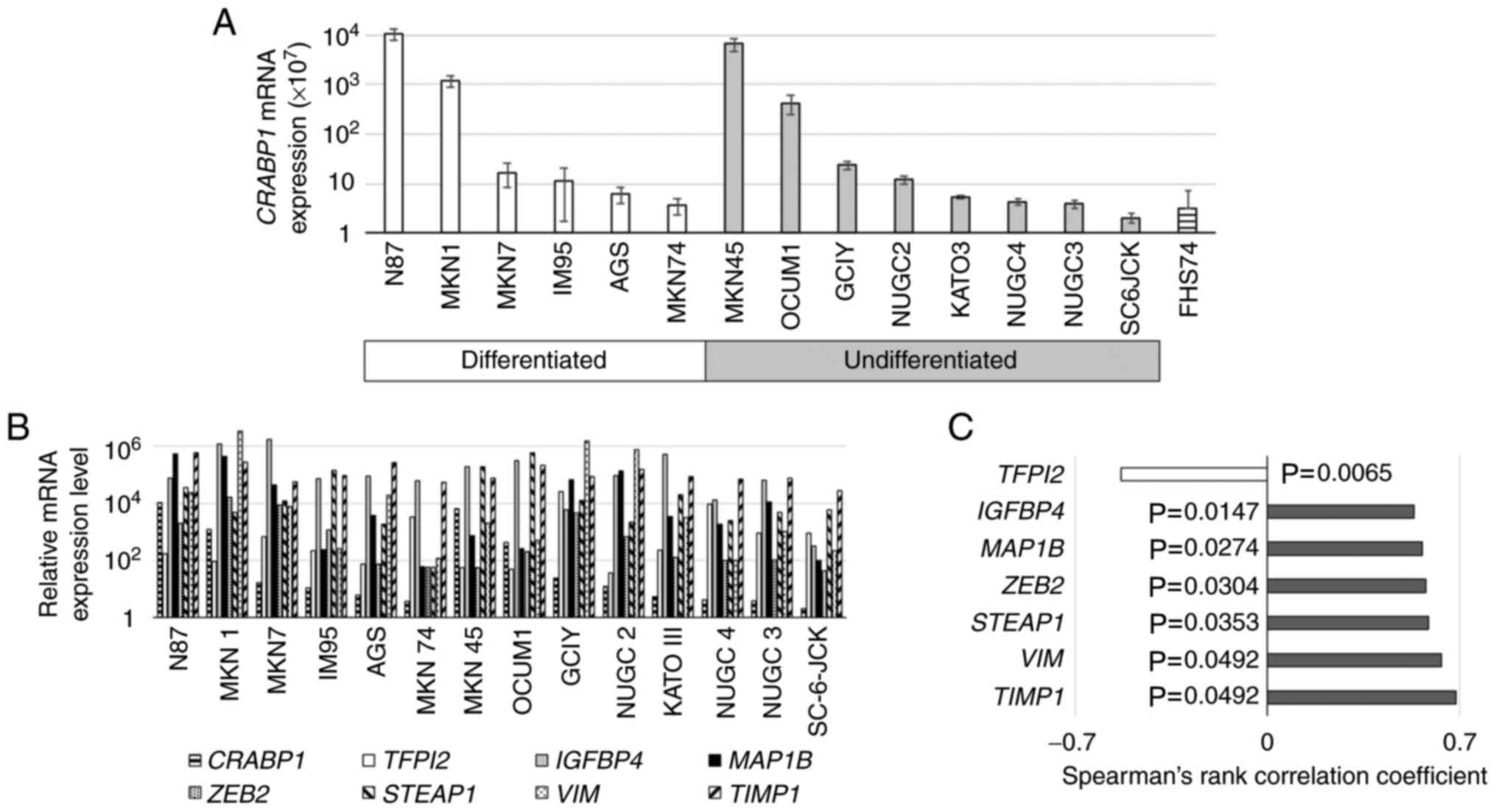
cancer cell lines are presented in Fig. 1B. There were large differences in
the levels of CRABP1 mRNA and those of other genes among
gastric cancer cell lines. CRABP1 mRNA levels positively
correlated with those encoding IGFBP4, MAP1B, ZEB2, STEAP1, VIM and
TIMP1 and negatively with TFPI2 (Fig.
1C).
Analyses of CRABP1 mRNA levels in gastric
cancer cell lines
To characterize CRABP1 in gastric cancer, the
levels of CRABP1 mRNA in 12 gastric cancer cell lines were
next compared with those of a nontumorigenic epithelial cell line.
CRABP1 mRNA levels were >2-fold higher in MKN1, MKN7,
N87, IM95, GCIY, MKN45, NUGC2 and OCUM1 cells compared with FHs74
cells (Fig. 1A). CRABP1
mRNA levels did not significantly differ according to the extent of
differentiation of the gastric cancer cells. MKN1, MKN45 and NUGC4
cells were selected for subsequent analyses, since MKN1 and MKN45
cells expressed relatively high levels of CRABP1 mRNA, and
these three cell lines were easy to use in functional analyses.
Effect of CRABP1 knockdown on the
biological activities of gastric cancer cells
The efficiency of CRABP1 knockdown by
transfection of siCRABP1-1 and siCRABP1-2 alone was
evaluated in MKN1, NUGC4 and MKN45 cells (Fig. S1). These two siRNAs were pooled
to constitute a CRABP1-specific siRNA. To evaluate the
function of CRABP1 in gastric cancer cells, MKN1 and NUGC4
cells were transfected with a CRABP1-specific siRNA. It was
first determined that the knockdown efficacy of the CRABP1
siRNA in MKN1, MKN45 and NUGC4 cells was sufficient for analysis
(Figs. 2A and S2). The proliferation of
siRNA-transfected MKN1, MKN45 and NUGC4 cells as well as the
invasiveness and migration of MKN1 and MKN45 cells were then
evaluated. The proliferation of MKN1, MKN45 and NUGC4 cells was
decreased as a result of CRABP1 knockdown starting from 72 h after
transfection compared with the siControl-transfected cells
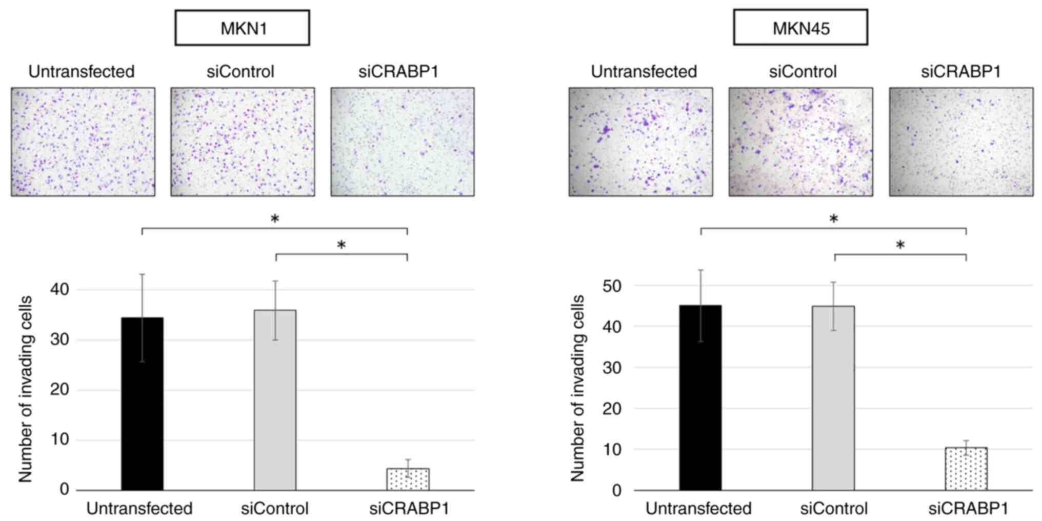
(Figs. 2B and S2). Furthermore, the invasiveness of
MKN1 and MKN45 cells was reduced by inhibiting CRABP1
expression (Fig. 3). The
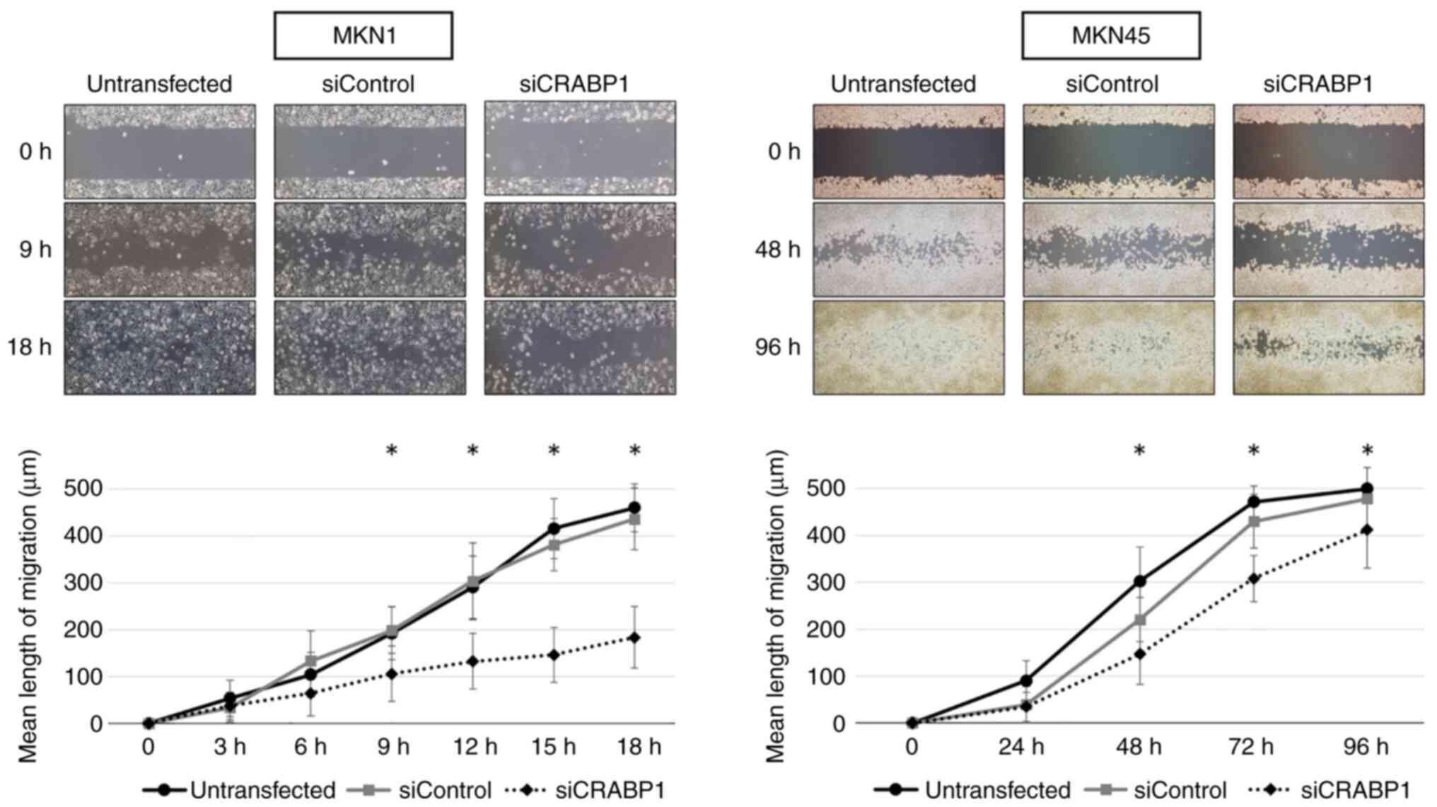
migration of MKN1 and MKN45 cells was reduced by inhibiting
CRABP1 expression (Fig.
4).
Effect of CRABP1 knockdown on peritoneal
metastasis in mouse xenograft models of gastric cancer
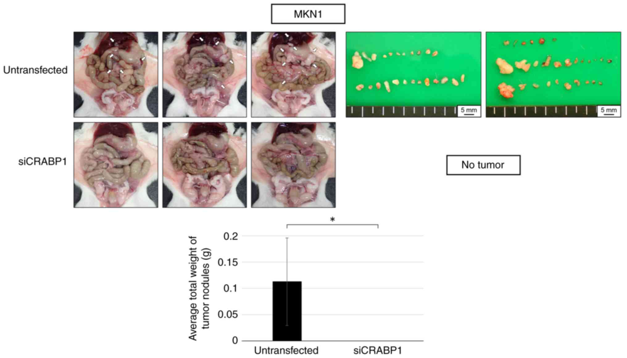
MKN1 and NUGC4 cells transfected with CRABP1
siRNA or untransfected were injected into mice to identify the
function of CRABP1 in recurrence and metastasis of gastric
cancer. Observations in the abdominal cavity of the mice were
performed after euthanasia. In the MKN1 xenograft model, peritoneal
dissemination was not observed in the siCRABP1 group
(Fig. 5). Peritoneal metastasis
in the NUGC4-model mice was disseminated to a smaller extent in the
siCRABP1 group compared with the untransfected group
(Fig. S3).
Prognostic impact of CRABP1
expression
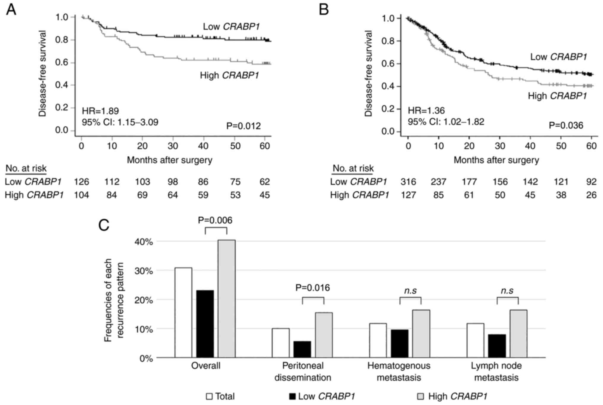
The DFS rate of the CRABP1-high group was
significantly lower compared with that of the CRABP1-low
group (5-year DFS rates; 59.6% and 77.8%, respectively; P=0.012)
(Fig. 6A) and were consistent
with those of the extra-validation cohort (Fig. 6B).
Next, gastric cancer recurrence patterns were
analyzed according to CRABP1 mRNA levels of 230 patients who
underwent R0 resection (stages I-III). Among them, 57 (24.7%)
experienced postoperative recurrence at 65 initial recurrence
sites. Analysis of recurrence patterns revealed that high
expression of CRABP1 mRNA was significantly associated with
peritoneal recurrence (P=0.016) (Fig.
6C), but not with the other two recurrence patterns.
The correlations between CRABP1 expression
and clinicopathological characteristics of patients were next
examined (Table III). High
CRABP1 expression was significantly associated with lymph
node metastasis. Univariate analysis of DFS demonstrated that
carbohydrate antigen 19-9 (37 IU/ml), tumor size ≥50 mm,
macroscopic type (Borrmann type 4/5), pT4, lymphatic involvement,
vascular invasion, invasive growth, lymph node metastasis and high
CRABP1 mRNA expression in gastric cancer tissues were
significant prognostic factors for adverse outcomes (Table IV). Multivariable analysis
identified high CRABP1 mRNA expression as an independent
prognostic factor of poor outcome (hazard ratio 1.89; 95%
confidence interval, 1.15-3.09; P=0.012).
| Table IIICRABP1 expression and the clinical
characteristics of patients with gastric cancer. |
Table III
CRABP1 expression and the clinical
characteristics of patients with gastric cancer.
| Clinical
characteristics | Expression level of
CRABP1
| P-value |
|---|
| Low (n=126) | High (n=104) |
|---|
| Age, years | | | 0.687 |
| <70 | 74 | 64 | |
| ≥70 | 52 | 40 | |
| Sex | | | 0.769 |
| Male | 89 | 76 | |
| Female | 37 | 28 | |
| CEA (ng/ml) | | | 0.850 |
| ≤5 | 107 | 90 | |
| >5 | 19 | 14 | |
| CA19-9 (IU/ml) | | | 0.382 |
| ≤37 | 102 | 89 | |
| >37 | 24 | 15 | |
| Tumor location | | | 0.992 |
| Entire | 4 | 4 | |
| Upper third | 34 | 27 | |
| Middle third | 43 | 37 | |
| Lower third | 45 | 36 | |
| Tumor size
(mm) | | | 0.562 |
| <50 | 68 | 56 | |
| ≥50 | 58 | 48 | |
| Macroscopic
type | | | 0.376 |
| Borrmann type
4/5 | 10 | 12 | |
| Others | 116 | 92 | |
| Multifocal
lesions | | | 0.823 |
| Absent | 115 | 94 | |
| Present | 11 | 10 | |
| Tumor depth
(UICC) | | | 0.581 |
| pT1-3 | 83 | 64 | |
| pT4 | 43 | 40 | |
|
Differentiation | | | 1.000 |
|
Differentiated | 54 | 45 | |
|
Undifferentiated | 72 | 59 | |
| Lymphatic
involvement | | | 0.288 |
| Absent | 24 | 14 | |
| Present | 102 | 90 | |
| Vascular
invasion | | | 0.077 |
| Absent | 55 | 33 | |
| Present | 71 | 71 | |
| Infiltrative
growth | | | 0.886 |
| Absent | 58 | 29 | |
| Present | 68 | 75 | |
| Lymph node
metastasis | | | 0.006 |
| Absent | 58 | 29 | |
| Present | 68 | 75 | |
| UICC stage | | | 0.056 |
| I | 34 | 16 | |
| II | 40 | 31 | |
| III | 52 | 57 | |
| Table IVPrognostic factors for disease-free
survival of patients with gastric cancer. |
Table IV
Prognostic factors for disease-free
survival of patients with gastric cancer.
| Variables | Univariate
| Multivariable
|
|---|
| Hazard ratio | 95% CI | P-value | Hazard ratio | 95% CI | P-value |
|---|
| Age (≥70
years) | 0.82 | 0.50-1.34 | 0.420 | | | |
| Sex (female) | 1.06 | 0.63-1.76 | 0.834 | | | |
| CEA (>5
ng/ml) | 1.39 | 0.74-2.58 | 0.304 | | | |
| CA 19-9 (>37
IU/ml) | 2.35 | 1.37-4.03 | 0.002 | 1.82 | 1.03-3.22 | 0.040 |
| Tumor location
(lower third) | 0.76 | 0.46-1.27 | 0.297 | | | |
| Tumor size (≥50
mm) | 1.95 | 1.21-3.15 | 0.006 | 1.44 | 0.88-2.35 | 0.145 |
| Macroscopic type
(Borrmann type 4/5) | 2.32 | 1.27-4.24 | 0.007 | 1.26 | 0.65-2.45 | 0.487 |
| Multifocal
lesions | 0.91 | 0.39-2.09 | 0.816 | | | |
| Tumor depth (pT4,
UICC) | 2.55 | 1.59-4.08 | <0.001 | 1.63 | 0.96-2.78 | 0.073 |
| Tumor
differentiation (undifferentiated) | 1.59 | 0.97-2.60 | 0.068 | | | |
| Lymphatic
involvement | 4.12 | 1.50-11.30 | 0.006 | 0.93 | 0.29-3.04 | 0.932 |
| Vascular
invasion | 2.66 | 1.52-4.65 | <0.001 | 1.34 | 0.72-2.48 | 0.359 |
| Invasive
growth | 1.66 | 1.03-2.69 | 0.038 | 1.12 | 0.64-1.97 | 0.687 |
| Lymph node
metastasis | 7.97 | 3.63-17.49 | <0.001 | 4.94 | 2.03-12.03 | <0.001 |
| High CRABP1
expression | 2.07 | 1.28-3.35 | 0.003 | 1.89 | 1.15-3.09 | 0.012 |
Discussion
In the present study, biomarkers of the malignant
phenotype of gastric cancer that predict postoperative recurrence
were searched. As a result, it was identified that the expression
levels of CRABP1 mRNA correlated with those of genes
encoding EMT-related molecules. Furthermore, knockdown of
CRABP1 influenced the proliferation, invasiveness, and
migration of gastric cancer cell lines. The results of these in
vitro analyses are consistent with the demonstration that
CRABP1 expression in primary tumor tissues of gastric cancer
was an independent predictor for worse postoperative
recurrence-free survival, which significantly correlated with an
increased rate of peritoneal recurrence.
CRABP1 specifically binds retinoic acid, an
activator of ERK1/2, which in turn, activates protein phosphatase
2A through binding to CRABP1 to lengthen the cell cycle
(17). This effect sensitizes
cancer cells to apoptosis by triggering the homeostatic action of
retinoic acid on the genome via the retinoic acid receptor
(18). Thus, CRABP1 may
encode a tumor suppressor, as indicated by findings that
CRABP1 inhibits the growth of cancers such as those of the
esophagus and thyroid (19-21). Conversely, evidence has indicated
that the tumor suppressive effect of CRABP1 is independent
of its retinoic acid-binding activity and may contribute to the
malignant transformation of mesenchymal tumors (22). Moreover, these findings suggested
that high expression of CRABP1 is associated with lymph node
metastasis and poor differentiation/high grade of pancreatic
neuroendocrine tumors (22).
Furthermore, a previous study revealed that CRABP1
expression is associated with poor prognosis of patients with
breast cancer, which reflects high Ki67 immunoreactivity and a high
pathological grade (23). Thus,
the relationships between CRABP1 expression and cancer
varies among organs, suggesting that CRABP1 may possess
unidentified functions.
Metastasis that leads to cancer recurrence involves
factors such as adhesion, infiltration, and angiogenesis, as the
EMT contributes to cancer progression and metastasis (24-26). For example, the present PCR array
results showed that CRABP1 expression significantly and
positively correlated with that of numerous EMT-promoting factors.
Moreover, CRABP1 expression negatively correlated with the
expression of TFPI2, which is often suppressed during the
EMT; and the gene encoding TFPI2 is frequently methylated in
gastric cancers (27,28). These results suggested that
CRABP1 is coordinately expressed with cancer-related
molecules and may promote peritoneal dissemination of gastric
cancer through the EMT.
Furthermore, siRNA-mediated knockdown of
CRABP1 expression reduced the proliferative, invasive and
migratory capacities of gastric cancer cells. Proliferation and
invasion of gastric cancer cells are required for their migration
from the primary tumor site, passage through endothelial cells, and
invasion of lymphatic and blood vessels, which culminates in the
colonization of lymph nodes and target organs, as well as the
proliferation of cancer cells in the parenchyma (29).
In a mouse xenograft model of peritoneal metastasis
of gastric cancer, it was found that the total weight of
disseminated nodules was lower in the group, in which CRABP1
mRNA levels were knocked down compared with those of the
untransfected group. These results suggested that CRABP1 is
involved in the recurrence of peritoneal dissemination of gastric
cancer. In the present study, high expression of CRABP1 in
gastric cancer tissues was associated with a higher recurrence
rate, shorter DFS and significantly more frequent peritoneal
dissemination, leading to recurrence. These results indicated that
preoperative and intraoperative analysis of CRABP1
expression may predict the risk of peritoneal dissemination
recurrence after curative resection.
Thus, evaluating the expression of CRABP1 as
a biomarker of patients at high risk of peritoneal dissemination
may inform decisions on implementing a surveillance plan that
considers the course of peritoneal dissemination after surgery.
Specifically, closely spaced abdominal echocardiography and
computed tomography of the pelvis can be used to detect small
amounts of ascites and small peritoneal nodules. Furthermore, the
present data have important clinical implications for administering
adjuvant chemotherapy to patients with high tissue levels of
CRABP1 mRNA after resection of gastric cancer to reduce
their risk of recurrence.
There are several limitations to the present study.
First, the clinical impact of CRABP1 expression was
retrospectively evaluated. Second, the clinical samples of the
present study were insufficient to evaluate CRABP1 as a
biomarker to detect disseminated metastasis. A prospective
observational study of clinical samples, including disseminated
metastasis, is there- fore required to evaluate the prognostic
ability of CRABP1 expression levels. Third, the detailed
molecular mechanisms underlying the correlation between high
CRABP1 expres- sion and postoperative prognosis, including
disseminated recurrence, must be determined. Identification of the
relevant signal transduction pathways is required to fully
understand the role of CRABP1 in tumor progression. In
breast cancer cells, CRABP1 sequesters all-trans-retinoic
acid (atRA) in the cytosol, inhibiting its nuclear action (23). Evaluating the expression levels of
CRABP1 in gastric cancer cells and the effects of atRA on
the tumor may further illuminate their mechanism of action related
to malignancy.
In summary, it was revealed in the present study
that CRABP1 influenced the malignant phenotype of gastric
cancer cells and that its high expression in primary tumor tissues
may serve as a biomarker for determining the prognosis of
recurrence after curative resection, particularly that of patients
with peritoneal dissemination.
Supplementary Data
Availability of data and materials
The datasets used and/or analyzed during the current
study are available from the corresponding author upon reasonable
request.
Authors' contributions
KS, MK, and SN performed the experiments and data
analysis. KS, MK, DS, SN, YI, NH, MH, CT, GN, and YK collected
cases and clinical data. KS and MK confirm the authenticity of all
the raw data. KS and MK conceived and designed the study and
prepared the initial draft of the manuscript. YK supervised the
project. All authors contributed to the final manuscript. All
authors read and approved the final manuscript.
Ethics approval and consent to
participate
The present study conformed to the ethical
guidelines of the World Medical Association Declaration of Helsinki
Ethical Principles for Medical Research Involving Human Subjects
(2013). The present study was approved (approval no. 2014-0043) by
the Institutional Review Board of Nagoya University (Nagoya,
Japan). Written informed consent was obtained from all patients.
Animal experiments were approved (approval no. M210414-001) by the
Animal Research Committee of Nagoya University (Nagoya, Japan).
Patient consent for publication
Not applicable.
Competing interests
The authors declare that they have no competing
interests.
Acknowledgments
Not applicable.
Funding
No funding was received.
Abbreviations:
|
ATCC
|
American Type Culture Collection
|
|
atRA
|
all-trans-retinoic acid
|
|
CA
|
carbohydrate antigen
|
|
CRABP1
|
cellular retinoic acid-binding protein
1
|
|
CT
|
computed tomography
|
|
DFS
|
disease-free survival
|
|
EMT
|
epithelial-mesenchymal transition
|
|
GAPDH
|
glyceraldehyde-3-phosphate
dehydrogenase
|
|
JCRB
|
Japanese Collection of Research
Bioresources Cell Bank
|
|
OS
|
overall survival
|
|
PBS
|
phosphate-buffered saline
|
|
RT-qPCR
|
reverse transcription-quantitative
polymerase chain reaction
|
|
ROC
|
receiver operating characteristic
|
|
UICC
|
Union for International Cancer
Control
|
References
|
1
|
Bray F, Ferlay J, Soerjomataram I, Siegel
RL, Torre LA and Jemal A: Global cancer statistics 2018: GLOBOCAN
estimates of incidence and mortality worldwide for 36 cancers in
185 countries. CA Cancer J Clin. 68:394–424. 2018. View Article : Google Scholar : PubMed/NCBI
|
|
2
|
Wadhwa R, Song S, Lee JS, Yao Y, Wei Q and
Ajani JA: Gastric cancer-molecular and clinical dimensions. Nat Rev
Clin Oncol. 10:643–655. 2013. View Article : Google Scholar : PubMed/NCBI
|
|
3
|
McLean MH and El-Omar EM: Genetics of
gastric cancer. Nat Rev Gastroenterol Hepatol. 11:664–674. 2014.
View Article : Google Scholar : PubMed/NCBI
|
|
4
|
Dong D, Ruuska SE, Levinthal DJ and Noy N:
Distinct roles for cellular retinoic acid-binding proteins I and II
in regulating signaling by retinoic acid. J Biol Chem.
274:23695–23698. 1999. View Article : Google Scholar : PubMed/NCBI
|
|
5
|
Kanda M, Shimizu D, Tanaka H, Shibata M,
Iwata N, Hayashi M, Kobayashi D, Tanaka C, Yamada S, Fujii T, et
al: Metastatic pathway-specific transcriptome analysis identifies
MFSD4 as a putative tumor suppressor and biomarker for hepatic
metastasis in patients with gastric cancer. Oncotarget.
7:13667–13679. 2016. View Article : Google Scholar : PubMed/NCBI
|
|
6
|
Liu JY, Peng CW, Yang XJ, Huang CQ and Li
Y: The prognosis role of AJCC/UICC 8th edition staging
system in gastric cancer, a retrospective analysis. Am J Transl
Res. 10:292–303. 2018.
|
|
7
|
Kanda M, Murotani K, Kobayashi D, Tanaka
C, Yamada S, Fujii T, Nakayama G, Sugimoto H, Koike M, Fujiwara M
and Kodera Y: Postoperative adjuvant chemotherapy with S-1 alters
recurrence patterns and prognostic factors among patients with
stage II/III gastric cancer: A propensity score matching analysis.
Surgery. 158:1573–1580. 2015. View Article : Google Scholar : PubMed/NCBI
|
|
8
|
Foo M and Leong T: Adjuvant therapy for
gastric cancer: Current and future directions. World J
Gastroenterol. 20:13718–13727. 2014. View Article : Google Scholar : PubMed/NCBI
|
|
9
|
Kanda M, Nomoto S, Oya H, Takami H,
Shimizu D, Hibino S, Hashimoto R, Kobayashi D, Tanaka C, Yamada S,
et al: The expression of melanoma-associated antigen D2 both in
surgically resected and serum samples serves as clinically relevant
biomarker of gastric cancer progression. Ann Surg Oncol. 23(Suppl
2): S214–S221. 2016. View Article : Google Scholar
|
|
10
|
Umeda S, Kanda M, Miwa T, Tanaka H, Tanaka
C, Kobayashi D, Suenaga M, Hattori N, Hayashi M, Yamada S, et al:
Expression of sushi domain containing two reflects the malignant
potential of gastric cancer. Cancer Med. 7:5194–5204. 2018.
View Article : Google Scholar :
|
|
11
|
Kanda M, Shimizu D, Fujii T, Sueoka S,
Tanaka Y, Ezaka K, Takami H, Tanaka H, Hashimoto R, Iwata N, et al:
Function and diagnostic value of Anosmin-1 in gastric cancer
progression. Int J Cancer. 138:721–730. 2016. View Article : Google Scholar
|
|
12
|
Kanda M, Shimizu D, Tanaka H, Tanaka C,
Kobayashi D, Hayashi M, Iwata N, Niwa Y, Yamada S, Fujii T, et al:
Significance of SYT8 for the detection, prediction, and treatment
of peritoneal metastasis from gastric cancer. Ann Surg.
267:495–503. 2018. View Article : Google Scholar
|
|
13
|
Shimizu D, Kanda M, Tanaka H, Kobayashi D,
Tanaka C, Hayashi M, Iwata N, Niwa Y, Takami H, Yamada S, et al:
GPR155 serves as a predictive biomarker for hematogenous metastasis
in patients with gastric cancer. Sci Rep. 7:420892017. View Article : Google Scholar : PubMed/NCBI
|
|
14
|
Shimizu D, Kanda M, Sugimoto H, Shibata M,
Tanaka H, Takami H, Iwata N, Hayashi M, Tanaka C, Kobayashi D, et
al: The protein arginine methyltransferase 5 promotes malignant
phenotype of hepatocellular carcinoma cells and is associated with
adverse patient outcomes after curative hepatectomy. Int J Oncol.
50:381–386. 2017. View Article : Google Scholar : PubMed/NCBI
|
|
15
|
Kilkenny C, Browne WJ, Cuthill IC, Emerson
M and Altman DG: Improving bioscience research reporting: The
ARRIVE guidelines for reporting animal research. PLOS Biol.
8:e10004122010. View Article : Google Scholar : PubMed/NCBI
|
|
16
|
Szász AM, Lánczky A, Nagy Á, Förster S,
Hark K, Green JE, Boussioutas A, Busuttil R, Szabó A and Győrffy B:
Cross-validation of survival associated biomarkers in gastric
cancer using transcriptomic data of 1,065 patients. Oncotarget.
7:49322–49333. 2016. View Article : Google Scholar : PubMed/NCBI
|
|
17
|
Persaud SD, Park SW, Ishigami-Yuasa M,
Koyano-Nakagawa N, Kagechika H and Wei LN: All trans-retinoic acid
analogs promote cancer cell apoptosis through non-genomic Crabp1
mediating ERK1/2 phosphorylation. Sci Rep. 6:223962016. View Article : Google Scholar : PubMed/NCBI
|
|
18
|
Persaud SD: The functional role of
retinoic acid and the cellular retinoic acid binding protein 1
(Crabp1) in tumor suppression. 141:2018.
|
|
19
|
Tanaka K, Imoto I, Inoue J, Kozaki K,
Tsuda H, Shimada Y, Aiko S, Yoshizumi Y, Iwai T, Kawano T and
Inazawa J: Frequent methylation-associated silencing of a candidate
tumor-suppressor, CRABP1, in esophageal squamous-cell carcinoma.
Oncogene. 26:6456–6468. 2007. View Article : Google Scholar : PubMed/NCBI
|
|
20
|
Celestino R, Nome T, Pestana A, Hoff AM,
Gonçalves AP, Pereira L, Cavadas B, Eloy C, Bjøro T,
Sobrinho-Simões M, et al: CRABP1, C1QL1 and LCN2 are biomarkers of
differentiated thyroid carcinoma, and predict extrathyroidal
extension. BMC Cancer. 18:682018. View Article : Google Scholar : PubMed/NCBI
|
|
21
|
Huang Y, de la Chapelle A and Pellegata
NS: Hypermethylation, but not LOH, is associated with the low
expression of MT1G and CRABP1 in papillary thyroid carcinoma. Int J
Cancer. 104:735–744. 2003. View Article : Google Scholar : PubMed/NCBI
|
|
22
|
Kainov Y, Favorskaya I, Delektorskaya V,
Chemeris G, Komelkov A, Zhuravskaya A, Trukhanova L, Zueva E,
Tavitian B, Dyakova N, et al: CRABP1 provides high malignancy of
transformed mesenchymal cells and contributes to the pathogenesis
of mesenchymal and neuroendocrine tumors. Cell Cycle. 13:1530–1539.
2014. View Article : Google Scholar : PubMed/NCBI
|
|
23
|
Liu RZ, Garcia E, Glubrecht DD, Poon HY,
Mackey JR and Godbout R: CRABP1 is associated with a poor prognosis
in breast cancer: Adding to the complexity of breast cancer cell
response to retinoic acid. Mol Cancer. 14:1292015. View Article : Google Scholar : PubMed/NCBI
|
|
24
|
Lamouille S, Xu J and Derynck R: Molecular
mechanisms of epithelial-mesenchymal transition. Nat Rev Mol Cell
Biol. 15:178–196. 2014. View Article : Google Scholar : PubMed/NCBI
|
|
25
|
Pastushenko I and Blanpain C: EMT
transition states during tumor progression and metastasis. Trends
Cell Biol. 29:212–226. 2019. View Article : Google Scholar
|
|
26
|
Aiello NM and Kang Y: Context-dependent
EMT programs in cancer metastasis. J Exp Med. 216:1016–1026. 2019.
View Article : Google Scholar : PubMed/NCBI
|
|
27
|
Qu Y, Dang S and Hou P: Gene methylation
in gastric cancer. Clin Chim Acta Int J Clin Chem. 424:53–65. 2013.
View Article : Google Scholar
|
|
28
|
Takada H, Wakabayashi N, Dohi O, Yasui K,
Sakakura C, Mitsufuji S, Taniwaki M and Yoshikawa T: Tissue factor
pathway inhibitor 2 (TFPI2) is frequently silenced by aberrant
promoter hypermethylation in gastric cancer. Cancer Genet
Cytogenet. 197:16–24. 2010. View Article : Google Scholar : PubMed/NCBI
|
|
29
|
Kanda M and Kodera Y: Molecular mechanisms
of peritoneal dissemination in gastric cancer Molecular mechanisms
of peritoneal dissemination in gastric cancer. World J
Gastroenterol. 22:6829–6840. 2016. View Article : Google Scholar : PubMed/NCBI
|