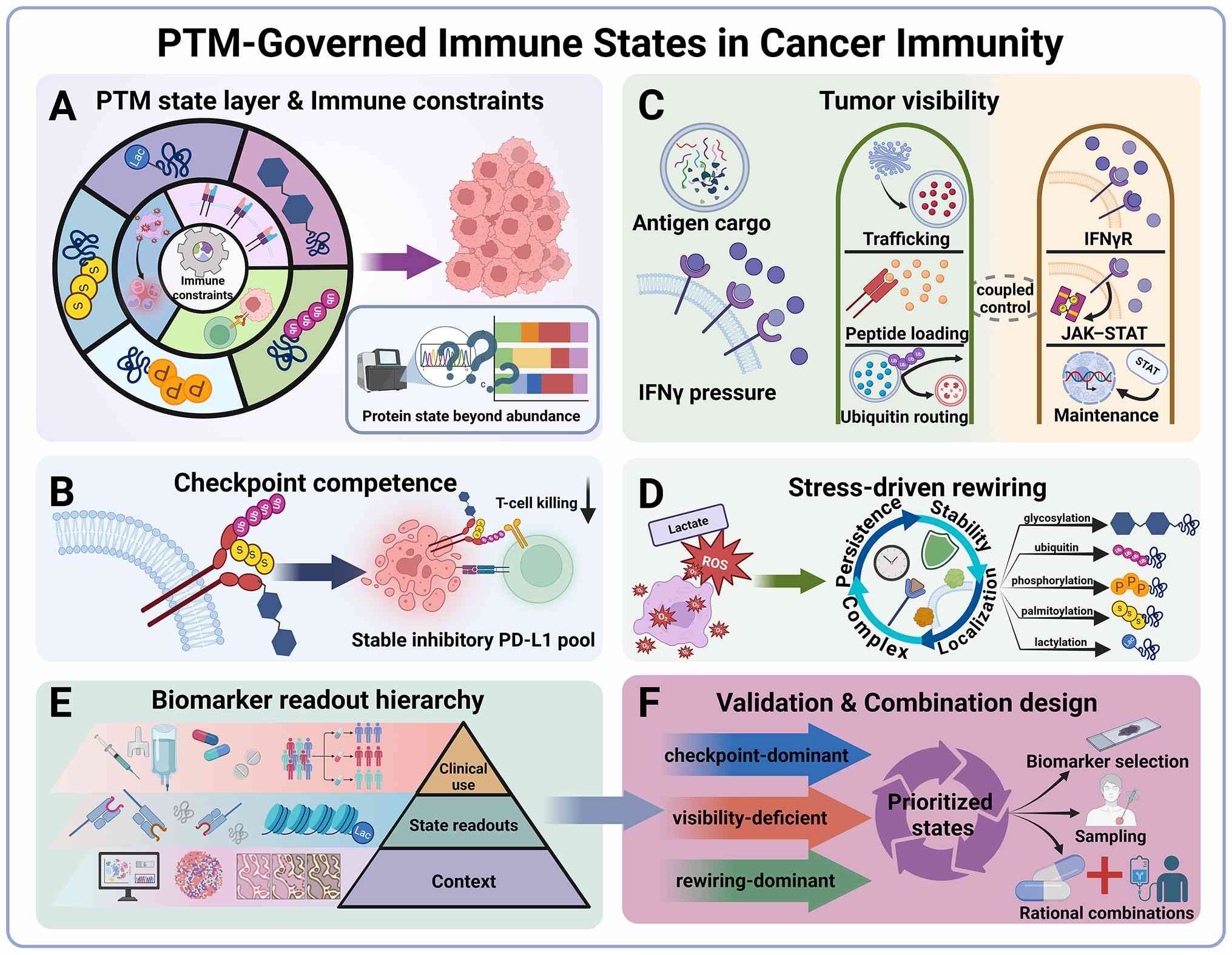
Cancer immunotherapy and multimodal regimens can
yield durable benefit in selected patients, yet immune escape and
therapeutic resistance remain major causes of progression (1-3).
Across clinical settings, treatment failure often reflects
disruption of a limited set of rate-limiting processes required for
sustained immune control, particularly checkpoint competence, tumor
visibility, and the maintenance of productive effector function
(2). Response heterogeneity is
frequently discordant with transcript-level immune activation, and
resistance can arise early during therapy despite limited genomic
change, indicating the involvement of regulatory layers that act on
short timescales, encode microenvironmental context, and directly
tune protein fate and signaling kinetics (4). Post-translational modifications
(PTMs) represent a major such layer because they regulate protein
stability, trafficking, subcellular localization, interaction
networks and signal duration, thereby converting tumor-intrinsic
stress and therapy-imposed selective pressure into adaptive immune
phenotypes and resistant states (5).
Although PTM-focused literature is extensive, most
syntheses remain organized either by modification class or by
individual targets, which can fragment immune regulation into
parallel mechanisms and obscure the shared functional constraints
that ultimately determine therapeutic response (6). In the present review, PTM biology
was instead interpreted through three recurrent immune constraints
that limit durable antitumor immunity: Checkpoint competence, tumor
visibility and stress-conditioned immune-state programming
(7-11). This framework allows
mechanistically distinct PTM events to be understood through
convergent functional outputs and more directly links molecular
regulation to biomarker logic and therapeutic resistance.
A central premise of the present review is that PTMs
rarely function as isolated switches. Rather, combinatorial
modification states and PTM crosstalk shape coupled changes in
protein stability, localization, complex assembly and signaling
persistence (5). Focus was
therefore addressed on exemplar circuits in which the evidence
chain is strongest and the translational implications are clearest.
Programmed death-ligand 1 (PD-L1) provides a paradigm for this
logic, as glycosylation, palmitoylation, and ubiquitination or
deubiquitination converge on a shared functional endpoint:
Stabilization of a membrane-resident suppressive state that is not
captured by expression alone (7,8,12-16). Therapy-stress-induced PTM rewiring
as an early resistance mechanism was also emphasized, because PTM
networks can be selected during treatment and subsequently
stabilized across treatment-naïve, on-treatment, and relapse phases
(2,5).
The present review is therefore not intended to
catalogue PTMs comprehensively across cancer immunity. Instead, it
addresses a narrower translational question: Which PTM-governed
protein states most directly shape checkpoint competence, tumor
visibility, and stress-conditioned immune-state programming, and
which of these states are most tractable for biomarker development
or therapeutic intervention. On this basis, focus was addressed on
five core exemplar PTM axes, glycosylation, palmitoylation,
ubiquitin editing, phosphorylation and lactylation, because they
repeatedly map to rate-limiting immune constraints and provide
mechanistically informative state-control examples, although their
assay and therapeutic maturity remain uneven across targets and
tumor contexts (17,18). Glycosylation, palmitoylation,
ubiquitin editing and phosphorylation are emphasized because they
recurrently govern checkpoint stability, antigen-presentation
competence, receptor trafficking and interferon (IFN)-linked
signaling persistence (19-24). Lactylation is considered
separately because, within this framework, its main significance
lies in linking metabolic stress to chromatin-coupled
immunosuppressive fixation (10,25,26). The present review therefore uses
these five core axes as the principal organizing examples for
interpreting immune-state constraints in cancer immunity.
Additional modification classes are discussed selectively where
they clarify the same functional state variables, reveal framework
boundaries, or provide relevant examples of
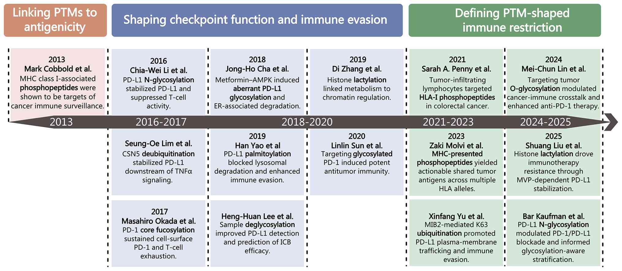
molecular-state-selective vulnerability. To orient the reader to
the historical emergence and conceptual scope of this framework,
selected milestones linking PTMs to tumor antigenicity,
checkpoint-state control, immune evasion and immunotherapy
resistance are summarized in Fig.
1.
PTMs influence cancer immune escape and therapeutic
resistance through multiple, closely interconnected regulatory
routes. To organize these mechanisms into a clinically
interpretable framework, this section examines how PTM-governed
protein states shape three recurrent immune constraints: Checkpoint
competence, tumor visibility and stress-conditioned immune-state
programming. It further considers the molecular coordinates,
multi-omics resources, and biomarker contexts required to translate
these states into measurable and potentially actionable
variables.
Immune escape and treatment resistance often arise
when a limited number of rate-limiting steps fail to convert immune
activation into durable tumor control (27-29). PTMs are well suited to this
problem because they rapidly tune protein fate and signal
propagation in a context-dependent manner, including under
therapy-imposed selective pressure (13,30). A constraint-centered synthesis
therefore provides a principled way to integrate PTM biology in
immuno-oncology while remaining anchored to processes that
repeatedly constrain clinical benefit.
A recurring challenge is that numerous
immune-relevant phenotypes are determined by protein state rather
than transcript abundance (31,32). Across these immune constraints,
diverse PTM chemistries converge on a restricted set of state
variables that define functional output, including proteostasis,
trafficking, complex assembly and signaling persistence (5). This convergence is what makes PTM
biology interpretable from a biomarker perspective, because it
allows mechanistically distinct PTM events to be read out through a
limited number of functionally aligned state variables. Using these
variables as mechanistic coordinates enables cross-study comparison
while keeping interpretation tied to analyzable control points
rather than pathway labels.
PTM-governed protein states should not be
interpreted as tissue-independent molecular outputs. Their
functional weight is conditioned by the cellular and tissue context
in which a modification is installed, maintained, or removed.
Pan-cancer proteogenomic analyses show that oncogenic effects can
diverge across RNA, protein and phosphoprotein layers and are
accompanied by distinct signaling states (33). Direct in vivo nutrient
tracing further demonstrates cell-programmed partitioning of
glucose and glutamine among myeloid cells, T cells and cancer cells
within tumors (34). Nutrient use
can also be translated into PTM-dependent transcriptional states.
In exhausted CD8+ T cells, altered acetate and citrate
utilization redirects acetyl-CoA production and locus-specific
histone acetylation, thereby shaping transcriptional
differentiation (35).
Accordingly, protein abundance or PTM occupancy measured by mass
spectrometry should be interpreted together with cellular origin
and metabolic context when assigning a dominant immune
constraint.
These mechanistic coordinates may also converge
within selected therapy-resistant or stem-like tumor cell states
rather than being distributed uniformly across malignant
populations. Recent studies have shown that immune selection can
enrich tumor cell populations in which stemness and immune-evasive
programs are coupled, while glioblastoma (GBM) stem cells can
sustain PD-L1-associated immunosuppression through a
ubiquitin-linked β-catenin signaling circuit (36,37). Within the present framework,
stem-like tumor states are therefore not considered a fourth
dominant immune constraint. Rather, they represent a resistant
cellular context in which checkpoint competence, impaired tumor
visibility, and adaptive state persistence may coexist or become
selectively reinforced. This distinction allows stemness-associated
immune escape to be integrated without replacing the functional
constraints that organize PTM-state interpretation.
Immune checkpoint blockade (ICB) ultimately depends
on the persistence and accessibility of the surface-competent
checkpoint pool, rather than on total expression alone. PTMs shape
functional checkpoint states by biasing proteostasis and
trafficking decisions that determine whether ligands or receptors
accumulate in suppressive membrane configurations and how long
these configurations are maintained (8). For PD-L1, such control is exerted
through coordinated regulation of N-glycosylation and ubiquitin
editing that stabilizes the ligand and modulates its commitment to
proteasomal or lysosomal degradation, together with
palmitoylation-dependent stabilization that limits ubiquitination
and proteasomal turnover (13,15,20).
This framework also highlights response
heterogeneity that can arise when modification state alters
antibody engagement independently of bulk abundance. PTM-dependent
processing can reshape epitope accessibility and thereby change the
effective pharmacology of blockade (38). In line with this, removal of
N-linked glycosylation increases anti-PD-L1 antibody binding
affinity and signal intensity and improves clinical outcome
prediction, while complementary glycoproteomic evidence
demonstrates that PD-L1 glycosylation state can measurably affect
binding to clinical antibodies (7,39).
Tumor visibility is a dynamic property defined by
the capacity to sustain immune recognition pressure over time. Two
components dominate this immune constraint: Competence of antigen
processing and presentation, and competence and persistence of IFN
and signal transducer and activator of transcription (STAT)
programs that reinforce visibility under immune pressure (29). PTMs modulate visibility by
regulating the stability, assembly and trafficking of antigen
presentation machinery and by tuning the kinetics and persistence
of IFN-linked signaling circuits (40,41). In antigen-presenting cells,
ubiquitin-dependent routing decisions can determine whether
peptide-loaded complexes are preferentially recycled or delivered
for lysosomal turnover, thereby controlling the durability of
surface display rather than merely its formation (42). In cancer, ubiquitin-dependent
control of major histocompatibility complex (MHC) class II
trafficking can indirectly reduce MHC I surface expression and
antigen presentation to CD8 T cells, indicating that PTM control
can directly reparameterize the tumor visibility axis at the level
of peptide display (22).
These mechanisms are especially consequential in
therapy-exposed tumors, where response failure can arise with
limited new genomic change and where transcriptomic inflammation
does not necessarily guarantee sustained antigen presentation
competence (43). Consistent with
this visibility-centered logic, longitudinal clinical analyses of
acquired resistance to PD-1 blockade have identified defects
converging on antigen presentation and IFN responsiveness,
underscoring that visibility can collapse through disruption of
either arm even after an initial response. Mechanistically,
tumor-intrinsic modulation of IFN pathway signal duration can be
achieved through phosphorylation and ubiquitin-controlled negative
feedback at the JAK-STAT module, including SOCS1-mediated
inhibition of JAK activity and SOCS box-dependent recruitment of
ubiquitin machinery that restricts sustained STAT1 signaling and
weakens IFN-driven MHC class I programs (44-46). Together, these data support a
competence-focused view that separates upstream immune activation
from the tumor's capacity to sustain presenting states and
IFN-reinforced visibility under therapeutic pressure, thereby
linking PTM regulation to impaired priming, insufficient
reinforcement and primary non-response under ICB (41,43).
A third recurrent immune constraint concerns whether
immune cells retain cytotoxic competence in microenvironments
shaped by hypoxia and mitochondrial oxidative stress, which can
accelerate exhaustion under sustained antigenic stimulation
(47). An important frontier is
the increasing mechanistic resolution of metabolite-sensitive PTMs
at the chromatin interface, where lactate-linked histone
lactylation can couple metabolic constraints to transcriptional
programs that tune CD8 T-cell fitness and effector output (3). At present, however, the
translational maturity of this axis remains uneven across tumor
contexts, and its strongest current value may lie in defining
emerging state-transition biomarkers rather than routine deployable
assays. Mechanistic interpretation of this axis requires explicit
attention to persistence and context because exhausted CD8 T cells
can adopt a fixed dysfunctional state with limited reprogramming
capacity after PD-1 blockade. Consistent with this, de novo
epigenetic programs can function as a barrier to checkpoint
blockade-mediated rejuvenation, supporting entrenched dysfunction
under therapeutic selection (48). Across these immune constraints,
functionally relevant regulation often reflects combinatorial PTM
states and crosstalk rather than isolated marks, converging on
shared state variables such as stability, localization, complex
competence and signaling persistence (13).
To consolidate this constraint-centered framework,
the dominant state constraints, their corresponding state
variables, representative PTM routes, and the supporting
experimental contexts are summarized in Table I.
As the preceding sections indicate, the immune
constraints that limit durable antitumor immunity are often not
adequately resolved through transcript-level profiling, because
they are more directly expressed through changes in protein state,
including altered abundance, localization, complex competence and
signaling persistence (49). This
distinction is especially important in PTM-oriented
immuno-oncology, because modification-dependent changes in protein
stability, trafficking and signaling architecture are more directly
captured by proteomic and phospho-proteomic than by transcriptomic
layers alone (49,50). The aforementioned developed
framework therefore requires data layers that can resolve these
state variables directly. From this perspective, PTM-governed
immune states are most effectively decoded through integrated
multi-omics rather than through any single modality in isolation.
Immuno-peptidomic analyses further show that PTM-associated changes
in the tumor proteome can reshape the MHC class I ligandome and
generate cancer-specific modified antigens, thereby linking
proteomic state to the antigenic information presented at the
tumor-immune interface (51).
Spatially resolved profiling adds a further dimension by defining
how tumor and immune states are organized within tissue and whether
they coincide with immune exclusion or immune-evasive niche
structure (52).
PTM-governed immune states should not be assumed to
be spatially uniform within a single lesion. Single-cell spatial
proteomics has revealed extensive region-specific proteomic
variation and immune-cell heterogeneity within pancreatic tumors,
while spatial profiling of ovarian cancer has identified locally
distinct immune phenotypes with differential abundance of PD-L1,
B2M, granzyme B and cluster of differentiation (CD)163 (53,54). Spatial multi-omics in GBM further
demonstrated that hypoxia and regionally segregated inflammatory or
metabolic stimuli are associated with spatially exclusive adaptive
programs and enhanced immunosuppressive tumor-myeloid interactions
(55). These findings suggest
that the protein-state variables used in the present framework to
infer checkpoint competence, tumor visibility and
stress-conditioned rewiring may be locally discordant within the
same lesion. Consistent with this concern, multi-region analysis of
treatment-naive esophageal squamous cell carcinoma showed that
limited biopsy specimens were insufficient to accurately
approximate whole-tumor PD-L1 combined positive scores (56). A single-core biopsy may therefore
capture a locally dominant state rather than the lesion-level
dominant state constraint. Dominant-state assignment should
accordingly be interpreted as a sampling-dependent estimate that
requires spatial contextualization rather than as an intrinsically
uniform tumor property.
The increasing availability of resource-oriented
platforms now makes such analyses feasible across molecular and
clinical scales. PTM-focused databases support site-level
annotation, functional interpretation and regulator-substrate
mapping, including kinase-substrate and E3 ligase-substrate
relationships (57). Public
cancer atlases provide harmonized genomic, transcriptomic,
proteomic, phospho-proteomic and clinical data within a shared
patient framework (50). Public
immuno-peptidomic resources further extend this landscape by
aggregating large-scale MHC-bound peptide data, including modified
antigens, into searchable atlases that support antigen
prioritization and cross-cohort comparison (58,59). The value of these resources lies
not in cataloguing molecular complexity for its own sake, but in
providing the layered resolution needed to define PTM-governed
immune states and nominate assay-compatible candidate readouts.
If PTM-governed immune states are to be captured
more directly through integrated proteomic, phospho-proteomic,
immuno-peptidomic and spatially resolved approaches, the next issue
is not simply how to detect them, but how to use them in clinical
decision-making (50,51,60). Within this framework, biomarker
classes are best distinguished by the clinical question they are
intended to answer rather than by platform alone (61). Importantly, the framework
distinguishes direct PTM-state readouts from proximal protein-state
and contextual companion readouts. Direct PTM measurements are
intended to indicate functional immune constraints, whereas
companion readouts provide supporting information on inflammation,
immune abundance, spatial organization, or treatment-associated
state change. Some baseline readouts are diagnostic, establishing
that a biologically relevant state is present in the tumor.
Representative examples include checkpoint-competent inhibitory
surface states shaped by PD-L1 glycosylation or palmitoylation,
attenuated antigenic visibility linked to impaired human leukocyte
antigen (HLA)-A expression or broader antigen-presentation
deficiency, and metabolically reinforced suppressive states coupled
to lactylation-driven macrophage programming (7,8,25,62,63). Others can function prognostically
by indicating whether such states are associated with more
persistent immune escape or more limited durability of disease
control, independent of the specific intervention chosen (61).
A different class is predictive and is intended to
guide treatment selection. Here, the most informative features are
those that remain mechanistically close to the dominant state
constraint and therefore capture a tractable dependency, such as
persistent PTM-shaped checkpoint competence, incomplete
antigen-presentation programs, or HLA-linked
neoantigen-presentation states that enrich for benefit from
immunotherapy (7,63,64). By contrast, pharmacodynamic
readouts are used to determine whether therapy is actually shifting
the relevant state over time (61). Their value lies in showing whether
a PTM-regulated immune program is being contracted, reprogrammed,
or stabilized during treatment, thereby helping distinguish
meaningful state transition from transient pathway perturbation
(65,66). These use contexts can overlap, but
they should remain analytically distinct, because the same
PTM-linked feature may carry one value at baseline, another during
on-treatment monitoring, and a third at progression or relapse
(65). In this sense, the
translational relevance of PTM biology lies not in documenting
modification events per se, but in assigning each readout to the
decision context in which it can most meaningfully support
stratification, monitoring and rational therapeutic design.
Operationally, a PTM-state feature should advance beyond
mechanistic interest only when it can be assigned preferentially to
one dominant state constraint, measured in a prespecified specimen
type and treatment window, and interpreted in a way that could
alter baseline stratification, on-treatment monitoring, or
relapse-state attribution.
To render this framework more operational from a
biomarker perspective, candidate PTM-state biomarker modules
aligned to the dominant state constraints in cancer immunotherapy
are summarized in Table II.
Clinical practice often treats PD-L1 as a static
biomarker sampled once to infer dependence on the programmed cell
death 1 axis, most commonly through immunohistochemistry (IHC)
workflows with substantial analytic and interpretive variability
across assays and settings (68).
Yet PD-L1 is better understood as a proteostasis-governed surface
signal whose functional output is continuously shaped by
trafficking and turnover (69,70). In numerous tumors, the critical
variable is therefore less PD-L1 transcription itself than the
ability to sustain an antibody-accessible, PD-1-binding pool at the
plasma membrane through signal-coupled control of stability and
routing (19,70,71). Bulk PD-L1 readouts are therefore
intrinsically ambiguous because they compress synthesis,
degradation and epitope accessibility into a single value (71).
A functional-state view reframes PD-L1 around
variables proximal to checkpoint output. Stability determines
whether transient inflammatory pulses are converted into durable
inhibitory tone by maintaining PD-L1 in a protected regime rather
than committing it to rapid clearance (19). Routing and membrane residence
determine whether PD-L1 is positioned at the tumor-immune interface
rather than distributed across internal pools shaped by endocytic
sorting and recycling (69).
Epitope accessibility governs what IHC detects and what therapeutic
antibodies can engage, so that a PD-L1 pool may be biologically
present yet analytically underestimated when glycan-dependent
features reduce effective recognition (71).
In this framework, PTMs are not ancillary
annotations on PD-L1 expression but state specifiers that set
residence time, routing, and molecular presentation at the
tumor-immune interface, thereby defining the synapse-competent pool
that constrains checkpoint blockade depth (19,70-72). The key question is therefore not
simply whether PD-L1 is present, but which proteoforms dominate,
how they are stabilized, and whether available assays capture the
pool that actually mediates inhibition. This also means that assay
choice, epitope dependence and sample processing become part of the
biomarker problem rather than secondary technical issues. This view
provides a mechanistic explanation for staining-response
discordance and supports stratification anchored to functional
protein state.
PD-L1 should be viewed not as a single molecular
entity but as a family of proteoforms whose post-translational
state dictates stability, trafficking, membrane residence and
epitope accessibility. Glycosylation, palmitoylation, ubiquitin
editing and vesicular recycling jointly determine whether PD-L1
remains stable, surface-resident, and functionally accessible at
the tumor-immune interface (21,73-76).
This protein-state view clarifies why PD-L1
intensity is an inconsistent surrogate for PD-1 axis dependence.
Routine staining compresses synthesis and degradation into a single
number, and it is additionally confounded by platform- and
clone-dependent analytic behavior that can shift classification
even when underlying biology is similar (77). Moreover, epitope presentation can
be remodeled by glycosylation, creating underestimation under
standard IHC that can be partially corrected by deglycosylation
workflows. The corrected signal has been reported to align more
closely with ICB outcomes in real-world patient material (78). These configurations are not
interchangeable because they suggest different state variables to
measure and different ways to lower checkpoint tone. Cohort-level
evidence adds translational weight when PTM-aware or protein-state
measurements map to the same variables and reproducibly track
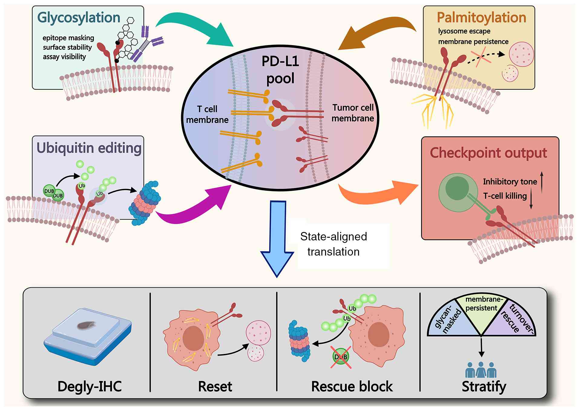
response or immune architecture (78). These convergent PTM axes can
therefore be viewed as a common state-control architecture that
determines whether PD-L1 is maintained as a synapse-competent pool
at the tumor-immune interface, as illustrated in Fig. 2, and how this state may be
translated into biomarker and intervention logic.
N-linked glycosylation is not simply a maturation
detail for PD-L1. Across experimental systems it acts as a
protein-state determinant that influences whether PD-L1 persists in
a surface-available configuration capable of engaging PD-1 or is
diverted toward turnover. Mechanistically, loss of N-glycan
protection can expose PD-L1 to phosphorylation-linked
ubiquitination programs, including a GSK-3β and β-TrCP route,
thereby accelerating clearance and contracting the persistent
surface fraction under inflammatory cycling (79).
Stem-like tumor populations provide a cellular
context in which this glycosylation-dependent stability program can
be selectively reinforced. In breast cancer (BC) stem-like cells,
epithelial-mesenchymal transition activates β-catenin-dependent
transcription of STT3 isoforms, with higher STT3 induction in
stem-like than non-stem populations. STT3-dependent PD-L1
N-glycosylation subsequently protects PD-L1 from
ubiquitin-proteasome-mediated degradation and enriches the stable
PD-L1 protein pool in cancer stem-like cells (14). Genetic depletion of STT3 reduced
PD-L1 enrichment and increased the susceptibility of these cells to
immune-mediated elimination, directly linking a stemness-associated
signaling state to PTM-governed checkpoint competence. This
mechanism illustrates how the same PD-L1 proteostasis axis can
acquire greater functional weight within a therapy-resistant
cellular subpopulation.
The clinical relevance of this layer follows because
glycan state can reshape assay observability. Proteomics and
antibody-binding analyses indicate that PD-L1 glycosylation can
alter recognition by clinically used antibodies, rendering signal
intensity a composite of biology and epitope accessibility
(39). In tissue workflows,
enzymatic deglycosylation has been shown to enhance PD-L1 IHC
detection in validation cohorts, with the effect most evident in
samples that appear low by standard staining (80). In a triple-negative BC cohort
treated with atezolizumab, tumor-cell membrane PD-L1 assessed after
enzymatic deglycosylation demonstrated improved correlation with
clinical response compared with conventional IHC, consistent with
glycan-dependent underestimation in at least a subset of specimens
(81).
These observations also constrain therapeutic
extrapolation. Because N-glycosylation is ubiquitous and context
dependent, broad inhibition is unlikely to be broadly tolerable or
informative. A more defensible translational approach is to treat
glycosylation as a stability and detectability axis, improving
measurement of the relevant PD-L1 state where misclassification is
plausible and prioritizing context-restricted interventions that
weaken the stabilizing program rather than targeting global
glycosylation itself. One proof-of-principle strategy is to target
glycosylated PD-L1 directly, which has been shown to drive
internalization and degradation and to augment antitumor immunity
in preclinical models (73).
Palmitoylation offers a direct entry point into
checkpoint competence by defining how PD-L1 is retained as a
synapse-competent pool at the tumor immune interface. In colorectal
cancer (CRC) models, PD-L1 palmitoylation in the cytosolic domain
stabilizes PD-L1 by limiting ubiquitination and suppressing
lysosome-dependent degradation, and ZDHHC3 is identified as a
principal enzymatic node whose inhibition by 2-bromopalmitate,
genetic depletion, or competitive interference reduces PD-L1
persistence and strengthens antitumor T cell immunity in
vitro and in vivo (8).
A complementary pharmacologic example links this
modification to endomembrane fate. Benzosceptrin C inhibits DHHC3
activity, prevents PD-L1 palmitoylation, and drives PD-L1 away from
membrane retention by blocking return through recycling endosomes
and triggering lysosome-mediated degradation, with enhanced
tumor-infiltrating T cell immunity and improved tumor control,
including in checkpoint blockade combinations (72).
Evidence also supports that palmitoylation-dependent
stabilization can extend beyond a single tumor context, and
clearance programs can independently set the residence time of
PD-L1 at the interface (20,82,83). Translationally, this framework
motivates interventions that collapse the persistent membrane
fraction rather than only lowering transcripts. A stapled peptide
proteolysis-targeting chimera (PROTAC) targeting ZDHHC3 has been
reported to downregulate PD-L1 and to increase effector cytokine
release in tumor and T-cell coculture systems, illustrating one
route to state resetting through the palmitoylation node (84). The operational challenge remains
to develop patient material measurements that report
palmitoylation-linked persistence and to establish when this state
variable is the dominant constraint on checkpoint blockade
depth.
If palmitoylation biases PD-L1 toward membrane
persistence, ubiquitination determines whether that persistence can
be sustained under degradative pressure. Ubiquitin marks encode a
fate decision that commits PD-L1 to regulated disposal, and an E3
ligase axis can drive PD-L1 proteasomal degradation with measurable
consequences for antitumor immunity (85). Deubiquitinases (DUBs) can rewrite
that commitment by removing ubiquitin chains and stabilizing PD-L1
even when degradation programs are engaged, as shown by
OTUB1-dependent protection of PD-L1 from ER-associated degradation
with corresponding effects on CD8 infiltration and tumor immunity
(21). This rationale becomes
especially relevant in inflammation-shaped tumors because
inflammatory cytokines can activate immune pressure while also
strengthening PD-L1 protein stability through CSN5-dependent
deubiquitination (15).
CSN5 provides a concrete illustration of how
inflammatory inputs are translated into a hardened PD-L1 state at
the protein level. In CRC models, macrophage-derived CCL5 induces
CSN5 via p65 and STAT3, and CSN5 deubiquitinates PD-L1 to prolong
ligand availability at the tumor immune interface and suppress
T-cell mediated elimination in vitro and in vivo
(86).
USP22 extends this threshold behavior from a single
node to a reinforcing circuit. USP22 directly deubiquitinates PD-L1
and also stabilizes CSN5 through deubiquitination, creating a
module that sustains PD-L1 protein stability and promotes immune
evasion while remaining partly uncoupled from transcript dynamics
(87). Complementary in
vivo evidence in CD274-amplified settings indicates that USP22
depletion increases tumor immunogenicity and tumor-infiltrating
lymphocytes and improves the efficacy of CD274-targeted
immunotherapy (88).
The development question is therefore best posed in
terms of state specificity. Additional DUB nodes have been reported
to stabilize PD-L1 in a tumor-context dependent manner, and
small-molecule strategies that target DUB dependence directly
provide a route to restore ubiquitin-proteasome disposal of PD-L1
and promote antitumor immunity in immune-competent models (89-91).
Across these three PTM axes, the central question is
whether PD-L1 is maintained as a durable surface-exposed fraction
capable of repeated PD-1 engagement at the tumor-immune interface.
A more actionable view is that these PTMs impose three distinct
constraints on the synapse-competent PD-L1 pool, namely engagement
and assay alignment, membrane persistence, and rescue from turnover
(21,72,85,86,92). Stratification should therefore
focus on the dominant tissue-level constraint rather than on PD-L1
abundance alone.
This convergence helps explain why similar PD-L1
staining intensities can accompany different inhibitory burdens. A
single clinical readout compresses production, turnover and
detectability into one score, and multicenter harmonization efforts
have shown that assay configuration and laboratory workflow can
shift PD-L1 status calls (93).
Clone- and platform-dependent sensitivity differences that move
samples across thresholds have also been documented across tumor
types, underscoring assay alignment as a prerequisite for
stratification (94-96). Consistent with glycan-dependent
detectability as a clinically relevant state variable,
deglycosylated PD-L1 assessment has been reported to align more
closely with ICB outcome than conventional PD-L1 readouts in
real-world BC patient material (78).
A functional PD-L1 state readout shifts
stratification from single regulators to state variables closest to
checkpoint output, including interface competence, surface
persistence and assay alignment. At the translational level, direct
PTM-state readouts such as deglycosylation-sensitive PD-L1 or
palmitoylation-linked persistence should be distinguished from
contextual companion readouts, including spatial immune
architecture or pathway activity scores, which support
interpretation but do not themselves define the PTM state. The
translational question is which constraint dominates in tissue and
therefore which lever is most likely to lower checkpoint tone.
Glycosylation-weighted states prioritize state-aligned measurement
and upstream destabilization rather than global glycosylation
inhibition. Palmitoylation-weighted states motivate state-reset
combinations that erode the persistent membrane reservoir.
Rescue-from-turnover states prioritize protein-state validation in
inflammation-shaped settings where persistence can outpace
transcription. The remaining gap is whether these state variables
converge into reproducible regimes in patient tissue that align
with immune architecture and ICB outcomes across cohorts. Spatial
receptor-ligand proximity offers one practical validation route,
and PD-1 to PD-L1 proximity in mismatch repair-deficient CRCs has
been reported to predict benefit from PD-1 blockade (97).
ICB can amplify antitumor immunity only when
antigenic information is effectively displayed and IFNγ signaling
is sustained enough to reinforce that display under immune pressure
(98-100). The resulting tumor visibility
constraint frequently reflects constraints in protein abundance,
trafficking and signal duration that diverge from transcript-level
immune activation (101,102). At the proximal level, it is
governed by whether peptide MHC complexes are generated and
maintained at relevant cellular interfaces, and whether IFNγ-STAT1
signaling reaches sufficient amplitude and persistence to install
and sustain an immunogenic presentation state in tumor and myeloid
compartments (40). PTMs operate
across both layers of this visibility architecture: They regulate
the routing, stability and surface residence of
antigen-presentation machinery, and they tune the amplitude,
persistence and compartmental enforcement of IFNγ-STAT1 signaling
required to maintain visible tumor states under immune pressure
(22,103).
A recurring clinical paradox is that measurable
immune infiltration does not necessarily translate into benefit
from checkpoint blockade (4,27).
Others remain immunologically cold with low effector density and
primary resistance across checkpoint regimens (4,104). Across these phenotypes, a common
proximate correlate is reduced antigen presentation capacity
together with impaired IFNγ-STAT1 pathway competence, including
failure to maintain antigen presentation outputs under immune
pressure (101,105). Although upstream causes differ,
the operational immune constraint is similar. The immune system
either receives too little antigenic information, or it cannot
sustain the IFN-driven program needed to keep antigen processing,
loading and display active (4).
Two features make this visibility constraint well
suited to a PTM-centered analysis. First, visibility is graded
rather than binary. It depends on the stability and surface
residence of antigen presentation machinery, which can be reset by
post-translational control of ubiquitination, endocytosis and
degradation (101,106). Second, failure modes can be
compartment specific. Tumor cells may downshift MHC class I display
through allele-specific HLA loss, whereas macrophage-rich regions
can reduce MHC class II abundance and antigen presentation to CD4 T
cells under immunosuppressive cues, weakening CD4-dependent support
for durable cytotoxic control (107-109). This compartment logic is
important mechanistically because it suggests that low visibility
can arise from distinct protein state constraints imposed in
different cellular contexts even when bulk transcript-level immune
activation appears superficially similar.
Therapy-resistant stem-like tumor populations
provide one such context in which visibility can be selectively
attenuated. In triple-negative BC, ICB was shown to select LCOR-low
cancer stem cells with reduced antigen-processing and presentation
machinery, thereby promoting immune escape and resistance to
checkpoint therapy (110).
Restoration of LCOR increased antigen-presentation programs and
tumor immunogenicity independently of canonical IFN signaling,
establishing that stem-like state selection can directly alter the
proximal display layer of tumor visibility. Importantly, this
mechanism is transcriptional rather than PTM-driven. Direct
evidence that specific PTM networks install or stabilize
visibility-deficient states selectively within cancer stem cell
populations remains limited. This unresolved interface between
stemness, PTM control, and antigen-presentation competence
represents an important mechanistic gap in the current
tumor-visibility framework.
Within this framework, the tumor visibility axis is
best resolved as a coupled two-layer system encompassing
antigen-presentation competence and IFNγ-STAT1 reinforcement. The
PTM mechanisms emphasized here represent proximal regulatory routes
within this architecture rather than exclusive determinants of
visibility failure. This distinction is supported by evidence that
broader cellular stress states can alter the same visibility
layers. Tumor microenvironmental (TME) peroxynitrite can remodel
the MHC-I-bound peptide repertoire and impair recognition of
peroxynitrite-sensitive antigens by cytotoxic T cells (111), whereas mitochondrial fission can
reduce cancer-cell surface MHC-I through an IRE1α-XBP1s-TPP2
pathway (112). Redox-associated
signaling can also modify IFN-linked responses. In melanoma cells,
IFNγ-induced neuronal nitric oxide synthase (nNOS) and nitric oxide
signaling contributed to STAT1 and STAT3 activation and PD-L1
induction, whereas nNOS inhibition attenuated these responses
(113). PTM-controlled protein
states should therefore be interpreted as mechanistically proximal
and analyzable components of tumor visibility whose functional
weight is conditioned by the broader metabolic, organelle-stress
and redox environment.
The second layer of the tumor visibility
architecture is IFNγ-STAT1 reinforcement, whose competence is
determined by signal amplitude and persistence rather than cytokine
availability alone (114,115).
The IFNγ-STAT1 module must reach sufficient activation and remain
engaged long enough to install and maintain antigen presentation
programs (44,116). These STAT1 kinetics function as
a maintenance signal for antigen processing and presentation
modules and for the turnover of MHC machinery, thereby shaping the
replenishment and surface residence of peptide-MHC (pMHC) complexes
over time (116). PTMs operate
at this control layer. Phosphorylation governs STAT1 activation and
nuclear transcriptional function, whereas ubiquitin-dependent
processes shape signal persistence by controlling complex stability
and turnover of pathway components (117,118).
Operationally, IFNγ pathway failure is often
expressed as kinetic patterns that indicate where the pathway is
gated. A frequent pattern is premature termination. Negative
regulators induced after IFNγ stimulation can recruit ubiquitin
machinery to accelerate turnover of signaling intermediates or
curtail sustained phosphorylation. The result is a brief STAT1
activation pulse that fails to maintain antigen processing and
loading programs (45,46). A corresponding state-aligned
readout is the dwell time of phosphorylated STAT1 together with the
persistence, rather than transient induction, of antigen
presentation modules.
In other contexts, the dominant constraint is
amplitude damping. Excessive dephosphorylation of upstream kinases
or STAT1 can flatten pathway gain and prevent threshold crossing
required for robust antigen presentation and chemokine programs
associated with immune recruitment and retention (119). Readouts that map to this
constraint include STAT1 phosphorylation intensity and nuclear
residency together with IFN response scores anchored to antigen
presentation rather than global inflammation.
A distinct but related constraint is spatial or
compartmental uncoupling. Pathway components can be diverted from
productive signaling locales or retained in non-productive states
that limit sustained transcriptional enforcement. In this setting,
receptor stability control can regulate complex assembly and
residence time, converting IFNγ exposure into ineffective signaling
(120). This framing emphasizes
that IFNγ non-responsiveness is not always explained by gene loss
or absent transcription. In some tumors it can reflect reversible
protein state constraints that are best interpreted using
state-aligned, compartment-aware measurements. Because these
constraints ultimately manifest as altered abundance and
composition of pMHC complexes at the immune interface, readouts
that directly interrogate displayed peptides provide a
complementary perspective on visibility (121,122).
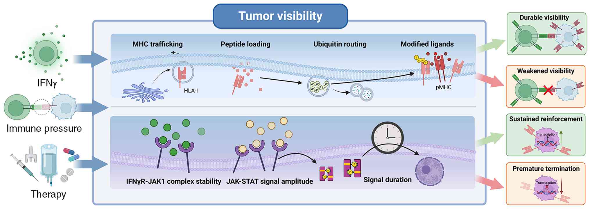
Taken together, tumor visibility is not determined
by antigen presentation alone, but by the coordinated coupling of
the presentation machinery with IFNγ-STAT1 reinforcement, a
two-layer architecture that defines whether antigenic information
is durably displayed at the tumor-immune interface, as summarized
in Fig. 3. In this sense,
antigen-presentation machinery defines the proximal display layer,
whereas IFNγ-STAT1 kinetics defines the reinforcement layer that
determines whether visible states are transiently induced or
durably sustained.
A direct way to read out the output of the
visibility axis is to measure presented peptides rather than
upstream regulators (123,124). This approach quantifies the
peptide cargo actually displayed as pMHC at tumor and myeloid
interfaces and therefore reports visibility more proximally than
transcript-based surrogates (58,125). In this sense, PTM
immuno-peptidomics is valuable not simply because it identifies
modified antigens, but because it reads out the antigenic state
that the immune system is actually being asked to recognize. PTMs
can create or alter epitopes that enter antigen processing, are
loaded onto MHC molecules, and are displayed at immune interfaces
(126). PTMs thus become not
only regulators of visibility, but also components of the displayed
antigenic substrate itself (127).
Phosphorylated peptides provide a parallel PTM class
that can expand the landscape captured by immuno-peptidomics and
expose additional layers of state dependent antigen display
(128). From a biomarker
perspective, PTM immuno-peptidomics provides a readout that is
substantially closer to mechanism than transcript-based surrogates,
because it interrogates the antigenic surface actually available
for immune recognition. It can determine whether tumor and myeloid
compartments present modified epitopes, quantify their abundance,
and support inference about whether antigenic information is being
effectively displayed under a given visibility state (129,130).
In this sense, PTM immuno-peptidomics does more than
catalogue displayed epitopes (51,129). By directly resolving naturally
presented HLA ligands, including modified tumor-associated
peptides, it connects the biochemical state of the tumor proteome
with the antigenic surface that is genuinely available for immune
surveillance (51,131,132). This feature becomes especially
important when transcript-based signatures indicate an immune
active or T-cell inflamed microenvironment yet remain insufficient
to account for effective recognition or durable clinical benefit,
because these outcomes also depend on intact antigen processing,
HLA presentation, and sustained tumor cell visibility to cytotoxic
T cells (29,62,63,100). Viewed in this way, PTM-defined
antigen display serves as a mechanism-proximal readout of the
visibility axis and provides a practical bridge from two-layer
visibility biology to biomarker refinement, stratification and
target prioritization in immunotherapy-oriented settings.
Beyond checkpoint regulation and tumor visibility,
antitumor immunity is also constrained by the ability of immune
cells to preserve functional competence under persistent
microenvironmental stress. This section examines how metabolic,
oxidative, hypoxic and antigenic pressures are translated through
PTM-dependent changes in protein state and signaling behavior, and
how these changes promote either reversible adaptation or more
persistent immune dysfunction.
In immunotherapy-relevant settings, this axis
concerns whether immune cells retain cytotoxic competence, and
whether that competence remains reversible, in microenvironments
shaped by hypoxia, lactate accumulation, mitochondrial oxidative
stress and sustained antigenic stimulation (133-136). Under these conditions, immune
dysfunction is not determined by inflammatory exposure alone, but
by whether stress is translated into durable protein-state
constraints that preserve effector function or stabilize
non-productive states.
To keep mechanistically heterogeneous evidence
interpretable, this chapter organizes the discussion around four
recurring state variables: stability, localization, complex
competence and signaling persistence. Glycosylation,
palmitoylation, phosphorylation and ubiquitin editing are
emphasized because they repeatedly converge on these variables and
thereby provide a practical scale for interpreting how stress is
translated into preserved cytotoxicity or fixed dysfunction
(137-140).
Hypoxia, lactate accumulation, mitochondrial
oxidative stress and sustained antigenic stimulation reshape
substrate availability, redox balance and signaling thresholds in
immune cells. In this framework, nutritional status is therefore
interpreted as an integrated metabolic state defined by nutrient
access, nutrient utilization and nutrient-coupled signaling rather
than by energy availability alone (141).
The metabolic state of the cancer cell is an
upstream determinant of this pressure landscape rather than a
parallel feature of tumor growth. Pharmacologic lactate
dehydrogenase inhibition in solid tumor models reduced glucose
uptake by cancer cells and redirected intratumoral glucose
availability toward infiltrating T cells, with corresponding gains
in T-cell function and antitumor activity (142). Lactate production can also be
translated into more persistent PTM-governed regulatory states. In
GBM, lactate produced by stem-like tumor cells and microglia or
macrophages increased tumor-cell histone lactylation and induced an
immunosuppressive transcriptional program that upregulated CD47 and
suppressed phagocytosis (143).
In pancreatic ductal adenocarcinoma, heightened glycolytic activity
and lactate accumulation promoted ENSA K63 lactylation, sustained
SRC and STAT3 phosphorylation, increased CCL2 production, and
reinforced macrophage-rich immunosuppression and resistance to ICB
(144). These findings position
cancer-cell metabolic routing as a generator of immune-state
pressure and lactylation as one metabolite-sensitive route through
which this pressure can be converted into a more persistent
regulatory state, as discussed further in Section 6.
Glycolytic reprogramming also generates reactive
dicarbonyl pressure beyond lactate accumulation. Methylglyoxal is a
reactive glycolytic byproduct whose biological effects are strongly
dependent on cellular context and exposure level. In glycolytic GBM
and BC models, methylglyoxal challenge induced NRF2 and GLO1
expression and enhanced detoxification capacity, whereas in
vivo exposure produced a biphasic growth response in which
lower concentrations promoted tumor growth and higher
concentrations suppressed growth (145). Methylglyoxal can also impose
discrete protein-fate changes. A recent study showed that
methylglyoxal triggers BRCA2 proteolysis and transiently
compromises BRCA2 tumor-suppressive function, linking glycolytic
carbon overflow to altered protein-state control (146). More broadly, non-enzymatic
glycation by reactive dicarbonyls can increase proteotoxic burden.
Quantitative proteomics identified more than 1,000 protein
carboxymethylation sites and showed that glycation induces a
proteotoxic response with secondary perturbation of
protein-degradation machinery (147).
Carbonyl stress can also suppress immune effectors
directly. Methylglyoxal accumulated in myeloid-derived suppressor
cells and was transferred to CD8+ T cells, imposing
metabolic paralysis, whereas neutralization of dicarbonyl activity
relieved T-cell suppression and improved checkpoint inhibition in
murine tumors (148). Dicarbonyl
stress therefore represents a non-enzymatic route of metabolic
state translation that is chemically distinct from the
enzyme-regulated PTM axes emphasized in the present review.
Accordingly, the PTM effectors considered below should be viewed as
proximal state-control routes embedded within a broader metabolic
and redox environment rather than as exclusive causes of
stress-conditioned immune-state programming.
The key consequence is not simply metabolic stress
itself, but the conversion of stress into altered receptor
availability, membrane organization, and signaling duration,
thereby narrowing the functional margin required to sustain
effector programs (149,150). In this setting, inflammatory
exposure and immune infiltration may coexist with weak cytotoxic
execution because the limiting defect lies in renewable effector
competence rather than immune entry alone (151). A central feature of this axis is
therefore context-dependent directionality. Similar inflammatory
cues may remain compatible with preserved elimination capacity in
relatively permissive environments, but can drive progressive
dysfunction when embedded in hypoxic, oxidatively stressed and
chronically stimulated niches (133,152,153). Under such conditions,
protein-state constraints stabilize non-productive configurations,
shorten productive signaling windows, and favor exhaustion
deepening despite apparent immune activation (154,155). For immunotherapy, this helps
explain why early resistance can emerge even in tumors that remain
inflamed and infiltrated (156).
Glycosylation, palmitoylation, phosphorylation and
ubiquitin editing are considered in the present review as
mechanistic routes that reshape stability, localization, complex
competence and signaling persistence under hypoxia, oxidative
stress and sustained stimulation, thereby influencing whether
cytotoxic programs remain renewable under immunotherapy pressure or
drift toward treatment-refractory dysfunction.
Glycosylation most directly affects stability and
surface residence by controlling receptor and ligand maturation,
trafficking and clearance (73).
Across immune contexts, altered glycosylation states are associated
with changes in effective receptor abundance and activation
threshold (157). Perturbation
studies further show that glycan processing and trimming can
reweight surface retention and ligand availability, thereby
changing the amplitude and duration of downstream signaling
(158). Under stress-conditioned
conditions, this becomes particularly relevant when
surface-competent pools are already limiting, such that even modest
reductions in mature receptor availability can shorten productive
signaling windows and accelerate functional decline.
Palmitoylation most directly affects localization
by controlling membrane partitioning and microdomain residency
(138). Human and ex vivo
data link preserved synapse integrity and membrane organization
with maintained effector function (159). Causal studies further show that
palmitoylation regulates microdomain residency, receptor clustering
and assembly of signaling-competent complexes (160,161). In stressed microenvironments,
this spatial control becomes particularly important because
activation may proceed either through synapse-aligned signaling or
through non-productive membrane configurations that dissipate
signals and weaken persistence.
Phosphorylation provides a dominant route for rapid
threshold control and for imposing temporal structure on
transcriptional output (162).
In numerous immune settings, the distinction between transient
activation and sustained effector reinforcement maps to differences
in phosphorylation kinetics, nuclear routing, and the dwell time of
activated transcription factors (163). Perturbation of kinases,
phosphatases, or adaptor nodes shows that relatively modest changes
in phosphorylation gain can shift cells from sustained signaling to
brief pulses that fail to maintain cytotoxic programs (164). Under stressed microenvironments,
this vulnerability is amplified because energetic limitation and
redox imbalance constrain sustained kinase-driven signaling and
favor premature signal damping (165).
Ubiquitination and deubiquitination control
turnover and signaling duration by editing degradation, recycling
and complex disassembly decisions (166). This role extends beyond antigen
processing and presentation. In mammalian protein quality control,
the UBR4-KCMF1 ubiquitin ligase complex recognizes
mono-ubiquitinated orphan subunits and extends K48-linked
polyubiquitin chains, enabling degradation of unassembled or
partially assembled protein-complex components (167). Ubiquitin-dependent control also
operates at immune-signaling nodes. In CD8+ T cells, a
CUL5 E3 ligase complex acting through PCMTD2 restrains TCR and IL-2
signaling, whereas CUL5 loss enhances TCR and cytokine signaling,
effector function and tumor control (168). Ubiquitin editing should
therefore be interpreted as a protein-fate and signaling-control
system that spans selective proteostasis and immune signaling
rather than as a mechanism restricted to antigen processing. Human
and TME studies associate weakened effector competence with
enhanced negative feedback and accelerated turnover of signaling
intermediates (169). Causal
perturbation studies further indicate that ubiquitin ligases and
DUBs determine complex lifetime and whether signaling is sustained
or prematurely terminated (150). Under chronic stimulation, this
editing function can convert persistent antigen exposure into
short-lived signaling pulses, thereby limiting transcriptional
reinforcement and stabilizing non-effector states (170).
Taken together, these PTM routes support a common
interpretation in which stress-driven immune dysfunction is not
explained by stimulation alone, but by progressive restriction of
stability, localization, complex competence and signaling
persistence.
Persistent antigenic stimulation superimposed on
hypoxia, lactate accumulation, and mitochondrial oxidative stress
can drive immune cells from adaptive dampening into a more fixed
dysfunctional state (133,134,154,171). Once this transition occurs,
limited recovery after checkpoint release is often better explained
by protein-state constraints than by receptor engagement alone
(172). The limiting step
becomes the ability to sustain productive signaling and the
transcriptional reinforcement required for durable effector
maintenance under stress. Reversibility therefore depends on
whether stressed cells can reestablish signaling persistence and
interface competence rather than merely undergo transient
activation, a distinction that helps explain why checkpoint release
may produce incomplete reinvigoration in stress-hardened tumors
(173).
Within the PTM scope considered in the present
review, locking is most directly interpreted through changes in
signaling persistence, membrane organization and protein turnover
(164). Glycosylation can reduce
the effective size of surface-competent pools during chronic
stimulation (73). Palmitoylation
can stabilize or misdirect synapse-relevant membrane partitioning
(160). Phosphorylation kinetics
can be compressed into weak or short-lived pulses when acidic and
metabolic constraints limit pathway gain (150). Ubiquitination and
deubiquitination can accelerate turnover and impose negative
feedback, thereby shortening complex lifetime and converting
persistent inputs into transient signaling (166,174). Together, these processes
reinforce long-lived configurations with reduced signaling
persistence and weakened complex competence, providing a
mechanistic basis for entrenched dysfunction and limited
reprogramming capacity (175).
Functional outcomes under stress are rarely
determined by a single PTM in isolation. Instead, glycosylation,
palmitoylation, phosphorylation and ubiquitin control frequently
converge on the same signaling modules or closely coupled nodes,
generating combinatorial PTM states that determine whether
signaling remains productive or collapses into dysfunction
(176,177).
These combinatorial states can be interpreted
through the same four variables used throughout this chapter
(176). N-glycosylation and
ubiquitin-controlled proteostasis shape the effective abundance and
turnover of key immune regulators. Palmitoylation governs membrane
partitioning and access to productive interfaces at the
immunological synapse (13,178). Phosphorylation, together with
ubiquitin-dependent braking, constrains signaling duration and
reinforcement (138,174,179). Accordingly, similar levels of
immune infiltration or inflammatory exposure may yield markedly
different cytotoxic output, exhaustion depth, and immunotherapy
sensitivity because the underlying PTM configuration differs. This
provides a mechanistic rationale for combination strategies aimed
at restoring renewable effector competence rather than simply
amplifying activation.
Metabolic enzymes further illustrate why PTM
interpretation should remain site- and state-variable specific.
Recent analyses of cancer glycolysis have emphasized that metabolic
enzymes including pyruvate kinase M2 (PKM2), phosphoglycerate
kinase 1 (PGK1), hexokinases, ketohexokinase A, and nucleoside
diphosphate kinases can acquire non-canonical regulatory functions
beyond their canonical metabolic roles (180). Primary studies increasingly
resolve this principle at defined modified residues. In cancer
cells, hydrogen sulfide-dependent sulfhydration of PKM2 at Cys326
destabilizes the tetrameric enzyme, reduces pyruvate kinase
activity, and increases PKM2-mediated transcriptional activation,
whereas blockade of this modification stabilizes the tetramer,
redirects glucose metabolism toward mitochondrial respiration, and
suppresses tumor growth (181).
Site-specific glycosylation provides a parallel mechanism.
O-GlcNAcylation of PGK1 at Thr255 enhances catalytic activity and
promotes mitochondrial translocation, where PGK1 suppresses
pyruvate oxidation and reinforces glycolytic metabolism in colon
cancer (182). Triosephosphate
isomerase (TPI) provides a particularly informative site-resolved
example. In non-small-cell lung cancer, PRKACA-mediated TPI Ser58
phosphorylation enhances enzymatic activity and glycolysis and
promotes tumor growth and metastasis, while elevated Ser58
phosphorylation is detected in multiple human tumors and correlates
with poorer survival (183).
Independent biochemical analysis further showed that
hemi-phosphorylation of the TPI dimer can enhance catalytic
activity, indicating that phosphorylation stoichiometry can
influence enzymatic output (184). A distinct phosphorylation state
couples TPI1 to nuclear metabolism. Nutrient-responsive mTORC1-CDK2
signaling phosphorylates TPI1 at Ser117 and promotes nuclear
translocation, which lowers nuclear dihydroxyacetone phosphate,
permits acetate accumulation, and supports nutrient- and
cell-cycle-dependent global histone acetylation (185). PTM crosstalk can also regulate
TPI1 protein fate. Although demonstrated in idiopathic pulmonary
fibrosis rather than cancer, HDAC11-mediated deacetylation of TPI1
at Lys69 reduces K48-linked polyubiquitination and increases
protein stability, providing cross-context evidence that
acetylation and ubiquitin-dependent turnover can converge on the
same metabolic enzyme (186).
Together, these findings indicate that metabolic enzymes can occupy
PTM-defined states that separately encode catalytic flux,
localization, chromatin coupling and proteostatic stability. Their
direct consequences for antitumor immune escape remain incompletely
resolved, but they identify metabolic enzymes as upstream
protein-state variables capable of reshaping the metabolic and
transcriptional environment in which immune-regulatory PTM programs
operate.
Among the aforementioned state variables,
lactylation is particularly informative because it provides a
mechanistic route through which metabolic pressure can be coupled
to chromatin regulation and translated into durable suppressive
output (10,25,187). This feature distinguishes
lactylation from a simple metabolic correlate, because
lactate-derived histone lactylation can directly stimulate gene
transcription from chromatin and reinforce stable immunosuppressive
programs in tumor-associated myeloid compartments (25,187). By placing a recurrent feature of
the TME at the level of transcriptional stabilization, lactylation
provides a direct framework for linking lactate-rich stress to
persistent changes in immune function (3). In this chapter, lactylation is
therefore considered a bridge mechanism that extends the
aforementioned protein-state logic into a chromatin-coupled layer
of immune regulation, with particular relevance to checkpoint
sensitivity, myeloid control of the effective presentation
environment, and stress-driven rewiring (26).
Lactate enrichment is increasingly recognized as a
recurrent and spatially heterogeneous feature of solid tumors,
arising from uneven metabolic activity and perfusion constraints
(188). Lactylation is discussed
separately not because it is already the most clinically mature PTM
axis in cancer immunity, but because it provides an unusually
coherent bridge between metabolic pressure, chromatin-state
remodeling, and immunotherapy resistance. In this setting,
lactylation is mechanistically informative because it links
metabolic pressure to chromatin regulation and thereby provides a
route by which similar environmental exposure can yield divergent
functional outcomes (10). The
value of this framing is not that lactylation replaces
transcriptional readouts, but that it adds a more proximal layer
for interpreting differences in checkpoint sensitivity, maintenance
of effective tumor visibility, and persistence of cytotoxic
competence.
Histone lactylation provides a concrete basis for
this interpretation. It has been established as a lactate-linked
chromatin mark capable of promoting gene transcription and
connecting glycolytic metabolism to chromatin regulation (10). Studies in immune contexts further
show that lactylation can shape CD8 T-cell metabolic fitness and
effector function and can reinforce immunosuppressive programs in
tumor-associated myeloid compartments (3,25).
Taken together, these observations support a state-based view in
which lactylation is most useful when treated as a translator of
metabolic pressure into durable transcriptional programs rather
than as a passive correlate of lactate exposure.
From a biomarker perspective, these properties make
lactylation particularly relevant as an exploratory stratification
and rewiring-state readout in metabolically stressed tumors with
suppressive immune wiring and reduced immunotherapy sensitivity
(26,189). Because lactate influences
immunity through multiple routes, the discussion in the present
review is restricted to lactylation at the chromatin interface as
the metabolite-coupled path most directly linked to durable
suppressive programming.
This focus should not be interpreted as implying
that lactylation is the only metabolite-sensitive route to
chromatin-state remodeling. Regulated nuclear relocalization and
chromatin recruitment of metabolic enzymes can themselves become
state-control variables that couple metabolic activity to
transcriptional output. Recent native chromatome profiling
identified more than 200 metabolic enzymes within the chromatin
environment and revealed tissue-specific patterns of chromatin
association. Experimental restriction of inosine monophosphate
dehydrogenase 2 to nuclear or cytoplasmic compartments further
produced distinct transcriptional programs, demonstrating that
subcellular localization can add a regulatory layer beyond bulk
enzyme abundance (190). A
PTM-dependent example is provided by ATP-citrate lyase in
endometrial cancer, where AKT-mediated Ser455 phosphorylation
promotes nuclear translocation, increases histone acetylation, and
upregulates pyrimidine-metabolism genes (191). Chromatin recruitment can provide
a complementary route. A nuclear fraction of
methylenetetrahydrofolate dehydrogenase, cyclohydrolase and
formyltetrahydrofolate synthetase 1 (MTHFD1) is recruited to
distinct genomic loci through direct interaction with
bromodomain-containing protein 4 (BRD4), and perturbation of either
MTHFD1 or BRD4 produces related changes in nuclear metabolite
composition and gene expression (192). These findings place regulated
protein localization and chromatin recruitment alongside histone
lactylation as mechanisms through which metabolic state can be
translated into transcriptional reprogramming. Lactylation is
therefore considered in the present review as one defined
chromatin-coupling route rather than the sole metabolite-sensitive
mechanism capable of stabilizing transcriptional state.
Lactylation provides a mechanistic route by which
lactate-associated pressure can be coupled to durable immune state
change (10,25). Two cellular contexts are
especially informative in this respect. Tumor-associated
macrophages and related myeloid cells shape the suppressive
interface through local immune conditioning and support for
productive immune recognition, whereas CD8 T cells determine
whether cytotoxic programs can be maintained under sustained
stimulation. Considered together, these lineages illustrate how
lactylation can reinforce suppression on one side of the interface
while limiting effector persistence on the other.
Within myeloid compartments, the most informative
consequence of lactylation is the stabilization of a suppressive
interface. Under lactate-rich pressure, chromatin-coupled
consolidation of suppressive programs can reduce the capacity of
tumor-infiltrating myeloid cells to sustain productive immune
recognition while reinforcing inhibitory mediator programs that
suppress T-cell activity (25,187). The central functional outcome is
the persistence of an interface in which T cells encounter
insufficient support for durable execution. In this way,
lactylation becomes relevant not simply because it marks metabolic
stress, but because it helps explain how stress is converted into a
myeloid state that remains suppressive even when inflammatory
activity is still detectable.
In CD8 T cells, the most informative consequence is
reduced persistence of cytotoxic competence. Histone lactylation
has been shown to shape CD8 T-cell metabolism and effector
function, and lactylation-aligned tumor programs are associated
with CD8 dysfunction and poor immunotherapy response (3,26).
Under metabolic stress, such coupling may bias CD8 T cells away
from durable effector maintenance and toward states that are
progressively harder to reverse. The resulting picture is not one
in which activation is absent, but one in which activation fails to
remain productive over time. Taken together, these two cellular
contexts support a unified interpretation in which lactylation
couples lactate-rich pressure to durable transcriptional programs
that maintain suppressive interfaces and erode sustained antitumor
killing.
Taken together, lactylation is best positioned in
the present review as an emerging bridge mechanism rather than as a
mature stand-alone clinical axis. Its relevance to checkpoint
competence is indirect and is mediated through chromatin-coupled
suppressive programming rather than through direct control of
ligand-state competence (25,26). Its relevance to tumor visibility
remains hypothesis-generating and should be interpreted as a
plausible link between metabolic stress and weakened
antigen-presentation competence rather than as a fully validated
visibility biomarker class (189). By contrast, its strongest
current support lies in stress-conditioned immune-state
programming, where myeloid suppression and CD8 dysfunction converge
under lactate-rich pressure (3,10,26). In this sense, lactylation is most
appropriately treated at present as an exploratory rewiring-state
readout and mechanistic prioritization node, not yet as a routine
clinical assay class equivalent in maturity to the aforementioned
checkpoint-oriented PTM axes (193,194).
Histone lactylation presents a distinct
pre-analytical challenge because the intended readout is a
metabolically responsive chromatin state rather than a constitutive
protein abundance measure. Isomer-resolved analyses have shown that
lysine L-lactylation is the dominant lactylation isomer on cellular
histones and responds dynamically to glycolytic perturbation
(195). This responsiveness
raises uncertainty as to whether signals measured after variable
tissue acquisition-to-fixation intervals faithfully preserve the
state present at sampling, because lactylation-specific stability
across routine pre-analytical and fixation conditions has not been
systematically qualified. Evidence from other histone PTMs further
shows that pre-analytical stability is modification dependent.
Several histone acetylation marks declined within 24 h of
post-mortem delay, whereas multiple histone methylation marks
remained comparatively stable (196). These observations do not
establish equivalent instability for lactylation, but they argue
against assuming uniform preservation fidelity across histone
modifications.
Analytical identity represents a separate
limitation. Lysine L-lactylation, lysine D-lactylation, and
Nε-carboxyethyl-lysine are structurally related modifications that
require methods capable of resolving their chemical identities
(195). A global lactyl-lysine
signal obtained without isomer-specific qualification should
therefore not automatically be interpreted as chemically resolved
histone L-lactylation. Site-specific readouts introduce an
additional reagent-validation requirement. Recent characterization
of H4K79 and H4K91 lactylation used mass-spectrometry-verified
modified peptides and validated site-specific antibodies by dot
blot and competitive enzyme-linked immunosorbent assay before
mechanistic analysis (197).
Formalin-fixed paraffin-embedded (FFPE) processing creates an
additional concern for mass-spectrometric interpretation.
Comparative analysis of FFPE and fresh-frozen tumor xenografts
identified persistent histone chemical modifications with mass
shifts corresponding to apparent methylation, dimethylation,
acetylation and ubiquitination that were more frequent in FFPE
material (198). This finding
does not demonstrate artifactual lactylation, but it establishes
that fixation-associated histone chemistry can mimic endogenous PTM
mass shifts and supports orthogonal confirmation of site-resolved
lactylation assignments in FFPE specimens.
Archived FFPE tissue remains technically accessible
for histone-state analysis, but analytical feasibility should not
be equated with lactylation-specific biomarker equivalence.
FACT-seq has enabled sensitive profiling of several histone
modifications in archived FFPE tissues and further demonstrated
that optimal epitope-retrieval conditions can differ among histone
marks (199). These findings
support mark-specific FFPE assay development while cautioning
against transferring analytical conditions to lactylation without
direct qualification. For retrospective lactylation cohorts,
incompletely documented acquisition and fixation histories may
therefore confound biological variation with unmeasured
pre-analytical variation. Initial assay qualification should
establish concordance between controlled FFPE and matched fresh or
fresh-frozen material, verify antibody isomer and site specificity,
and incorporate orthogonal mass-spectrometric confirmation in a
representative subset. Because lactylation-associated immune
programs can arise in tumor, myeloid, and lymphocyte compartments
(3,25,26), compartment-resolved detection
should accompany global lactylation measurements. Until
preservation fidelity and assay equivalence are established,
archived FFPE cohorts are best suited to hypothesis generation and
association testing rather than the definition of clinically
portable lactylation thresholds.
The preceding sections support a practical
conclusion. Under sustained metabolic and inflammatory stress, the
limiting determinant of immune control is often not expression
level itself, but the state in which key proteins are stabilized,
positioned and maintained over time. Distinct PTMs can converge on
the same functional constraint by stabilizing inhibitory
checkpoints, weakening effective visibility, or reinforcing
suppressive chromatin programs, even when abundance alone is an
incomplete guide to function (8,13,26,178,189). What becomes clinically
actionable is therefore not the modification class in isolation,
but the dominant state variable through which inhibitory signaling
is maintained, visibility is weakened, or suppressive programs are
stabilized.
From a translational standpoint, intervention
priorities are best organized across the three dominant state
constraints: Checkpoint competence, tumor visibility and
stress-conditioned immune-state programming. One recurrent
situation is continued checkpoint dominance in tumors that already
show immune infiltration and inflammatory activity. In such
settings, the limiting problem is often the persistence of a
functionally competent inhibitory surface state rather than
checkpoint abundance alone. As discussed earlier for PD-L1, the
clinically consequential variable is the fraction that remains
surface resident, interface competent, and available for inhibitory
engagement (8,14,178). Once this state is maintained,
inhibitory signaling can remain dominant even when transcript-level
measures appear similar across tumors. The intervention implication
is therefore to contract the persistent inhibitory surface pool and
shorten the lifetime of synapse competent checkpoint states, rather
than to rely on bulk reduction in expression as a sufficient
objective. Different modification layers may influence this state
through effects on membrane residence, partitioning, or turnover,
but the translational question remains the same, namely whether
functional inhibitory availability can be reduced enough to permit
durable reinvigoration. Readouts are most informative when they
remain close to this biology and capture surface competence,
routing and turnover behavior, and interface level engagement in
the relevant compartments, together with immune proximal endpoints
that reflect persistence of cytotoxic programs rather than
transient activation alone.
A second situation is characterized by limited
immune visibility, in which immune cells may be present but
recognition and amplification remain insufficient for durable
control. Here the key constraint lies in the effective presentation
interface and in the persistence of IFN-linked reinforcement.
Antigen processing and presentation may be induced without being
durably maintained, and IFN exposure may occur without sustained
transcriptional enforcement of the programs needed for recognition,
recruitment and execution (189,200,201). In this setting, intervention is
most appropriately directed toward stabilizing presentation
competence and prolonging IFN-linked reinforcement rather than
merely increasing inflammatory tone. This perspective is important
because different modification dependent mechanisms can converge on
the same visibility problem by weakening antigen-presenting
machinery, damping pathway gain, or accelerating termination of
reinforcing signals. The clinically relevant question is therefore
whether the tumor becomes durably visible to effectors, not simply
whether inflammatory signals can be transiently detected. The most
informative readouts in this setting should therefore reflect
presentation competence, persistence of IFN responsive programs,
and preservation of effector access to tumor nests. When available,
measurements closer to the displayed antigenic interface can
further strengthen this interpretation.
A third situation emerges when suppressive
programming becomes stabilized early and proves increasingly
resistant to reversal. In this context, the central problem is not
only checkpoint signaling or poor visibility, but the progressive
writing of stress into durable transcriptional states that erode
cytotoxic persistence despite ongoing stimulation. The discussion
of metabolite sensitive chromatin coupling is particularly relevant
here because it provides a mechanism by which sustained
lactate-rich pressure can be converted into durable suppressive
output (10). Lactylation offers
a coherent example of this transition. Here, however, lactylation
should be interpreted as a mechanistically coherent bridge example
rather than as evidence that all rewiring-state biomarkers are
already equally mature for routine deployment or therapeutic
targeting. Its translational importance lies less in lactate
exposure itself than in whether a chromatin-coupled suppressive
state has formed in myeloid and CD8 compartments and whether that
state is already constraining effector persistence (26,202). Intervention in this setting is
therefore most appropriately conceived as preventing early fixation
of stress imprinted programs and resetting chromatin-coupled
suppressive states before they become clinically entrenched. The
most informative readouts are those that capture early switching in
myeloid and CD8 compartments and interpret lactylation-aligned
chromatin signals together with suppressive and effector modules
that distinguish durable rewiring from transient fluctuation.
These dominant constraints do not carry equal
weight across treatment phases. Before therapy, the central task is
to identify which state is most limiting and to select an initial
combination logic accordingly. During treatment, the priority
shifts to detecting early state changes that indicate movement
toward durable control or toward suppressive rewiring. At relapse,
the key question becomes whether the observed program remains
reversible or has acquired a more fixed character that will require
strategies directed at persistence rather than acute
activation.
The deployment of PTM-state biomarkers is
constrained not only by biological complexity but also by
analytical barriers that directly shape interpretability and
clinical use (203-205). Sample compatibility is a first
limitation, because some PTM readouts are best captured in fresh or
rapidly processed material, whereas others must ultimately be
inferred from formalin-fixed tissues under crosslinking-related
constraints on molecular resolution (206-208). Assay dependence is equally
important, as IHC, mass spectrometry, spatial proteomics, and
immuno-peptidomics interrogate different analytical layers of the
same biology, making platform choice part of the biomarker
definition rather than a secondary technical detail (51,209). PTM lability adds a further layer
of difficulty, particularly for states influenced by turnover,
metabolic flux, or compartmental redistribution, where
pre-analytical handling can alter the signal that is intended to be
captured (208,209). Finally, cross-context
transferability cannot be assumed, because a PTM state that is
informative at one treatment phase or in one sampling context may
not retain the same meaning in another (205,210). For this reason, PTM-centered
biomarker development requires analytically disciplined readout
design in which intended use, sample context and platform
constraints are specified in advance rather than addressed
retrospectively. These general constraints become especially
consequential when candidate readouts introduce selective
enrichment or biochemical pretreatment into established analytical
workflows. HLA-I immuno-peptidomics and deglycosylation-assisted
PD-L1 IHC illustrate distinct standardization problems in the
transition from discovery-oriented measurement to multicenter
clinical deployment.
For HLA-I immuno-peptidomic readouts,
standardization must encompass upstream sample and ligand
processing, LC-MS/MS acquisition and computational analysis as an
integrated analytical procedure. Different immunoprecipitation
formats can recover substantial method-exclusive ligand subsets
with distinct physicochemical properties despite broadly comparable
overall peptide yields, indicating that upstream isolation can
alter the apparent immunopeptidome rather than only its analytical
depth (211). The challenge is
greater for PTM-bearing ligands, because modified peptides may
occur at low stoichiometric abundance and the modification space
permitted during database searching can directly constrain their
identification (126).
Computational processing is an additional source of
non-equivalence. Direct benchmarking of four data-independent
acquisition pipelines demonstrated differences in immunopeptidome
coverage, replicate reproducibility and experimental false-positive
control (212), whereas
large-scale aggregation of heterogeneous immuno-peptidomic datasets
has required explicit batch-effect control and data-type-specific
processing workflows before ligand integration (213). Multicenter translation will
therefore require prespecified immunoaffinity workflows, common
quality-control criteria, harmonized acquisition and computational
procedures, and reporting rules that distinguish exploratory
peptide discovery from analytically validated PTM-state readouts.
Semi-automated HLA isolation and data-independent acquisition
workflows provide a practical route to reduce manual handling and
increase throughput (214).
Formal cross-platform and interlaboratory bridging will
nevertheless be required before such measurements can be treated as
clinically interchangeable.
Deglycosylation-assisted PD-L1 IHC presents a
different standardization problem because enzymatic pretreatment
becomes part of the analytical procedure used to define the
readout. A dedicated patient-sample protocol has incorporated
enzymatic removal of N-glycans from FFPE tissue sections into the
conventional IHC workflow before PD-L1 assessment (215). The analytical consequence of
deglycosylation is also dependent on antibody recognition
characteristics. Pre-analytical stress and deglycosylation can
differentially affect PD-L1 antibodies according to the location
and conformational properties of their binding epitopes (216). Consistent with this principle,
deglycosylation increased PD-L1 detection with 28-8, CAL10 and
SP142 but slightly reduced detection with 73-10 in lung cancer
tissues, and the same study identified protocol standardization,
clinically applicable cutoffs, and alignment between detection and
therapeutic antibodies as unresolved requirements (71). More broadly, a comparison of 22C3,
28-8 and SP142 across 418 routine clinical specimens found
concordant positive or negative tumor-cell classification with all
three assays in only 60% of cases (95). Multicenter validation of 22C3
laboratory-developed tests further showed that centrally
disseminated protocols did not by themselves ensure diagnostic
accuracy and that iterative protocol modification was required to
improve laboratory performance (217). Thus, deglycosylation conditions,
antibody clone, staining workflow and scoring definition should be
locked as a single assay configuration, with interlaboratory
reproducibility and prospectively defined thresholds established
before routine deployment. Increased signal after deglycosylation
should not by itself be equated with clinical assay
interchangeability. A defined workflow must first demonstrate
reproducible patient classification across laboratories and
clinically relevant cohorts.
An intervention framework centered on state
constraints is only useful if the accompanying measurements track
the biology that actually governs response. The goal is not to
accumulate a large number of loosely related markers, but to define
a compact set of readouts that remain proximal to functional output
across platforms, cohorts and treatment phases. At minimum, a
candidate PTM-state biomarker should satisfy four conditions before
being prioritized for validation: It should map preferentially to
one dominant state constraint, remain close to functional output
rather than distal correlation, be measurable in a prespecified
specimen type and treatment window, and carry a plausible
consequence for baseline stratification, pharmacodynamic
interpretation, or relapse-state attribution.
Within this framework, checkpoint competence is
more informatively represented by readouts that remain close to the
functional inhibitory interface, including surface-accessible PD-L1
state, membrane persistence and effective receptor-ligand
engagement, rather than by bulk ligand abundance measures. By
contrast, the visibility axis is best captured by durable
antigen-presentation competence, sustained IFN-responsive
reinforcement, and evidence that effectors can productively engage
tumor tissue, because benefit tracks with preservation of these
programs whereas resistant lesions recurrently lose them (29,43,66,98,218).
The rewiring axis is best represented by
chromatin-linked suppressive remodeling, including
lactylation-aligned signals in the relevant myeloid and CD8
compartments, because histone lactylation has been directly
implicated in immunosuppressive macrophage activity and in
transcriptional control of CD8 T-cell function (3,25).
Spatial heterogeneity should be treated as a
validation variable rather than an optional contextual layer when
PTM-state biomarkers are used to assign a dominant state
constraint. Multiregional single-cell profiling has shown that
cellular composition and T-cell states can vary among regions
within the same lesion, whereas selected TME communication networks
remain conserved across regions, indicating that regional
concordance can help distinguish recurrent tumor-level features
from local variation (219).
Spatial analysis under immune-checkpoint therapy further
demonstrated that resistant malignant subclones can occupy distinct
niches characterized by different immunosuppressive
microenvironments (220).
Accordingly, where tissue access permits, validation should
incorporate spatially separated tumor regions and spatially
resolved profiling to determine whether a candidate PTM-state
readout is regionally restricted or recurrent across the lesion.
Spatial discordance should be retained as biologically informative
rather than collapsed into a single binary assignment, whereas
recurrent patterns across regions provide stronger support for
lesion-level dominant-state classification. Temporal heterogeneity
requires a complementary design. Longitudinal single-cell and
spatial multi-omics in melanoma have identified
treatment-associated emergence of resistant tumor subclones and
remodeling of spatial immune structures (221), while paired pretreatment and
early on-treatment profiling has linked expansion of a
TCF4-governed mesenchymal-like tumor state to resistance to ICB
(222). Multiregional assessment
and serial sampling should therefore be considered complementary
components of PTM-state biomarker validation.
A practical validation path follows naturally from
these considerations. A minimal validation path should prespecify
the intended use context, one fit-for-purpose and analytically
valid PTM readout, the companion variables required for biological
interpretation, and the sampling timepoint most relevant to the
intended clinical decision (205,210,223). Pretreatment material is needed
to test whether prespecified state variables stratify subsequent
outcome. Paired on-treatment samples are needed to determine
whether early changes in visibility, checkpoint-relevant immune
state, or suppressive rewiring accompany benefit, as longitudinal
analyses have shown dynamic tumor and microenvironment remodeling
during nivolumab therapy and stronger predictive performance for
on-treatment signatures than for pretreatment signatures (43,66).
Relapse-associated samples are needed to define
whether treatment failure is accompanied by stabilized defects in
IFN responsiveness, antigen presentation, or immune contexture, as
acquired resistance has repeatedly been linked to lesions in IFN
pathway signaling, β2-microglobulin, HLA class I antigen processing
and presentation, and broader genomic or immunophenotypic rewiring
at progression (29,98,218).
Clinical endpoints are therefore most informative
when interpreted together with immune proximal endpoints,
particularly response durability and resistance timing, because the
value of this framework lies in linking measurable state change to
sustained immune control.
To translate the state-centered framework outlined
above into a practical validation strategy, PTM-state-aligned and
companion readouts, assay platforms, sampling windows,
immune-proximal endpoints and validation uses across treatment
phases are summarized in Table
III.
As the preceding sections indicate, the
translational value of PTM biology depends not only on mechanistic
relevance but also on whether a pathway can be modulated with
sufficient selectivity and an acceptable therapeutic window
(224,225). Because PTM-directed
interventions often act close to core proteostasis and signaling
nodes, translational design must weigh biomarker proximity against
systemic liability, rather than treating efficacy logic and
toxicity logic as separable questions. Kinase-directed therapy
provides the most mature example of clinically tractable PTM-linked
intervention, yet long-term clinical experience with tyrosine
kinase inhibitors also demonstrates that effective target
engagement does not preclude substantial treatment-related toxicity
(224-226). Epigenetic regulators pose a
related but distinct challenge. Chromatin-directed therapies can
reshape tumor and immune states, and clinical studies combining
epigenetic agents with ICB have underscored the practical
importance of tolerability, schedule and dose optimization
(227,228). More recently, targeted protein
degradation has expanded the translational scope of PTM biology by
harnessing the ubiquitin-proteasome system to eliminate
disease-relevant proteins rather than merely inhibit catalytic
activity (229,230). The therapeutic window is likely
to narrow as intervention moves from a substrate-selective PTM
dependency toward broadly shared proteostasis or phosphorylation
machinery.
Modification-defined proteoforms may provide a more
selective therapeutic entry point when a disease-enriched protein
state creates a pharmacologically distinct vulnerability. In Jurkat
T-cell acute lymphoblastic leukemia cells, deamidated TPI
accumulated under the experimental conditions and was not detected
in the normal T lymphocytes examined. Disulfiram and curcumin
preferentially bound and inhibited recombinant deamidated TPI,
while cellular exposure reduced endogenous TPI activity and
viability in Jurkat cells without significant corresponding effects
in normal T lymphocytes. Sodium dichloroacetate pretreatment
further enhanced the cytotoxic effects of both compounds and
reduced the concentrations required to achieve comparable cytotoxic
activity (231). Complementary
evidence from BC models showed accumulation of deamidated human TPI
in MDA-MB-231 cells but not primary mammary epithelial cells.
Rabeprazole and auranofin markedly impaired TPI activity and
viability in the cancer cells while exerting substantially smaller
effects in the normal-cell comparator, and rabeprazole suppressed
tumor growth in a xenograft model (232). These findings support the
concept that disease-enriched deamidated proteoforms can create
molecular-state vulnerabilities that may be more selective than
global inhibition of TPI or broadly shared PTM machinery.
Modification-state-directed intervention is also becoming
experimentally tractable through induced-proximity approaches.
Epidermal growth factor receptor (EGFR)-targeting phosphorylation
targeting chimeras promoted targeted receptor dephosphorylation,
and a covalent construct selective for mutant EGFR inhibited
dysregulated EGFR signaling and reduced cancer-cell viability
(233). Collectively, these
studies identify molecular-state selectivity as a plausible route
to therapeutic-window engineering, while emphasizing that
proteoform enrichment, on-target dependence, and selectivity in
relevant normal tissues require direct validation before clinical
extrapolation. However, these examples primarily establish the
feasibility of molecular-state-selective intervention in cancer
cells; their direct relevance to antitumor immunity, immunotherapy
response, or clinically improved immunotherapeutic windows remains
to be demonstrated.
Clinical proteasome inhibition provides a
cautionary boundary case for the systemic liability of broad
proteostasis intervention. In the phase III APEX trial, bortezomib
improved outcomes in relapsed multiple myeloma, but grade 3 or 4
adverse events occurred in 75% of treated patients (234). Phosphorylation-directed therapy
presents a distinct selectivity problem because clinically
evaluated kinase drugs frequently engage broader target spectra
than their nominal targets. Chemical proteomic profiling of 243
clinically evaluated kinase drugs identified previously
unrecognized targets and both kinase and non-kinase off-target
interactions (235). At the
pathway level, the phase III BELLE-3 trial provides a separate
example of the therapeutic-window challenge. The pan-class I PI3K
inhibitor buparlisib improved progression-free survival, yet grade
3 or 4 transaminase elevations and hyperglycaemia were frequent,
and its safety profile did not support further development in that
setting (236). These
observations argue against inferring clinical tractability from
pathway-level target engagement alone. Targeted degradation may
improve selectivity, but this advantage is conditional rather than
intrinsic. Mutant-selective BRAF PROTACs preferentially degraded
mutant BRAF while largely sparing wild-type RAF proteins through
differential ternary-complex formation in cells (237), whereas a trastuzumab-PROTAC
conjugate restricted BRD4 degradation to HER2-positive BC cells
while sparing HER2-negative cells (238). Conversely, MD-224, originally
developed as an MDM2 degrader, was subsequently shown to degrade
PXR and additional nuclear receptors through a non-canonical
binding surface (239).
Proteome-wide screening has further demonstrated that subtle
chemical changes can markedly alter neo-substrate specificity and
that some degraders induce substantially broader protein regulation
than others (240).
Therapeutic-window engineering should therefore prioritize
substrate or molecular-state selectivity, tumor-directed delivery,
and proteome-wide degradation profiling before targeted degradation
is assumed to reduce systemic toxicity. To make this framework more
operational from a biomarker perspective, Fig. 4 maps dominant state constraints to
the corresponding readout layer, assay layer, and treatment-phase
context in which each state is most informatively captured.
Within the framework proposed in the present
review, rational combination design should therefore be guided by
the prioritized state constraint or combination of co-dominant
constraints rather than by PTM class alone. Checkpoint-dominant
states are most plausibly addressed by strategies that reduce the
persistence or effective engagement of the inhibitory surface pool
(8). Visibility-deficient states
are more likely to benefit from interventions that restore antigen
presentation and preserve IFN-linked reinforcement (24,200). Rewiring-dominant states, by
contrast, are more likely to require early interruption of
suppressive stabilization before dysfunction becomes fixed
(26). Toxicity should
accordingly be treated as part of the design logic rather than as a
downstream complication, because clinically useful translation will
depend on demonstrating that the intended PTM-state dependency can
be perturbed at exposures that remain tolerable in relevant normal
tissues and in combination regimens that avoid unnecessary overlap
of immune-mediated or organ-specific adverse effects. A
toxicity-aware framework is therefore essential if PTM-directed
strategies are to move from mechanistic plausibility toward
clinically usable combination design.
With these resource layers in place, a supporting
practical task is to determine which PTM-linked signals merit
downstream validation, and which should remain at the level of
mechanistic interest. Once a dominant state constraint and
candidate PTM readout have been defined, PTM-aware public-data
analysis can serve as a compact and reproducible framework for
prioritizing features for deeper functional and clinical
validation. In practice, candidate PTM enzymes, readers, or
modification-sensitive substrates can first be anchored to curated
PTM resources that provide site-level annotation and
regulator-substrate relationships (57). These candidates can then be
evaluated in proteogenomic cohorts that integrate proteomic,
phospho-proteomic, transcriptomic and clinical layers, allowing
alignment with immune phenotypes, pathway activity and clinical
state (49,50). Proteogenomic concordance alone
does not establish whether a candidate PTM directly alters
molecular recognition or complex stability. Structural
bioinformatics can therefore provide a complementary triage layer
between molecular association and empirical validation. For
glycosylated proteins, PTM-aware structural reconstruction and
glycan conformational modeling can restore missing
three-dimensional glycan information and estimate
modification-dependent changes in protein surface accessibility
(241,242). Molecular modeling of
glycosylated TIM-3 further showed that N-glycan conformation and
composition can alter ligand stabilization at an immune-checkpoint
binding site, illustrating the value of docking-based analysis for
prioritizing modification-sensitive binding sites (243). For palmitoylated membrane
proteins, membrane-aware molecular dynamics can resolve structural
effects that depend on bilayer context. In PD-L1, microsecond-scale
all-atom simulations supported by FRET and immune-eliminating
experiments linked palmitoylation-dependent raft partitioning to
altered membrane orientation and PD-1 association (244). Structural predictions should
therefore be used to prioritize PTM-state mechanisms with plausible
effects on epitope accessibility, ligand recognition, or complex
assembly. They do not establish functional causality and require
biochemical, biophysical and cell-based confirmation before
translational prioritization. Where available, immuno-peptidomic
atlases can determine whether PTM-linked variation is accompanied
by altered presentation of modified peptides or broader shifts in
displayed antigenic content (51,58). Spatially resolved datasets can
further test whether the inferred states localize to tumor cells,
myeloid compartments, or effector lymphocytes and whether
interface-level organization is linked to immune outcome (52,97).
The analytical value of this strategy lies in
moving beyond descriptive association toward constraint-resolved
inference. A practical validation framework should establish
whether a candidate feature maps preferentially to one dominant
state constraint, remains reproducible across more than one
molecular layer, and tracks with treatment phase in pretreatment,
on-treatment, or progression-associated settings (43,49,66). Features that satisfy these
criteria are better positioned to support biomarker refinement,
patient stratification, and rational combination design. Framed in
this way, public-data and structural analyses provide complementary
triage layers between PTM mechanism and clinically oriented
validation, sharpening candidate prioritization without displacing
fit-for-purpose experimental confirmation.
Several boundaries of the proposed framework should
be acknowledged. First, the five core exemplar PTM axes emphasized
in the present review represent a selective set of mechanistically
supported state-control routes rather than an exhaustive account of
PTM regulation in cancer immunity. Additional modification classes,
non-enzymatic protein-state changes, and higher-order PTM crosstalk
are discussed selectively when they illuminate the same functional
state variables or define the boundaries of the framework; they are
not intended to constitute equally developed core axes of the
synthesis. Second, checkpoint competence, tumor visibility and
stress-conditioned immune-state programming are intended as
translational coordinates rather than a complete taxonomy of
antitumor immunity. Evidence remains uneven across immune
compartments, tumor lineages, and tissue contexts and is currently
weighted toward selected checkpoint, antigen-presentation, T-cell
and myeloid programs. Third, prioritizing a dominant state
constraint necessarily simplifies a biologically coupled and
dynamic system. Multiple constraints may coexist within the same
lesion, differ across spatial compartments, or shift between
pretreatment, on-treatment and resistant states (56,219-222). Accordingly, state assignment
should not force mutually exclusive classification: Mixed,
co-dominant and indeterminate states should be retained when the
available spatial, molecular, or longitudinal evidence does not
support a single predominant constraint. Finally, much of the
mechanistic evidence discussed in the present review derives from
defined experimental models or selected cancer cohorts. Prospective
clinical validation will therefore require prespecified intended
use, analytically valid PTM-state readouts, and longitudinal
sampling strategies aligned with the relevant clinical decision
point (205,210). These boundaries constrain
immediate clinical extrapolation and should be considered when
selecting candidate readouts or intervention targets. The proposed
framework should therefore be regarded as a prioritization strategy
for organizing mechanistically proximal and measurable protein
states rather than as a comprehensive inventory of immune effectors
or PTM-dependent mechanisms. Its translational value will
ultimately depend on whether the proposed state variables remain
reproducible, contextually interpretable, and clinically
informative across independent prospective cohorts (Fig. 5).
The present review argues that the biological and
translational significance of PTMs is best understood through the
immune constraints they regulate, not through an isolated catalogue
of modification chemistries. From this perspective, diverse PTM
programs converge on three interconnected processes: Checkpoint
competence, tumor visibility and stress-conditioned immune-state
programming. Across these processes, distinct PTM classes regulate
a limited set of functional state variables, including protein
stability, subcellular localization, complex assembly and signaling
persistence (245,246). This framework helps explain why
PD-L1 should be interpreted as a protein-state problem rather than
a simple abundance marker, why tumor visibility depends on
sustained antigen-presentation competence and IFN-linked
reinforcement rather than inflammatory tone alone, and why
lactylation represents a mechanistically coherent route by which
metabolic stress is translated into durable immunosuppressive
programs. The central implication is that the most actionable unit
for biomarker discovery, validation and therapeutic intervention is
not the PTM class itself, but the prioritized state constraint, or
combination of co-dominant constraints, that sustains immune
evasion and informs patient stratification across treatment
phases.
Future studies should therefore move beyond static
abundance-based measurements and establish phase-aware biomarker
workflows anchored to functional state across pretreatment,
on-treatment and progression settings. Key priorities include the
discovery of compact PTM-state candidates in spatially and
proteogenomically resolved clinical samples, validation of
fit-for-purpose readouts linked to prioritized or co-dominant state
constraints, and clarification of which PTM-regulated immune states
remain reversible, and which become fixed during resistance.
Equally important is the development of clinically interpretable
biomarker panels that support patient stratification, response
monitoring and rational combination selection rather than
descriptive PTM cataloguing alone. The next decisive step is not
further expansion of PTM inventories, but prospective validation of
compact PTM-state biomarker panels that can guide stratified
immunotherapy in real clinical use contexts.
Not applicable.
JP, BL, ZH and YC conceived the review topic and
designed the overall conceptual framework. JP and BL drafted the
manuscript. RL, CF and LYL contributed to literature collection,
organization and critical revision of the manuscript. ZH and YC
supervised the work and revised the manuscript for important
intellectual content. All authors read and approved the final
version of the manuscript. Data authentication is not
applicable.
Not applicable.
Not applicable.
The authors declare that they have no competing
interests.
Not applicable.
No funding was received.
|
1
|
Ribas A and Wolchok JD: Cancer
immunotherapy using checkpoint blockade. Science. 359:1350–1355.
2018. View Article : Google Scholar : PubMed/NCBI
|
|
2
|
Sharma P, Hu-Lieskovan S, Wargo JA and
Ribas A: Primary, adaptive, and acquired resistance to cancer
immunotherapy. Cell. 168:707–723. 2017. View Article : Google Scholar : PubMed/NCBI
|
|
3
|
Raychaudhuri D, Singh P, Chakraborty B,
Hennessey M, Tannir AJ, Byregowda S, Natarajan SM, Trujillo-Ocampo
A, Im JS and Goswami S: Histone lactylation drives CD8+
T cell metabolism and function. Nat Immunol. 25:2140–2151. 2024.
View Article : Google Scholar : PubMed/NCBI
|
|
4
|
Jiang P, Gu S, Pan D, Fu J, Sahu A, Hu X,
Li Z, Traugh N, Bu X, Li B, et al: Signatures of T cell dysfunction
and exclusion predict cancer immunotherapy response. Nat Med.
24:1550–1558. 2018. View Article : Google Scholar : PubMed/NCBI
|
|
5
|
Deribe YL, Pawson T and Dikic I:
Post-translational modifications in signal integration. Nat Struct
Mol Biol. 17:666–672. 2010. View Article : Google Scholar : PubMed/NCBI
|
|
6
|
Dai X, Gao Y and Wei W: Post-translational
regulations of PD-L1 and PD-1: Mechanisms and opportunities for
combined immunotherapy. Semin Cancer Biol. 85:246–252. 2022.
View Article : Google Scholar
|
|
7
|
Lee HH, Wang YN, Xia W, Chen CH, Rau KM,
Ye L, Wei Y, Chou CK, Wang SC, Yan M, et al: Removal of N-linked
glycosylation enhances PD-L1 detection and predicts anti-PD-1/PD-L1
therapeutic efficacy. Cancer Cell. 36:168–178.e4. 2019. View Article : Google Scholar : PubMed/NCBI
|
|
8
|
Yao H, Lan J, Li C, Shi H, Brosseau JP,
Wang H, Lu H, Fang C, Zhang Y, Liang L, et al: Inhibiting PD-L1
palmitoylation enhances T-cell immune responses against tumours.
Nat Biomed Eng. 3:306–317. 2019. View Article : Google Scholar : PubMed/NCBI
|
|
9
|
Cho KJ and Roche PA: Regulation of MHC
class II-peptide complex expression by ubiquitination. Front
Immunol. 4:3692013. View Article : Google Scholar : PubMed/NCBI
|
|
10
|
Zhang D, Tang Z, Huang H, Zhou G, Cui C,
Weng Y, Liu W, Kim S, Lee S, Perez-Neut M, et al: Metabolic
regulation of gene expression by histone lactylation. Nature.
574:575–580. 2019. View Article : Google Scholar : PubMed/NCBI
|
|
11
|
Britt EC, John SV, Locasale JW and Fan J:
Metabolic regulation of epigenetic remodeling in immune cells. Curr
Opin Biotechnol. 63:111–117. 2020. View Article : Google Scholar : PubMed/NCBI
|
|
12
|
Lemma EY, Letian A, Altorki NK and McGraw
TE: Regulation of PD-L1 trafficking from synthesis to degradation.
Cancer Immunol Res. 11:866–874. 2023. View Article : Google Scholar : PubMed/NCBI
|
|
13
|
Li CW, Lim SO, Xia W, Lee HH, Chan LC, Kuo
CW, Khoo KH, Chang SS, Cha JH, Kim T, et al: Glycosylation and
stabilization of programmed death ligand-1 suppresses T-cell
activity. Nat Commun. 7:126322016. View Article : Google Scholar : PubMed/NCBI
|
|
14
|
Hsu JM, Xia W, Hsu YH, Chan LC, Yu WH, Cha
JH, Chen CT, Liao HW, Kuo CW, Khoo KH, et al: STT3-dependent PD-L1
accumulation on cancer stem cells promotes immune evasion. Nat
Commun. 9:19082018. View Article : Google Scholar : PubMed/NCBI
|
|
15
|
Lim SO, Li CW, Xia W, Cha JH, Chan LC, Wu
Y, Chang SS, Lin WC, Hsu JM, Hsu YH, et al: Deubiquitination and
stabilization of PD-L1 by CSN5. Cancer Cell. 30:925–939. 2016.
View Article : Google Scholar : PubMed/NCBI
|
|
16
|
Cha JH, Yang WH, Xia W, Wei Y, Chan LC,
Lim SO, Li CW, Kim T, Chang SS, Lee HH, et al: Metformin promotes
antitumor immunity via endoplasmic-reticulum-associated degradation
of PD-L1. Mol Cell. 71:606–620.e7. 2018. View Article : Google Scholar : PubMed/NCBI
|
|
17
|
Li Y, Zhang R and Hei H: Advances in
post-translational modifications of proteins and cancer
immunotherapy. Front Immunol. 14:12293972023. View Article : Google Scholar : PubMed/NCBI
|
|
18
|
Zhang H, Yan Q, Jiang S, Hu D, Lu P, Li S,
Sandai D, Zhang H, Zhang W and Zhu C: Protein post-translational
modifications and tumor immunity: A pan-cancer perspective. Phys
Life Rev. 55:142–209. 2025. View Article : Google Scholar : PubMed/NCBI
|
|
19
|
Chan LC, Li CW, Xia W, Hsu JM, Lee HH, Cha
JH, Wang HL, Yang WH, Yen EY, Chang WC, et al: IL-6/JAK1 pathway
drives PD-L1 Y112 phosphorylation to promote cancer immune evasion.
J Clin Invest. 129:3324–3338. 2019. View Article : Google Scholar : PubMed/NCBI
|
|
20
|
Yang Y, Hsu JM, Sun L, Chan LC, Li CW, Hsu
JL, Wei Y, Xia W, Hou J, Qiu Y and Hung MC: Palmitoylation
stabilizes PD-L1 to promote breast tumor growth. Cell Res.
29:83–86. 2019. View Article : Google Scholar
|
|
21
|
Zhu D, Xu R, Huang X, Tang Z, Tian Y,
Zhang J and Zheng X: Deubiquitinating enzyme OTUB1 promotes cancer
cell immunosuppression via preventing ER-associated degradation of
immune checkpoint protein PD-L1. Cell Death Differ. 28:1773–1789.
2021. View Article : Google Scholar
|
|
22
|
Wilson KR, Liu H, Healey G, Vuong V,
Ishido S, Herold MJ, Villadangos JA and Mintern JD: MARCH1-mediated
ubiquitination of MHC II impacts the MHC I antigen presentation
pathway. PLoS One. 13:e02005402018. View Article : Google Scholar : PubMed/NCBI
|
|
23
|
Liu H, Wilson KR, Firth AM, Macri C,
Schriek P, Blum AB, Villar J, Wormald S, Shambrook M, Xu B, et al:
Ubiquitin-like protein 3 (UBL3) is required for MARCH
ubiquitination of major histocompatibility complex class II and
CD86. Nat Commun. 13:19342022. View Article : Google Scholar : PubMed/NCBI
|
|
24
|
Apriamashvili G, Vredevoogd DW, Krijgsman
O, Bleijerveld OB, Ligtenberg MA, de Bruijn B, Boshuizen J, Traets
JJH, D'Empaire Altimari D, van Vliet A, et al: Ubiquitin ligase
STUB1 destabilizes IFNγ-receptor complex to suppress tumor IFNγ
signaling. Nat Commun. 13:19232022. View Article : Google Scholar
|
|
25
|
De Leo A, Ugolini A, Yu X, Scirocchi F,
Scocozza D, Peixoto B, Pace A, D'Angelo L, Liu JKC, Etame AB, et
al: Glucose-driven histone lactylation promotes the
immunosuppressive activity of monocyte-derived macrophages in
glioblastoma. Immunity. 57:1105–1123.e8. 2024. View Article : Google Scholar : PubMed/NCBI
|
|
26
|
Wang R, Li C, Cheng Z, Li M, Shi J, Zhang
Z, Jin S and Ma H: H3K9 lactylation in malignant cells facilitates
CD8+ T cell dysfunction and poor immunotherapy response.
Cell Rep. 43:1149572024. View Article : Google Scholar
|
|
27
|
Hugo W, Zaretsky JM, Sun L, Song C, Moreno
BH, Hu-Lieskovan S, Berent-Maoz B, Pang J, Chmielowski B, Cherry G,
et al: Genomic and transcriptomic features of response to anti-PD-1
therapy in metastatic melanoma. Cell. 165:35–44. 2016. View Article : Google Scholar : PubMed/NCBI
|
|
28
|
Meng K and Lu H: Clinical application of
high-LET radiotherapy combined with immunotherapy in malignant
tumors. Precis Radiat Oncol. 8:42–46. 2024. View Article : Google Scholar : PubMed/NCBI
|
|
29
|
Zaretsky JM, Garcia-Diaz A, Shin DS,
Escuin-Ordinas H, Hugo W, Hu-Lieskovan S, Torrejon DY,
Abril-Rodriguez G, Sandoval S, Barthly L, et al: Mutations
associated with acquired resistance to PD-1 blockade in melanoma. N
Engl J Med. 375:819–829. 2016. View Article : Google Scholar : PubMed/NCBI
|
|
30
|
Mukai K, Konno H, Akiba T, Uemura T,
Waguri S, Kobayashi T, Barber GN, Arai H and Taguchi T: Activation
of STING requires palmitoylation at the Golgi. Nat Commun.
7:119322016. View Article : Google Scholar : PubMed/NCBI
|
|
31
|
Mertins P, Mani DR, Ruggles KV, Gillette
MA, Clauser KR, Wang P, Wang X, Qiao JW, Cao S, Petralia F, et al:
Proteogenomics connects somatic mutations to signalling in breast
cancer. Nature. 534:55–62. 2016. View Article : Google Scholar : PubMed/NCBI
|
|
32
|
Gillette MA, Satpathy S, Cao S,
Dhanasekaran SM, Vasaikar SV, Krug K, Petralia F, Li Y, Liang WW,
Reva B, et al: Proteogenomic characterization reveals therapeutic
vulnerabilities in lung adenocarcinoma. Cell. 182:200–225.e35.
2020. View Article : Google Scholar : PubMed/NCBI
|
|
33
|
Li Y, Porta-Pardo E, Tokheim C, Bailey MH,
Yaron TM, Stathias V, Geffen Y, Imbach KJ, Cao S, Anand S, et al:
Pan-cancer proteogenomics connects oncogenic drivers to functional
states. Cell. 186:3921–3944.e25. 2023. View Article : Google Scholar : PubMed/NCBI
|
|
34
|
Reinfeld BI, Madden MZ, Wolf MM, Chytil A,
Bader JE, Patterson AR, Sugiura A, Cohen AS, Ali A, Do BT, et al:
Cell-programmed nutrient partitioning in the tumour
microenvironment. Nature. 593:282–288. 2021. View Article : Google Scholar : PubMed/NCBI
|
|
35
|
Ma S, Dahabieh MS, Mann TH, Zhao S,
McDonald B, Song WS, Chung HK, Farsakoglu Y, Garcia-Rivera L,
Hoffmann FA, et al: Nutrient-driven histone code determines
exhausted CD8+ T cell fates. Science. 387:eadj30202025.
View Article : Google Scholar
|
|
36
|
Rozalén C, Sangrador I, Avalle S,
Blasco-Benito S, Tzortzi P, Sanz-Flores M, Palomeque JÁ,
Torren-Duran P, Dalmau M, Brunel H, et al: TIM3+ breast
cancer cells license immune evasion during micrometastasis
outbreak. Cancer Cell. 43:1549–1567.e9. 2025. View Article : Google Scholar
|
|
37
|
Guo M, Yuan Z, Jin X, Li X, Deng Y, Zhou
S, Niu R, Bae J, Park JB, Shi B and Yin J: Inhibition of ICAM1
diminishes stemness and enhances antitumor immunity in glioblastoma
via β-catenin/PD-L1 signaling. Nat Commun. 16:86422025. View Article : Google Scholar
|
|
38
|
Chu CW, Čaval T, Alisson-Silva F,
Tankasala A, Guerrier C, Czerwieniec G, Läubli H and Schwarz F:
Variable PD-1 glycosylation modulates the activity of immune
checkpoint inhibitors. Life Sci Alliance. 7:e2023023682024.
View Article : Google Scholar : PubMed/NCBI
|
|
39
|
Benicky J, Sanda M, Brnakova Kennedy Z,
Grant OC, Woods RJ, Zwart A and Goldman R: PD-L1 glycosylation and
its impact on binding to clinical antibodies. J Proteome Res.
20:485–497. 2021. View Article : Google Scholar
|
|
40
|
Manguso RT, Pope HW, Zimmer MD, Brown FD,
Yates KB, Miller BC, Collins NB, Bi K, LaFleur MW, Juneja VR, et
al: In vivo CRISPR screening identifies Ptpn2 as a cancer
immunotherapy target. Nature. 547:413–418. 2017. View Article : Google Scholar : PubMed/NCBI
|
|
41
|
Lee JH, Shklovskaya E, Lim SY, Carlino MS,
Menzies AM, Stewart A, Pedersen B, Irvine M, Alavi S, Yang JYH, et
al: Transcriptional downregulation of MHC class I and melanoma
de-differentiation in resistance to PD-1 inhibition. Nat Commun.
11:18972020. View Article : Google Scholar
|
|
42
|
Cho KJ, Walseng E, Ishido S and Roche PA:
Ubiquitination by March-I prevents MHC class II recycling and
promotes MHC class II turnover in antigen-presenting cells. Proc
Natl Acad Sci USA. 112:10449–10454. 2015. View Article : Google Scholar : PubMed/NCBI
|
|
43
|
Riaz N, Havel JJ, Makarov V, Desrichard A,
Urba WJ, Sims JS, Hodi FS, Martín-Algarra S, Mandal R, Sharfman WH,
et al: Tumor and microenvironment evolution during immunotherapy
with nivolumab. Cell. 171:934–949.e16. 2017. View Article : Google Scholar : PubMed/NCBI
|
|
44
|
Luo N, Formisano L, Gonzalez-Ericsson PI,
Sanchez V, Dean PT, Opalenik SR, Sanders ME, Cook RS, Arteaga CL,
Johnson DB and Balko JM: Melanoma response to anti-PD-L1
immunotherapy requires JAK1 signaling, but not JAK2.
Oncoimmunology. 7:e14381062018. View Article : Google Scholar : PubMed/NCBI
|
|
45
|
Liau NPD, Laktyushin A, Lucet IS, Murphy
JM, Yao S, Whitlock E, Callaghan K, Nicola NA, Kershaw NJ and Babon
JJ: The molecular basis of JAK/STAT inhibition by SOCS1. Nat
Commun. 9:15582018. View Article : Google Scholar : PubMed/NCBI
|
|
46
|
Zhang JG, Metcalf D, Rakar S, Asimakis M,
Greenhalgh CJ, Willson TA, Starr R, Nicholson SE, Carter W,
Alexander WS, et al: The SOCS box of suppressor of cytokine
signaling-1 is important for inhibition of cytokine action in vivo.
Proc Natl Acad Sci USA. 98:13261–13265. 2001. View Article : Google Scholar : PubMed/NCBI
|
|
47
|
Alicea Pauneto CDM, Riesenberg BP, Gandy
EJ, Kennedy AS, Clutton GT, Hem JW, Hurst KE, Hunt EG, Green JM,
Miller BC, et al: Intra-tumoral hypoxia promotes CD8+ T
cell dysfunction via chronic activation of integrated stress
response transcription factor ATF4. Immunity. 58:2489–2504.e8.
2025. View Article : Google Scholar
|
|
48
|
Ghoneim HE, Fan Y, Moustaki A, Abdelsamed
HA, Dash P, Dogra P, Carter R, Awad W, Neale G, Thomas PG and
Youngblood B: De novo epigenetic programs inhibit PD-1
blockade-mediated T cell rejuvenation. Cell. 170:142–157.e19. 2017.
View Article : Google Scholar : PubMed/NCBI
|
|
49
|
Petralia F, Ma W, Yaron TM, Caruso FP,
Tignor N, Wang JM, Charytonowicz D, Johnson JL, Huntsman EM, Marino
GB, et al: Pan-cancer proteogenomics characterization of tumor
immunity. Cell. 187:1255–1277.e27. 2024. View Article : Google Scholar : PubMed/NCBI
|
|
50
|
Li Y, Dou Y, Da Veiga Leprevost F, Geffen
Y, Calinawan AP, Aguet F, Akiyama Y, Anand S, Birger C, Cao S, et
al: Proteogenomic data and resources for pan-cancer analysis.
Cancer Cell. 41:1397–1406. 2023. View Article : Google Scholar : PubMed/NCBI
|
|
51
|
Kacen A, Javitt A, Kramer MP, Morgenstern
D, Tsaban T, Shmueli MD, Teo GC, da Veiga Leprevost F, Barnea E, Yu
F, et al: Post-translational modifications reshape the antigenic
landscape of the MHC I immunopeptidome in tumors. Nat Biotechnol.
41:239–251. 2023. View Article : Google Scholar
|
|
52
|
Yeh CY, Aguirre K, Laveroni O, Kim S, Wang
A, Liang B, Zhang X, Han LM, Valbuena R, Bassik MC, et al: Mapping
spatial organization and genetic cell-state regulators to target
immune evasion in ovarian cancer. Nat Immunol. 25:1943–1958. 2024.
View Article : Google Scholar : PubMed/NCBI
|
|
53
|
Xu Y, Wang X, Li Y, Mao Y, Su Y, Mao Y,
Yang Y, Gao W, Fu C, Chen W, et al: Multimodal single cell-resolved
spatial proteomics reveal pancreatic tumor heterogeneity. Nat
Commun. 15:101002024. View Article : Google Scholar : PubMed/NCBI
|
|
54
|
Mateiou C, Lokhande L, Diep LH, Knulst M,
Carlsson E, Ek S, Sundfeldt K and Gerdtsson A: Spatial tumor immune
microenvironment phenotypes in ovarian cancer. NPJ Precis Oncol.
8:1482024. View Article : Google Scholar : PubMed/NCBI
|
|
55
|
Ravi VM, Will P, Kueckelhaus J, Sun N,
Joseph K, Salié H, Vollmer L, Kuliesiute U, von Ehr J, Benotmane
JK, et al: Spatially resolved multi-omics deciphers bidirectional
tumor-host interdependence in glioblastoma. Cancer Cell.
40:639–655.e13. 2022. View Article : Google Scholar : PubMed/NCBI
|
|
56
|
Yu B, Qi C, Liu Z, Ma N, Tian C, Wang Y,
Li C and Li Z: Spatial heterogeneity of PD-L1 expression influence
its assessment in esophageal squamous cell carcinoma. Transl Oncol.
59:1024422025. View Article : Google Scholar : PubMed/NCBI
|
|
57
|
Chung CR, Tang Y, Chiu YP, Li S, Hsieh WK,
Yao L, Chiang YC, Pang Y, Chen GT, Chou KC, et al: dbPTM 2025
update: Comprehensive integration of PTMs and proteomic data for
advanced insights into cancer research. Nucleic Acids Res. 53(D1):
D377–D386. 2025. View Article : Google Scholar :
|
|
58
|
Yi X, Liao Y, Wen B, Li K, Dou Y, Savage
SR and Zhang B: caAtlas: An immunopeptidome atlas of human cancer.
iScience. 24:1031072021. View Article : Google Scholar : PubMed/NCBI
|
|
59
|
Huang X, Gan Z, Cui H, Lan T, Liu Y, Caron
E and Shao W: The SysteMHC Atlas v2.0, an updated resource for mass
spectrometry-based immunopeptidomics. Nucleic Acids Res. 52(D1):
D1062–D1071. 2024. View Article : Google Scholar :
|
|
60
|
Aung TN, Monkman J, Warrell J, Vathiotis
I, Bates KM, Gavrielatou N, Trontzas IP, Tan CW, Fernandez AI,
Moutafi M, et al: Spatial signatures for predicting immunotherapy
outcomes using multi-omics in non-small cell lung cancer. Nat
Genet. 57:2482–2493. 2025. View Article : Google Scholar : PubMed/NCBI
|
|
61
|
Califf RM: Biomarker definitions and their
applications. Exp Biol Med (Maywood). 243:213–221. 2018. View Article : Google Scholar : PubMed/NCBI
|
|
62
|
Huang L, Malu S, McKenzie JA, Andrews MC,
Talukder AH, Tieu T, Karpinets T, Haymaker C, Forget MA, Williams
LJ, et al: The RNA-binding protein MEX3B mediates resistance to
cancer immunotherapy by downregulating HLA-A expression. Clin
Cancer Res. 24:3366–3376. 2018. View Article : Google Scholar : PubMed/NCBI
|
|
63
|
Thompson JC, Davis C, Deshpande C, Hwang
WT, Jeffries S, Huang A, Mitchell TC, Langer CJ and Albelda SM:
Gene signature of antigen processing and presentation machinery
predicts response to checkpoint blockade in non-small cell lung
cancer (NSCLC) and melanoma. J Immunother Cancer. 8:e0009742020.
View Article : Google Scholar : PubMed/NCBI
|
|
64
|
Han J, Dong Y, Zhu X, Reuben A, Zhang J,
Xu J, Bai H, Duan J, Wan R, Zhao J, et al: Assessment of human
leukocyte antigen-based neoantigen presentation to determine
pan-cancer response to immunotherapy. Nat Commun. 15:11992024.
View Article : Google Scholar : PubMed/NCBI
|
|
65
|
Vilain RE, Menzies AM, Wilmott JS,
Kakavand H, Madore J, Guminski A, Liniker E, Kong BY, Cooper AJ,
Howle JR, et al: Dynamic changes in PD-L1 expression and immune
infiltrates early during treatment predict response to PD-1
blockade in melanoma. Clin Cancer Res. 23:5024–5033. 2017.
View Article : Google Scholar : PubMed/NCBI
|
|
66
|
Du K, Wei S, Wei Z, Frederick DT, Miao B,
Moll T, Tian T, Sugarman E, Gabrilovich DI, Sullivan RJ, et al:
Pathway signatures derived from on-treatment tumor specimens
predict response to anti-PD1 blockade in metastatic melanoma. Nat
Commun. 12:60232021. View Article : Google Scholar : PubMed/NCBI
|
|
67
|
Burr ML, Sparbier CE, Chan YC, Williamson
JC, Woods K, Beavis PA, Lam EYN, Henderson MA, Bell CC, Stolzenburg
S, et al: CMTM6 maintains the expression of PD-L1 and regulates
anti-tumour immunity. Nature. 549:101–105. 2017. View Article : Google Scholar : PubMed/NCBI
|
|
68
|
Vranic S and Gatalica Z: PD-L1 testing by
immunohistochemistry in immuno-oncology. Biomol Biomed. 23:15–25.
2023. View Article : Google Scholar :
|
|
69
|
Ye Z, Xiong Y, Peng W, Wei W, Huang L, Yue
J, Zhang C, Lin G, Huang F, Zhang L, et al: Manipulation of PD-L1
endosomal trafficking promotes anticancer immunity. Adv Sci
(Weinh). 10:e22064112023. View Article : Google Scholar :
|
|
70
|
Zhang J, Bu X, Wang H, Zhu Y, Geng Y,
Nihira NT, Tan Y, Ci Y, Wu F, Dai X, et al: Cyclin D-CDK4 kinase
destabilizes PD-L1 via cullin 3-SPOP to control cancer immune
surveillance. Nature. 553:91–95. 2018. View Article : Google Scholar :
|
|
71
|
Mei J, Xu J, Yang X, Gu D, Zhou W, Wang H
and Liu C: A comparability study of natural and deglycosylated
PD-L1 levels in lung cancer: Evidence from immunohistochemical
analysis. Mol Cancer. 20:112021. View Article : Google Scholar : PubMed/NCBI
|
|
72
|
Wang Q, Wang J, Yu D, Zhang Q, Hu H, Xu M,
Zhang H, Tian S, Zheng G, Lu D, et al: Benzosceptrin C induces
lysosomal degradation of PD-L1 and promotes antitumor immunity by
targeting DHHC3. Cell Rep Med. 5:1013572024. View Article : Google Scholar : PubMed/NCBI
|
|
73
|
Li CW, Lim SO, Chung EM, Kim YS, Park AH,
Yao J, Cha JH, Xia W, Chan LC, Kim T, et al: Eradication of
triple-negative breast cancer cells by targeting glycosylated
PD-L1. Cancer Cell. 33:187–201.e10. 2018. View Article : Google Scholar : PubMed/NCBI
|
|
74
|
Shi YY, Fan G, Tan R, Li S, Sun HB, Li R,
Yang M, Gao S, Liu M and Dai MY: Treating ICB-resistant cancer by
inhibiting PD-L1 via DHHC3 degradation induced by cell penetrating
peptide-induced chimera conjugates. Cell Death Dis. 15:7012024.
View Article : Google Scholar : PubMed/NCBI
|
|
75
|
Shi F, Li GJ, Liu Y, Zhou HM, Zhang Y, Wei
SY, Zan BJ, Gao M, Chen FS, Li BX, et al: USP19 deficiency enhances
T-cell-mediated antitumor immunity by promoting PD-L1 degradation
in colorectal cancer. Pharmacol Res. 214:1076682025. View Article : Google Scholar : PubMed/NCBI
|
|
76
|
Dong T, Niu H, Chu Z, Zhou C, Gao Y, Jia
M, Sun B, Zheng X, Zhang W, Zhang J, et al: Targeting VPS18 hampers
retromer trafficking of PD-L1 and augments immunotherapy. Sci Adv.
10:eadp49172024. View Article : Google Scholar : PubMed/NCBI
|
|
77
|
Rimm DL, Han G, Taube JM, Yi ES, Bridge
JA, Flieder DB, Homer R, West WW, Wu H, Roden AC, et al: A
prospective, multi-institutional, pathologist-based assessment of 4
immunohistochemistry assays for PD-L1 expression in non-small cell
lung cancer. JAMA Oncol. 3:1051–1058. 2017. View Article : Google Scholar : PubMed/NCBI
|
|
78
|
Wang SC, Shen YC, Pan MR, Ou-Yang F, Liao
TY, Liu LC, Rau KM, Lu LS, Huang CC, Dai MS, et al: Deglycosylated
PD-L1 is a biomarker for immune checkpoint blockade response: A
real-world study in breast cancer patients in Taiwan. Cancer Lett.
631:2179122025. View Article : Google Scholar : PubMed/NCBI
|
|
79
|
Shu Z, Dwivedi B, Switchenko JM, Yu DS and
Deng X: PD-L1 deglycosylation promotes its nuclear translocation
and accelerates DNA double-strand-break repair in cancer. Nat
Commun. 15:68302024. View Article : Google Scholar : PubMed/NCBI
|
|
80
|
Xu J, Yang X, Mao Y, Mei J, Wang H, Ding J
and Hua D: Removal of N-linked glycosylation enhances PD-L1
detection in colon cancer: Validation research based on
immunohistochemistry analysis. Technol Cancer Res Treat.
20:153303382110194422021. View Article : Google Scholar : PubMed/NCBI
|
|
81
|
Ou-Yang F, Li CL, Chen CC, Shen YC, Moi
SH, Luo CW, Xia WY, Wang YN, Lee HH, Wang LH, et al:
De-glycosylated membrane PD-L1 in tumor tissues as a biomarker for
responsiveness to atezolizumab (Tecentriq) in advanced breast
cancer patients. Am J Cancer Res. 12:123–137. 2022.PubMed/NCBI
|
|
82
|
Wang H, Yao H, Li C, Shi H, Lan J, Li Z,
Zhang Y, Liang L, Fang JY and Xu J: HIP1R targets PD-L1 to
lysosomal degradation to alter T cell-mediated cytotoxicity. Nat
Chem Biol. 15:42–50. 2019. View Article : Google Scholar
|
|
83
|
Zhou Z, Lu Y, Li P, Liu X, Cheng W, Chen
HN, Dai L and Ren H: The lipid-metabolic enzyme HSD17B12 drives
lysosomal degradation of PD-L1 potentiating anti-tumor immunity in
a mouse model. PLoS Biol. 24:e30036032026. View Article : Google Scholar : PubMed/NCBI
|
|
84
|
Shi YY, Wang AJ, Liu XL, Dai MY and Cai
HB: Stapled peptide PROTAC induced significantly greater anti-PD-L1
effects than inhibitor in human cervical cancer cells. Front
Immunol. 14:11932222023. View Article : Google Scholar : PubMed/NCBI
|
|
85
|
Wu Y, Zhang C, Liu X, He Z, Shan B, Zeng
Q, Zhao Q, Zhu H, Liao H, Cen X, et al: ARIH1 signaling promotes
anti-tumor immunity by targeting PD-L1 for proteasomal degradation.
Nat Commun. 12:23462021. View Article : Google Scholar : PubMed/NCBI
|
|
86
|
Liu C, Yao Z, Wang J, Zhang W, Yang Y,
Zhang Y, Qu X, Zhu Y, Zou J, Peng S, et al: Macrophage-derived CCL5
facilitates immune escape of colorectal cancer cells via the
p65/STAT3-CSN5-PD-L1 pathway. Cell Death Differ. 27:1765–1781.
2020. View Article : Google Scholar
|
|
87
|
Wang Y, Sun Q, Mu N, Sun X, Wang Y, Fan S,
Su L and Liu X: The deubiquitinase USP22 regulates PD-L1
degradation in human cancer cells. Cell Commun Signal. 18:1122020.
View Article : Google Scholar : PubMed/NCBI
|
|
88
|
Huang X, Zhang Q, Lou Y, Wang J, Zhao X,
Wang L, Zhang X, Li S, Zhao Y, Chen Q, et al: USP22 deubiquitinates
CD274 to suppress anticancer immunity. Cancer Immunol Res.
7:1580–1590. 2019. View Article : Google Scholar : PubMed/NCBI
|
|
89
|
Wang Z, Kang W, Li O, Qi F, Wang J, You Y,
He P, Suo Z, Zheng Y and Liu HM: Abrogation of USP7 is an
alternative strategy to downregulate PD-L1 and sensitize gastric
cancer cells to T cells killing. Acta Pharm Sin B. 11:694–707.
2021. View Article : Google Scholar : PubMed/NCBI
|
|
90
|
Jingjing W, Wenzheng G, Donghua W, Guangyu
H, Aiping Z and Wenjuan W: Deubiquitination and stabilization of
programmed cell death ligand 1 by ubiquitin-specific peptidase 9,
X-linked in oral squamous cell carcinoma. Cancer Med. 7:4004–4011.
2018. View Article : Google Scholar : PubMed/NCBI
|
|
91
|
Zhang Y, Huang Y, Yu D, Xu M, Hu H, Zhang
Q, Cai M, Geng X, Zhang H, Xia J, et al: Demethylzeylasteral
induces PD-L1 ubiquitin-proteasome degradation and promotes
antitumor immunity via targeting USP22. Acta Pharm Sin B.
14:4312–4328. 2024. View Article : Google Scholar : PubMed/NCBI
|
|
92
|
Kaufman B, Abu-Ahmad M, Radinsky O, Gharra
E, Manko T, Bhattacharya B, Gologan D, Erlichman N, Meshel T, Nuta
Y, et al: N-glycosylation of PD-L1 modulates the efficacy of immune
checkpoint blockades targeting PD-L1 and PD-1. Mol Cancer.
24:1402025. View Article : Google Scholar : PubMed/NCBI
|
|
93
|
Adam J, Le Stang N, Rouquette I, Cazes A,
Badoual C, Pinot-Roussel H, Tixier L, Danel C, Damiola F, Damotte
D, et al: Multicenter harmonization study for PD-L1 IHC testing in
non-small-cell lung cancer. Ann Oncol. 29:953–958. 2018. View Article : Google Scholar : PubMed/NCBI
|
|
94
|
Krigsfeld GS, Prince EA, Pratt J,
Chizhevsky V, William Ragheb J, Novotny J Jr and Huron D: Analysis
of real-world PD-L1 IHC 28-8 and 22C3 pharmDx assay utilisation,
turnaround times and analytical concordance across multiple tumour
types. J Clin Pathol. 73:656–664. 2020. View Article : Google Scholar : PubMed/NCBI
|
|
95
|
Maule JG, Clinton LK, Graf RP, Xiao J,
Oxnard GR, Ross JS and Huang RSP: Comparison of PD-L1 tumor cell
expression with 22C3, 28-8, and SP142 IHC assays across multiple
tumor types. J Immunother Cancer. 10:e0055732022. View Article : Google Scholar : PubMed/NCBI
|
|
96
|
Paliogiannis P, Lobrano R, Bella MA, Fara
A, Uras MG, Pinna MA, Tedde A, Madonia M, Zinellu A and Cossu A:
PD-L1 immunohistochemical expression in bladder urothelial cancer
with SP263, SP142 and 22C3 antibodies: A comparative study. Ann
Diagn Pathol. 69:1522672024. View Article : Google Scholar : PubMed/NCBI
|
|
97
|
Saberzadeh-Ardestani B, Graham RP, McMahon
S, Ahanonu E, Shi Q, Williams C, Hubbard A, Zhang W, Muranyi A, Yan
D, et al: Immune marker spatial distribution and clinical outcome
after PD-1 blockade in mismatch repair-deficient, advanced
colorectal carcinomas. Clin Cancer Res. 29:4268–2477. 2023.
View Article : Google Scholar : PubMed/NCBI
|
|
98
|
Gettinger S, Choi J, Hastings K, Truini A,
Datar I, Sowell R, Wurtz A, Dong W, Cai G, Melnick MA, et al:
Impaired HLA class I antigen processing and presentation as a
mechanism of acquired resistance to immune checkpoint inhibitors in
lung cancer. Cancer Discov. 7:1420–1435. 2017. View Article : Google Scholar : PubMed/NCBI
|
|
99
|
Shin DS, Zaretsky JM, Escuin-Ordinas H,
Garcia-Diaz A, Hu-Lieskovan S, Kalbasi A, Grasso CS, Hugo W,
Sandoval S, Torrejon DY, et al: Primary resistance to PD-1 blockade
mediated by JAK1/2 mutations. Cancer Discov. 7:188–201. 2017.
View Article : Google Scholar
|
|
100
|
Ayers M, Lunceford J, Nebozhyn M, Murphy
E, Loboda A, Kaufman DR, Albright A, Cheng JD, Kang SP, Shankaran
V, et al: IFN-γ-related mRNA profile predicts clinical response to
PD-1 blockade. J Clin Invest. 127:2930–2940. 2017. View Article : Google Scholar : PubMed/NCBI
|
|
101
|
Kaul S, Cole NB, Bandola-Simon J, Lu Y,
Tondini E, Walseng E and Roche PA: CD83 suppresses endogenous
March-I-dependent MHC class II ubiquitination, endocytosis, and
degradation. Proc Natl Acad Sci USA. 122:e25040771222025.
View Article : Google Scholar : PubMed/NCBI
|
|
102
|
Kim HJ, Bandola-Simon J, Ishido S, Wong
NW, Koparde VN, Cam M and Roche PA: Ubiquitination of MHC class II
by March-I regulates dendritic cell fitness. J Immunol.
206:494–504. 2021. View Article : Google Scholar
|
|
103
|
Zhang Y, Chen Y, Liu Z and Lai R: ERK is a
negative feedback regulator for IFN-γ/STAT1 signaling by promoting
STAT1 ubiquitination. BMC Cancer. 18:6132018. View Article : Google Scholar
|
|
104
|
Huang HC, Huang YL, Chen YJ, Wu HY, Hsu
CL, Kao HF, Liao BC, Hsieh MS, Lin NY, Liao YH, et al: The branched
N-glycan of PD-L1 predicts immunotherapy responses in patients with
recurrent/metastatic HNSCC. Oncogenesis. 13:362024. View Article : Google Scholar : PubMed/NCBI
|
|
105
|
Sade-Feldman M, Jiao YJ, Chen JH, Rooney
MS, Barzily-Rokni M, Eliane JP, Bjorgaard SL, Hammond MR, Vitzthum
H, Blackmon SM, et al: Resistance to checkpoint blockade therapy
through inactivation of antigen presentation. Nat Commun.
8:11362017. View Article : Google Scholar : PubMed/NCBI
|
|
106
|
Liu H, Jain R, Guan J, Vuong V, Ishido S,
La Gruta NL, Gray DH, Villadangos JA and Mintern JD: Ubiquitin
ligase MARCH 8 cooperates with CD83 to control surface MHC II
expression in thymic epithelium and CD4 T cell selection. J Exp
Med. 213:1695–1703. 2016. View Article : Google Scholar : PubMed/NCBI
|
|
107
|
McGranahan N, Rosenthal R, Hiley CT, Rowan
AJ, Watkins TBK, Wilson GA, Birkbak NJ, Veeriah S, Van Loo P,
Herrero J, et al: Allele-specific HLA loss and immune escape in
lung cancer evolution. Cell. 171:1259–1271.e11. 2017. View Article : Google Scholar : PubMed/NCBI
|
|
108
|
Mittal SK, Cho KJ, Ishido S and Roche PA:
Interleukin 10 (IL-10)-mediated immunosuppression: March-I
induction regulates antigen presentation by macrophages but not
dendritic cells. J Biol Chem. 290:27158–27167. 2015. View Article : Google Scholar : PubMed/NCBI
|
|
109
|
Alspach E, Lussier DM, Miceli AP,
Kizhvatov I, DuPage M, Luoma AM, Meng W, Lichti CF, Esaulova E,
Vomund AN, et al: MHC-II neoantigens shape tumour immunity and
response to immunotherapy. Nature. 574:696–701. 2019. View Article : Google Scholar : PubMed/NCBI
|
|
110
|
Pérez-Núñez I, Rozalén C, Palomeque JÁ,
Sangrador I, Dalmau M, Comerma L, Hernández-Prat A, Casadevall D,
Menendez S, Liu DD, et al: LCOR mediates interferon-independent
tumor immunogenicity and responsiveness to immune-checkpoint
blockade in triple-negative breast cancer. Nat Cancer. 3:355–370.
2022. View Article : Google Scholar : PubMed/NCBI
|
|
111
|
Tcyganov EN, Sanseviero E, Marvel D, Beer
T, Tang HY, Hembach P, Speicher DW, Zhang Q, Donthireddy LR,
Mostafa A, et al: Peroxynitrite in the tumor microenvironment
changes the profile of antigens allowing escape from cancer
immunotherapy. Cancer Cell. 40:1173–1189.e6. 2022. View Article : Google Scholar : PubMed/NCBI
|
|
112
|
Lei X, Lin H, Wang J, Ou Z, Ruan Y,
Sadagopan A, Chen W, Xie S, Chen B, Li Q, et al: Mitochondrial
fission induces immunoescape in solid tumors through decreasing
MHC-I surface expression. Nat Commun. 13:38822022. View Article : Google Scholar : PubMed/NCBI
|
|
113
|
Tong S, Cinelli MA, El-Sayed NS, Huang H,
Patel A, Silverman RB and Yang S: Inhibition of
interferon-gamma-stimulated melanoma progression by targeting
neuronal nitric oxide synthase (nNOS). Sci Rep. 12:17012022.
View Article : Google Scholar : PubMed/NCBI
|
|
114
|
Marijt KA, Sluijter M, Blijleven L,
Tolmeijer SH, Scheeren FA, van der Burg SH and van Hall T:
Metabolic stress in cancer cells induces immune escape through a
PI3K-dependent blockade of IFNγ receptor signaling. J Immunother
Cancer. 7:1522019. View Article : Google Scholar
|
|
115
|
Benci JL, Xu B, Qiu Y, Wu TJ, Dada H,
Twyman-Saint Victor C, Cucolo L, Lee DSM, Pauken KE, Huang AC, et
al: Tumor interferon signaling regulates a multigenic resistance
program to immune checkpoint blockade. Cell. 167:1540–1554.e12.
2016. View Article : Google Scholar : PubMed/NCBI
|
|
116
|
Chandrasekaran S, Sasaki M, Scharer CD,
Kissick HT, Patterson DG, Magliocca KR, Seykora JT, Sapkota B,
Gutman DA, Cooper LA, et al: Phosphoinositide 3-kinase signaling
can modulate MHC class I and II expression. Mol Cancer Res.
17:2395–2409. 2019. View Article : Google Scholar : PubMed/NCBI
|
|
117
|
Shuai K, Stark GR, Kerr IM and Darnell JE
Jr: A single phosphotyrosine residue of Stat91 required for gene
activation by interferon-gamma. Science. 261:1744–1746. 1993.
View Article : Google Scholar : PubMed/NCBI
|
|
118
|
Lawrence DW and Kornbluth J: E3 ubiquitin
ligase NKLAM ubiquitinates STAT1 and positively regulates
STAT1-mediated transcriptional activity. Cell Signal. 28:1833–1841.
2016. View Article : Google Scholar : PubMed/NCBI
|
|
119
|
Zhu Z, Tang R, Huff S, Kummetha IR, Wang
L, Li N and Rana TM: Small-molecule PTPN2 inhibitors sensitize
resistant melanoma to Anti-PD-1 immunotherapy. Cancer Res Commun.
3:119–129. 2023. View Article : Google Scholar : PubMed/NCBI
|
|
120
|
Mizuno S, Mizuno Y, Abe K, Macy AM, Chong
KK, Kobayashi Y, Hastings KT, Hoon DSB and Bustos MA:
Interferon-gamma-inducible protein 30 prevents IFN-γ-receptor 1
degradation to maintain PD-L1 and MHC-II levels in metastatic
melanoma. Cell Commun Signal. 24:1822026. View Article : Google Scholar
|
|
121
|
Kraemer AI, Chong C, Huber F, Pak H,
Stevenson BJ, Müller M, Michaux J, Altimiras ER, Rusakiewicz S,
Simó-Riudalbas L, et al: The immunopeptidome landscape associated
with T cell infiltration, inflammation and immune editing in lung
cancer. Nat Cancer. 4:608–628. 2023. View Article : Google Scholar : PubMed/NCBI
|
|
122
|
Hoenisch Gravel N, Nelde A, Bauer J,
Mühlenbruch L, Schroeder SM, Neidert MC, Scheid J, Lemke S,
Dubbelaar ML, Wacker M, et al: TOFIMS mass
spectrometry-based immunopeptidomics refines tumor antigen
identification. Nat Commun. 14:74722023. View Article : Google Scholar
|
|
123
|
Salek M, Förster JD, Becker JP, Meyer M,
Charoentong P, Lyu Y, Lindner K, Lotsch C, Volkmar M, Momburg F, et
al: optiPRM: A targeted immunopeptidomics LC-MS workflow with
ultra-high sensitivity for the detection of mutation-derived tumor
neoepitopes from limited input material. Mol Cell Proteomics.
23:1008252024. View Article : Google Scholar :
|
|
124
|
Phulphagar KM, Ctortecka C, Jacome ASV,
Klaeger S, Verzani EK, Hernandez GM, Udeshi ND, Clauser KR, Abelin
JG and Carr SA: Sensitive, high-throughput HLA-I and HLA-II
immunopeptidomics using parallel accumulation-serial fragmentation
mass spectrometry. Mol Cell Proteomics. 22:1005632023. View Article : Google Scholar : PubMed/NCBI
|
|
125
|
Cui Y, Phuong K, Abdelfattah NS, Temple
HM, Maiorino L, Kim BJ, Dye J, Yu KKH, Spranger S, Irvine DJ and
White FM: Quantitative cell type-specific immunopeptidome analysis
of macrophage and tumor coevolution reveals therapeutic MHC-I
peptides in glioblastoma. Cancer Res. 85:4958–4976. 2025.
View Article : Google Scholar : PubMed/NCBI
|
|
126
|
Mangalaparthi KK, Madugundu AK, Ryan ZC,
Garapati K, Peterson JA, Dey G, Prakash A and Pandey A: Digging
deeper into the immunopeptidome: Characterization of
post-translationally modified peptides presented by MHC I. J
Proteins Proteom. 12:151–160. 2021. View Article : Google Scholar : PubMed/NCBI
|
|
127
|
Faridi P, Woods K, Ostrouska S, Deceneux
C, Aranha R, Duscharla D, Wong SQ, Chen W, Ramarathinam SH, Lim Kam
Sian TCC, et al: Spliced Peptides and cytokine-driven changes in
the immunopeptidome of melanoma. Cancer Immunol Res. 8:1322–1334.
2020. View Article : Google Scholar : PubMed/NCBI
|
|
128
|
Solleder M, Racle J, Guillaume P, Coukos
G, Bassani-Sternberg M and Gfeller D: Deciphering the landscape of
phosphorylated HLA-II ligands. iScience. 25:1042152022. View Article : Google Scholar : PubMed/NCBI
|
|
129
|
Molvi Z, Klatt MG, Dao T, Urraca J,
Scheinberg DA and O'Reilly RJ: The landscape of MHC-presented
phosphopeptides yields actionable shared tumor antigens for cancer
immunotherapy across multiple HLA alleles. J Immunother Cancer.
11:e0068892023. View Article : Google Scholar : PubMed/NCBI
|
|
130
|
Engelhard VH, Obeng RC, Cummings KL,
Petroni GR, Ambakhutwala AL, Chianese-Bullock KA, Smith KT, Lulu A,
Varhegyi N, Smolkin ME, et al: MHC-restricted phosphopeptide
antigens: Preclinical validation and first-in-humans clinical trial
in participants with high-risk melanoma. J Immunother Cancer.
8:e0002622020. View Article : Google Scholar : PubMed/NCBI
|
|
131
|
Bassani-Sternberg M, Bräunlein E, Klar R,
Engleitner T, Sinitcyn P, Audehm S, Straub M, Weber J,
Slotta-Huspenina J, Specht K, et al: Direct identification of
clinically relevant neoepitopes presented on native human melanoma
tissue by mass spectrometry. Nat Commun. 7:134042016. View Article : Google Scholar : PubMed/NCBI
|
|
132
|
Fritsche J, Rakitsch B, Hoffgaard F, Römer
M, Schuster H, Kowalewski DJ, Priemer M, Stos-Zweifel V, Hörzer H,
Satelli A, et al: Translating immunopeptidomics to
immunotherapy-decision-making for patient and personalized target
selection. Proteomics. 18:e17002842018. View Article : Google Scholar :
|
|
133
|
Scharping NE, Rivadeneira DB, Menk AV,
Vignali PDA, Ford BR, Rittenhouse NL, Peralta R, Wang Y, Wang Y,
DePeaux K, et al: Mitochondrial stress induced by continuous
stimulation under hypoxia rapidly drives T cell exhaustion. Nat
Immunol. 22:205–215. 2021. View Article : Google Scholar : PubMed/NCBI
|
|
134
|
Peralta RM, Xie B, Lontos K, Nieves-Rosado
H, Spahr K, Joshi S, Ford BR, Quann K, Frisch AT, Dean V, et al:
Dysfunction of exhausted T cells is enforced by MCT11-mediated
lactate metabolism. Nat Immunol. 25:2297–2307. 2024. View Article : Google Scholar : PubMed/NCBI
|
|
135
|
Vardhana SA, Hwee MA, Berisa M, Wells DK,
Yost KE, King B, Smith M, Herrera PS, Chang HY, Satpathy AT, et al:
Impaired mitochondrial oxidative phosphorylation limits the
self-renewal of T cells exposed to persistent antigen. Nat Immunol.
21:1022–1033. 2020. View Article : Google Scholar : PubMed/NCBI
|
|
136
|
Beltra JC, Manne S, Abdel-Hakeem MS,
Kurachi M, Giles JR, Chen Z, Casella V, Ngiow SF, Khan O, Huang YJ,
et al: Developmental relationships of four exhausted
CD8+ T cell subsets reveals underlying transcriptional
and epigenetic landscape mechanisms. Immunity. 52:825–841.e8. 2020.
View Article : Google Scholar
|
|
137
|
Shi X, Zhang D, Li F, Zhang Z, Wang S,
Xuan Y, Ping Y and Zhang Y: Targeting glycosylation of PD-1 to
enhance CAR-T cell cytotoxicity. J Hematol Oncol. 12:1272019.
View Article : Google Scholar : PubMed/NCBI
|
|
138
|
Morrison E, Wegner T, Zucchetti AE,
Álvaro-Benito M, Zheng A, Kliche S, Krause E, Brügger B, Hivroz C
and Freund C: Dynamic palmitoylation events following T-cell
receptor signaling. Commun Biol. 3:3682020. View Article : Google Scholar : PubMed/NCBI
|
|
139
|
Gallagher MP, Conley JM, Vangala P, Garber
M, Reboldi A and Berg LJ: Hierarchy of signaling thresholds
downstream of the T cell receptor and the Tec kinase ITK. Proc Natl
Acad Sci USA. 118:e20258251182021. View Article : Google Scholar : PubMed/NCBI
|
|
140
|
Hu H, Wang H, Xiao Y, Jin J, Chang JH, Zou
Q, Xie X, Cheng X and Sun SC: Otud7b facilitates T cell activation
and inflammatory responses by regulating Zap70 ubiquitination. J
Exp Med. 213:399–414. 2016. View Article : Google Scholar : PubMed/NCBI
|
|
141
|
Guo C, You Z, Shi H, Sun Y, Du X, Palacios
G, Guy C, Yuan S, Chapman NM, Lim SA, et al: SLC38A2 and glutamine
signalling in cDC1s dictate anti-tumour immunity. Nature.
620:200–208. 2023. View Article : Google Scholar : PubMed/NCBI
|
|
142
|
Verma S, Budhu S, Serganova I, Dong L,
Mangarin LM, Khan JF, Bah MA, Assouvie A, Marouf Y, Schulze I, et
al: Pharmacologic LDH inhibition redirects intratumoral glucose
uptake and improves antitumor immunity in solid tumor models. J
Clin Invest. 134:e1776062024. View Article : Google Scholar : PubMed/NCBI
|
|
143
|
Wang S, Huang T, Wu Q, Yuan H, Wu X, Yuan
F, Duan T, Taori S, Zhao Y, Snyder NW, et al: Lactate reprograms
glioblastoma immunity through CBX3-regulated histone lactylation. J
Clin Invest. 134:e1768512024. View Article : Google Scholar : PubMed/NCBI
|
|
144
|
Sun K, Zhang X, Shi J, Huang J, Wang S, Li
X, Lin H, Zhao D, Ye M, Zhang S, et al: Elevated protein
lactylation promotes immunosuppressive microenvironment and
therapeutic resistance in pancreatic ductal adenocarcinoma. J Clin
Invest. 135:e1870242025. View Article : Google Scholar : PubMed/NCBI
|
|
145
|
Nokin MJ, Durieux F, Bellier J, Peulen O,
Uchida K, Spiegel DA, Cochrane JR, Hutton CA, Castronovo V and
Bellahcène A: Hormetic potential of methylglyoxal, a side-product
of glycolysis, in switching tumours from growth to death. Sci Rep.
7:117222017. View Article : Google Scholar : PubMed/NCBI
|
|
146
|
Kong LR, Gupta K, Wu AJ, Perera D,
Ivanyi-Nagy R, Ahmed SM, Tan TZ, Tan SL, Fuddin A, Sundaramoorthy
E, et al: A glycolytic metabolite bypasses 'two-hit' tumor
suppression by BRCA2. Cell. 187:2269–2287.e16. 2024. View Article : Google Scholar
|
|
147
|
Di Sanzo S, Spengler K, Leheis A,
Kirkpatrick JM, Rändler TL, Baldensperger T, Dau T, Henning C,
Parca L, Marx C, et al: Mapping protein carboxymethylation sites
provides insights into their role in proteostasis and cell
proliferation. Nat Commun. 12:67432021. View Article : Google Scholar : PubMed/NCBI
|
|
148
|
Baumann T, Dunkel A, Schmid C, Schmitt S,
Hiltensperger M, Lohr K, Laketa V, Donakonda S, Ahting U,
Lorenz-Depiereux B, et al: Regulatory myeloid cells paralyze T
cells through cell-cell transfer of the metabolite methylglyoxal.
Nat Immunol. 21:555–566. 2020. View Article : Google Scholar : PubMed/NCBI
|
|
149
|
Elia I, Rowe JH, Johnson S, Joshi S,
Notarangelo G, Kurmi K, Weiss S, Freeman GJ, Sharpe AH and Haigis
MC: Tumor cells dictate anti-tumor immune responses by altering
pyruvate utilization and succinate signaling in CD8+ T
cells. Cell Metabol. 34:1137–1150.e6. 2022. View Article : Google Scholar
|
|
150
|
Tsai YL, Arias-Badia M, Kadlecek TA, Lwin
YM, Srinath A, Shah NH, Wang ZE, Barber D, Kuriyan J, Fong L and
Weiss A: TCR signaling promotes formation of an STS1-Cbl-b complex
with pH-sensitive phosphatase activity that suppresses T cell
function in acidic environments. Immunity. 56:2682–2698.e9. 2023.
View Article : Google Scholar
|
|
151
|
Thommen DS, Koelzer VH, Herzig P, Roller
A, Trefny M, Dimeloe S, Kiialainen A, Hanhart J, Schill C, Hess C,
et al: A transcriptionally and functionally distinct
PD-1+ CD8+ T cell pool with predictive
potential in non-small-cell lung cancer treated with PD-1 blockade.
Nat Med. 24:994–1004. 2018. View Article : Google Scholar : PubMed/NCBI
|
|
152
|
Bannoud N, Dalotto-Moreno T, Kindgard L,
García PA, Blidner AG, Mariño KV, Rabinovich GA and Croci DO:
Hypoxia supports differentiation of terminally exhausted CD8 T
cells. Front Immunol. 12:6609442021. View Article : Google Scholar : PubMed/NCBI
|
|
153
|
Schietinger A, Philip M, Krisnawan VE,
Chiu EY, Delrow JJ, Basom RS, Lauer P, Brockstedt DG, Knoblaugh SE,
Hämmerling GJ, et al: Tumor-specific T cell dysfunction is a
dynamic antigen-driven differentiation program initiated early
during tumorigenesis. Immunity. 45:389–401. 2016. View Article : Google Scholar : PubMed/NCBI
|
|
154
|
Philip M, Fairchild L, Sun L, Horste EL,
Camara S, Shakiba M, Scott AC, Viale A, Lauer P, Merghoub T, et al:
Chromatin states define tumour-specific T cell dysfunction and
reprogramming. Nature. 545:452–456. 2017. View Article : Google Scholar : PubMed/NCBI
|
|
155
|
Thommen DS and Schumacher TN: T cell
dysfunction in cancer. Cancer Cell. 33:547–562. 2018. View Article : Google Scholar : PubMed/NCBI
|
|
156
|
Trujillo JA, Sweis RF, Bao R and Luke JJ:
T cell-inflamed versus non-T cell-inflamed tumors: A conceptual
framework for cancer immunotherapy drug development and combination
therapy selection. Cancer Immunol Res. 6:990–1000. 2018. View Article : Google Scholar : PubMed/NCBI
|
|
157
|
Azevedo CM, Xie B, Gunn WG, Peralta RM,
Dantas CS, Fernandes-Mendes H, Joshi S, Dean V, Almeida P, Wilfahrt
D, et al: Reprogramming CD8+ T-cell branched N-glycosylation limits
exhaustion, enhancing cytotoxicity and tumor killing. Cancer
Immunol Res. 13:1655–1673. 2025. View Article : Google Scholar : PubMed/NCBI
|
|
158
|
Sun L, Li CW, Chung EM, Yang R, Kim YS,
Park AH, Lai YJ, Yang Y, Wang YH, Liu J, et al: Targeting
glycosylated PD-1 induces potent antitumor immunity. Cancer Res.
80:2298–2310. 2020. View Article : Google Scholar : PubMed/NCBI
|
|
159
|
Lu B, Sun YY, Chen BY, Yang B, He QJ, Li J
and Cao J: zDHHC20-driven S-palmitoylation of CD80 is required for
its costimulatory function. Acta Pharmacol Sin. 45:1214–1223. 2024.
View Article : Google Scholar : PubMed/NCBI
|
|
160
|
Tewari R, Shayahati B, Fan Y and
Akimzhanov AM: T cell receptor-dependent S-acylation of ZAP-70
controls activation of T cells. J Biol Chem. 296:1003112021.
View Article : Google Scholar : PubMed/NCBI
|
|
161
|
Bieerkehazhi S, Fan Y, West SJ, Tewari R,
Ko J, Mills T, Boehning D and Akimzhanov AM:
Ca2+-dependent protein acyltransferase DHHC21 controls
activation of CD4+ T cells. J Cell Sci. 135:jcs2581862022.
View Article : Google Scholar
|
|
162
|
Voisinne G, Locard-Paulet M, Froment C,
Maturin E, Menoita MG, Girard L, Mellado V, Burlet-Schiltz O,
Malissen B, Gonzalez de Peredo A and Roncagalli R: Kinetic
proofreading through the multi-step activation of the ZAP70 kinase
underlies early T cell ligand discrimination. Nat Immunol.
23:1355–1364. 2022. View Article : Google Scholar : PubMed/NCBI
|
|
163
|
Wither MJ, White WL, Pendyala S, Leanza
PJ, Fowler DM and Kueh HY: Antigen perception in T cells by
long-term Erk and NFAT signaling dynamics. Proc Natl Acad Sci USA.
120:e23083661202023. View Article : Google Scholar : PubMed/NCBI
|
|
164
|
O'Donoghue GP, Bugaj LJ, Anderson W,
Daniels KG, Rawlings DJ and Lim WA: T cells selectively filter
oscillatory signals on the minutes timescale. Proc Natl Acad Sci
USA. 118:e20192851182021. View Article : Google Scholar : PubMed/NCBI
|
|
165
|
Scharping NE, Menk AV, Moreci RS,
Whetstone RD, Dadey RE, Watkins SC, Ferris RL and Delgoffe GM: The
tumor microenvironment represses T cell mitochondrial biogenesis to
drive intratumoral T cell metabolic insufficiency and dysfunction.
Immunity. 45:701–703. 2016. View Article : Google Scholar : PubMed/NCBI
|
|
166
|
Jahan AS, Lestra M, Swee LK, Fan Y, Lamers
MM, Tafesse FG, Theile CS, Spooner E, Bruzzone R, Ploegh HL and
Sanyal S: Usp12 stabilizes the T-cell receptor complex at the cell
surface during signaling. Proc Natl Acad Sci USA. 113:E705–E714.
2016. View Article : Google Scholar : PubMed/NCBI
|
|
167
|
Carrillo Roas S, Yagita Y, Murphy P,
Kurzbauer R, Clausen T, Zavodszky E and Hegde RS: Convergence of
orphan quality control pathways at a ubiquitin chain-elongating
ligase. Mol Cell. 85:815–828.e10. 2025. View Article : Google Scholar : PubMed/NCBI
|
|
168
|
Liao X, Li W, Zhou H, Rajendran BK, Li A,
Ren J, Luan Y, Calderwood DA, Turk B, Tang W, et al: The CUL5 E3
ligase complex negatively regulates central signaling pathways in
CD8+ T cells. Nat Commun. 15:6032024. View Article : Google Scholar
|
|
169
|
Kumar J, Kumar R, Kumar Singh A, Tsakem
EL, Kathania M, Riese MJ, Theiss AL, Davila ML and Venuprasad K:
Deletion of Cbl-b inhibits CD8+ T-cell exhaustion and
promotes CAR T-cell function. J Immunother Cancer. 9:e0016882021.
View Article : Google Scholar
|
|
170
|
Wagle MV, Marchingo JM, Howitt J, Tan SS,
Goodnow CC and Parish IA: The ubiquitin ligase adaptor NDFIP1
selectively enforces a CD8+ T cell tolerance checkpoint
to high-dose antigen. Cell Rep. 24:577–584. 2018. View Article : Google Scholar : PubMed/NCBI
|
|
171
|
Yu YR, Imrichova H, Wang H, Chao T, Xiao
Z, Gao M, Rincon-Restrepo M, Franco F, Genolet R, Cheng WC, et al:
Disturbed mitochondrial dynamics in CD8+ TILs reinforce
T cell exhaustion. Nat Immunol. 21:1540–1551. 2020. View Article : Google Scholar : PubMed/NCBI
|
|
172
|
Pauken KE, Sammons MA, Odorizzi PM, Manne
S, Godec J, Khan O, Drake AM, Chen Z, Sen DR, Kurachi M, et al:
Epigenetic stability of exhausted T cells limits durability of
reinvigoration by PD-1 blockade. Science. 354:1160–1165. 2016.
View Article : Google Scholar : PubMed/NCBI
|
|
173
|
Abdel-Hakeem MS, Manne S, Beltra JC,
Stelekati E, Chen Z, Nzingha K, Ali MA, Johnson JL, Giles JR,
Mathew D, et al: Epigenetic scarring of exhausted T cells hinders
memory differentiation upon eliminating chronic antigenic
stimulation. Nat Immunol. 22:1008–1019. 2021. View Article : Google Scholar : PubMed/NCBI
|
|
174
|
Eggert J, Zinzow-Kramer WM, Hu Y, Kolawole
EM, Tsai YL, Weiss A, Evavold BD, Salaita K, Scharer CD and
Au-Yeung BB: Cbl-b mitigates the responsiveness of naive
CD8+ T cells that experience extensive tonic T cell
receptor signaling. Sci Signal. 17:eadh04392024. View Article : Google Scholar
|
|
175
|
Khan O, Giles JR, McDonald S, Manne S,
Ngiow SF, Patel KP, Werner MT, Huang AC, Alexander KA, Wu JE, et
al: TOX transcriptionally and epigenetically programs
CD8+ T cell exhaustion. Nature. 571:211–218. 2019.
View Article : Google Scholar : PubMed/NCBI
|
|
176
|
Leutert M, Entwisle SW and Villén J:
Decoding post-translational modification crosstalk with proteomics.
Mol Cell Proteomics. 20:1001292021. View Article : Google Scholar : PubMed/NCBI
|
|
177
|
Minguez P, Letunic I, Parca L,
Garcia-Alonso L, Dopazo J, Huerta-Cepas J and Bork P: PTMcode v2: A
resource for functional associations of post-translational
modifications within and between proteins. Nucleic Acids Res.
43(Database Issue): D494–D502. 2015. View Article : Google Scholar :
|
|
178
|
Okada M, Chikuma S, Kondo T, Hibino S,
Machiyama H, Yokosuka T, Nakano M and Yoshimura A: Blockage of core
fucosylation reduces cell-surface expression of PD-1 and promotes
anti-tumor immune responses of T cells. Cell Rep. 20:1017–1028.
2017. View Article : Google Scholar : PubMed/NCBI
|
|
179
|
Hundt M, Tabata H, Jeon MS, Hayashi K,
Tanaka Y, Krishna R, De Giorgio L, Liu YC, Fukata M and Altman A:
Impaired activation and localization of LAT in anergic T cells as a
consequence of a selective palmitoylation defect. Immunity.
24:513–522. 2006. View Article : Google Scholar : PubMed/NCBI
|
|
180
|
Fukushi A, Kim HD, Chang YC and Kim CH:
Revisited metabolic control and reprogramming cancers by means of
the warburg effect in tumor cells. Int J Mol Sci. 23:100372022.
View Article : Google Scholar : PubMed/NCBI
|
|
181
|
Wang RH, Chen PR, Chen YT, Chen YC, Chu
YH, Chien CC, Chien PC, Lo SY, Wang ZL, Tsou MC, et al: Hydrogen
sulfide coordinates glucose metabolism switch through destabilizing
tetrameric pyruvate kinase M2. Nat Commun. 15:74632024. View Article : Google Scholar : PubMed/NCBI
|
|
182
|
Nie H, Ju H, Fan J, Shi X, Cheng Y, Cang
X, Zheng Z, Duan X and Yi W: O-GlcNAcylation of PGK1 coordinates
glycolysis and TCA cycle to promote tumor growth. Nat Commun.
11:362020. View Article : Google Scholar : PubMed/NCBI
|
|
183
|
Duan Y, Li J, Wang F, Wei J, Yang Z, Sun
M, Liu J, Wen M, Huang W, Chen Z, et al: Protein modifications
throughout the lung cancer proteome unravel the cancer-specific
regulation of glycolysis. Cell Rep. 37:1101372021. View Article : Google Scholar : PubMed/NCBI
|
|
184
|
Schachner LF, Soye BD, Ro S, Kenney GE,
Ives AN, Su T, Goo YA, Jewett MC, Rosenzweig AC and Kelleher NL:
Revving an engine of human metabolism: Activity enhancement of
triosephosphate isomerase via hemi-phosphorylation. ACS Chem Biol.
17:2769–2780. 2022. View Article : Google Scholar : PubMed/NCBI
|
|
185
|
Zhang JJ, Fan TT, Mao YZ, Hou JL, Wang M,
Zhang M, Lin Y, Zhang L, Yan GQ, An YP, et al: Nuclear
dihydroxyacetone phosphate signals nutrient sufficiency and cell
cycle phase to global histone acetylation. Nat Metab. 3:859–875.
2021. View Article : Google Scholar : PubMed/NCBI
|
|
186
|
Li Y, Shi X, Zhang F, Zhou X, Zhu X, Chen
J, Fu K, Chen J, Yang J, Chen Z, et al: HDAC11-mediated
deacetylation of triosephosphate isomerase 1 promotes idiopathic
pulmonary fibrosis. Research (Wash D C). 8:09532025.PubMed/NCBI
|
|
187
|
Xiong J, He J, Zhu J, Pan J, Liao W, Ye H,
Wang H, Song Y, Du Y, Cui B, et al: Lactylation-driven
METTL3-mediated RNA m6A modification promotes
immunosuppression of tumor-infiltrating myeloid cells. Mol Cell.
82:1660–1677.e10. 2022. View Article : Google Scholar
|
|
188
|
Tan Y, Tan W, Liang Y, Long Y, Chen S, Hu
Q, Ou Y, Fu J, Chen H, Ren F, et al: Machine learning-enabled
spatial multi-omics uncovers lactate-driven targets and tumor
microenvironmental reprogramming in cancer. NPJ Digit Med.
9:1092025. View Article : Google Scholar : PubMed/NCBI
|
|
189
|
Zhang B, Xu A, Wang H, Yan W, Zhang Q and
Wang S: MPC-mediated lactate production drives histone lactylation
in dendritic cells to affect tumor progression and immunotherapy.
Cell Mol Life Sci. 82:3712025. View Article : Google Scholar : PubMed/NCBI
|
|
190
|
Kourtis S, Gañez Zapater A, Elbæk CR,
Schmidt A, Ghose R, Coll Manzano A, Espinar Calvo L, Guirola M,
Pardo-Lorente N, Garcia-Cao M, et al: Native chromatome profiling
reveals hundreds of metabolic enzymes in the nucleus across
tissues. Nat Commun. 17:16552026. View Article : Google Scholar : PubMed/NCBI
|
|
191
|
Dai M, Yang B, Chen J, Liu F, Zhou Y, Zhou
Y, Xu Q, Jiang S, Zhao S, Li X, et al: Nuclear-translocation of
ACLY induced by obesity-related factors enhances pyrimidine
metabolism through regulating histone acetylation in endometrial
cancer. Cancer Lett. 513:36–49. 2021. View Article : Google Scholar : PubMed/NCBI
|
|
192
|
Sdelci S, Rendeiro AF, Rathert P, You W,
Lin JMG, Ringler A, Hofstätter G, Moll HP, Gürtl B, Farlik M, et
al: MTHFD1 interaction with BRD4 links folate metabolism to
transcriptional regulation. Nat Genet. 51:990–998. 2019. View Article : Google Scholar : PubMed/NCBI
|
|
193
|
Liu S, Pan Y, Liu W, Bu X, Shao R, Wang Q,
Wu J, Wu C, Hu W, Xu J, et al: Lactylation-driven MVP upregulation
boosts immunotherapy resistance by inhibiting PD-L1 degradation in
hepatocellular carcinoma. J Immunother Cancer. 13:e0122302025.
View Article : Google Scholar : PubMed/NCBI
|
|
194
|
Zong Z, Zhang L and Zhou F: Lactylation in
cancer: Advances, challenges, and future perspectives. Cancer Res.
85:3192–3195. 2025. View Article : Google Scholar : PubMed/NCBI
|
|
195
|
Zhang D, Gao J, Zhu Z, Mao Q, Xu Z, Singh
PK, Rimayi CC, Moreno-Yruela C, Xu S, Li G, et al: Lysine
L-lactylation is the dominant lactylation isomer induced by
glycolysis. Nat Chem Biol. 21:91–99. 2025. View Article : Google Scholar
|
|
196
|
Jarmasz JS, Stirton H, Davie JR and Del
Bigio MR: DNA methylation and histone post-translational
modification stability in post-mortem brain tissue. Clin
Epigenetics. 11:52019. View Article : Google Scholar : PubMed/NCBI
|
|
197
|
Liu J, Zhao L, Yan M, Jin S, Shang L, Wang
J, Wang Q, Zhao S, Shen Z, Liu T, et al: H4K79 and H4K91 histone
lactylation, newly identified lactylation sites enriched in breast
cancer. J Exp Clin Cancer Res. 44:2522025. View Article : Google Scholar : PubMed/NCBI
|
|
198
|
Bauden M, Kristl T, Andersson R,
Marko-Varga G and Ansari D: Characterization of histone-related
chemical modifications in formalin-fixed paraffin-embedded and
fresh-frozen human pancreatic cancer xenografts using LC-MS/MS. Lab
Invest. 97:279–288. 2017. View Article : Google Scholar
|
|
199
|
Zhao L, Xing P, Polavarapu VK, Zhao M,
Valero-Martínez B, Dang Y, Maturi N, Mathot L, Neves I, Yildirim I,
et al: FACT-seq: Profiling histone modifications in formalin-fixed
paraffin-embedded samples with low cell numbers. Nucleic Acids Res.
49:e1252021. View Article : Google Scholar : PubMed/NCBI
|
|
200
|
Pitter MR, Kryczek I, Zhang H, Nagarsheth
N, Xia H, Wu Z, Tian Y, Okla K, Liao P, Wang W, et al: PAD4
controls tumor immunity via restraining the MHC class II machinery
in macrophages. Cell Rep. 43:1139422024. View Article : Google Scholar : PubMed/NCBI
|
|
201
|
Zuo Y, Feng Q, Jin L, Huang F, Miao Y, Liu
J, Xu Y, Chen X, Zhang H, Guo T, et al: Regulation of the linear
ubiquitination of STAT1 controls antiviral interferon signaling.
Nat Commun. 11:11462020. View Article : Google Scholar : PubMed/NCBI
|
|
202
|
Ugolini A, De Leo A, Yu X, Scirocchi F,
Liu X, Peixoto B, Scocozza D, Pace A, Perego M, Gardini A, et al:
Functional reprogramming of neutrophils within the brain tumor
microenvironment by hypoxia-driven histone lactylation. Cancer
Discov. 15:1270–1296. 2025. View Article : Google Scholar : PubMed/NCBI
|
|
203
|
Lee JW, Devanarayan V, Barrett YC, Weiner
R, Allinson J, Fountain S, Keller S, Weinryb I, Green M, Duan L, et
al: Fit-for-purpose method development and validation for
successful biomarker measurement. Pharm Res. 23:312–328. 2006.
View Article : Google Scholar : PubMed/NCBI
|
|
204
|
Masucci GV, Cesano A, Hawtin R, Janetzki
S, Zhang J, Kirsch I, Dobbin KK, Alvarez J, Robbins PB, Selvan SR,
et al: Validation of biomarkers to predict response to
immunotherapy in cancer: Volume I-pre-analytical and analytical
validation. J Immunother Cancer. 4:762016. View Article : Google Scholar
|
|
205
|
Dobbin KK, Cesano A, Alvarez J, Hawtin R,
Janetzki S, Kirsch I, Masucci GV, Robbins PB, Selvan SR, Streicher
HZ, et al: Validation of biomarkers to predict response to
immunotherapy in cancer: Volume II-clinical validation and
regulatory considerations. J Immunother Cancer. 4:772016.
View Article : Google Scholar
|
|
206
|
Dressler FF, Schoenfeld J, Revyakina O,
Vogele D, Kiefer S, Kirfel J, Gemoll T and Perner S: Systematic
evaluation and optimization of protein extraction parameters in
diagnostic FFPE specimens. Clin Proteomics. 19:102022. View Article : Google Scholar : PubMed/NCBI
|
|
207
|
Friedrich C, Schallenberg S, Kirchner M,
Ziehm M, Niquet S, Haji M, Beier C, Neudecker J, Klauschen F and
Mertins P: Comprehensive micro-scaled proteome and phosphoproteome
characterization of archived retrospective cancer repositories. Nat
Commun. 12:35762021. View Article : Google Scholar : PubMed/NCBI
|
|
208
|
Vassilakopoulou M, Parisi F, Siddiqui S,
England AM, Zarella ER, Anagnostou V, Kluger Y, Hicks DG, Rimm DL
and Neumeister VM: Preanalytical variables and phosphoepitope
expression in FFPE tissue: quantitative epitope assessment after
variable cold ischemic time. Lab Invest. 95:334–341. 2015.
View Article : Google Scholar
|
|
209
|
Haragan A, Liebler DC, Das DM, Soper MD,
Morrison RD, Slebos RJC, Ackermann BL, Fill JA, Schade AE, Gosney
JR and Gruver AM: Accelerated instability testing reveals
quantitative mass spectrometry overcomes specimen storage
limitations associated with PD-L1 immunohistochemistry. Lab Invest.
100:874–886. 2020. View Article : Google Scholar : PubMed/NCBI
|
|
210
|
Wang W, He Y, Yuan X, Weng L, Bai J, Liu
X, Han L, Xu J, Wang S, Lu S, et al: Protocol for Chinese lung
cancer evolution and microenvironment tracking under therapy study.
J Transl Int Med. 13:599–609. 2025. View Article : Google Scholar : PubMed/NCBI
|
|
211
|
Wacker M, Bauer J, Wessling L, Dubbelaar
M, Nelde A, Rammensee HG and Walz JS: Immunoprecipitation methods
impact the peptide repertoire in immunopeptidomics. Front Immunol.
14:12197202023. View Article : Google Scholar : PubMed/NCBI
|
|
212
|
Shahbazy M, Ramarathinam SH, Illing PT,
Jappe EC, Faridi P, Croft NP and Purcell AW: Benchmarking
bioinformatics pipelines in data-independent acquisition mass
spectrometry for immunopeptidomics. Mol Cell Proteomics.
22:1005152023. View Article : Google Scholar : PubMed/NCBI
|
|
213
|
Ran Z, Mu M, Lin S, Wang T, Zeng J, Kuang
L, Chen K, Suo S, Yuan K and Xu H: Deciphering the MHC
immunopeptidome of human cancers with ligand.MHC atlas. Brief
Bioinform. 26:bbaf3142025. View Article : Google Scholar : PubMed/NCBI
|
|
214
|
Lim Kam Sian TCC, Goncalves G, Steele JR,
Shamekhi T, Bramberger L, Jin D, Shahbazy M, Purcell AW,
Ramarathinam S, Stoychev S and Faridi P: SAPrIm, a semi-automated
protocol for mid-throughput immunopeptidomics. Front Immunol.
14:11075762023. View Article : Google Scholar : PubMed/NCBI
|
|
215
|
Wang YN, Lee HH, Wei Y, Chu YY, Xia W,
Wang M, Yu D and Hung MC: An optimized protocol for PD-L1
pathological assessment with patient sample deglycosylation to
improve correlation with therapeutic response. STAR Protoc.
3:1011982022. View Article : Google Scholar : PubMed/NCBI
|
|
216
|
Lawson NL, Scorer PW, Williams GH,
Vandenberghe ME, Ratcliffe MJ and Barker C: Impact of
decalcification, cold ischemia, and deglycosylation on performance
of programmed cell death ligand-1 antibodies with different binding
epitopes: Comparison of 7 clones. Mod Pathol. 36:1002202023.
View Article : Google Scholar : PubMed/NCBI
|
|
217
|
Torlakovic E, Albadine R, Bigras G, Boag
A, Bojarski A, Cabanero M, Camilleri-Broët S, Cheung C, Couture C,
Craddock KJ, et al: Canadian multicenter project on standardization
of programmed death-ligand 1 immunohistochemistry 22C3
laboratory-developed tests for pembrolizumab therapy in NSCLC. J
Thorac Oncol. 15:1328–1337. 2020. View Article : Google Scholar : PubMed/NCBI
|
|
218
|
Ricciuti B, Lamberti G, Puchala SR,
Mahadevan NR, Lin JR, Alessi JV, Chowdhury A, Li YY, Wang X, Spurr
L, et al: Genomic and immunophenotypic landscape of acquired
resistance to PD-(L)1 blockade in non-small-cell lung cancer. J
Clin Oncol. 42:1311–1321. 2024. View Article : Google Scholar : PubMed/NCBI
|
|
219
|
Ma L, Heinrich S, Wang L, Keggenhoff FL,
Khatib S, Forgues M, Kelly M, Hewitt SM, Saif A, Hernandez JM, et
al: Multiregional single-cell dissection of tumor and immune cells
reveals stable lock-and-key features in liver cancer. Nat Commun.
13:75332022. View Article : Google Scholar : PubMed/NCBI
|
|
220
|
Kamatani T, Umeda K, Iwasawa T, Miya F,
Matsumoto K, Mikami S, Hara K, Shimoda M, Suzuki Y, Nishino J, et
al: Clonal diversity shapes the tumour microenvironment leading to
distinct immunotherapy responses in metastatic urothelial
carcinoma. Nat Commun. 16:79952025. View Article : Google Scholar : PubMed/NCBI
|
|
221
|
Quek C, Pratapa A, Bai X, Al-Eryani G,
Pires da Silva I, Mayer A, Bartonicek N, Harvey K, Maher NG, Conway
JW, et al: Single-cell spatial multiomics reveals tumor
microenvironment vulnerabilities in cancer resistance to
immunotherapy. Cell Rep. 43:1143922024. View Article : Google Scholar : PubMed/NCBI
|
|
222
|
Pozniak J, Pedri D, Landeloos E, Van Herck
Y, Antoranz A, Vanwynsberghe L, Nowosad A, Roda N, Makhzami S,
Bervoets G, et al: A TCF4-dependent gene regulatory network confers
resistance to immunotherapy in melanoma. Cell. 187:166–183.e25.
2024. View Article : Google Scholar : PubMed/NCBI
|
|
223
|
Zhai W, Lin Y, Zhou Y, Wang Q, Zhao Z,
Feng S, Rao B, Sun W, Wang Y, Zhou Z, et al: A signature of plasma
exosomal miRNAs predict therapeutic efficacy to neoadjuvant
immunotherapy in patients with non-small cell lung cancer. J Transl
Int Med. 13:614–617. 2025. View Article : Google Scholar : PubMed/NCBI
|
|
224
|
Druker BJ, Talpaz M, Resta DJ, Peng B,
Buchdunger E, Ford JM, Lydon NB, Kantarjian H, Capdeville R,
Ohno-Jones S and Sawyers CL: Efficacy and safety of a specific
inhibitor of the BCR-ABL tyrosine kinase in chronic myeloid
leukemia. N Engl J Med. 344:1031–1037. 2001. View Article : Google Scholar : PubMed/NCBI
|
|
225
|
Takeda K, Umezawa R, Yamamoto T, Takahashi
N, Suzuki Y, Kishida K, Omata S and Jingu K: Lymphocytopenia
following adjuvant radiotherapy for breast cancer. Precis Radiat
Oncol. 8:22–29. 2024. View Article : Google Scholar : PubMed/NCBI
|
|
226
|
Cortes JE, Kim DW, Pinilla-Ibarz J, le
Coutre PD, Paquette R, Chuah C, Nicolini FE, Apperley JF, Khoury
HJ, Talpaz M, et al: Ponatinib efficacy and safety in Philadelphia
chromosome-positive leukemia: final 5-year results of the phase 2
PACE trial. Blood. 132:393–404. 2018. View Article : Google Scholar : PubMed/NCBI
|
|
227
|
Baretti M, Danilova L, Durham JN, Betts
CB, Cope L, Sidiropoulos DN, Tandurella JA, Charmsaz S, Gross N,
Hernandez A, et al: Entinostat in combination with nivolumab in
metastatic pancreatic ductal adenocarcinoma: A phase 2 clinical
trial. Nat Commun. 15:98012014. View Article : Google Scholar
|
|
228
|
Han J, Hua Y, Wu X, Tang X and Liu C:
Chemoradio-immunotherapy versus chemoimmunotherapy or
chemoradiotherapy in extensive-stage small-cell lung cancer: A
retrospective analysis of survival outcomes. Precis Radiat Oncol.
9:295–303. 2025. View Article : Google Scholar :
|
|
229
|
Zheng H, Cui K, Mao J, Wan Y and Zheng J:
Chimeric degraders in neurodegenerative diseases: Challenges and
future directions. J Transl Int Med. 13:508–511. 2025. View Article : Google Scholar : PubMed/NCBI
|
|
230
|
Mahadevan D, Barve M, Mahalingam D, Parekh
J, Kurman M, Strauss J, Tremaine L, Hromas R, Sills J, McCulloch J,
et al: First in human phase 1 study of DT2216, a selective BCL-xL
degrader, in patients with relapsed/refractory solid malignancies.
J Hematol Oncol. 18:982025. View Article : Google Scholar : PubMed/NCBI
|
|
231
|
Flores-López LA, De la Mora-De la Mora I,
Malagón-Reyes CM, García-Torres I, Martínez-Pérez Y, López-Herrera
G, Hernández-Alcántara G, León-Avila G, López-Velázquez G,
Olaya-Vargas A, et al: Selective inhibition of deamidated
triosephosphate isomerase by disulfiram, curcumin, and sodium
dichloroacetate: synergistic therapeutic strategies for T-cell
acute lymphoblastic leukemia in jurkat cells. Biomolecules.
14:12952024. View Article : Google Scholar : PubMed/NCBI
|
|
232
|
Enríquez-Flores S, Flores-López LA, De la
Mora-De la Mora I, García-Torres I, Gracia-Mora I,
Gutiérrez-Castrellón P, Fernández-Lainez C, Martínez-Pérez Y,
Olaya-Vargas A, de Vos P and López-Velázquez G: Naturally occurring
deamidated triosephosphate isomerase is a promising target for
cell-selective therapy in cancer. Sci Rep. 12:40282022. View Article : Google Scholar : PubMed/NCBI
|
|
233
|
Hu Z, Chen PH, Li W, Krone M, Zheng S,
Saarbach J, Velasco IU, Hines J, Liu Y and Crews CM: EGFR targeting
PhosTACs as a dual inhibitory approach reveals differential
downstream signaling. Sci Adv. 10:eadj72512024. View Article : Google Scholar : PubMed/NCBI
|
|
234
|
Richardson PG, Sonneveld P, Schuster MW,
Irwin D, Stadtmauer EA, Facon T, Harousseau JL, Ben-Yehuda D,
Lonial S, Goldschmidt H, et al: Bortezomib or high-dose
dexamethasone for relapsed multiple myeloma. N Engl J Med.
352:2487–2498. 2005. View Article : Google Scholar : PubMed/NCBI
|
|
235
|
Klaeger S, Heinzlmeir S, Wilhelm M, Polzer
H, Vick B, Koenig PA, Reinecke M, Ruprecht B, Petzoldt S, Meng C,
et al: The target landscape of clinical kinase drugs. Science.
358:eaan43682017. View Article : Google Scholar : PubMed/NCBI
|
|
236
|
Di Leo A, Johnston S, Lee KS, Ciruelos E,
Lønning PE, Janni W, O'Regan R, Mouret-Reynier MA, Kalev D, Egle D,
et al: Buparlisib plus fulvestrant in postmenopausal women with
hormone-receptor-positive, HER2-negative, advanced breast cancer
progressing on or after mTOR inhibition (BELLE-3): A randomised,
double-blind, placebo-controlled, phase 3 trial. Lancet Oncol.
19:87–100. 2018. View Article : Google Scholar
|
|
237
|
Alabi S, Jaime-Figueroa S, Yao Z, Gao Y,
Hines J, Samarasinghe KTG, Vogt L, Rosen N and Crews CM:
Mutant-selective degradation by BRAF-targeting PROTACs. Nat Commun.
12:9202021. View Article : Google Scholar : PubMed/NCBI
|
|
238
|
Maneiro MA, Forte N, Shchepinova MM,
Kounde CS, Chudasama V, Baker JR and Tate EW: Antibody-PROTAC
conjugates enable HER2-dependent targeted protein degradation of
BRD4. ACS Chem Biol. 15:1306–1312. 2020. View Article : Google Scholar : PubMed/NCBI
|
|
239
|
Huber AD, Lin W, Jung YH, Poudel S, Yang
G, Wu J, Carrigan AG, Pagala V, Wang W, Fu Y, et al: PROTAC
repurposing uncovers a noncanonical binding surface that mediates
chemical degradation of nuclear receptors. Nat Commun. 16:98052025.
View Article : Google Scholar : PubMed/NCBI
|
|
240
|
Steger M, Nishiguchi G, Wu Q, Schwalb B,
Shashikadze B, McGowan K, Actis M, Aggarwal A, Shi Z, Price J, et
al: Unbiased mapping of cereblon neosubstrate landscape by
high-throughput proteomics. Nat Commun. 16:77732025. View Article : Google Scholar : PubMed/NCBI
|
|
241
|
Ives CM, Singh O, D'Andrea S, Fogarty CA,
Harbison AM, Satheesan A, Tropea B and Fadda E: Restoring protein
glycosylation with GlycoShape. Nat Methods. 21:2117–2127. 2024.
View Article : Google Scholar : PubMed/NCBI
|
|
242
|
Tsai YX, Chang NE, Reuter K, Chang HT,
Yang TJ, von Bülow S, Sehrawat V, Zerrouki N, Tuffery M, Gecht M,
et al: Rapid simulation of glycoprotein structures by grafting and
steric exclusion of glycan conformer libraries. Cell.
187:1296–1311.e26. 2024. View Article : Google Scholar : PubMed/NCBI
|
|
243
|
Vergoten G and Bailly C: N-glycosylation
reinforces interaction of immune checkpoint TIM-3 with a small
molecule ligand. Comput Biol Chem. 104:1078522023. View Article : Google Scholar : PubMed/NCBI
|
|
244
|
Zhang S, Wang HY, Tao X, Chen Z, Levental
I and Lin X: Palmitoylation of PD-L1 regulates its membrane
orientation and immune evasion. Langmuir. 41:5170–5178. 2025.
View Article : Google Scholar : PubMed/NCBI
|
|
245
|
Zhang Z, Ren C, Xiao R, Ma S, Liu H, Dou
Y, Fan Y, Wang S, Zhan P, Gao C, et al: Palmitoylation of TIM-3
promotes immune exhaustion and restrains antitumor immunity. Sci
Immunol. 9:eadp73022024. View Article : Google Scholar : PubMed/NCBI
|
|
246
|
Kalinichenko A, Huemer J, Humer T, Haimel
M, Svaton M, Socquet-Juglard N, Casoni GP, Prakash C, von der Linde
M, Pazmandi J, et al: Protein palmitoylation and sphingolipid
metabolism control regulated exocytosis in cytotoxic lymphocytes.
Sci Immunol. 10:eado38252025. View Article : Google Scholar : PubMed/NCBI
|