Introduction
Lung cancer is the leading malignant tumor in terms
of incidence and mortality globally, and is primarily treated with
surgery, chemotherapy, targeted therapy and immunotherapy (1). However, these treatments face
challenges such as tumor recurrence, metastasis and drug resistance
(2,3). A growing body of evidence suggests
that lung cancer stem cells (LCSCs) are a rare subpopulation with
the ability to self-renew and differentiate in multiple directions
and may be a key factor contributing to tumor heterogeneity,
metastasis and drug resistance (4). A study has demonstrated that LCSCs
drive tumor growth, proliferation, metastasis, and recurrence
(5). The self-renewal ability of
LCSCs maintains the lung cancer cell population, even during
treatment, while their multilineage differentiation potential
causes tumor heterogeneity, complicating treatment efficacy
(6).
Xeroderma pigmentosum group C (XPC) binds to damaged
DNA to initiate repair and is vital in nucleotide excision repair
(NER) (7,8). XPC has binding sites for OCT-4 and
SOX2 (9,10). As the most genomically diverse
malignant tumor, lung cancer relies on DNA repair pathways,
especially NER, which are required for maintaining tumor diversity
(11,12). XPC dysfunction or abnormal
expression increases DNA damage accumulation, leading to genomic
instability and tumor development (13). In numerous tumor types, low XPC
gene expression is linked to poor prognosis, highlighting its role
in preserving genomic stability and suppressing tumor growth
(14-16).
The JAK/STAT signaling pathway, particularly the
JAK2/STAT3 pathway, has been widely recognized as a key regulator
of stemness in various malignant tumors (17-19). Sustained activation of STAT3
maintains the self-renewal capacity of cancer stem cells and
promotes their resistance to conventional chemotherapy (20,21). Notably, SOX2, a key stemness
marker directly regulated by STAT3, has been shown to be involved
in the maintenance of LCSCs (22-24). Given the interaction between XPC
and SOX2, and the fact that STAT3 regulates SOX2 expression through
transcription, it was hypothesized that XPC may modulate LCSC
characteristics via the JAK2/STAT3/SOX2 axis (25).
Although previous studies have confirmed that XPC is
a tumor suppressor gene in lung adenocarcinoma and demonstrated its
influence on chemotherapy sensitivity, the specific role of XPC in
the biology of LCSCs and its underlying molecular mechanisms remain
unclear (26-30). In particular, three questions
remain unresolved: Whether XPC overexpression inhibits LCSC
self-renewal and tumorigenicity; whether XPC specifically regulates
the chemotherapeutic sensitivity of the LCSCs population; and which
signaling pathways mediates XPC-dependent LCSCs regulation. Our
previous study (15) has shown
that downregulating XPC in tumor cells via short hairpin (sh)RNA
enhances tumor stemness and upregulates related molecular markers.
A549 (lung adenocarcinoma) and H460 (large cell lung carcinoma) are
well-established non-small cell lung cancer (NSCLC) cell lines with
moderate endogenous XPC expression, making them suitable for
investigating the effects of XPC overexpression on stem cell
characteristics and chemotherapy sensitivity. Therefore, the
present study established XPC-overexpressing cell lines for in
vitro functional and in vivo xenograft model studies and
it was demonstrated that elevated XPC expression attenuates the
characteristics of LCSCs manifested as reduced spheroid formation,
decreased expression of stem cell markers (SOX2, OCT-4, CD133) and
impaired tumor-forming ability in nude mouse models. Furthermore,
at the mechanistic level, it was determined that XPC overexpression
is closely associated with reduced JAK2/STAT3 phosphorylation
levels and attenuated stem cell properties, suggesting that XPC may
influence lung cancer stem cell characteristics by regulating the
JAK2/STAT3 signaling pathway. This association provides new
insights into the link between DNA repair capacity and cancer stem
cell properties. These findings establish XPC as a dual-function
regulator that can both inhibit the maintenance of lung cancer stem
cells and enhance the efficacy of chemotherapy, providing a
promising therapeutic target for overcoming drug resistance in
NSCLC.
Materials and methods
Cell culture
The human lung cancer cell line A549 (cat. no.
TCHu150) and H460 (cat. no. TCHu205) were provided by the National
Collection of Authenticated Cell Cultures. All cells were cultured
in Dulbecco's modified Eagle's medium (DMEM; Pricella; Elabscience
Bionovation Inc.; cat. no. PM150210) with 10% fetal bovine serum
(FBS; Shanghai ExCell Biology, Inc.; cat. no. FSP500) and 1%
penicillin-streptomycin (PS; Beijing Solarbio Science &
Technology Co., Ltd.; cat. no. P1400) in a 5% CO2
incubator at 37°C. The two cell lines were certified as
mycoplasma-free by the supplier upon receipt. During cell culture,
mycoplasma contamination was routinely monitored using PCR-based
detection methods, and no contamination was detected throughout the
present study.
Reverse transcription-quantitative (RT-q)
PCR
Total RNA was extracted from 1×10⁶ cells
using the Super FastPure Cell RNA Isolation Kit (cat. no. RC102-01;
Vazyme Biotech Co., Ltd.) according to the instructions in the
manual.
RNA samples were removed from -80°C and thawed at
4°C. The reverse transcription reaction mixture was prepared
according to the instructions for the ReverTra Ace qPCR RT Kit
(cat. no. FSQ-101; Toyobo Co., Ltd.). After mixing all samples, the
reverse transcription reaction was incubated at 37°C for 15 min,
followed by inactivation of the reverse transcriptase by incubation
at 98°C for 5 min.
qPCR amplification was performed using the cDNA
obtained from reverse transcription as a template. The reaction
system used SYBR Green PCR Master Mix (cat. no. A57156, Applied
Biosystems; Thermo Fisher Scientific, Inc.) and detection was
performed on the QuantStudio 3 Real-Time Fluorescent Quantitative
PCR System (Applied Biosystems; Thermo Fisher Scientific, Inc.).
The forward primer sequence for the XPC gene was
5'-CTTCGGAGGGCGATGAAAC-3' and the reverse primer sequence was
5'-TTGAGAGGTAGTAGGTGTCCAC-3'; The forward primer sequence for the
internal control gene GAPDH was 5'-GGAGCGAGATCCCTCCAAAAT-3', and
the reverse primer sequence was 5'-GGCTGTTGTCATACTTCTCATGG-3'. The
thermal cycling conditions were: An initial pre-denaturation step
at 95°C for 2 min, followed by 40 cycles, each consisting of 15 sec
of denaturation at 95°C and 60 sec of annealing at 60°C. After the
program ended, the Cq values for each group were recorded, and
quantitative analysis was performed using the 2-ΔΔCq
method (31).
Animals
In the present study, 10 BALB/c-nu athymic nude mice
(18-20 g, 6-week-old) were purchased from Jinan Pengyue Animal
Breeding Co., Ltd [License No. SCXK (Lu) 2022 0006].
The animals were housed in the SPF-grade animal
facility at the Experimental Animal Center of Weifang Second
People's Hospital, with the temperature maintained at 20-26°C and
relative humidity stabilized at 40-70%. This temperature and
humidity range aligns with the physiological requirements of nude
mice, facilitating their metabolism and physiological functions
while preventing health issues caused by inappropriate temperature
and humidity conditions. The lighting used a system that simulates
natural circadian rhythms, providing a 12-h light/dark cycle. The
ventilation system was efficient, with air rigorously filtered to
meet SPF-grade standards, effectively removing dust,
microorganisms, and harmful gases. The air exchange rate was 10-20
times per h, ensuring fresh and high-quality air to safeguard the
respiratory health of experimental animals. The feed consisted of
irradiated, SPF-grade experimental animal feed, and drinking water
was supplied via a purified, sterile water system, which was
provided to nude mice after high-pressure sterilization. The
present study was approved by the Experimental Animal Ethics
Committee of Weifang No.2 People's Hospital (approval no.
2024LAC019).
Bioinformatics analysis
The bioinformatics data came from clinical data on
patients with lung adenocarcinoma patients in The Cancer Genome
Atlas (TCGA) database and samples collected from unaffected tissue
sites in the Genotype-Tissue Expression (GTEx) database, both date
were from the UCSC Xena project (xena.ucsc.edu). Gene Expression Profiling Interactive
Analysis (GEPIA; gepia.cancer-pku.cn) was used for data
visualization.
Lentiviral transfection and efficiency
detection
Stable cell lines with high XPC expression were
constructed via lentiviral transfection. XPC-overexpressing
lentivirus (gene ID: NM_004628, with GFP and puromycin resistance)
and control lentivirus were purchased from WZ Biosciences Inc (Cat.
no. CH894514). For transduction, lentivirus was added to A549 and
H460 cells at a multiplicity of infection (MOI) of 10. The culture
plates were incubated at 37°C in a humidified environment
containing 5% CO2 for 24 h. Afterward, the
virus-containing medium was removed and replaced it with fresh
complete growth medium to minimize cytotoxicity. Purinomycin
screening begins 48 h after transduction, with a final
concentration of 1.0 μg/ml for A549 cells and 1.0
μg/ml for H460 cells; The screening medium was changed every
two days, and untransduced cells maintained in parallel at the same
puromycin concentration served as negative controls to confirm that
uninfected cells had been completely eliminated. The screening
process continued for 10 to 14 days, until all cells in the
negative control wells had shed and died.
Western blotting
Protein was extracted using RIPA lysis buffer
(Beyotime Biotechnology; cat. no P0013C). The BCA standard curve
was determined using Pierce BCA Protein Assay Kits from Thermo
Fisher Scientific, Inc. The protein (10 μg per lane) was
separated using the Future PAGE 4-20% Protein Prep Gel (ACE
Biotechnology Co., Ltd.; cat. no ET15420LGel) and transferred to a
nitrocellulose membrane. After blocking with 5% skimmed milk powder
for 1 h at room temperature, the membrane was incubated with the
primary antibodies at 4°C overnight. Following primary antibody
incubation, the membranes were incubated with the secondary
antibody for 1 h at room temperature. The proteins were visualized
using an imaging system after immersion in ECL luminescent solution
(Beyotime Biotechnology; cat. no P0018AM) for 2-3 sec.
Semi-quantitative analyses were performed by ImageJ version 1.53t
(National Institutes of Health).
Antibodies
The following primary antibodies were used in the
present study: GAPDH mouse monoclonal antibody (cat. no. AB181602;
1:5,000), XPC rabbit monoclonal antibody (cat. no. AB309129;
1:1,000), SOX2 rabbit monoclonal antibody (cat. no. AB92494;
1:1,000), JAK2 rabbit monoclonal antibody (cat. no. AB108596;
1:1,000), phosphorylated (p-) JAK2 rabbit monoclonal antibody (cat.
no. AB32101; 1:1,000), and p-STAT3 rabbit monoclonal antibody (cat.
no. AB76315; 1:1,000) were purchased from Abcam (Shanghai) Trading
Co., Ltd. STAT3 rabbit monoclonal antibody (cat. no. 4904S;
1:1,000) was purchased from Cell Signaling Technology (Shanghai)
Biological Reagents Co., Ltd.
The following secondary antibodies were used in the
present study: HRP-conjugated Goat Anti-Rabbit IgG(H+L) (cat. no.
SA00001-2; 1:5,000), HRP-conjugated Goat Anti-Mouse IgG(H+L) (cat.
no. SA00001-1; 1:5,000) was purchased from Proteintech (Wuhan)
Group, Inc.
Cell-based immunofluorescence assay
Cells (5×10⁴) were grown on glass
coverslips and harvested after 24 h of culture. Following fixation
with 4% paraformaldehyde (15 min, room temperature) and
permeabilization with 0.1% Triton X-100 at room temperature,
non-specific sites were blocked with 5% donkey serum (Mei5bio; cat.
no MF835-01). Samples were incubated with the primary antibody
(1:100 dilution) overnight at 4°C, washed three times with PBST (10
min each, room temperature), and then incubated with the
AF555-conjugated secondary antibody (1:100 dilution) at room
temperature for 60 min. After another three washes with PBST (10
min each, room temperature), the cell nuclei were counterstained
with DAPI for 15 min at room temperature. Images were observed and
collected using a fluorescence inverted microscope (20× objective
lens), and were analyzed and quantified using ImageJ version
1.53t.
Cell proliferation and migration
assay
Cell growth curves
Cells in the logarithmic growth phase were seeded at
a density of 2×10⁴ cells into a 24-well plate. After
plating the cells were cultured in a 37°C, 5% CO2
incubator. Each day, a sample was taken from one group of cells,
which were digested and centrifuged (800 × g; 5 min at room
temperature.), then resuspend in PBS. The cells were then counted
and the cell counts at each time point were recorded, a growth
curve was plotted with time on the x-axis and cell count on the
y-axis and the differences in cell proliferation between the two
groups were compared.
Wound healing assay
To minimize the interference of cell proliferation
on wound healing, scratch wound healing assays were conducted under
serum-free conditions. Cells were grown to 90% confluence, and
wounds were created using a sterile 200 μl pipette tip.
After washing with PBS to remove detached cells, the medium was
replaced with serum-free DMEM. Images of the wound area were taken
at 0 and 24 h using an inverted microscope. The wound closure rate
was calculated using the following formula:
Transwell migration
Cell migration assays were performed using 24-well
Transwell chambers made from polycarbonate membranes with a pore
size of 8.0 μm. A total of 2×10⁴ cells in 200
μl of serum-free DMEM were seeded into the upper chamber,
while 600 μl of DMEM containing 10% FBS (as a chemotactic
factor) was added to the lower chamber. After incubation for 24 h
in a humidified incubator at 37°C and 5% CO2, uninvaded
cells on the upper surface were gently removed with a cotton swab.
Cells that had migrated through the Transwell and adhered to the
bottom were fixed with 4% formaldehyde for 15 min at room
temperature, followed by staining with DAPI for 20 min at room
temperature. Images of the invading cells were captured at 200×
magnification using a fluorescence inverted microscope and five
random fields of view were selected from each membrane for
counting. Each experiment was repeated three times.
Colony formation
A total of 1×10³ cells were grown in the
logarithmic growth phase into a 60-mm culture dish, incubated in a
37°C, 5% CO2 incubator, the medium changed every 3 days.
The medium was discarded after 14 days, washed three times with
PBS, stained with crystal violet for 30 min at room temperature and
finally washed 2-3 times with PBS. The plate was then imaged and
the colonies (≥0.5 mm in diameter) were counted.
Drug sensitivity testing
The MTS assay was used to detect cell viability
after treating cells with different concentrations of drugs and the
IC50 value was calculated to determine the sensitivity
of cells to the drug. After adding the cells into in a 96-well
plate and incubating for 24 h, the model drug [doxorubicin (DOX) or
cisplatin (CDDP)] was diluted in a 2-fold gradient at a maximum
concentration of 6 μg/ml or 100 μg/ml, respectively.
After 72 h, MTS was used to detect different cell activities, and
GraphPad 8 software (Dotmatics) was used to fit the IC50
curve of the cells. Subsequently, cell activity was measured at 24
and 48 h, with the IC50 value as the intermediate
point.
Apoptosis assay
Cells were seeded at a density of 2×10⁵
cells per well in a 6-well plate and treated with CDDP for 72 h.
Apoptosis was detected using the Annexin V-YSFluor™ 647/7-AAD
Apoptosis Detection Kit (Shanghai Yeasen Biotechnology Co., Ltd.;
cat. no. 40312ES60) according to the manufacturer's instructions.
Briefly the steps were as follows: The cells were harvested, washed
with cold PBS and resuspend in 1X binding buffer. Annexin
V-YSFluor™ 647 and 7-AAD, were added and incubated for 15 min at
room temperature in the dark. Flow cytometric analysis was
performed using the RaiseCyte2L6C flow cytometer and the data was
analyzed using FlowJo software (v 10.8.1l BD Biosciences), and
early apoptotic cells (Annexin V+/7-AAD-) and
late apoptotic/necrotic cells (Annexin
V+/7-AAD+) were quantified.
DNA damage detection (TUNEL assay)
DNA damage was assessed using a TUNEL assay kit
(Beyotime Biotechnology; cat. no. C1090). Cells were treated with
CDDP for 72 h, fixed with 4% glutaraldehyde, permeabilized with
0.3% Tween X-100, and incubated with the TUNEL reaction mixture at
37°C for 60 min. Images were captured under a fluorescence
microscope, and TUNEL-positive cells were quantified using ImageJ
version 1.53t (National Institutes of Health).
Drug accumulation
Cells were seeded at a density of 3×10⁴
cells into a 24-well plate and were treated with 10 μg/ml
DOX for 2, 4 and 6 h. After washing three times with ice-cold PBS,
the cells were fixed with 4% paraformaldehyde for 15 min at room
temperature. DOX fluorescence was observed using an inverted
fluorescence microscope and quantitative analysis of the average
fluorescence intensity at each time point was performed using
ImageJ version 1.53t (National Institutes of Health).
Sphere assay
Sphere assay were performed using AggreWell™ 400
(STEMCELL Technologies; cat. no.: 34411). Each well was seeded with
1×10³ cells and cultured in serum-free DMEM medium
containing B27 (1X), EGF (20 ng/ml), and bFGF (20 ng/ml). The
medium was changed every 2 days. At the end of the culture period,
images were captured and the number of spheroids with a diameter
≥50 μm was counted to assess the cells ability of the cells
to grow in suspension and their self-renewal capacity.
Observation of tumor proliferation
capacity in vivo
The present study was conducted using 6-week-old,
male, nude mice, all of which were male, weighing 18-20 g. The mice
were randomly divided into two groups, with five mice in each
group. A549-CTR and A549-XPC cells in the logarithmic growth phase
were diluted to 1×10⁷ cells/ml using DMEM medium
containing 1% FBS. Tumor cells were stored on ice. and 200
μl of the cell suspension (2×10⁶ cells/mice) was
injected subcutaneously into the dorsal aspect of the left hind
limb of the nude mice using a 1 ml syringe, labeled as the A549-CTR
group and A549-XPC group. After 5 min of rest, the injection site
was disinfected with a sterile cotton swab dipped in 75%
alcohol.
After establishing the xenograft tumor model, the
physiological status and tumor growth of the nude mice were
observed daily. Once the tumor was visible to the naked eye
(longest diameter >2 mm), the longest and shortest sides of the
subcutaneous tumor were measured every 2 days using a Vernier
caliper, denoted as La and Lb, respectively.
Tumor volume was calculated using the following formula:
V=La × Lb2/2, where V denotes
tumor volume; La, tumor long axis; and Lb,
tumor short axis.
After 12 measurements were taken, the nude mice were
sacrificed using carbon dioxide asphyxiation (carbon dioxide was
injected into the euthanasia device at a volume replacement rate of
30%/min). The tumor tissue was collected for image capture and
weighing and the excised tumor tissue was stored in 4%
paraformaldehyde. In the subcutaneous tumor implantation
experiments with nude mice conducted in this study, the following
humane endpoints were established: The diameter of the tumor in any
dimension must not exceed 15 mm (maximum diameter 11.04 mm), the
tumor volume must not exceed 2,000 mm3 (maximum volume
452.10 mm3), and the tumor weight must not exceed 10% of
the animal's body weight. During the experiment, the animals' body
weight and tumor growth were monitored daily; when any of these
parameters reached the aforementioned thresholds, the experimental
animals were immediately sacrificed. No animals reached the
aforementioned thresholds.
Statistical analysis
To compare the means of two independent samples,
unpaired independent-samples t-test were employed. This test is
suitable for determining whether there is a statistically
significant difference between the means of two groups. For
comparisons involving multiple groups, one-way analysis of variance
(ANOVA) was used to detect significant differences among the means
of multiple groups and Tukey's multiple comparisons test was used
for post hoc tests. For longitudinal tumor volume data collected at
multiple time points from the same group of animals and
longitudinal drug uptake data collected at multiple time points
from the same group of cells, a two-factor repeated-measures ANOVA
was performed, with time as a within-group factor and different
cell types as a between-group factor. In the post hoc analysis, the
Sidak multiple comparison test was used to perform pairwise
comparisons between groups at each time point. For survival
analysis, Kaplan-Meier survival curves were plotted using JASP
0.98.1, and the log-rank test was used to assess differences in
survival rates among groups. All quantitative data are presented as
mean ± standard deviation (SD) unless otherwise stated. P<0.05
was considered to indicate a statistically significant difference.
Statistical analyses were performed using GraphPad Prism 8.0.2
(Dotmatics).
Results
XPC suppresses proliferation and
metastasis of NSCLC
First the transcription level of XPC was analyzed in
lung cancer tissues from the TCGA database using the GEPIA
platform. The results showed that XPC expression was decreased in
human LUAD tissues (Fig. 1A;
P<0.001). Furthermore, survival analysis of lung cancer patients
with different XPC expression levels via the Kmplot platform
(https://kmplot.com/analysis/) showed
that the median survival of patients with low XPC expression was 52
months, which was only 61.90% of that in patients with high XPC
expression. This exploratory bioinformatics analysis suggests a
possible association between reduced XPC expression and poor
survival prognosis (Fig. 1B;
P<0.001).
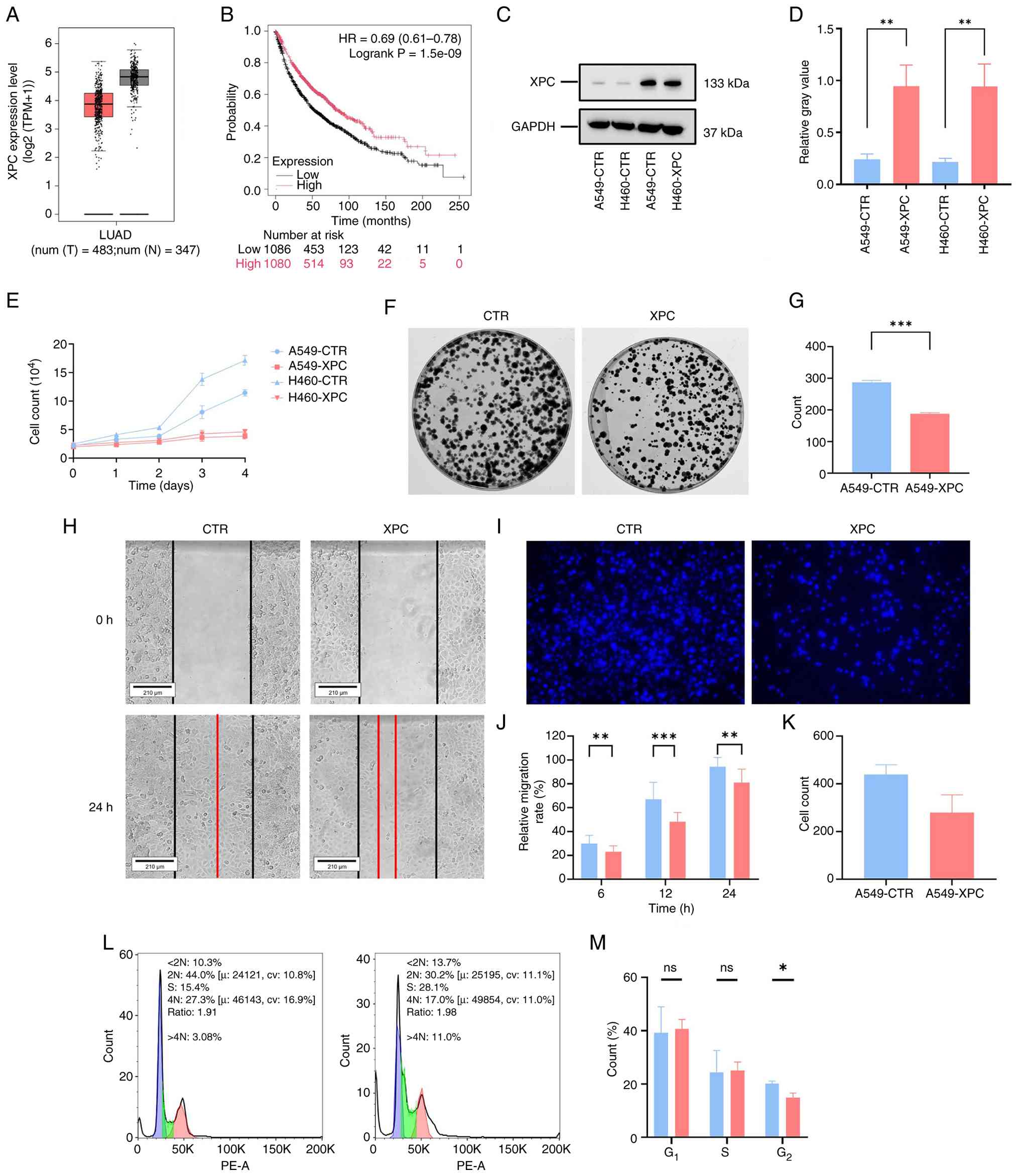 | Figure 1XPC inhibits cell proliferation,
migration and induces cell cycle arrest in non-small cell lung
cancer. (A) Analysis of XPC transcript levels in lung
adenocarcinoma tissues from TCGA database using GEPIA. (B) Survival
analysis of lung cancer patients with different XPC expression
levels via Km-plot platform. (C) Western blotting of XPC protein
expression in XPC-overexpressing cell lines. (D) Quantitative
analysis of western blotting results. (E) Cell growth curves (n=3).
(F) Representative images of cell colony formation assay. (G)
Quantitative analysis of colony formation (n=3). (H) Representative
images of scratch wound healing assay (scale bar, 200μm),
the red line indicates the cell boundary at the end of the
experiment. (I) Representative images of Transwell cell migration
assay (scale bar, 100 μm). (J) Quantitative analysis of
wound healing rate (n=3). (K) Quantitative analysis of cell
migration in Transwell assay (n=3). (L) Cell cycle distribution
analyzed by flow cytometry. (M) Quantitative analysis of cell cycle
phase proportions (n=3). All data are presented as mean ± SD.
*P<0.05, **P<0.01,
***P<0.001; ns, no significant difference. XPC,
xeroderma pigmentosum group C; TCGA, The Cancer Genome Atlas;
GEPIA, Gene Expression Profiling Interactive Analysis; LUAD, lung
adenocarcinoma; HR, Hazard Ratio; CTR, control. |
To investigate the functional effect of XPC
expression levels in tumors, the present study established two
XPC-overexpressing cell lines, A549-XPC and H460-XPC, using
lentiviral transduction. RT-qPCR results showed that the XPC
transcript level in A549-XPC cells was 10.75 times higher than in
A549-CTR cells (H460-XPC vs. H460-CTR, 8.76 times; Fig. S1A; P<0.01). Furthermore,
western blotting confirmed that the XPC protein level in A549-XPC
cells was 3.90 times that of the control group (H460-XPC vs.
H460-CTR, 4.35 times; Fig. 1C and
D; P<0.001). Additionally, immunofluorescence assays
indicated that the overexpressed XPC was primarily localized in the
nucleus, which is consistent with previously reported expression
patterns of XPC (Fig. S1B and C)
(32). These results confirmed
the successful establishment of the two XPC-overexpressing cell
lines, A549-XPC and H460-XPC and their control lines, which can be
used to study the functional differences mediated by XPC in tumor
cells.
The growth curves of the different cell lines during
cell culture were recorded. Within the recording period, the
control groups entered the exponential growth phase by day 2,
whereas the XPC-overexpressing groups did not enter the exponential
growth phase even by day 4. At day 4, the cell number in the
control groups was significantly higher than in the experimental
groups, suggesting that XPC overexpression inhibits the
proliferation of tumor cells (Fig.
1E; P<0.001). The in vitro cell colony formation
assay further supported the view that XPC overexpression inhibits
tumor cell proliferation ability (Fig. 1F and G; P<0.001; Fig. S2A and B). The number of cell
colonies formed by XPC-overexpressing cells was only 65.31% of that
in the control groups.
Migration is a crucial capability for tumor cell
metastasis; therefore, the effect of XPC overexpression on tumor
cell migration was investigated. The scratch wound healing assay
showed that the wound closure in the experimental cell lines after
24 h of culture was significantly lower than in the control groups.
The wound healing capacity was reduced by 0.77-fold compared with
the control groups (Fig. 1H-J;
P<0.001; Fig. S3A and B).
Furthermore, the invasive capability of the cells was assessed
using the Transwell assay. As expected, fewer A549-XPC cells
migrated through the membrane in the same amount of time (Fig. 1I-K; P<0.001; Fig. S3C and D). Subsequently, the tumor
cells were labeled with PE and analyzed the cell cycle of the
different groups using flow cytometry. The results showed a
significant reduction in the proportion of cells in the
G2/M phase in the XPC-overexpressing groups (Fig. 1L and M; P<0.05; Fig. 2C and D), suggesting that XPC
inhibits tumor cell proliferation by reinforcing the
G2/M checkpoint. These results indicated that reduced
expression of XPC in tumor cells enhanced their proliferation and
migration capabilities, leading to increased tumor malignancy, and
that XPC is a key negative regulator of NSCLC growth and
metastasis.
Mechanisms of XPC-mediated modulation of
sensitivity to CDDP and DOX in lung cancer cells
CDDP and DOX are front-line cytotoxic agents against
NSCLC and their primary mode of action is the induction of DNA
damage. XPC, the key sensor of helix-distorting adducts, initiates
global-genome nucleotide excision repair. It was therefore
hypothesized that elevated XPC could potentiate the lethal effects
of these drugs.
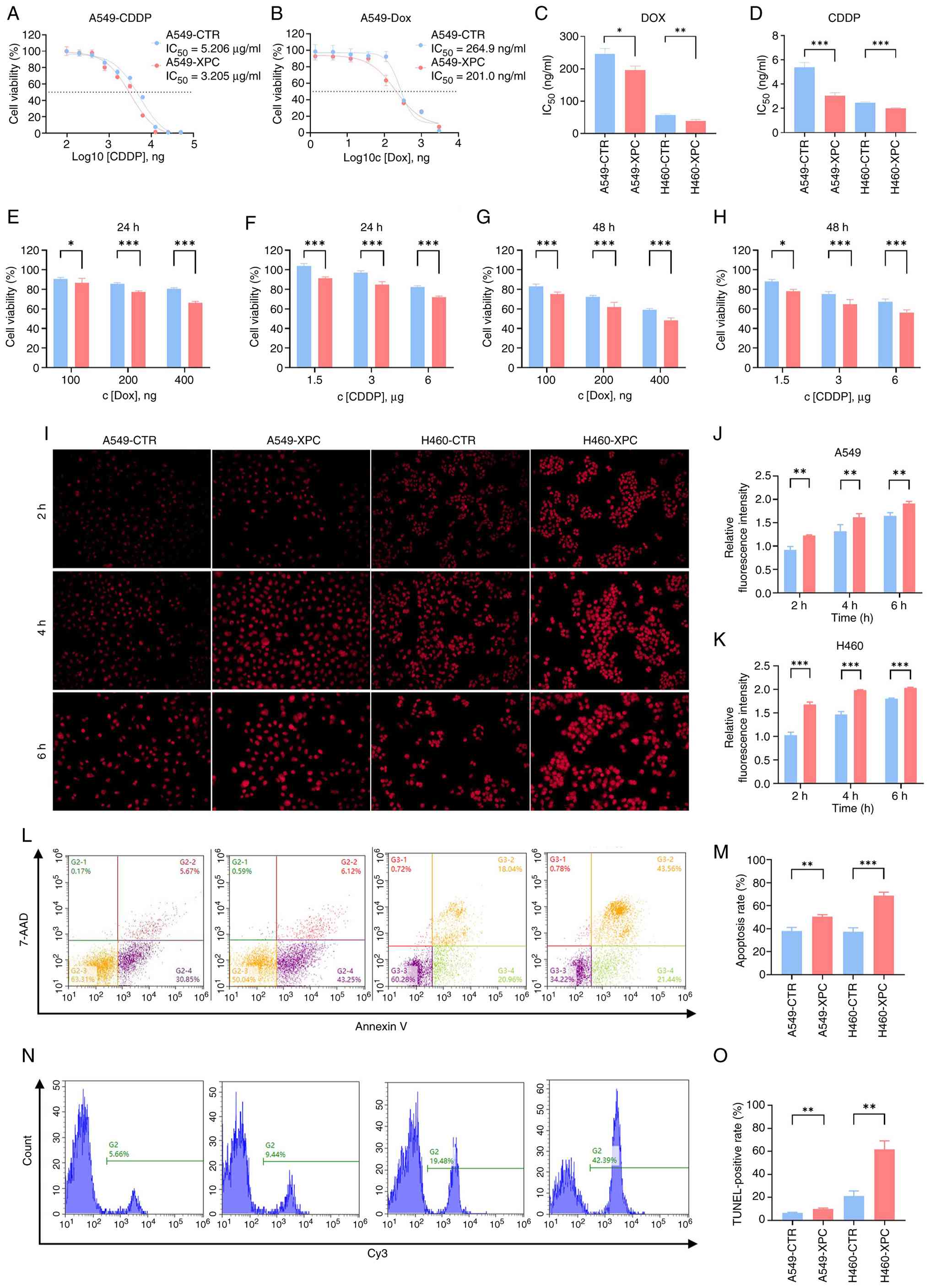
Indeed, 72 h exposure to either CDDP or DOX yielded
markedly lower half-maximal inhibitory concentrations
(IC50) in XPC-over-expressing cell sub-lines compared
with empty-vector controls (Fig. 2A
and B and Fig. S4A and B).
Subsequent time- and dose-response profiling revealed that
cytotoxicity increased in a strictly concentration- and
schedule-dependent manner for both agents (Fig. 2C-H and Fig. S4C-F). Notably, at every
concentration and time point examined, XPC-high cells exhibited
consistently greater sensitivity to CDDP or DOX than their XPC-low
counterparts.
To elucidate the mechanism underlying XPC-mediated
chemotherapy sensitization, the present study assessed cell
apoptosis, DNA damage burden and drug accumulation. Analysis of the
time course of DOX accumulation revealed that, compared with the
control group, A549-XPC cells exhibited significantly elevated
intracellular fluorescence intensity (relating to DOX uptake) at 2,
4 and 6 h, with the most significant difference observed at 4 h
(Fig. 2I-K; P<0.01). Following
treatment with cisplatin, the apoptosis rate in XPC-overexpressing
cells was significantly higher compared to the control group
(Fig. 2L and M; P<0.01). TUNEL
staining revealed significantly increased DNA damage in
XPC-overexpressing cells following drug exposure (Fig. 2N and O; P<0.01). These data
suggested that XPC-mediated chemotherapy sensitivity involves
enhanced apoptosis induction, an increased DNA damage burden, and
elevated intracellular drug accumulation.
XPC-regulated expression of
stemness-associated markers and JAK/STAT signaling in lung cancer
cells
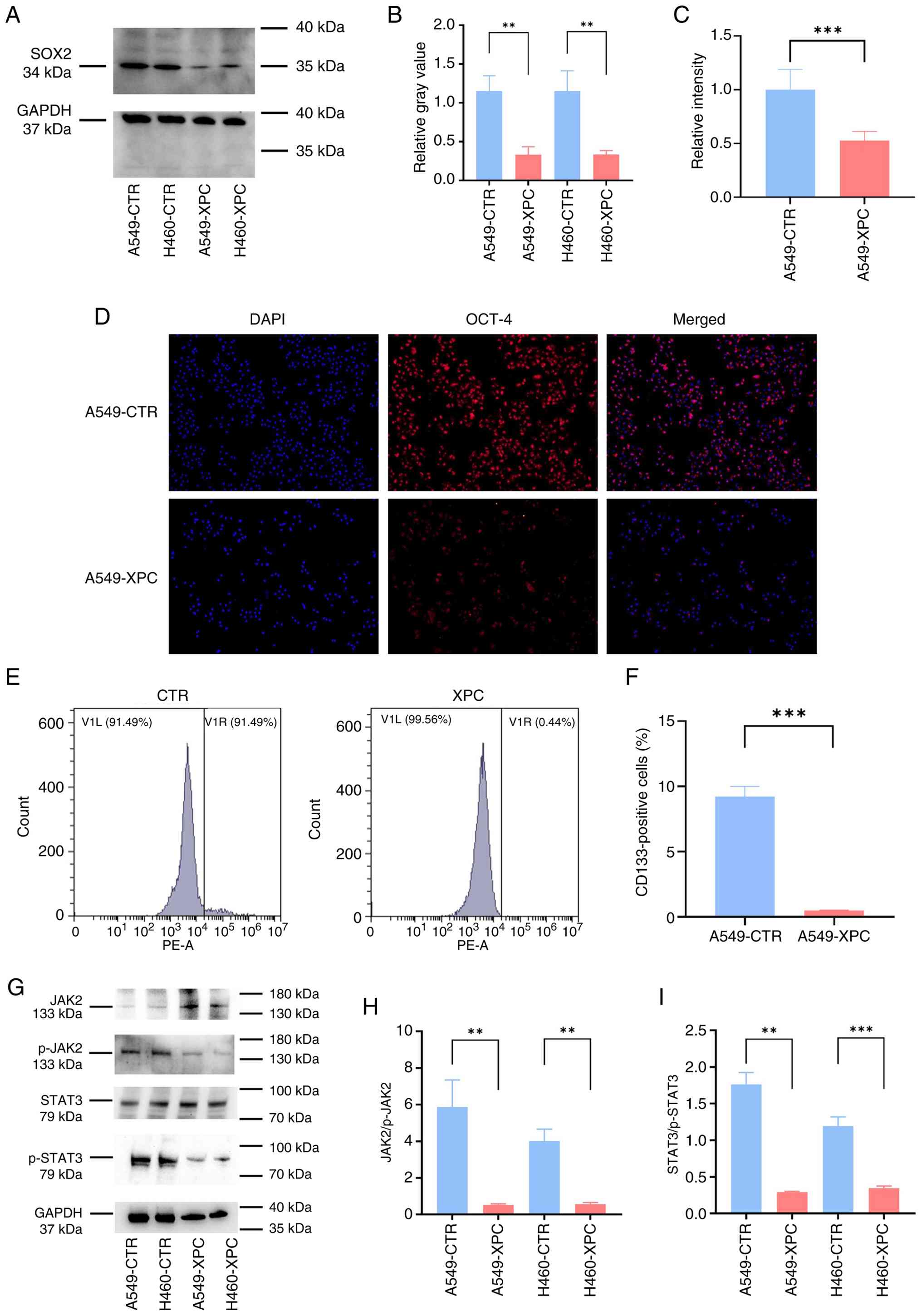
To determine whether XPC abundance modulated the
stem-like properties of NSCLC cells, the expression of SOX2 (34
kDa) was first examined. Western blotting revealed a pronounced
reduction in SOX2 protein in XPC-overexpressing cell sub-lines
relative to vector controls (Fig. 3A
and B and Fig. S5A and B),
suggesting altered expression of stemness-associated markers and
flow-cytometric analyses corroborated this finding: OCT-4 levels
were markedly lower, and the CD133+ fraction was
significantly decreased in XPC-high cell populations (P<0.01;
Fig. 3C and D and Fig. S5C and D). Collectively, these
data indicated that enforced XPC expression reduces the expression
of stemness-associated markers (SOX2, OCT-4 and CD133) in NSCLC
cells.
Mechanistically, interrogation of the JAK/STAT axis
showed that XPC overexpression selectively impaired pathway
activity: p-JAK2 and p-STAT3 were both substantially reduced,
without a concomitant decline in total STAT3 (Fig. 3G-I). Surprisingly, total JAK2
protein was markedly elevated, suggesting a compensatory feedback
loop that attempts to restore signaling capacity in the face of
persistent p-JAK2 attenuation. Thus, XPC reduces SOX2-driven
stemness, at least in part, by dampening JAK/STAT3 signaling in
NSCLC cells.
Effects of XPC on clonogenic ability and
serum-dependent growth of lung cancer cells
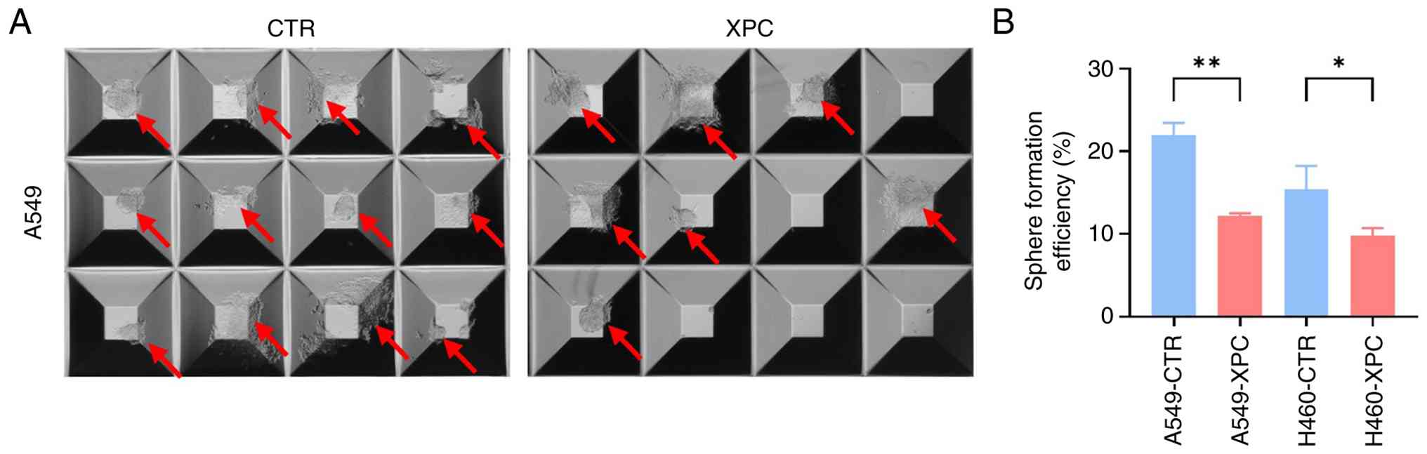
To quantitatively assess the effect of XPC on tumor
initiation capacity, a spheroid formation assay was conducted in
serum-free, ultra-low-adhesion 24-well round-bottom plates
(Fig. 4). A total of 8 days after
seeding, the spheroids formed by A549-XPC cells were markedly
smaller than those in the A549-CTR control group, and their
spheroid formation efficiency was also markedly lower than that of
the A549-CTR control group. The same trend was observed in H460
cells, confirming that elevated XPC levels inhibit the self-renewal
capacity of NSCLC cells with different genetic backgrounds.
Roles of XPC in tumorigenesis and tumor
growth characteristics
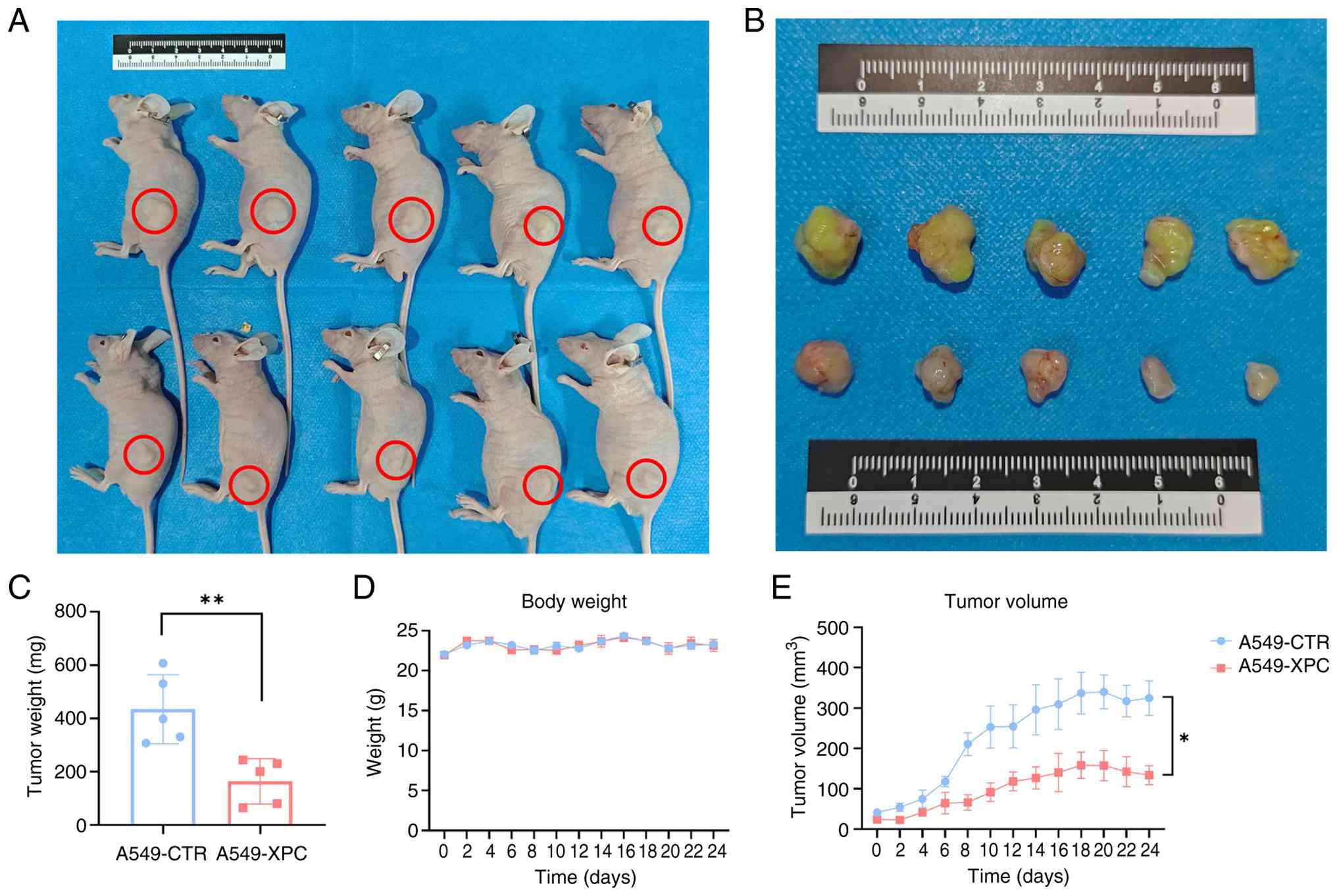
Tumor growth was analyzed using two-way
repeated-measures ANOVA, which revealed a significant main effect
of XPC overexpression on tumor volume over time (Fig. 5; P<0.05). A549-XPC lesions grew
slower than the controls, yielding markedly smaller volumes from
day 9 onward, the final measurement taken on day 24 showed that the
tumor volume (134.04±46.77 mm3) in the A549-XPC group
was 41.28% of that in the control group (324.64±85.31
mm3), representing a significant reduction. At the
endpoint, the excised tumors were weighed. The mean mass in the
XPC-overexpression group (164.24±76.16 mg) was 62% lower than the
control (434.70±115.72 mg). These data demonstrated that elevated
XPC suppresses the in vivo tumorigenic potential of A549
cells.
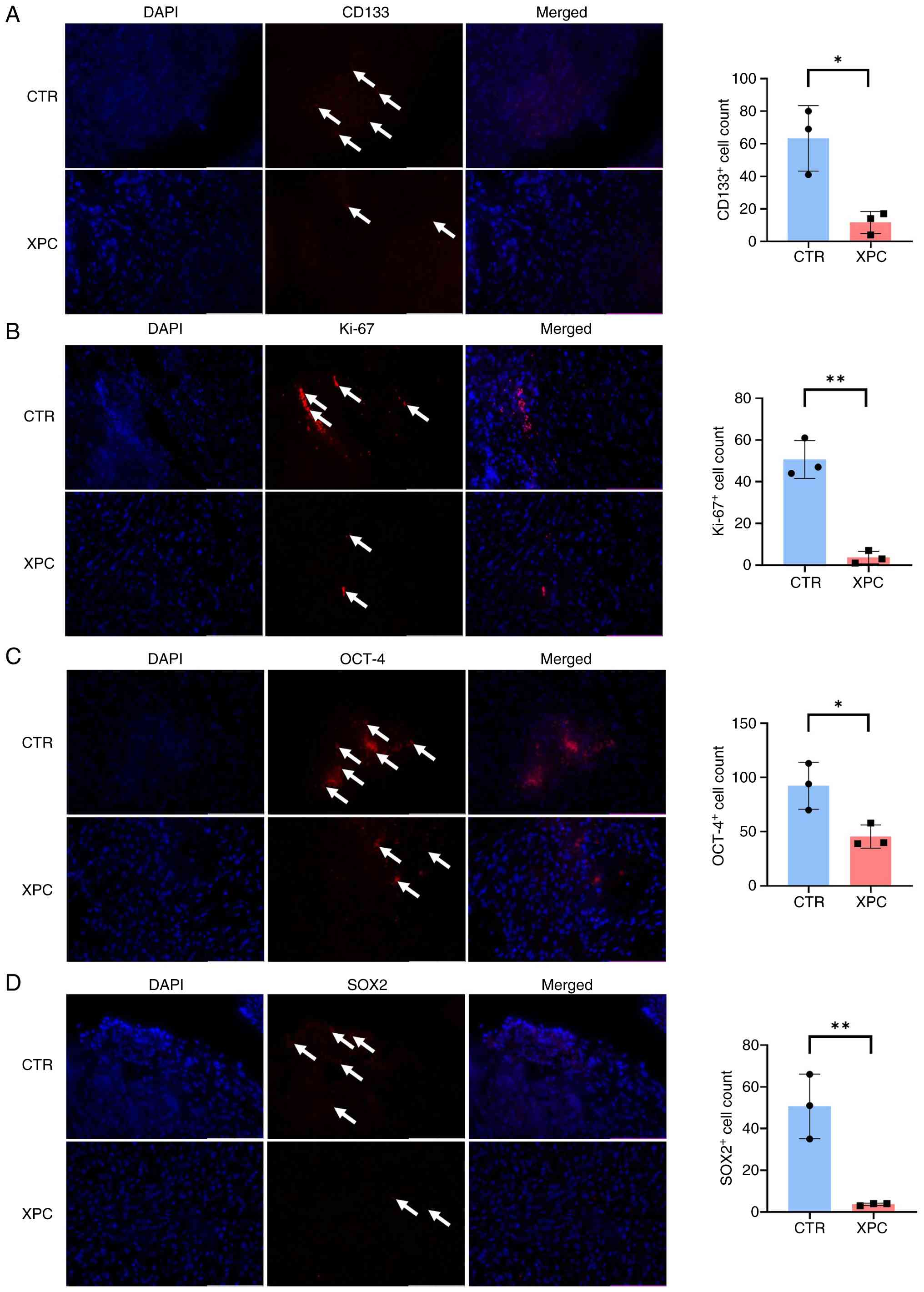
In the tissues with immunofluorescence staining, the
expression levels of tumor stem cell markers such as CD133, OCT4
and SOX2 were detected in different tissues (Fig. 6 and Fig. S6). The results showed that the
expression levels of these markers were markedly reduced in tumor
tissues with overexpressed XPC. Additionally, the proliferative
activity of tumor tissues was demonstrated through Ki-67 staining,
and the results showed a significant decrease in Ki-67 expression
levels in tumor tissues with high XPC expression. This result
illustrated the inhibitory effect of XPC on the stemness and
proliferative capacity of malignant tumor cells.
Discussion
XPC is the primary sensor of DNA adducts and
initiates genome-wide nucleotide excision repair (33-37). The present study investigated the
role of XPC mRNA expression levels as a potential prognostic marker
for treatment outcomes in NSCLC cell lines; while bioinformatic
analyses of patient datasets have suggested associations, these
associations still need to be validated in prospective studies. Our
previous study found that downregulating XPC in tumor cells via
shRNA leads to enhanced tumor stemness and upregulation of related
molecular markers (15). It was
hypothesized that XPC may exert its anti-tumorigenic, anti-invasive
and anti-metastatic effects, as well as improve the prognosis of
lung adenocarcinoma patients, by regulating the stemness of lung
cancer cells. However, the specific mechanisms underlying these
effects require further investigation.
The present study established A549 and H460 NSCLC
cell lines overexpressing XPC using lentiviral transfection and
assessed XPC expression at the mRNA and protein levels. Functional
assays demonstrated that XPC overexpression inhibits cell
proliferation, migration and clonogenic capacity, while enhancing
sensitivity to CDDP and DOX. Mechanistically, cells with high XPC
expression exhibited S-phase arrest, reduced SOX2 expression and
attenuated JAK2/STAT3 phosphorylation. Notably, the present data
indicated that overexpression of XPC enhanced cellular sensitivity
to CDDP and DOX. The potential involvement of p53 in this
sensitization effect was not examined in the present study and
remains an important limitation to be addressed in future work. The
present mechanistic studies have revealed the cause of this
seemingly paradoxical phenomenon: Cells with high XPC expression
exhibit enhanced drug-induced apoptosis, an increased DNA damage
burden and elevated intracellular drug accumulation. This is
consistent with the findings of certain studies, which suggest that
under genotoxic conditions, the overactivation or dysregulation of
DNA repair can exacerbate replicative stress and apoptotic
signaling (38,39). In vivo, XPC overexpression
markedly suppressed tumor formation in a subcutaneous xenograft
model. The present findings indicated that elevated XPC expression
suppressed lung cancer stem cell characteristics and enhanced
sensitivity to CDDP and DOX chemotherapy. The increased sensitivity
observed in XPC-overexpressing cells is associated with enhanced
intracellular accumulation of DOX, suggesting that, in addition to
its known role in DNA damage recognition, XPC may also promote drug
uptake or retention. These elevated intracellular drug
concentrations, combined with XPC-mediated initiation of DNA
repair, likely enhance DNA damage-mediated cell death during
genotoxic chemotherapy. Furthermore, XPC-mediated inhibition of the
JAK2/STAT3 pathway and downregulation of stemness markers (SOX2,
OCT-4 and CD133) may lower the drug resistance threshold by
depleting the cancer stem cell pool, which inherently resists
conventional chemotherapy (40,41).
In addition, the present study examined the
activation status of the JAK/STAT pathway in NSCLC cells following
XPC overexpression. The results showed that the phosphorylation
levels of the JAK2/STAT3 pathway were markedly reduced in cells
with high XPC expression, concomitant with decreased SOX2
expression levels. Thus, regulating XPC expression levels within
tumor cells may play a crucial role in mitigating the progression
and drug resistance of NSCLC. These observations suggested that XPC
may be associated with the modulation of JAK2/STAT3 signaling and
participate in regulating lung cancer stemness; however, this
association is currently associative and requires further
mechanistic validation. Additionally, the role of p53 in
XPC-mediated effects on chemotherapy sensitivity and stem cell
characteristics was not investigated in the present study,
representing a further limitation that should be explored in
subsequent research.
These preclinical findings suggested that XPC
expression may warrant further investigation in a clinical setting;
however, any speculation regarding its role as a prognostic marker
or therapeutic target is based on exploratory bioinformatics
analyses and in vitro/in vivo models, and independent
clinical or prospective cohort validation is required before such
conclusions can be drawn. Based on current preclinical data,
partially restoring XPC expression in tumors through
pharmacological means may be a potential future strategy for
overcoming chemotherapy resistance.
The present study had several limitations. Although
the present study demonstrated the effect of highly expressed XPC
on lung cancer stem cell characteristics and its association with
inhibition of the JAK/STAT signaling pathway, a causal relationship
between XPC and JAK2/STAT3-mediated regulation of lung cancer
stemness remains to be established. In addition, the specific
mechanisms by which XPC functions in DNA damage repair processes
within lung cancer stem cells remain poorly understood. As a key
protein in the NER pathway, the role of XPC in lung cancer stem
cells may extend beyond the recognition and repair of DNA damage
(42). Further research is needed
to determine whether XPC regulates the characteristics of lung
cancer stem cells by affecting specific DNA repair protein
complexes and whether this regulation is associated with the
inhibition of the JAK/STAT signaling pathway.
In conclusion, high expression of XPC in lung cancer
cells is associated with reduced expression of stemness-associated
markers (SOX2, OCT-4, CD133), decreased phosphorylation of JAK2 and
STAT3 and attenuated stem cell-like characteristics. While these
findings suggested a potential link between XPC and
JAK2/STAT3-mediated regulation of lung cancer stemness, further
mechanistic studies are required to establish causality. In
vitro studies showed that high expression of XPC reduced the
proliferation and migration capacity of lung cancer cells; in
vivo studies indicated that high expression of XPC decreases
the tumorigenic potential of tumor cells in vivo.
Additionally, high XPC expression enhanced the sensitivity of lung
cancer cells to DNA-damaging chemotherapeutic agents, improving the
antitumor efficacy of drugs such as CDDP and DOX. Future studies
should aim to further explore the molecular mechanisms of XPC in
lung cancer development, potentially leading to the development of
new tumor treatment strategies based on XPC.
Supplementary Data
Availability of data and materials
The data generated in the present study may be
requested from the corresponding author.
Authors' contributions
MQ and SW performed conceptual design. BY conducted
methodology and performed experiments. The data were quantified and
analyzed by YY, WW and YbW. HW, LQ and YmW performed bioinformatics
analyses. SS examined the physiological indicators of the animals.
BY and YY supervised and participated in the cell experiments. BY,
SW and MQ wrote the original manuscript. MQ and BY reviewed and
edited the article, supervised by MQ. MQ was responsible for
project management. SW and MQ confirm the authenticity of all the
raw data. All authors read and approved the final manuscript.
Ethics approval and consent to
participate
The present study has been approved by the
Laboratory Animal Ethics Committee of Weifang Second People's
Hospital (approval no. 2024LAC019).
Patient consent for publication
Not applicable.
Competing interests
The authors declare that they have no competing
interests.
Use of artificial intelligence tools
During the preparation of this work artificial
intelligence tools were used to improve the readability and
language of the manuscript and subsequently the authors reviewed
and edited the content produced by the artificial intelligence
tools as necessary, taking full responsibility for the ultimate
content of the publication.
Acknowledgements
Not applicable.
Funding
The present study was supported by the National Natural Science
Foundation of China, (grant no. 81871892), the Weifang City Science
and Technology Project Plan (grant nos. 2024JZ0018 and 2023YX077)
and Weifang Municipal Health Commission Scientific Research Program
(grant nos. WFWSJK-2022-008 and WFZYY2025-4-008).
References
|
1
|
Vaccaro A, Rahal Z, Kadara H and Cascone
T: A roadmap to precision immunotherapy for early-stage non-small
cell lung cancer. Cancer Discov. 15:884–889. 2025. View Article : Google Scholar : PubMed/NCBI
|
|
2
|
Qu T, Song L, Xu J, Lu X, Yin D, Dai J,
Zhang C, Guo R and Zhang E: MYLK-AS1 enhances glutamine metabolism
to promote EGFR inhibitor resistance in non-small cell lung cancer.
Cancer Res. 85:3052–3071. 2025. View Article : Google Scholar : PubMed/NCBI
|
|
3
|
Kenmotsu H, Yamamoto N, Misumi T, Yoh K,
Saito H, Sugawara S, Yamazaki K, Nakagawa K, Sugio K, Seto T, et
al: Five-year overall survival analysis of the JIPANG study:
Pemetrexed or vinorelbine plus cisplatin for resected stage II-IIIA
nonsquamous non-small-cell lung cancer. J Clin Oncol. 41:5242–5246.
2023. View Article : Google Scholar : PubMed/NCBI
|
|
4
|
Guevara Lopez ML, Gebo A, Parodi M,
Persano S, Maus-Conn J, Mingari MC, Loiacono F, Orecchia P, Sivori
S, Cantoni C, et al: CD56bright cytokine-induced
memory-like NK cells and NK-cell engagers synergize against
non-small cell lung cancer cancer-stem cells. J Immunother Cancer.
13:e0102052025. View Article : Google Scholar
|
|
5
|
Shi L, Li B, Tan J, Zhu L, Zhang S, Zhang
Y, Xiang M, Li J, Chen Y, Han X, et al: Exosomal lncRNA Mir100hg
from lung cancer stem cells activates H3K14 lactylation to enhance
metastatic activity in non-stem lung cancer cells. J
Nanobiotechnology. 23:1562025. View Article : Google Scholar : PubMed/NCBI
|
|
6
|
Testa U, Castelli G and Pelosi E: Lung
cancers: Molecular characterization, clonal heterogeneity and
evolution, and cancer stem cells. Cancers (Basel). 10:2482018.
View Article : Google Scholar : PubMed/NCBI
|
|
7
|
Zebian A, Shaito A, Mazurier F, Rezvani HR
and Zibara K: XPC beyond nucleotide excision repair and skin
cancers. Mutat Res Rev Mutat Res. 782:1082862019. View Article : Google Scholar : PubMed/NCBI
|
|
8
|
Lindsey-Boltz LA, Yang Y, Kose C, Deger N,
Eynullazada K, Kawara H and Sancar A: Nucleotide excision repair in
human cell lines lacking both XPC and CSB proteins. Nucleic Acids
Res. 51:6238–6245. 2023. View Article : Google Scholar : PubMed/NCBI
|
|
9
|
Zhang ET, He Y, Grob P, Fong YW, Nogales E
and Tjian R: Architecture of the human XPC DNA repair and stem cell
coactivator complex. Proc Natl Acad Sci U S A. 112:14817–14822.
2015. View Article : Google Scholar : PubMed/NCBI
|
|
10
|
Liu L, Peng Z, Xu Z and Wei X: XPC
Promotes pluripotency of human dental pulp cells through regulation
of Oct-4/Sox2/c-Myc. Stem Cells Int. 2016:34548762016. View Article : Google Scholar : PubMed/NCBI
|
|
11
|
Sears CR: DNA repair as an emerging target
for COPD-lung cancer overlap. Respir Investig. 57:111–121. 2019.
View Article : Google Scholar : PubMed/NCBI
|
|
12
|
Jlassi A, Rejaibi R, Manai M, Sahraoui G,
Guerfali FZ, Charfi L, Mezlini A, Manai M, Mrad K and Doghri R:
VISTA/CTLA4/PD1 coexpression on tumor cells confers a favorable
immune microenvironment and better prognosis in high-grade serous
ovarian carcinoma. Front Oncol. 14:13520532024. View Article : Google Scholar : PubMed/NCBI
|
|
13
|
Zhang X, He N, Gu D, Wickliffe J, Salazar
J, Boldogh I and Xie J: Genetic evidence for XPC-KRAS interactions
during lung cancer development. J Genet Genomics. 42:589–596. 2015.
View Article : Google Scholar : PubMed/NCBI
|
|
14
|
Crișan AS, Tripon F, Bogliș A, Crauciuc
GA, Trifa AP, Lázár E, Macarie I, Gabor MR and Bănescu C: The role
of DNA repair (XPC, XPD, XPF, and XPG) gene polymorphisms in the
development of myeloproliferative neoplasms. Medicina (Kaunas).
60:5062024. View Article : Google Scholar
|
|
15
|
Wang W, Ma S, Ding Z, Yang Y, Wang H, Yang
K, Cai X, Li H, Gao Z and Qu M: XPC protein improves lung
adenocarcinoma prognosis by inhibiting lung cancer cell stemness.
Front Pharmacol. 12:7079402021. View Article : Google Scholar : PubMed/NCBI
|
|
16
|
Hu LB, Chen Y, Meng XD, Yu P, He X and Li
J: Nucleotide excision repair factor XPC ameliorates prognosis by
increasing the susceptibility of human colorectal cancer to
chemotherapy and ionizing radiation. Front Oncol. 8:2902018.
View Article : Google Scholar : PubMed/NCBI
|
|
17
|
Dolatabadi S, Jonasson E, Lindén M,
Fereydouni B, Bäcksten K, Nilsson M, Martner A, Forootan A, Fagman
H, Landberg G, et al: JAK-STAT signalling controls cancer stem cell
properties including chemotherapy resistance in myxoid liposarcoma.
Int J Cancer. 145:435–449. 2019. View Article : Google Scholar : PubMed/NCBI
|
|
18
|
Chen L, Wang Y and Zhang B:
Hypermethylation in the promoter region inhibits AJAP1 expression
and activates the JAK/STAT pathway to promote prostate cancer cell
migration and stem cell sphere formation. Pathol Res Pract.
241:1542242023. View Article : Google Scholar
|
|
19
|
Liu SC, Huang CM, Bamodu OA, Lin CS, Liu
BL, Tzeng YM, Tsai JT, Lee WH and Chen TM: Ovatodiolide suppresses
nasopharyngeal cancer by targeting stem cell-like population,
inducing apoptosis, inhibiting EMT and dysregulating JAK/STAT
signaling pathway. Phytomedicine. 56:269–278. 2019. View Article : Google Scholar : PubMed/NCBI
|
|
20
|
Loftus AEP, Romano MS, Phuong AN, McKinnel
BJ, Muir MT, Furqan M, Dawson JC, Avalle L, Douglas AT, Mort RL, et
al: An ILK/STAT3 pathway controls glioblastoma stem cell
plasticity. Dev Cell. 59:3197–3212.e7. 2024. View Article : Google Scholar : PubMed/NCBI
|
|
21
|
Wang T, Fahrmann JF, Lee H, Li YJ,
Tripathi SC, Yue C, Zhang C, Lifshitz V, Song J, Yuan Y, et al:
JAK/STAT3-regulated fatty acid β-oxidation is critical for breast
cancer stem cell self-renewal and chemoresistance. Cell Metab.
27:136–150.e5. 2018. View Article : Google Scholar
|
|
22
|
Li J, Wang Y, Wang Z, Wei Y, Diao P, Wu Y,
Wang D, Jiang H, Wang Y and Cheng J: Super-enhancer driven
LIF/LIFR-STAT3-SOX2 regulatory feedback loop promotes cancer
stemness in head and neck squamous cell carcinoma. Adv Sci (Weinh).
11:e24044762024. View Article : Google Scholar : PubMed/NCBI
|
|
23
|
Zhao D, Pan C, Sun J, Gilbert C,
Drews-Elger K, Azzam DJ, Picon-Ruiz M, Kim M, Ullmer W, El-Ashry D,
et al: VEGF drives cancer-initiating stem cells through
VEGFR-2/Stat3 signaling to upregulate Myc and Sox2. Oncogene.
34:3107–3119. 2015. View Article : Google Scholar
|
|
24
|
Liu N, Ning X, Zhang X, Zhou Z, Fu M, Wang
Y and Wu T: Gradient galectin-1 coating technology: Bionic
multichannel nerve guidance conduits promote neural cell migration.
Adv Technol Neurosci. 1:276–289. 2024.
|
|
25
|
Xiao Z, Ding L, Yu Y, Ma C, Lei C, Liu Y,
Chang X, Chen Y, He Y, Zhu Y and Zhang H: Tanreqing injection
inhibits stemness and enhances sensitivity of non-small cell lung
cancer models to gefitinib through ROS/STAT3 signaling pathway. J
Cancer. 15:4259–4274. 2024. View Article : Google Scholar : PubMed/NCBI
|
|
26
|
Kahnamouei SA, Narouie B, Sotoudeh M,
Mollakouchekian MJ, Simforoosh N, Ziaee SA, Samzadeh M, Afshari M,
Jamaldini SH, Imeni M and Hasanzad M: Association of XPC gene
polymorphisms with prostate cancer risk. Clin Lab. 62:1009–1015.
2016. View Article : Google Scholar : PubMed/NCBI
|
|
27
|
Yang J, Xu Z, Li J, Zhang R, Zhang G, Ji
H, Song B and Chen Z: XPC epigenetic silence coupled with p53
alteration has a significant impact on bladder cancer outcome. J
Urol. 184:336–343. 2010. View Article : Google Scholar : PubMed/NCBI
|
|
28
|
Yue AM, Xie ZB, Zhao HF, Guo SP, Shen YH
and Wang HP: Associations of ABCB1 and XPC genetic polymorphisms
with susceptibility to colorectal cancer and therapeutic prognosis
in a Chinese population. Asian Pac J Cancer Prev. 14:3085–3091.
2013. View Article : Google Scholar : PubMed/NCBI
|
|
29
|
MacDonagh L, Gray SG, Breen E, Cuffe S,
Finn SP, O'Byrne KJ and Barr MP: Lung cancer stem cells: The root
of resistance. Cancer Lett. 372:147–156. 2016. View Article : Google Scholar : PubMed/NCBI
|
|
30
|
Wang X, Chen Y, Wang X, Tian H, Wang Y,
Jin J, Shan Z, Liu Y, Cai Z, Tong X, et al: Stem cell factor SOX2
confers ferroptosis resistance in lung cancer via upregulation of
SLC7A11. Cancer Res. 81:5217–5229. 2021. View Article : Google Scholar : PubMed/NCBI
|
|
31
|
Livak KJ and Schmittgen TD: Analysis of
relative gene expression data using real-time quantitative PCR and
the 2(-Delta Delta C(T)) Method. Methods. 25:402–408. 2001.
View Article : Google Scholar
|
|
32
|
Wang H, Huang Y, Shi J, Zhi Y, Yuan F, Yu
J, Chen Z and Yang J: XPC deficiency leads to centrosome
amplification by inhibiting BRCA1 expression upon
cisplatin-mediated DNA damage in human bladder cancer. Cancer Lett.
444:136–146. 2019. View Article : Google Scholar
|
|
33
|
Zhao B, Huang Y, Shi J, Zhou X, Fagman JB,
Wang L, Liu S, Wang W, Liu Y, Chen Z and Yang J: XPC deficiency
activate cisplatin-mediated autophagy in bladder cancer by limiting
novel PHRF1-mediated ubiquitination of the p53 protein. Adv Sci
(Weinh). 13:e175632025. View Article : Google Scholar : PubMed/NCBI
|
|
34
|
Wu Z, Wang M, Dong X, Sun Y, Zhang L, Tang
M, Jiang X, Guo Y, Wang J, Wang W, et al: The deubiquitinase OTUD1
orchestrates cisplatin chemosensitivity of non-small cell lung
cancer through destabilizing RAD23B/XPC. Oncogene. 44:4855–4867.
2025. View Article : Google Scholar : PubMed/NCBI
|
|
35
|
Lee GY, Jang JS, Lee SY, Jeon HS, Kim KM,
Choi JE, Park JM, Chae MH, Lee WK, Kam S, et al: XPC polymorphisms
and lung cancer risk. Int J Cancer. 115:807–813. 2005. View Article : Google Scholar : PubMed/NCBI
|
|
36
|
Zhou H, Saliba J, Sandusky GE and Sears
CR: XPC protects against smoking- and carcinogen-induced lung
adenocarcinoma. Carcinogenesis. 40:403–411. 2019. View Article : Google Scholar : PubMed/NCBI
|
|
37
|
Gao Y, Mu J, Liu K and Wang M: Integrating
molecular fingerprints with machine learning for accurate
neurotoxicity prediction: An observational study. 2:109–115.
2025.
|
|
38
|
Acharya G, Mani C, Sah N, Saamarthy K,
Young R, Reedy MB, Sobol RW and Palle K: CHK1 inhibitor induced
PARylation by targeting PARG causes excessive replication and
metabolic stress and overcomes chemoresistance in ovarian cancer.
Cell Death Discov. 10:2782024. View Article : Google Scholar : PubMed/NCBI
|
|
39
|
Manic G, Musella M, Corradi F, Sistigu A,
Vitale S, Soliman Abdel Rehim S, Mattiello L, Malacaria E, Galassi
C, Signore M, et al: Control of replication stress and mitosis in
colorectal cancer stem cells through the interplay of PARP1, MRE11
and RAD51. Cell Death Differ. 28:2060–2082. 2021. View Article : Google Scholar : PubMed/NCBI
|
|
40
|
Liu M, Wu H and Xu C: Targeting cancer
stem cell pathways for lung cancer therapy. Curr Opin Oncol.
35:78–85. 2023. View Article : Google Scholar
|
|
41
|
Wang J, Chen Y, Wang C and Ren K: Research
status and controversy on non-small cell lung cancer stem cells.
Adv Clin Exp Med. 34:633–640. 2025. View Article : Google Scholar
|
|
42
|
Bai Y, Xu L, Yang X, Hu Z, Yuan J, Wang F,
Shao M, Yuan W, Qian J, Ma H, et al: Sequence variations in DNA
repair gene XPC is associated with lung cancer risk in a Chinese
population: A case-control study. BMC Cancer. 7:812007. View Article : Google Scholar : PubMed/NCBI
|