Introduction
Lung cancer, the most lethal malignancy worldwide,
continues to place an increasing burden on healthcare systems.
Globally, 2022 witnessed >2.8 million new cases and 1.8 million
deaths, accounting for almost 18% of all cancer-related fatalities
(1). Despite therapeutic
advances, several barriers continue to hinder the long-term
survival of patients with lung cancer, including resistance to
targeted therapies, off-target effects, low response rates to
immune checkpoint inhibitors and chemotherapy-related toxicity
(2). Hence, an in-depth
exploration of intercellular communication mechanisms within the
tumor microenvironment (TME) and the identification of novel
intervention targets are of utmost importance for overcoming
therapeutic bottlenecks in lung cancer. Macrophages represent the
most abundant immune cell population infiltrating the lung cancer
microenvironment. Characterized by high plasticity, they can
polarize into anti-tumor M1 or pro-tumor M2 phenotypes (3). Macrophage polarization exhibits
dynamic evolution during lung cancer progression: It is initially
dominated by the M1 phenotype, exerting antitumor effects; however,
as the tumor advances, it gradually shifts toward the M2 phenotype,
driving immune escape through mechanisms such as promoting tumor
cell metastasis, angiogenesis, and inhibiting T-cell function,
ultimately contributing to a tumor-promoting microenvironment
(4). Therefore, targeting the
polarization of M1/M2 macrophages is a key therapeutic target for
lung cancer. Certain drugs, such as artemether and CLMSR
nanoparticles can induce M1/M2 reprogramming, restore and enhance
their phagocytic function and antigen-presenting ability, thereby
activating antitumor immune responses and exerting potent anti-lung
cancer activity (5,6). MicroRNAs (miRNAs/miRs) are a class
of non-coding RNAs ~22 nucleotides in length, which regulate the
expression of target genes at the post-transcriptional level and
are widely involved in biological processes including cell
differentiation, proliferation, and immune response (7). A previous study demonstrated that
miRNAs serve as key molecular switches that regulate macrophage
polarization (8). Moreover,
several tumor-suppressing miRNAs, such as miR-7, miR-34a and the
let-7 family, have been proven to inhibit the proliferation and
migration of lung cancer cells and induce their apoptosis, thereby
exerting anti-cancer effects (9,10).
Thus, modulating miRNA expression to restore the M1/M2 balance may
produce beneficial anti-lung cancer outcomes.
Overview of miRNAs
Biosynthesis of miRNAs. miRNAs are a class of
small non-coding RNAs widely present in eukaryotes. They negatively
regulate gene expression at the post-transcriptional level by
binding to target mRNAs, thereby promoting mRNA degradation or
inhibiting translation (11). The
biosynthesis of miRNAs is an orderly multi-step process. Firstly,
in the cell nucleus, miRNA genes are transcribed by RNA polymerase
II to generate primary miRNA (pri-miRNA) with a stem-loop
structure; subsequently, these pri-miRNAs are cleaved by the Drosha
ribonuclease III-DiGeorge syndrome critical region 8 complex to
form precursor miRNAs (pre-miRNAs) with a length of ~70
nucleotides. Secondly, these pre-miRNAs are transported from the
cell nucleus to the cytoplasm through a mechanism dependent on
Exportin-5. Finally, in the cytoplasm, these pre-miRNAs are further
cleaved by the Dicer enzyme, ultimately forming mature miRNA double
strands with ~22 nucleotides. One of these strands is selectively
loaded onto the RNA-induced silencing complex (RISC). Functionally,
the RISC carrying the mature miRNA mainly mediates partial
complementarity with the) of the target mRNA, promoting the
demethylation-dependent degradation of the target mRNA and
inhibiting its translation, thereby achieving post-transcriptional
negative regulation of gene expression. Through this mechanism,
miRNAs are widely involved in processes, such as cell
proliferation, differentiation, apoptosis and the maintenance of
cell homeostasis (12,13).
Regulatory role of miRNAs in the
occurrence and development of lung cancer
As a key regulator of gene expression, miRNAs are
involved in diverse biological processes, including cell
proliferation, differentiation, apoptosis and immune regulation.
The aberrant expression of miRNAs is closely associated with
various diseases (14). In the
context of malignant tumors, miRNA plays a critical role in tumor
initiation, progression, proliferation and metastasis, exhibiting
both tumor-suppressive and oncogenic functions, and thus hold
potential as tools for diagnosis, treatment and prognostic
assessment (15). Furthermore,
miRNAs can interact with extracellular vesicles, high-density
lipoproteins and RISC effector proteins, allowing them to remain
stable in extracellular fluids and positioning them as promising
disease biomarkers with broad application potential (16). A growing body of evidence has
identified multiple miRNAs as key biomarkers for lung cancer
(17), where they exert key
regulatory functions in processes, such as apoptosis, drug
resistance, proliferation, invasion and migration, thereby actively
participating in lung cancer pathogenesis and progression (18-20).
Overview of macrophage polarization
Macrophages, first discovered in 1908 for their
phagocytic activity, are essential components of the innate immune
response (21). Beyond their
well-established roles in phagocytosis, exogenous antigen
presentation and cytokine secretion, macrophages also participate
in systemic metabolism, hematopoiesis, angiogenesis and malignant
tumor progression (22). Under
distinct microenvironmental stimuli, macrophages polarize into
classical activated M1 or alternative activated M2 phenotypes. M1
macrophages, induced by granulocyte-macrophage colony-stimulating
factor (GM-CSF), Toll-like receptor (TLR) ligands, or T-helper 1
cell (Th1) cytokines, exhibit a high expression of pro-inflammatory
cytokines (e.g., TNF-α, IL-1β, IL-6, IL-12 and IL-23) and surface
molecules (CD40, CD64, CD68, CD80, CD86 and HLA-DR). They exhibit
potent antigen-presenting capacity, driving inflammation, pathogen
clearance and antitumor immunity (23). M2 macrophages are primarily
induced by Th2-type cytokines (IL-4/IL-13), immune complexes,
glucocorticoids, or adenosine, and play central roles in
anti-inflammatory responses, immune regulation and tissue repair
and remodeling. According to the nature of inducing stimuli and
resultant functional profiles, M2 macrophages are categorized into
four subtypes: M2a, M2b, M2c and M2d. Specifically, M2a
macrophages, induced by IL-4 or IL-13, highly expresses CD206 and
CD209 and is primarily involved in Th2-type immune responses,
allergic reactions and anti-parasitic immunity. M2b macrophages,
induced by immune complexes combined with TLR ligands, exhibit both
pro-inflammatory and anti-inflammatory characteristics, with a high
expression of IL-10 and TNF-α, playing a unique role in immune
regulation. M2c macrophages, induced by IL-10, transforming growth
factor-β (TGF-β), or glucocorticoids, represent the primary subtype
associated with acquired immune regulation, highly expressing CD163
and CD206, and various scavenger receptors, and are crucial for the
phagocytosis of apoptotic cells and tissue remodeling. M2d
macrophages, induced by TLR ligands in combination with adenosine
analogs, are primarily associated with angiogenesis and tumor
progression, with a high expression of vascular endothelial growth
factor (VEGF) and IL-10 (24). At
the molecular level, M2 macrophages secrete a range of
anti-inflammatory cytokines and chemokines, including TGF-β, IL-10,
C-C motif chemokine ligand (CCL)18 and CCL22. They also highly
express characteristic surface markers, such as CD163, CD206,
CD209, scavenger receptor A, scavenger receptor class B type I and
CCL2. Through these molecules, M2 macrophages not only efficiently
clear apoptotic cells and debris, but also participate in tissue
repair and matrix remodeling by secreting pro-fibrotic factors,
such as TGF-β and fibronectin (25,26). Studies have shown that in
early-stage lung cancer, M1 macrophages predominate and inhibit
lung cancer cell growth through the expression of various
pro-inflammatory cytokines, chemokines and effector molecules. By
contrast, advanced-stage lung cancer is characterized by a
progressive shift toward the M2 phenotype, in which M2 macrophages
promote tumor growth, invasion and metastasis (27). Therefore, targeting the inhibition
of the M2 phenotype and inducing M2-to-M1 repolarization to correct
the imbalance of macrophage polarization represents a promising
strategy for the prevention and treatment of lung cancer. However,
it should be noted that this strategy still faces several
challenges in practical application (28). Firstly, the TME is characterized
by hypoxia, the accumulation of lactic acid and various
immunosuppressive factors, such as IL-4, IL-10 and TGF-β (29). These factors may continuously
hinder the reversal of M2 to M1 polarization state, thereby
severely reducing the efficiency of re-polarization. Secondly, the
re-polarized M1 phenotype may lack long-term stability. The TME
continuously activates signaling pathways, such as STAT6 and
PI3K-AKT-mTOR, which prompt macrophages to transform into the M2
phenotype (30). This not only
reduces the initial re-polarization effect, but also diminishes the
ability of the polarized M1 cells to maintain a stable antitumor
state under the influence of dynamic microenvironmental signals
(31). Furthermore, the excessive
activation of M1-type macrophages can induce tissue inflammatory
damage and, in certain advanced tumor contexts, may even
paradoxically promote malignant progression (32). Therefore, future investigations
are warranted to elucidate the molecular regulatory networks
governing polarization reversal, systematically assess its
maintainability and safety within the complex in vivo
microenvironment, and explore optimization strategies in
combination with other therapeutic modalities. This may provide a
more robust theoretical foundation for the clinical translation of
this approach. The mechanisms of macrophage polarization are
illustrated in Fig. 1.
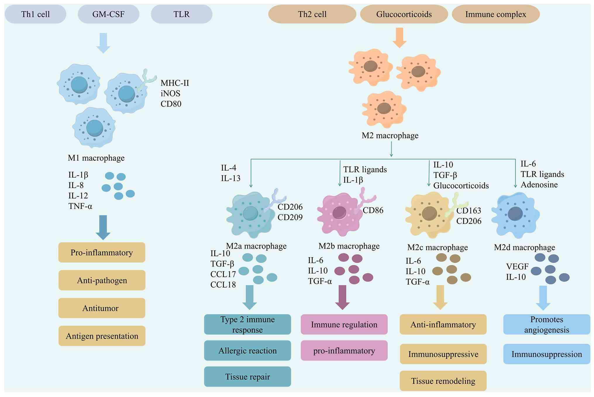 | Figure 1Mechanisms of macrophage
polarization. The image presents the complete regulatory network
for macrophage polarization. Under the guidance of Th1 and Th2
cells and GM-CSF and other signals, macrophages can polarize into
the pro-inflammatory M1 type (induced by TLR ligands, expressing
CD80/CD86/iNOS, secreting IL-1β, TNF-α, etc., with anti-pathogen
and anti-tumor functions), as well as four M2 subtypes: M2a
(induced by IL-4/IL-13, involved in type 2 immunity and allergy),
M2b (induced by immune complexes, having both immunoregulatory and
pro-inflammatory effects), M2c (induced by glucocorticoids/IL-10,
mediating immunosuppression and tissue repair), and M2d (induced by
TLR ligands/adenosine, promoting angiogenesis). The figure
systematically reveals the functional diversity of macrophages in
inflammation, immune regulation and tissue remodeling. GM-CSF,
granulocyte-macrophage colony-stimulating factor; TLR, Toll-like
receptor; iNOS, inducible nitric oxide synthase; MHC-II, major
histocompatibility complex class II. |
Regulatory effects of macrophage
polarization on lung cancer
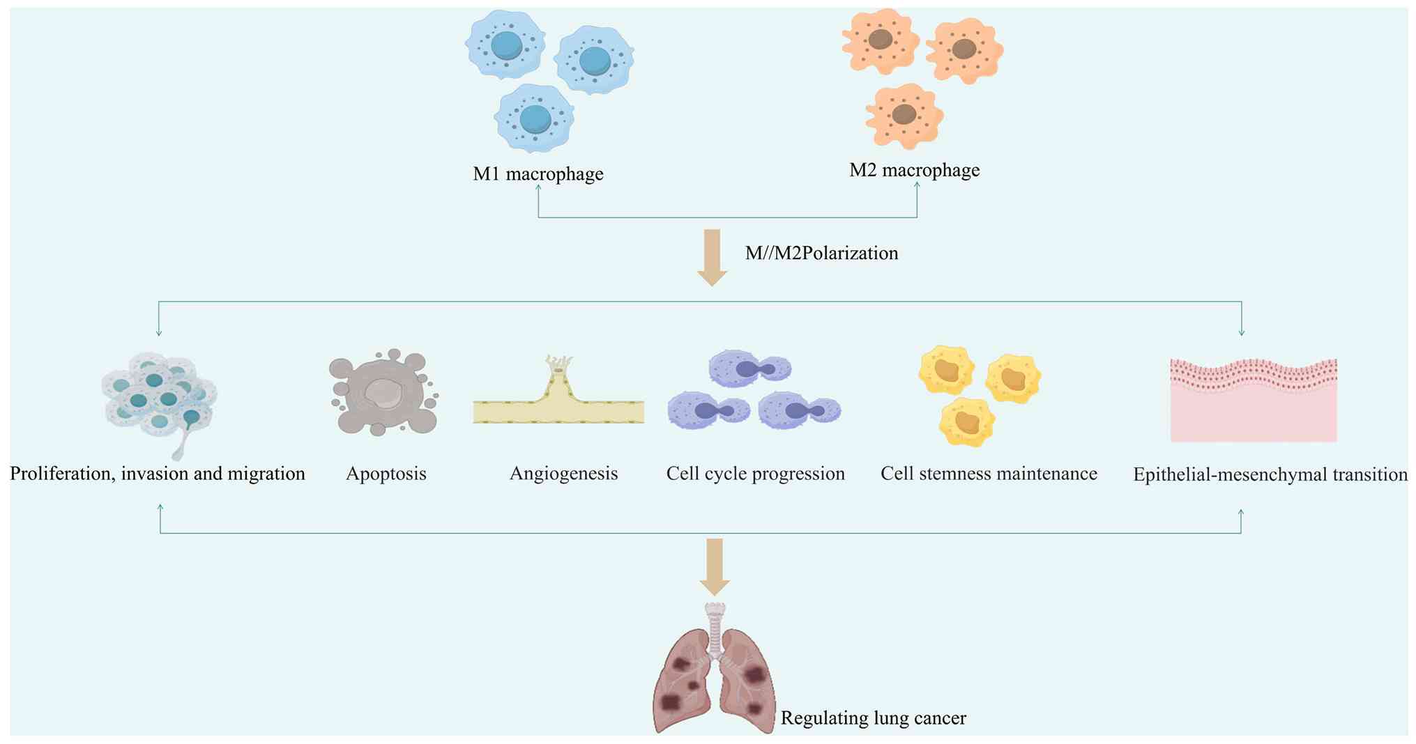
Macrophage polarization profoundly affects the
occurrence, development and therapeutic efficacy of lung cancer by
regulating multiple biological processes, such as proliferation and
invasion of lung cancer cells, apoptosis, angiogenesis, cell cycle
progression, the maintenance of tumor stemness and
epithelial-mesenchymal transition (EMT). The mechanisms by which
macrophage polarization regulates lung cancer are illustrated in
Fig. 2.
Regulation of the proliferation, invasion
and migration of lung cancer cells
The proliferation, invasion and migration of lung
cancer cells represent core processes driving malignant tumor
progression (33). In non-small
cell lung cancer (NSCLC), exosomes derived from M2 macrophages have
been shown to enhance tumor cell viability, migration and invasion
by carrying miRNAs, such as miR-155 and miR-196a-5p (34). C-X-C motif chemokine ligand 9
(CXCL9) inhibits the proliferation, migration, metastasis and
invasion of lung cancer cells by suppressing the polarization and
function of M2 macrophages (35).
Furthermore, octamer-binding transcription factor 4
(OCT4)-expressing lung cancer cells can induce macrophage
polarization toward the M2 phenotype by upregulating CSF levels,
thereby facilitating tumor growth, infiltration and metastasis
(36).
Regulation of the apoptosis of lung
cancer cells
Apoptosis is a genetically regulated program of cell
death that plays a critical role in maintaining tissue homeostasis,
eliminating abnormal cells and modulating tumor development. The
induction of apoptosis in tumor cells represents a key mechanism
underlying chemotherapy, targeted therapy and immunotherapy for
lung cancer (37). The
polarization state of macrophages serves as a critical
microenvironmental factor that regulates apoptosis in lung cancer
cells, with M1 and M2 macrophages exerting opposing pro-apoptotic
and anti-apoptotic effects, respectively, through distinct
molecular mechanisms. M1 macrophages secrete interferon-γ (IFN-γ),
which activates the Janus Kinase 1(JAK1)/signal transducer and
activator of transcription 1 (STAT1) signaling pathway. This
activation promotes interaction between STAT1 and tumor protein
p53, reduces p53 binding to mouse double minute 2 homolog, inhibits
p53 ubiquitination and degradation, and enhances p53 stability and
transcriptional activity, leading to the upregulation of
pro-apoptotic molecules, such as PUMA and initiation of the
mitochondrial apoptotic pathway (38). Furthermore, IFN-γ also induces an
increase in reactive oxygen species levels within lung cancer
cells, causing DNA damage and activating the ataxia telangiectasia
mutated/checkpoint kinase 2 pathway. These two pathways converge at
the p53/PUMA axis, synergistically amplifying apoptotic signaling
and collectively inducing lung cancer cell death (39). By contrast, M2 macrophages secrete
anti-inflammatory cytokines, including IL-10 and TGF-β, which
suppress antitumor immune responses and attenuate immune
cell-mediated tumor cell killing, thereby inhibiting tumor cell
apoptosis and promoting tumor cell survival (40).
Regulation of angiogenesis in lung
cancer
Angiogenesis plays a critical role in the
progression and metastasis of lung cancer. Intervening in tumor
angiogenesis and promoting vascular normalization have emerged as
effective strategies to inhibit LUAD growth (41). In lung cancer tissues,
tumor-associated macrophages (TAMs) typically polarize toward the
M2 phenotype, thereby promoting abnormal blood vessel formation and
accelerating tumor invasion and dissemination (42). Macrophage polarization exerts
opposing effects on tumor angiogenesis: M2 macrophages generally
promote aberrant vascularization through the secretion of
pro-angiogenic factors, thereby driving tumor progression, whereas
M1 macrophages typically suppress angiogenesis and exert antitumor
effects (43,44). Exosomes secreted by M2 macrophages
can be taken up by LUAD cells, and the miRNAs encapsulated within
these exosomes further promote the invasion, migration and aberrant
vascularization of LUAD cells (45). In LUAD, membrane-spanning
4-domains subfamily A member 1 has been shown to regulate M1
polarization, macrophage migration and angiogenesis, thereby
exerting an inhibitory effect on blood vessel formation (46).
Regulation of cell cycle progression in
lung cancer cells
The cell cycle is a core biological process
governing cell growth, division and proliferation. Dysregulation of
the cell cycle represents a key driving force in the initiation and
progression of lung cancer, and arresting cell cycle progression in
lung cancer cells can delay malignant tumor progression (47). Macrophage polarization profoundly
influences the proliferative fate of lung cancer cells by
modulating the expression of cell cycle checkpoints and
cycle-related proteins. Accumulating evidence indicates that M1 and
M2 macrophages exert opposing effects on the cell cycle of lung
cancer cells. Promoting M1 macrophage polarization can induce cell
cycle arrest (e.g., S-phase arrest), thereby exerting antitumor
effects (48,49). By contrast, inducing M2
polarization promotes G1/S transition and accelerates cell cycle
progression, driving lung cancer proliferation (50,51).
Regulation of stemness maintenance in
lung cancer cells
Emerging evidence indicates that cell stemness is
not only involved in the initiation and progression of lung cancer,
but also represents a major contributor to treatment failure
(52). The polarization state of
macrophages plays a critical regulatory role in maintaining the
stemness of lung cancer cells. M2 macrophages induce the expression
of stemness markers in lung cancer cells by secreting cytokines and
activating associated signaling pathways, thereby enhancing their
self-renewal capacity and tumorigenicity. By contrast, M1
polarization helps suppress the acquisition of stemness
characteristics (53). Tumor
necrosis factor-α-induced protein-8-like factor 2 (TIPE2) has been
shown to exert antitumor effects by inhibiting M2 macrophage
polarization, thereby suppressing stemness in lung cancer cells
(54). Furthermore, M2
macrophages can significantly upregulate the expression of
stemness-related genes in lung cancer cells, further promoting
disease progression (55).
Regulation of EMT in lung cancer
cells
EMT is a critical process through which lung cancer
cells acquire migratory and invasive capabilities, driving the
aggressive behavior of NSCLC and representing a core mechanism
underlying poor prognosis and high mortality (56). Macrophage polarization, through
its distinct subtypes, plays a crucial regulatory role in EMT of
lung cancer cells. M2 macrophages have been shown to directly
induce EMT in A549 cells, characterized by the upregulation of
vimentin and fibronectin, and the downregulation of E-cadherin, a
process closely associated with the activation of the NF-κB pathway
(57). Additionally, IL-6
secreted by M2 macrophages enhances the expression of
cyclooxygenase-2 and prostaglandin E2 (PGE2) in lung cancer cells,
with PGE2 subsequently promoting β-catenin nuclear translocation
and inducing EMT (58). Within
the lung cancer microenvironment, M2 macrophages initiate the EMT
program through the secretion of a panel of cytokines, among which
TGF-β, IL-6 and CCL2 have been identified as key mediators. These
factors act directly on lung cancer cells to activate
EMT-associated transcriptional programs (59). The polarized M2 macrophages
secrete cytokines such as IL-10 and TGF-β, which further promote
the EMT of NSCLC cells (60).
Moreover, M1 macrophages can inhibit the EMT process by modulating
the IFN-γ/JAK1/STAT1 pathway and reducing the expression of
EMT-related transcription factors (61).
miRNA-mediated regulation of macrophage
polarization in lung cancer
Therapeutic roles of miRNAs in regulating
macrophage polarization in lung cancer
Researchers have found that miR-99b and
miR-let-7b-5p, as well as the upregulation of miR-135a-5p, miR-770
and miR-613 can exert an attenuating effect on the growth and
metastasis of lung cancer. The mechanisms by which miRNAs regulate
macrophage polarization for lung cancer therapy are illustrated in
Fig. 3.
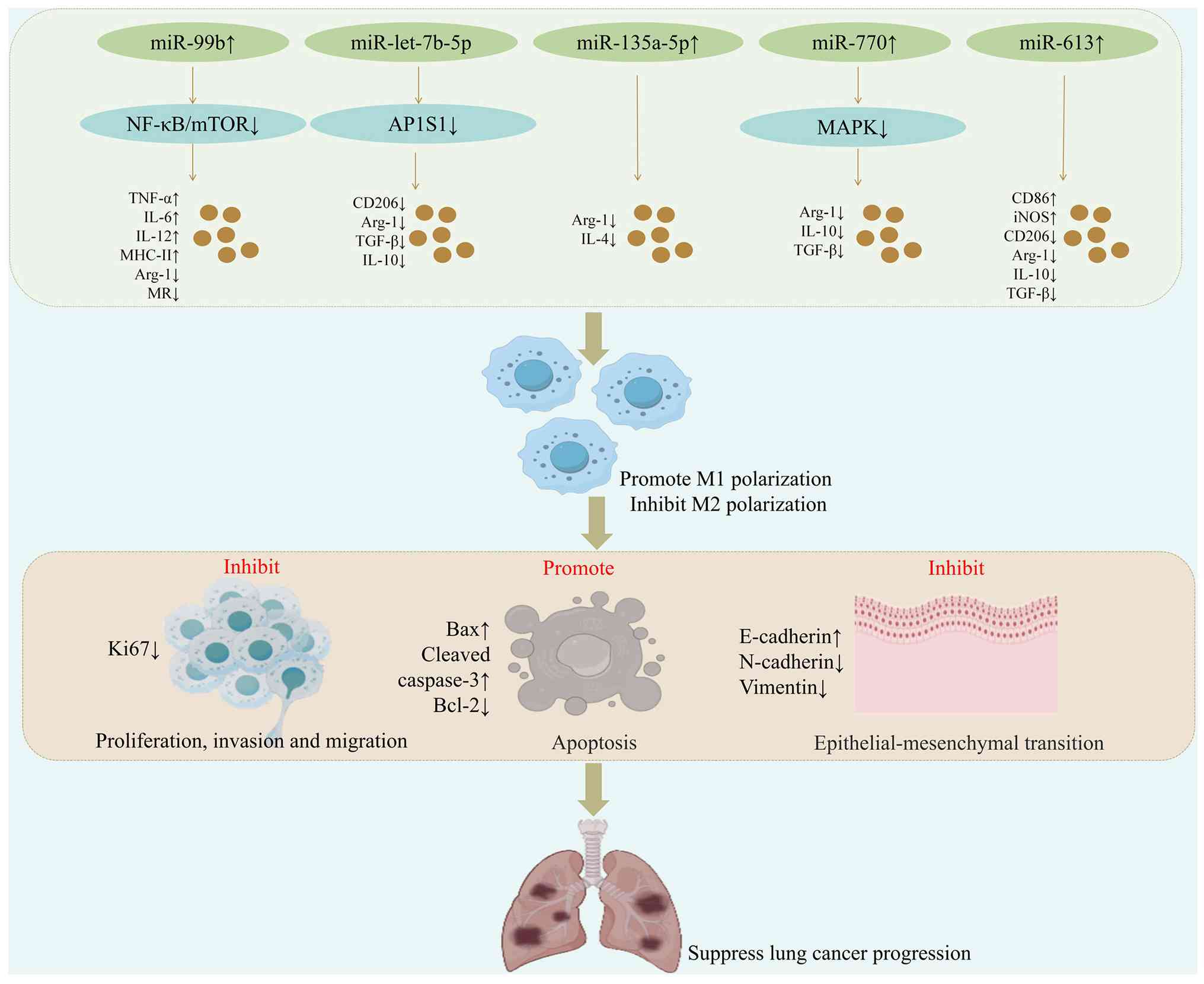 | Figure 3Mechanisms of miRNA-mediated
regulation of macrophage polarization in lung cancer therapy. The
image illustrates that miR-99b, miR-let-7b-5p, as well as the
upregulation of miR-135a-5p, miR-770 and miR-613 expression can
induce M1 polarization and inhibit M2 polarization, thereby
synergistically inhibiting the proliferation, invasion and
migration of lung cancer cells, inhibiting epithelial-mesenchymal
transition, and promoting cell apoptosis, thus significantly
delaying the growth and metastasis of lung cancer. MHC-II, major
histocompatibility complex class II; Arg-1, arginase-1; MR, mannose
receptor. |
miR-99b has been shown to participate in the
progression of various tumors owing to its potent immunomodulatory
functions (62). In the
microenvironment of lung cancer, miR-99b regulates the polarization
state of macrophages, thereby influencing the efficiency of
anti-tumor immune response. NF-κB and mTOR are key downstream
molecules through which miR-99b regulates macrophage polarization,
with mTOR identified as a direct target gene of miR-99b (63). The upregulation of miR-99b
suppresses κB-Ras2, subsequently activating NF-κB signaling and
boosting TNF-α, IL-6 and IL-12, and major histocompatibility
complex class II expression, all of which promote M1 polarization.
Concurrently, the elevated expression of miR-99b inhibits mTOR and
its downstream interferon regulatory factor (IRF)4, blocking the
mTOR/IRF4 axis and reducing arginase-1 (Arg-1) and Mannose receptor
(MR) expression, thereby restraining M2 polarization (64). Moreover, these changes improve
macrophage phagocytosis and antigen presentation, significantly
delaying tumor growth in a mouse transplanted tumor model (64). The let-7 family of miRNAs is
recognized as a class of tumor suppressors that play critical
regulatory roles in the initiation and progression of various
cancers (65). Specifically,
let-7b-5p has been shown to inhibit M2 polarization and enhance the
phagocytic activity of macrophages, thereby suppressing tumor
growth (66). The adaptor-related
protein complex 1 subunit sigma 1 (AP1S1) functions as a key
regulator of intracellular vesicle transport, and its aberrant
expression is closely associated with tumor progression, immune
evasion and malignant behavior (67). In lung cancer, the dysregulation
of AP1S1 may interfere with the antitumor immune response by
affecting vesicle transport. A previous study co-cultured
A549-derived extracellular vesicles (EVs) with phorbol 12-myristate
13-acetate (PMA)-induced differentiated THP-1 macrophages for 48 h
in vitro. They observed that the EV-delivered hsa-let-7b-5p
could directly target and inhibit the expression of AP1S1, thereby
regulating the AP1S1/p53 signaling pathway (68). Specifically, the EVs can target
and inhibit AP1S1 expression, leading to upregulation of CD86 and
inducible nitric oxide synthase (iNOS), the downregulation of
CD206, Arg-1, TGF-β and IL-10, and the subsequent inhibition of M2
polarization. Additionally, this regulation increases E-cadherin
expression, decreases N-cadherin and vimentin levels and suppresses
EMT, while also inhibiting the proliferation, invasion and
migration of A549 and H1299 cells (68). The activation of the STAT
signaling pathway mediates IL-4 secretion regulated by GATA binding
protein 3 (GATA3), a process that plays a crucial role in inducing
M2 macrophage polarization (69).
In the microenvironment of lung cancer, M2-type TAMs maintain an
immunosuppressive state by secreting cytokines such as IL-4,
thereby facilitating tumor immune escape. miR-135a-5p has been
demonstrated to exert tumor-suppressive effects in various
malignancies (70,71). The upregulation of miR-135a-5p
expression reduces the levels of STAT6, p-STAT6, GATA3 and IL-4,
thereby inhibiting the STAT6/GATA3/IL-4 signaling pathway, which in
turn decreases Arg-1 expression and suppresses M2 macrophage
polarization, thereby alleviating the immunosuppression mediated by
M2-type TAMs in lung cancer. Additionally, this upregulation
increases E-cadherin expression and decreases vimentin expression,
thereby attenuating the proliferative, invasive and migratory
abilities of tumor cells (72).
Furthermore, in vivo experiments using xenograft models have
confirmed that the upregulation of miR-135a-5p expression
downregulates Arg-1 levels in tumor tissues, inhibits M2 macrophage
polarization, and significantly impedes tumor growth and metastasis
(72). This confirms that
miR-135a-5p exerts a dual antitumor effect in lung cancer by
simultaneously reversing the immunosuppressive microenvironment and
inhibiting the malignant behavior of tumor cells (72). miR-770 has been shown to inhibit
tumorigenesis and EMT in NSCLC (73). The MAPK signaling pathway is known
to be involved in tumor cell growth, migration and invasion, In
NSCLC, the abnormal activation of the MAPK pathway is closely
associated with the malignant progression of the tumor and poor
prognosis (74). The upregulation
of miR-770 expression targets and downregulates MAP3K1, thereby
inhibiting the MAPK signaling pathway. This leads to reduced
expression of Arg-1, IL-10 and TGF-β, resulting in suppression of
M2 macrophage polarization, and this regulation helps to reverse
the immunosuppressive microenvironment. Additionally, miR-770
upregulation increases E-cadherin expression while decreasing
vimentin and N-cadherin expression, thereby inhibiting EMT, and
this is of utmost significance in preventing the epithelial-derived
tumor cells of NSCLC from acquiring mesenchymal phenotypes and
migratory abilities. Moreover, it induces apoptosis in K-MES-1,
A549 and NCI-H1650 cells, significantly reducing their viability
and suppressing proliferation, invasion and migration (75). In xenograft mouse models, the
upregulation of miR-770 expression has been shown to downregulate
CD206 expression and inhibit M2 macrophage polarization, while also
inducing apoptosis in tumor tissues and reducing Ki-67 expression,
thereby suppressing tumor growth (75). This indicates that this miRNA
exerts an antitumor effect in NSCLC by simultaneously promoting
immune remodeling and directly inhibiting the malignant phenotype
of the tumor (75). miR-613
exerts antitumor effects in various malignancies (76). The upregulation of miR-613
expression reduces the levels of CD206, Arg-1, IL-10 and TGF-β,
while increasing the expression of CD86 and iNOS, thereby
inhibiting M2 macrophage polarization and promoting M1
polarization, and this polarization remodeling helps to shift TAMs
from a tumor-promoting phenotype to a tumor-suppressing phenotype
(77). Additionally, miR-613
upregulation suppresses the proliferation, invasion and migration
of A549, HCC827, H1299 and H1650 cells, while also increasing the
expression of Bax and cleaved caspase-3, and decreasing Bcl-2
expression, thereby promoting the apoptosis of these cells
(77). This indicates that
miR-613 exerts a dual tumor-suppressing effect in lung cancer by
reprogramming the polarization balance of macrophages and directly
inducing apoptosis of tumor cells (77).
Pathogenic roles of miRNAs in regulating
macrophage polarization in lung cancer
During the progression of lung cancer, the
dysregulation of miRNA expression plays a crucial role in
regulating macrophage polarization. On the one hand, the
upregulation of miR-106a-5p, miR-21-5p, miR-3153, miR-146a, etc.,
can inhibit M1 polarization, promote M2 polarization, and promote
the proliferation, invasion and migration of lung cancer cells,
induce EMT, inhibit lung cancer cell apoptosis, and promote lung
cancer angiogenesis. On the other hand, the downregulation of
miR-335-5p, miR-4319, miR-103a, miR-155, miR-132-3p, etc., can
inhibit M1 polarization, promote M2 polarization and
synergistically enhance the maintenance of lung cancer cell
stemness, cell cycle progression and EMT, ultimately accelerating
tumor growth and metastasis. The mechanisms by which miRNAs
regulate macrophage polarization to promote lung cancer are
illustrated in Fig. 4.
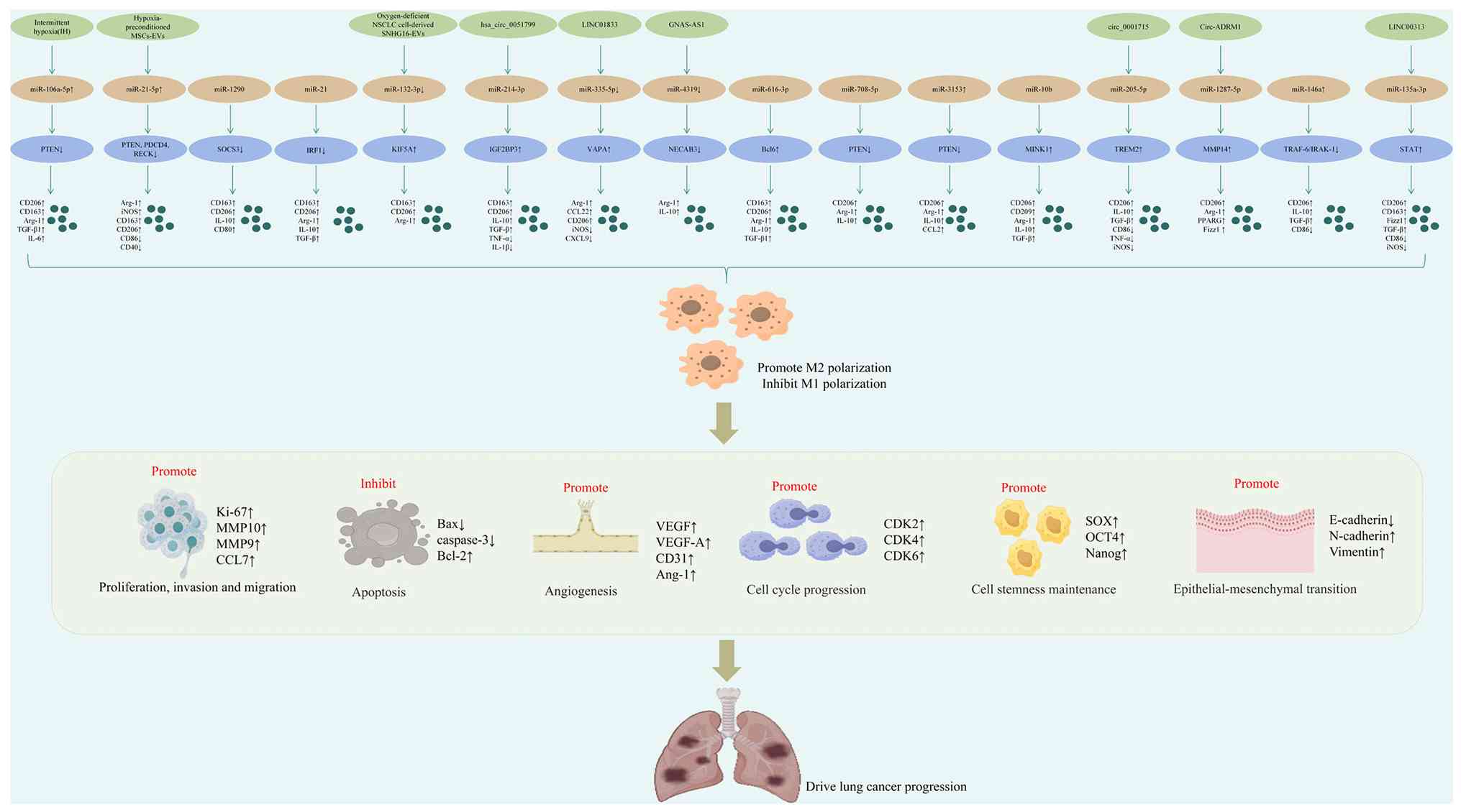 | Figure 4Mechanisms by which miRNAs regulate
macrophage polarization to promote lung cancer progression. The
image illustrates shows that the upregulation of miR-106a-5p,
miR-21-5p, miR-3153 and miR-146a can inhibit M1 polarization,
promote M2 polarization, and promote the proliferation, invasion
and migration of lung cancer cells, induce EMT, and inhibit lung
cancer cell apoptosis, while promoting lung cancer angiogenesis. In
addition, downregulation of miR-335-5p, miR-4319, miR-103a,
miR-155, and miR-132-3p can inhibit M1 polarization, promote M2
polarization, and synergistically enhance the maintenance of lung
cancer cell stemness, cell cycle progression and EMT, ultimately
accelerating tumor growth and metastasis. EMT,
epithelial-mesenchymal transition; IGF2BP3, insulin-like growth
factor 2 mRNA-binding protein 3; VAPA, vesicle-associated membrane
protein-associated protein A; NECAB3, N-terminal EF-hand type
calcium-binding protein 3; PTEN, phosphatase and tensin homolog;
MINK1, misshapen-like kinase 1; TREM2, triggering receptor
expressed on myeloid cells 2; TRAF6, TNF receptor-associated factor
6; IRAK1, interleukin-1 receptor-associated kinase 1. |
Regulation of macrophage polarization by
miRNAs driven by the hypoxic microenvironment
Obstructive sleep apnea (OSA) is one of the most
common sleep-disordered breathing conditions, characterized by
intermittent hypoxia (IH). Previous research has demonstrated that
OSA is closely associated with the progression of lung cancer
(78). On the one hand, exosomes
derived from the blood of patients with OSA can directly promote
the proliferation, invasion and migration of lung cancer TC1 cells
(78). On the other hand,
intermittent hypoxia, as the core pathological feature of OSA, has
been demonstrated to indirectly enhance the malignant phenotype of
TC1 cells by regulating macrophage polarization (79). miR-106a-5p expression has been
reported to be elevated in NSCLC (80). IH can upregulate miR-106a-5p
expression, inhibits phosphatase and tensin homolog (PTEN) and
activates the STAT3 signaling pathway. This cascade leads to the
upregulation of CD206, CD163, Arg-1, TGF-β1, IL-6, CXCR4 and CCL2,
along with the downregulation of iNOS, collectively promoting
macrophage polarization toward the M2 phenotype (81). Consequently, this enhances the
proliferation, invasion and migration of A549 and NCI-H226 cells,
while simultaneously downregulating E-cadherin expression and
upregulating N-cadherin and vimentin expression, thereby inducing
EMT and ultimately driving NSCLC progression (81). These findings reveal the molecular
mechanism by which OSA-related intermittent hypoxia reprograms
macrophage polarization through the miR-106a-5p/PTEN/STAT3 axis and
promotes the malignant phenotypic transformation of tumors,
providing a theoretical basis for targeted intervention in patients
with OSA and NSCLC. Hypoxia is a hallmark feature of the TME.
Tumor-derived exosomes under hypoxic conditions can remodel the
TME, promoting tumor progression, angiogenesis and pre-metastatic
niche formation (82). Suppressor
of cytokine signaling 3 (SOCS3) is a key regulator of innate and
adaptive immunity and plays a particularly crucial role in
macrophage polarization. In lung cancer, the dysregulation of SOCS3
expression may disrupt the immune balance and thereby promote
immune escape (83). miR-1290
targets and inhibits SOCS3 expression, leading to the activation of
the STAT3 signaling pathway (84). This results in increased
expression of CD163, CD206 and IL-10, along with the decreased
expression of CD80 and monocyte chemoattractant protein-1 (MCP-1),
thereby promoting macrophage polarization toward the M2 phenotype,
and this polarization process further exacerbated the
immunosuppressive state in the lung cancer microenvironment
(84). Additionally, the
upregulation of miR-1290 significantly enhances the proliferation,
invasion and migration of A549 and H1299 lung cancer cells. In a
mouse xenograft model, the overexpression of miR-1290 markedly
accelerates tumor growth. These findings further confirm that the
hypoxia-induced miR-1290/SOCS3/STAT3 axis promotes tumor formation
by reprogramming macrophages to the M2 phenotype and directly
facilitating the malignant behavior of lung cancer cells (84). IRF1 is a key transcription factor
involved in macrophage polarization and is regulated by multiple
miRNAs (85). miR-21 has been
shown to regulate lung cancer progression and EMT (86). The abnormal activation of the
PI3K/AKT pathway is closely associated with cell survival,
proliferation, and the establishment of an immunosuppressive
microenvironment in lung cancer (87). Under hypoxic conditions, miR-21
targets and inhibits IRF1 expression, leading to the increased
phosphorylation of PI3K and AKT, and the subsequent activation of
the PI3K/AKT signaling pathway. This cascade upregulates the
expression of CD163, CD206, Arg-1, IL-10 and TGF-β, thereby
promoting M2 macrophage polarization; this polarization process
further strengthens the immunosuppressive microenvironment,
creating favorable conditions for the immune escape and malignant
progression of lung cancer; at the same time, it enhances the
proliferative, invasive and migratory abilities of H1299 lung
cancer cells (87). In a mouse
subcutaneous tumor model, the overexpression of miR-21 under
hypoxic conditions was shown to significantly enhance the
proliferative capacity of H1299 cells and accelerate tumor growth
(87). This further confirms that
the hypoxia-induced miR-21/IRF1/PI3K/AKT axis, by inhibiting the
antitumor immune response mediated by IRF1 and promoting M2
polarization, collaboratively drives the malignant progression of
lung cancer. miR-214-3p can directly target oncogenes, thereby
inhibiting the malignant progression of LUAD (88). The hypoxic microenvironment
promotes the release of exosomal hsa_circ_0051799; this suggests
that hypoxia may impair the tumor-suppressive function of
miR-214-3p through a circRNA-mediated mechanism. Further
mechanistic investigations have revealed that hsa_circ_0051799
directly targets miR-214-3p, leading to the upregulation of
insulin-like growth factor 2 mRNA-binding protein 3 (IGF2BP3) and
the subsequent activation of the JAK/STAT3 signaling pathway, as
evidenced by the increased p-JAK2/JAK and p-STAT3/STAT ratios. This
cascade results in the upregulated expression of CD163, CD206,
IL-10, and TGF-β, thereby promoting M2 macrophage polarization,
while concurrently downregulating TNF-α, IL-1β, MCP-1 and IL-12,
thereby inhibiting M1 macrophage polarization; this polarization
imbalance has significantly exacerbated the immunosuppressive state
in the microenvironment of LUAD. Additionally, it upregulates
vimentin expression and downregulates E-cadherin expression,
inducing EMT. Furthermore, it significantly inhibits apoptosis in
A549, PC-9, H1975 and H1395 cells, while enhancing their
proliferative, invasive and migratory capabilities (89). These findings were further
validated in a xenograft model using nude mice (89). The aforementioned results confirm
that the hypoxia-induced
hsa_circ_0051799/miR-214-3p/IGF2BP3/JAK-STAT3 axis drives the
malignant progression of LUAD through multiple mechanisms, such as
reprogramming macrophage polarization balance, inducing EMT and
promoting tumor cell survival. Long non-coding RNA GNAS-AS1 has
been shown to play a role in tumor progression (90). Bioinformatics predictions have
indicated that miR-4319 is a potential binding target of GNAS-AS1.
N-terminal EF-hand type calcium-binding protein 3 (NECAB3) plays a
critical role in tumorigenesis by promoting the activation of
hypoxia-inducible factor-1 (HIF-1) (91). In lung cancer, the abnormal
activation of the HIF-1 signaling pathway is one of the core steps
that enable the tumor to adapt to the hypoxic microenvironment,
promote metabolic reprogramming, and drive malignant progression.
As a competitive endogenous RNA (ceRNA), GNAS-AS1 can sponge and
downregulate the expression of miR-4319, thereby relieving the
translation inhibitory effect of miR-4319 on NECAB3 and
subsequently upregulating the expression of NECAB3. This cascade
increases the expression of Arg-1 and IL-10, thereby promoting M2
macrophage polarization; this polarization process further
intensifies the immunosuppressive state in the lung cancer
microenvironment, creating favorable conditions for tumor immune
escape and malignant progression, while also enhancing the
proliferation, invasion and migration of A549 and H1299 cells
(92).
Regulation of macrophage polarization by
miRNAs mediated by lung cancer-derived exosomes
Mesenchymal stem cells (MSCs) are a group of highly
heterogeneous precursor cells characterized by their
anti-inflammatory, wound-healing-promoting and
homeostasis-maintaining properties (93). MSCs are also recruited to the TME,
where they secrete EVs that potently suppress antitumor immunity,
ultimately creating a favorable milieu for tumor progression
(94). As a well-established
anti-apoptotic and oncogenic miRNA, miR-21-5p serves not only as a
prognostic marker for lung cancer (95), but also, when downregulated,
promotes M1 macrophage polarization, thereby enhancing
pro-inflammatory responses and anti-tumor immunity (96). A previous comparative study
investigated the effects and mechanisms of exosomes derived from
MSCs under normoxic (N-EV) and short-term hypoxic preconditioning
(H-EV) conditions on NSCLC progression (97). The results demonstrated that
exosomes derived from MSCs subjected to hypoxic preconditioning
carry miR-21-5p, which targeted and reduced the expression of PTEN,
programmed cell death 4 and reversion-inducing cysteine-rich
protein with kazal motifs, consequently activating the Akt and
STAT3 signaling pathways (97).
This led to the increased expression of Arg-1, CD163, CD206, IL-10
and TGF-β, along with the decreased expression of CD86 and CD40,
collectively promoting M2 macrophage polarization, while inhibiting
M1 polarization; this polarization imbalance has significantly
exacerbated the immunosuppressive state in the microenvironment of
NSCLC, creating favorable conditions for tumor immune escape
(97). Furthermore, H-EVs
inhibited apoptosis in A549 and H23 cells, promoted their
proliferation, invasion and migration, and increased N-cadherin and
vimentin expression, thereby facilitating EMT. In vivo
xenograft models further confirmed that H-EVs significantly
increased Ki-67 and CD31 expression levels in tumor tissues,
promoting tumor cell proliferation and angiogenesis, respectively
(97). EVs serve as key mediators
of intercellular communication, and the miRNAs they carry can be
delivered to recipient cells, where they exert biological functions
by regulating the expression of multiple target genes (98). Notably, the hypoxic state within
the TME is not only a key driver of cancer growth and progression
(99), but is also closely
associated with immunosuppression (100). miR-103a is considered to
participate in hypoxia-induced reprogramming within the TME,
particularly in key processes, such as angiogenesis and immune
editing (101). In this context,
a previous study isolated EVs from human lung cancer CL1-5 cells
and investigated the impact of hypoxic conditions on their
functions. The expression of miR-103a in exosomes derived from lung
cancer cells was significantly downregulated under hypoxic
conditions, which targets and inhibits PTEN, leading to the
activation of the PI3K/AKT and STAT3 signaling pathways (102). Given that PTEN is one of the
most common inactivated tumor suppressor genes in lung cancer, its
functional deficiency leads to the continuous activation of the
PI3K/AKT pathway. Moreover, STAT3, as a key transcription factor
for inflammation and immune regulation, is also frequently
abnormally activated in lung cancer. The synergistic signal
transduction of these two factors constitutes a core molecular
event that drives the malignant progression of lung cancer and the
remodeling of the immune microenvironment. This cascade increases
the expression of Arg-1, IL-10, CCL18 and CCL22, thereby promoting
M2 macrophage polarization (102). This polarization process
significantly exacerbated the immunosuppressive state in the lung
cancer microenvironment, creating favorable conditions for tumor
immune escape and malignant progression. Additionally, hypoxic EVs
enhance the proliferation, invasion and migration of lung cancer
cells, while upregulating the expression of VEGF-A and
angiopoietin-1, thus promoting tumor angiogenesis (102). Long non-coding RNA small
nucleolar RNA host gene 16 (SNHG16), a newly identified oncogene,
has been shown to promote the progression of NSCLC (103). Kinesin family member 5A (KIF5A)
has been characterized as an oncogene that promotes invasiveness in
hepatocellular carcinoma, and its oncogenic role in LUAD has also
been validated (104).
miR-132-3p, which serves as a bridging molecule connecting SNHG16
and KIF5A, exhibits significant tumor-suppressive effects (96). As previously demonstrated,
exosomes derived from hypoxic NSCLC cells carrying SNHG16
(SNHG16-EVs) downregulate miR-132-3p expression, leading to the
upregulation of KIF5A. This cascade increases the expression of
CD163, CD206 and Arg-1, thereby promoting M2 macrophage
polarization; this polarization process further exacerbates the
immunosuppressive microenvironment of NSCLC. Additionally, it
enhances the expression of stemness markers, SOX2, OCT4 and Nanog,
thereby strengthening the stemness of A549 and H1299 cells. Since
tumor stem cells are closely associated with drug resistance,
recurrence and metastasis in NSCLC, their enhanced stemness confers
stronger self-renewal capabilities and treatment resistance to
tumor cells. Moreover, it increases N-cadherin and vimentin
expression, while decreasing E-cadherin expression, thereby
inducing EMT (60). Furthermore,
it upregulates cyclin-dependent kinase (CDK)2, CDK4 and CDK6
levels, accelerating cell cycle progression (60). Given that CDK2/4/6 are the core
regulatory factors for the G1/S phase transition, their abnormal
high expression in NSCLC drives the rapid proliferation of tumor
cells and is an important molecular basis for the malignant
progression of lung cancer. Radiotherapy is one of the primary
treatment modalities for NSCLC, with >50% of patients receiving
radiotherapy to achieve local tumor control, symptom relief, or
disease stabilization (105).
However, resistance to radiotherapy is a significant challenge that
limits its clinical efficacy. Moreover, immune cells within the
TME, particularly TAMs, play a crucial role in mediating
radiotherapy resistance. miR-616-3p has been reported to be
aberrantly expressed in various malignancies, including gastric,
breast and colon cancers (106).
PTEN plays a critical role in regulating macrophage polarization,
and it is one of the most common inactivated tumor suppressor genes
in NSCLC. Its functional deficiency leads to the continuous
activation of the PI3K/AKT pathway (107). The PTEN/PI3K/AKT signaling
pathway is also key to both macrophage polarization and the
progression of NSCLC, and the abnormal activation of this pathway
is closely related to cell survival, proliferation and the
establishment of an immunosuppressive microenvironment in NSCLC,
and constitutes a core molecular event driving the progression of
malignancy (108). miR-616-3p
expression is upregulated in NSCLC tissues and is further elevated
following radiotherapy, potentially contributing to radiotherapy
resistance by influencing macrophage polarization, this
polarization process not only exacerbates the immunosuppressive
microenvironment, but also directly weakens the cytotoxic effect of
radiotherapy through paracrine mechanisms, ultimately accelerating
the malignant progression of NSCLC. Mechanistically, miR-616-3p in
exosomes of NSCLC cells induced by radiotherapy can target and
inhibit PTEN expression, upregulate the p-PI3K/PI3K and p-AKT/AKT
ratios, and activate the PI3K/AKT signaling pathway. This
subsequently increases the expression of M2 polarization markers,
such as CD163, CD206, Arg-1, IL-10 and TGF-β1, promoting macrophage
polarization toward the M2 phenotype and ultimately accelerating
the malignant progression of NSCLC. These findings suggest that the
miR-616-3p/PTEN/PI3K/AKT axis induced by radiotherapy reprograms
macrophages towards M2 polarization, establishing an
immunosuppressive and survival-promoting microenvironment, thereby
driving the radiotherapy resistance and malignant progression of
NSCLC (109). Targeting this
axis may provide a novel therapeutic strategy for overcoming
resistance to radiotherapy (109). The activation of the programmed
cell death protein 1 (PD-1)/programmed cell death 1 ligand 1
(PD-L1) signaling axis suppresses T-cell-mediated immune responses
(110). IFN-γ upregulates PD-L1
expression, and the overexpression of PD-L1 on the surface of tumor
cells can induce the apoptosis of tumor-infiltrating T-cells,
thereby inhibiting antitumor immunity (111). A previous study demonstrated
that the expression of miR-708-5p in exosomes derived from LUAD
cells was significantly upregulated, and this suggests that this
miRNA may exert multiple regulatory effects in the LUAD
microenvironment through exosome-mediated intercellular
communication (112). Further
investigations revealed that miR-708-5p targets and inhibits PTEN
expression, increases the p-AKT/AKT and p-mTOR/mTOR ratios, and
activates the AKT/mTOR signaling pathway, thereby upregulating the
expression of M2 polarization markers, such as CD206, Arg-1 and
IL-10, ultimately inducing macrophage polarization toward the M2
phenotype; this polarization process significantly exacerbated the
immunosuppressive state in the microenvironment of LUAD.
Concurrently, miR-708-5p induces PD-L1 transcription and
translation, enhancing the proliferation, invasion and migration of
H1299 cells, and promoting immune escape. Mechanistic analyses
indicate that exosome-derived miR-708-5p stimulates IFN-γ
production in T-cells, while concurrently inhibiting
CD8+ T-cell function, exerting an immunomodulatory
effect (112). Furthermore, in
an Lewis lung cancer cells (LLCs) tumor transplantation model,
miR-708-5p was confirmed to accelerate transplanted tumor growth by
upregulating PD-L1 expression (112). The aforementioned results
indicate that the miR-708-5p/PTEN/AKT/mTOR axis drives the
oncogenic function of LUAD malignancy by simultaneously inducing
M2-type macrophage polarization and upregulating PD-L1-mediated
immune escape (112).
Regulation of macrophage polarization by
long non-coding RNAs mediated by miRNAs
Cancer-associated fibroblasts (CAFs), as core
components of the TME, promote tumor growth and metastasis through
the secretion of exosomes (113). miR-335-5p plays an oncogenic
role in NSCLC (114). The
expression of CAF-derived LINC01833 is significantly upregulated in
NSCLC tissues, and its high expression is closely associated with a
poor patient prognosis; this suggests that it may play a key role
as a carcinogenic factor in the progression of NSCLC (115). Vesicle-associated membrane
protein-associated protein A (VAPA) is involved in intracellular
transport and microtubule-related processes, and its dysregulation
can lead to cellular dysfunction (116). Mechanistic study has revealed
that LINC01833 downregulates miR-335-5p expression, leading to
upregulation of VAPA. This cascade reduces the expression of iNOS
and CXCL9, while increasing the expression of Arg-1, CCL22, and
CD206, thereby promoting M2 macrophage polarization and inhibiting
M1 polarization; this polarization significantly exacerbates the
immunosuppressive state in the microenvironment of NSCLC.
Consequently, it enhances the proliferation, invasion, and
migration of NSCLC cells (117).
The aforementioned findings indicate that the
LINC01833/miR-335-5p/VAPA axis derived from CAFs reprograms
macrophages to the M2 phenotype and directly promotes the malignant
behavior of tumor cells, playing a dual oncogenic role in the
progression of NSCLC. Recent studies have demonstrated that the
high expression of the long non-coding RNA LINC00313 is
significantly associated with a poor prognosis of patients with
cancer, suggesting its role as a pro-tumorigenic factor that drives
tumor progression (118,119). Conversely, miR-135a-3p functions
as a tumor suppressor (120).
STAT6 is the core transcription factor of the IL-4/IL-13 signaling
pathway. Its activation serves as a key molecular switch that
drives the polarization of M2-type macrophages. In the lung cancer
microenvironment, the abnormal activation of STAT6 can induce the
transcription of a series of immunosuppressive factors, thereby
promoting tumor immune escape (121). Research has revealed that
LINC00313 can function as a ceRNA sponge-like molecule that binds
to miR-135a-3p to upregulate STAT6 expression, leading to the
increased expression of M2-associated markers, such as CD206,
CD163, found in inflammatory zone 1 (Fizz1) and TGF-β, thereby
promoting M2 macrophage polarization. Concurrently, it reduces the
expression of M1-associated markers including CD86, iNOS, TNF-α and
IL-1β, inhibiting M1 macrophage polarization; this polarization
process significantly exacerbates the immunosuppressive state in
the lung cancer microenvironment. Additionally, LINC00313 promotes
the proliferation, invasion and migration of A549, NCI-H1299 and
H1975 cells (121). Results from
in vivo xenograft models are highly consistent with in
vitro findings, further confirming the central pro-tumorigenic
role of LINC00313, and this indicates that the
LINC00313/miR-135a-3p/STAT6 axis reprograms macrophages to the M2
phenotype through reprogramming and directly promotes the malignant
behavior of lung cancer cells, thereby exerting a dual carcinogenic
function (121).
Regulation of macrophage polarization by
circular RNA (circRNA)-encoded miRNAs
The JAK/STAT3 pathway plays a critical regulatory
role in tumor development and macrophage polarization. In LUAD, the
continuous phosphorylation activation of STAT3 is one of the key
molecular events that drive the malignant progression of the tumor
(such as lymphatic metastasis). A study has shown that IGF2BP3 can
stabilize SRC mRNA through m6A-dependent mechanisms,
thereby triggering the activation of the STAT3 pathway and exerting
a carcinogenic effect in lung cancer. Its abnormal high expression
is closely related to poor patient prognosis (122). circRNAs, as a distinct class of
non-coding RNA molecules, exhibit differential expression between
LUAD tissues and adjacent normal tissues, and play critical roles
in tumor proliferation, metastasis and prognosis (123). For instance, circ_0001715
promotes the growth and metastasis of NSCLC cells by regulating the
miR-1249-3p/FGF5 signaling axis (124). Moreover, miR-205-5p functions as
a tumor suppressor in various types of cancer, and its deficiency
or dysfunction significantly accelerates the malignant progression
of lung cancer (125). Further
research has revealed that circ_0001715 directly targets
miR-205-5p, leading to triggering receptor expressed on myeloid
cells-2 upregulation and the increased expression of M2-associated
markers, such as CD206, IL-10 and TGF-β, thereby promoting M2
macrophage polarization (126).
Concurrently, it reduces the expression of M1-associated markers
including CD86, TNF-α and iNOS, inhibiting M1 macrophage
polarization; this polarization process significantly exacerbates
the immunosuppressive state in the microenvironment of LUAD.
Moreover, circ_0001715 significantly enhances the proliferation,
invasion and migration of A549 and H1975 cells (126). These results indicate that the
circ_0001715/miR-205-5p/IGF2BP3/JAK-STAT3 axis reprograms the
polarization balance of macrophages and directly promotes the
malignant phenotype of LUAD cells, exerting a dual carcinogenic
effect (126). Matrix
metalloproteinase (MMP)14, a key member of the MMP family, has been
shown to promote cancer cell migration and invasion, thereby
accelerating malignant tumor progression. In lung cancer, MMP14 can
promote tumor infiltration and metastasis through multiple
mechanisms such as degrading the extracellular matrix (ECM),
releasing growth factors, and activating other members of the MMP
family (127). miR-1287-5p
functions as a tumor suppressor in various malignancies, and its
dysregulation is closely associated with tumor initiation and
progression (128). A previous
study revealed that MMP14 expression levels were significantly
negatively associated with the overall survival of patients with
LUAD; this indicates that MMP14 is a key negative regulatory factor
for the prognosis of LUAD (129). Further mechanistic
investigations demonstrated that circ-ADRM1 functioned as a ceRNA
sponge-like molecule that binds to miR-1287-5p and recruited the
deubiquitinating enzyme USP12, synergistically upregulating MMP14
expression. This led to the increased expression of M2-associated
markers such as CD206, Arg-1, peroxisome proliferator-activated
receptor (PPAR)G and Fizz1, thereby promoting M2 macrophage
polarization; this further exacerbated the immunosuppressive state
mediated by M2-type TAMs in the LUAD microenvironment (129). Additionally, circ-ADRM1 enhanced
the proliferation, invasion and migration of A549, PC-9, H1975,
H1435 and H2030 cells. The aforementioned results indicate that the
circ-ADRM1/miR-1287-5p/USP12/MMP14 axis synergistically promotes
the malignant progression of LUAD through multiple mechanisms,
including promoting ECM degradation, driving M2 polarization of
macrophages, and directly enhancing the malignant phenotype of
tumor cells (129).
Regulation of macrophage polarization by
miRNAs mediated by other factors
Cypermethrin (CYM) exhibits estrogen receptor
activity and has been shown to induce cell proliferation and
promote tumor metastasis (130).
Previous research has demonstrated that the upregulation of miR-155
promotes M1 macrophage polarization (131). The upregulation of Bcl6
expression can promote immunosuppressive signals, and the
inhibition of the JNK pathway is closely related to the remodeling
of the immune microenvironment in lung cancer. CYM can downregulate
miR-155 expression, leading to the upregulation of Bcl6 and tge
subsequent downregulation of mitogen-activated protein kinase 4
(MKK4), which in turn inhibits the JNK signaling pathway. This
cascade results in the decreased expression of TNF-α, IL-1β, and
iNOS, along with the increased expression of Arg-1, Fizz1 and
macrophage galactose N-acetyl-galactosamine specific lectin,
thereby promoting M2 macrophage polarization, while inhibiting M1
polarization; this polarization significantly exacerbates the
immunosuppressive state in the lung cancer microenvironment.
Additionally, CYM upregulates the expression of MMP10, MMP9, and
CCL7, thereby promoting the invasion and metastasis of LLCs. In
lung cancer mouse models, CYM has been confirmed to drive TAMs
toward the metastasis-promoting M2 phenotype and significantly
increase the number of lung metastases (132). The aforementioned results
indicate that CYM reprograms the imbalance of macrophage
polarization through the miR-155/Bcl6/MKK4/JNK axis, upregulates
invasion-related molecules and strengthens the immunosuppressive
microenvironment, thereby synergistically driving the invasion and
metastasis of lung cancer (132). Misshapen-like kinase 1 (MINK1)
has been identified as an activator of the JNK pathway (133), which is associated with M2
macrophage polarization (134).
miR-3153 is differentially expressed in the plasma of patients with
NSCLC; this suggests that it may serve as a diagnostic or
prognostic biomarker and be involved in the progression of the
disease (135). It has been
demonstrated that the upregulation of miR-3153 activates the JNK
signaling pathway by targeting and enhancing MINK1 expression,
leading to the increased expression of M2 polarization markers,
such as CD206, Arg-1, IL-10 and CCL2, thereby promoting M2
macrophage polarization, further exacerbating the immunosuppressive
microenvironment in NSCLC. Additionally, miR-3153 upregulation
enhances the proliferation, invasion, and migration of A549 and
Calu3 cells. In mouse models, elevated miR-3153 expression
significantly promotes tumor growth (136). The aforementioned results
indicate that the miR-3153/MINK1/JNK axis promotes the polarization
of macrophages towards a tumor-promoting M2 phenotype through
coordinated reprogramming and directly enhances the malignant
behavior of tumor cells, thereby driving the malignant progression
of NSCLC (136). miR-10b
expression is significantly upregulated in NSCLC tissues compared
with adjacent normal tissues; this suggests that it may play a
crucial role in promoting cancer development and progression in
NSCLC (137). Further research
has revealed that miR-10b promotes M2 macrophage polarization, as
evidenced by increased expression of CD206, CD209, Arg-1, TNF-α,
IL-6, IL-10, and TGF-β. Additionally, miR-10b enhances the
proliferation, invasion, and migration of A549, NCI-H1650, and
NCI-H1299 cells. Moreover, it increases N-cadherin expression,
while decreasing E-cadherin expression, thereby inducing EMT
(138). The aforementioned
results indicate that miR-10b in NSCLC promotes cancer development
by reprogramming macrophages to the M2 phenotype and inducing EMT
transformation in tumor cells, exerting a dual carcinogenic effect
(138). Within the TME, miR-146a
has been shown to promote M2 macrophage polarization, thereby
exerting tumor-promoting effects (139,140). TNF receptor-associated factor 6
(TRAF6) and interleukin-1 receptor-associated kinase 1 (IRAK1) play
a central role in promoting M1-type polarization and antitumor
immune responses. It has been revealed that the upregulation of
miR-146a targets and downregulates TRAF6 and IRAK1, leading to the
increased expression of M2-associated markers, such as CD206,
IL-10, and TGF-β, which promotes M2 macrophage polarization; this
polarization process significantly exacerbated the
immunosuppressive state in the lung cancer microenvironment.
Conversely, it reduces CD86 expression, inhibiting M1 macrophage
polarization (141).
Additionally, miR-146a suppresses apoptosis of H1299 cells, while
enhancing their proliferative, invasive and migratory abilities.
These results indicate that miR-146a in lung cancer exerts a
synergistic carcinogenic effect by inhibiting the
TRAF6/IRAK1-mediated M1 polarization signal, reprogramming
macrophages to the M2 phenotype, and directly promoting the
survival and malignant behavior of tumor cells (141). The mechanisms by which miRNAs
regulate macrophage polarization in lung cancer are summarized in
Table I.
 | Table ISummary of the mechanisms of the
regulation of macrophage polarization in lung cancer by miRNAs. |
Table I
Summary of the mechanisms of the
regulation of macrophage polarization in lung cancer by miRNAs.
| Regulator | miRNA | Expression
change | Direct targets and
changes | Lung cancer study
model | Regulation of
macrophage polarization | Effect on lung
cancer | (Refs.) |
|---|
| Unspecified | miR-99b | Upregulation | NF-κB, mTOR ↓ | Lung cancer
subcutaneous tumor model | Promote M1
polarization; Inhibit M2 polarization | Suppress lung
cancer progression | (64) |
| Unspecified | miR-let-7b-5p | Unspecified | AP1S1 ↓ | A549 and H1299
cells | Promote M1
polarization; Inhibit M2 polarization | Suppress lung
cancer progression | (68) |
| Unspecified | miR-135a-5p | Upregulation | Unspecified | A549, H1299 and
H460 cells | Promote M2
polarization; Inhibit M1 polarization | Suppress lung
cancer progression | (72) |
| Unspecified | miR-770 | Upregulation | MAPK ↓ | K-MES-1, A549 and
NCI-H1650 cells; transgenic mouse model for
xenotransplantation | Promote M1
polarization; Inhibit M2 polarization | Suppress lung
cancer progression | (75) |
| Unspecified | miR-613 | Upregulation | Unspecified | NSCLC patients;
A549, HCC827, H1299 and H1650 cells | Promote M1
polarization; Inhibit M2 polarization | Suppress lung
cancer progression | (77) |
| Intermittent
hypoxia (IH) | miR-106a-5p | Upregulation | PTEN↓ | NSCLC and OSAS
patients; Tumor xenograft model of nude mouse; A549 and NCI-H226
cells | Promote M2
polarization; Inhibit M1 polarization | Drive lung cancer
progression | (81) |
| Unspecified | miR-1290 | Unspecified | SOCS3 ↓ | A549 and H1299
cells; Tumor xenograft model of nude mouse | Promote M2
polarization; Inhibit M1 polarization | Drive lung cancer
progression | (84) |
| Unspecified | miR-21 | Unspecified | IRF1 ↓ | NSCLC patients;
H1299 cells; Subcutaneous tumor mouse model | Promote M2
polarization; Inhibit M1 polarization | Drive lung cancer
progression | (87) |
|
hsa_circ_0051799 | miR-214-3p | Unspecified | IGF2BP3 ↑ | A549, PC-9, H1975
and H1395 cells; Tumor xenograft model of nude mouse | Promote M2
polarization; Inhibit M1 polarization | Drive lung cancer
progression | (89) |
| GNAS-AS1 | miR-4319 | Downregulation | NECAB3 ↓ | NSCLC patients;
A549 and H1299 cells | Promote M2
polarization; Inhibit M1 polarization | Drive lung cancer
progression | (92) |
|
Hypoxia-preconditioned MSCs-EVs | miR-21-5p | Upregulation | PTEN, PDCD4, RECK
↓ | A549 and H23 cells;
tumor xenograft model of nude mouse | Promote M2
polarization; Inhibit M1 polarization | Drive lung cancer
progression | (97) |
| Hypoxic EVs | miR-103a | Downregulation | PTEN ↓ | CL1-5 cells;
NCI-H2087, NCI-H1792 and NCI-H1437 | Promote M2
polarization; Inhibit M1 polarization | Drive lung cancer
progression | (102) |
| Oxygen-deficient
NSCLC cell-derived SNHG16-EVs | miR-132-3p | Downregulation | KIF5A ↑ | NSCLC patients;
A549 and H1299 cells | Promote M2
polarization; Inhibit M1 polarization | Drive lung cancer
progression | (60) |
| Unspecified | miR-616-3p | Unspecified | PTEN ↓ | NSCLC and OSAS
patients; A549 and H1299 cells | Promote M2
polarization; Inhibit M1 polarization | Drive lung cancer
progression | (109) |
| Unspecified | miR-708-5p | Unspecified | PTEN ↓ | A549, H1299 and
H358 cells | Promote M2
polarization; Inhibit M1 polarization | Drive lung cancer
progression | (112) |
| LINC01833 | miR-335-5p | Downregulation | VAPA ↑ | NSCLC patients;
Tumor xenograft model of nude mouse | Promote M2
polarization; Inhibit M1 polarization | Drive lung cancer
progression | (117) |
| LINC00313 | miR-135a-3p | Upregulation | STAT ↑ | A549, NCl-H1299 and
H1975 cells; Tumor xenograft model of nude mouse | Promote M2
polarization; Inhibit M1 polarization | Drive lung cancer
progression | (121) |
| circ_0001715 | miR-205-5p | Unspecified | TREM2 ↑ | A549 and H1975
cells | Promote M2
polarization; Inhibit M1 polarization | Drive lung cancer
progression | (126) |
| Circ-ADRM1 | miR-1287-5p | Unspecified | MMP14 ↑ | A549, PC-9, H1975,
H1435 and H2030 cells | Promote M2
polarization; Inhibit M1 polarization | Drive lung cancer
progression | (129) |
| CYM | miR-155 | Downregulation | Bcl6 ↑ | LLCs | Promote M2
polarization; Inhibit M1 polarization | Drive lung cancer
progression | (132) |
| Unspecified | miR-3153 | Upregulation | MINK1 ↑ | LUAD patients; A549
and Calu3 cells | Promote M2
polarization; Inhibit M1 polarization | Drive lung cancer
progression | (136) |
| Unspecified | miR-10b | Unspecified | Unspecified | A549, NCI-H1650 and
NCI-H1299 cells | Promote M2
polarization; Inhibit M1 polarization | Drive lung cancer
progression | (137) |
| Unspecified | miR-146a | Upregulation | TRAF-6, IRAK-1
↓ | H1299 cells | Promote M2
polarization; Inhibit M1 polarization | Drive lung cancer
progression | (141) |
Synopsis
The prevention and treatment of lung cancer remain
major research challenges, and the occurrence and progression of
this malignancy are closely linked to macrophage polarization.
Regulating the balance between M1 and M2 polarization has thus
emerged as a key intervention strategy. Various miRNAs modulate the
polarization balance of macrophages through alterations in their
own expression and exert anti-lung cancer effects via
multi-targeted synergistic mechanisms. Through summarization and
analysis, it was found that:
As regards macrophage polarization and changes in
miRNA expression, the upregulation of miR-99b (64), miR-let-7b-5p (68), miR-135a-5p (72), miR-770 (75) and miR-613 (77) promotes M1 polarization, while
inhibiting M2 polarization. By contrast, the upregulation of
miR-106a-5p (81), miR-21-5p
(97), miR-3153 (136) and miR-146a (141), as well as the downregulation of
miR-335-5p (117), miR-4319
(92), miR-103a (102), miR-155 (132) and miR-132-3p (60), inhibits M1 polarization and
promotes M2 polarization. The underlying basis for these
differential effects lies in the specificity of target genes and
signaling pathways, and their realization is regulated by cell type
specificity, temporally specific expression, and local
microenvironmental signals. In terms of the specificity of target
genes and signaling pathways, miRNAs can regulate different
polarization directions through specific targets. For instance, in
M1-type macrophages activated by TLR4, miR-155 negatively regulates
the negative regulatory factors of signaling pathways, such as
SHIP1 and SOCS1, thereby promoting M1 polarization. However, in M2
polarization induced by IL-4, the expression pattern of miR-155 is
completely opposite to that observed in M1: miR-155 is
significantly upregulated in M1 polarization, whereas its
expression remains largely unchanged or mildly decreased in M2
polarization. This opposite regulatory direction suggests that the
function of miR-155 is highly dependent on the specific activated
signaling pathways and target gene networks involved (142). The time-specific expression is
manifested in the temporal regulation of macrophage polarization
process by miRNAs. During the 8-h dynamic process of bone
marrow-derived macrophages polarizing into the M1 type, M1-related
miRNAs can be classified into the early response group and late
response group based on the sequence of their expression peaks.
Among these, miR-1931, miR-3473e and miR-5128 have been verified as
early response miRNAs, suggesting that they play a key regulatory
role in the initiation stage of polarization (143). Furthermore, the target sites of
miR-155 in activated macrophages, dendritic cells and B-cells are
~20 to 75% cell-specific. This specificity persists in the
co-expression of miR-155 with its targets SOCS1 and SHIP1, which
exhibited a coordinated expression in single macrophages and tissue
niches, reflecting fine-tuning rather than binary regulation. This
difference is not due to the variable polyadenylation of the 3'UTR,
but is determined by the intrinsic cellular environment (such as
transcription factors, chromatin state) (144).
Signaling pathway regulation exhibits distinct
patterns. miRNAs regulate the polarization state of macrophages by
targeting multiple key signaling pathways, thereby influencing the
progression and prognosis of lung cancer. According to the existing
literature, these pathways mainly involve the NF-κB pathway, the
PTEN/PI3K/AKT pathway, the MAPK pathway and the JAK/STAT pathway,
etc. The regulatory patterns of each pathway exhibit certain
regular characteristics. Among these, the PTEN/PI3K/AKT pathway is
the core hub for miRNA to regulate M2 polarization, and it is
particularly crucial in hypoxic microenvironments. The NF-κB and
PI3K/AKT pathways are predominantly activated, whereas the MAPK
pathway is generally inhibited. Notably, the JNK and STAT-related
pathways exert dual regulatory effects. For instance, miR-155
downregulation inhibits JNK (132), while miR-3153 upregulation
activates it (136); both
promote M2 polarization and lung cancer progression. Similarly, the
upregulation of miR-106a-5p (81), miR-21-5p (97), miR-1290 (84) and miR-214-3p (89), along with tge downregulation of
miR-103a (102), activates STAT
pathways, favoring M2 polarization and tumor progression. By
contrast, tge upregulation of miR-135a-5p (72) inhibits the STAT6/GATA3/IL-4 axis,
suppressing M2 polarization and exerting an anti-lung cancer
effect. The differential regulatory effects of these pathways may
be attributed to factors, such as the specificity of target genes
and signaling pathways, cell type and microenvironment dependence,
distinct nodal points within the pathways, as well as the dose
effects and the network balance of gene expression. These pathways
do not exist independently, but are intertwined to form a complex
regulatory network. Microenvironmental factors, such as hypoxia and
inflammation can significantly affect the activity of these
pathways, thereby altering the regulatory direction of miRNAs.
Specifically, lung cancer cells release EVs containing miR-20a-5p
under hypoxic conditions, which can target and inhibit PTEN,
activate AKT, and promote M2 phenotypic polarization (145). Additionally, lung adenocarcinoma
M2 macrophages highly express AQP3. Through the PPAR-γ/NF-κB axis,
they promote M2 polarization and upregulate IL-6, thereby affecting
tumor growth and migration (146). Future research is required to
further clarify the interactions among these pathways and their
specificity in different stages and subtypes of lung cancer,
providing a theoretical basis for targeted precision treatment of
imbalanced macrophage polarization.
As regards the regulatory coordination mechanisms,
the main aspects involved include proliferation, invasion and
migration (64,68,72,77,81,84,87,92,97,102,109, 112,117,121,126,129,132,136,138,141), apoptosis (75,77,141), angiogenesis (97,102), cell cycle regulation (60), maintenance of stemness (60) and EMT (60,68,81,97,138). Among these, studies on
proliferation, invasion and migration, EMT and apoptosis are more
prevalent, whereas investigations into angiogenesis, cell cycle
regulation, and cancer stemness are relatively limited. To date,
there have been no systematic reports in the literature regarding
miRNAs regulating iron death or disulfide bond death, which are
novel forms of cell death, by modulating macrophage polarization,
thereby influencing the complete regulatory network of the
malignant progression of lung cancer. The observed heterogeneity in
the focus of these coordinated regulatory mechanisms may be closely
related to limitations in existing model systems and detection
methods, inconsistencies in the depth of mechanistic interpretation
across different phenotypes, and potential biases in target
recognition during functional screening. For instance, in the TME
of commonly used mouse models, the infiltration of T-cells is
significantly lower than that in human tumors, while the density of
macrophages is abnormally high (147). Additionally, two-dimensional
co-culture or tumor organoid models often fail to accurately
reflect the complex regulatory environment of multiple factors in
the body due to the lack of complete immune cell components
(148). More importantly,
genome-wide CRISPR screening relies on in vitro
immortalization or bone marrow-derived macrophages, whose genetic
epigenetic states are different from those of primary TAMs
(149). This makes it prone to
screening out targets whose functions are masked or compensated
in vivo, resulting in clinical transformation
deviations.
Research depth varies significantly across miRNAs.
The most extensive studies, i.e, those covering cellular, animal
and clinical levels, are available for miR-106a-5p and miR-21.
miR-616-3p, miR-3153, miR-4319 and miR-132-3p have been validated
in both cellular systems and clinical samples. miR-1290, miR-214-3p
and miR-770 have been examined in cell-based assays and xenograft
models. In contrast, most other miRNAs (e.g., miR-let-7b-5p,
miR-155) remain at the cellular stage. These substantial
differences in research depth may be attributed to factors, such as
the expression abundance of individual miRNAs, the difficulty of
detection in clinical samples, the extent to which their potential
as therapeutic targets drives research investment, the
accessibility of sample types and the feasibility of establishing
corresponding animal models. It is worth noting that the current
research methodology has certain limitations. In terms of cell
experiments, monolayer culture lacks the three-dimensional spatial
structure and complete components of the tumor microenvironment
(immune cells, CAFs, vascular endothelial cells and the cytokine
network), making it difficult to assess the immune killing effect
(150). In terms of animal
models, species differences lead to fundamental differences in
immunity, metabolism and pharmacokinetics between mice and humans,
resulting in deviations in result transformation; at the same time,
subcutaneous transplanted tumors, orthotopic transplanted tumors,
chemical induction models, and genetically engineered mice all have
a disconnection from the natural occurrence and progression process
of lung cancer, restricting the clinical translatability of the
results (151).
In the hypoxic TME, the expression of multiple
miRNAs becomes dysregulated, collectively exerting pro-tumorigenic
effects on lung cancer by modulating macrophage polarization.
Specifically, under hypoxic conditions, the expression of
miR-106a-5p and miR-21-5p is upregulated, whereas the expression of
miR-103a, miR-132-3p, miR-1290, miR-21 and miR-214-3p is
downregulated. These alterations promote M2 polarization, while
inhibiting M1 polarization, thereby driving the malignant
progression of lung cancer. Collectively, these findings suggest
that hypoxia, as a hallmark feature of the lung cancer
microenvironment, induces miRNA dysregulation, which in turn drives
macrophage polarization toward the M2 phenotype. This process
reshapes the tumor immune microenvironment, promotes angiogenesis
and tumor invasion, and exacerbates local hypoxia, ultimately
forming a closed-loop positive feedback regulatory network.
A single miRNA can simultaneously intervene in
multiple key aspects of tumor progression through its
multi-targeting characteristics. For example, in terms of
pro-tumorigenic synergy, the upregulation of miR-21-5p promotes
proliferation, invasion, migration, angiogenesis and EMT, while
inhibiting apoptosis (97); the
downregulation of miR-103a enhances proliferation, invasion,
migration and angiogenesis (102); miR-10b promotes proliferation,
invasion and migration, while inducing EMT (138). In terms of tumor-suppressive
synergy, miR-let-7b-5p (68) and
the upregulation of miR-135a-5p (72) both inhibit proliferation,
invasion, migration and EMT; the upregulation of miR-146a inhibits
proliferation, invasion and migration, while promoting apoptosis
(141); and the upregulation of
miR-770 inhibits proliferation, invasion and migration, induces
EMT, and promotes apoptosis (75). The diversity and complexity of
these miRNA functions essentially reflect the comprehensive output
presented by the miRNA regulatory network under specific temporal
and spatial conditions, and this output is constrained by many
factors. For instance, in terms of molecular target competition, in
NSCLC, lncRNA MALAT1 has been proven to function as a ceRNA,
directly binding to miR-206, thereby blocking the inhibitory effect
of miR-206 on MCP-1 and promoting the occurrence of lung cancer
(152). In terms of cellular
plasticity, miR-146a can exert tumor-suppressing effects by
inhibiting the NF-κB pathway, and can also promote tumor
progression by remodeling the functions of TAM through
extracellular vesicles (153).
Furthermore, in terms of dose-threshold effects, the bone
marrow-derived macrophages of mice with miR-23a-27a-24-2 cluster
knocked out showed a weakened inflammatory response (M1 type) upon
lipopolysaccharide stimulation, while the M2 type response was
enhanced; however, the overexpression of this cluster produces the
opposite effect (154). In terms
of upstream regulatory RNA networks, the main regulation is through
circular RNAs acting as 'sponges' for miRNAs. In LUAD, circHIF1A
acts as a sponge to absorb miR-486-5p, upregulating GRHL2,
promoting the malignant phenotype of the tumor and inducing M2
polarization through IL-10 (155). This complexity also suggests
that when developing miRNA-based diagnostic markers or therapeutic
agents, comprehensive analyses need to be conducted in the context
of specific cellular backgrounds and lung cancer subtypes.
In terms of regulatory targets, studies related to
PTEN are relatively abundant. For example, miR-708-5p and
miR-616-3p can downregulate PTEN expression; similarly, the
downregulation of miR-106a-5p and miR-21-5p, as well as the
upregulation of miR-103a, also target and inhibit PTEN expression,
thereby inducing M2 polarization and promoting lung cancer
progression. This may be attributed to PTEN serving as a central
metabolic checkpoint in macrophage polarization, with its 3'UTR
region rich in miRNA response elements, and to the functional
redundancy and coordinated regulation among multiple miRNAs
(156).
Additionally, miR-135a-5p and miR-135a-3p both
originate from different arms of the same precursor pre-miR-135a
(namely the 5p arm and the 3p arm). However, the upregulation of
miR-135a-5p can inhibit lung cancer, while the upregulation of
miR-135a-3p may promote lung cancer. It is considered that this
difference may be related to the preference of Dicer/AGO2 for the
processing of the two arms, the cell type-specific regulatory
mechanisms, changes in arm transformation or arm processing
regulation, and the differences in the target gene networks of the
two. The functions of miR-135a-5p and miR-135a-3p in lung cancer
exhibit notable differences. Specifically, miR-135a-5p can promote
the growth, migration and invasion of lung cancer cells by
targeting and inhibiting LOXL4 (157). However, miR-135a-3p can target
and inhibit the expression of KLF8, thereby exerting an anticancer
effect (158). A more in-depth
analysis of these mechanisms will help to comprehensively elucidate
the complex regulatory network of miR-135a in lung cancer and
provide a theoretical basis for precise intervention strategies
based on miR-135a.
The current clinical translation of miRNA
combination therapy still faces the following challenges: i) Low
delivery efficiency: miRNAs are prone to degradation by nucleases
in the body and difficult to achieve tumor-targeted delivery across
biological barriers. For instance, a study on the targeted delivery
of chemically modified miR-34a to prostate cancer clearly indicated
that the clinical application of tumor suppressive miRNAs as
anticancer drugs is constrained by three major bottlenecks: The
lack of efficient delivery vectors, the capture of miRNAs in
endocytic compartments preventing their release, and the rapid
degradation of nucleases in the circulatory system (159). ii) Significant off-target
effects: In the majority of cases, a single miRNA can regulate
multiple target genes. Although this extensive regulatory function
enables miRNAs to control various cellular processes, it also
increases the possibility of off-target effects on non-target
genes. Such off-target interactions may have harmful effects by
interfering with normal cellular functions or initiating oncogenic
signals (160). Therefore, how
to effectively avoid the off-target effect has become one of the
key challenges in the development of miRNA mimics and inhibitors.
iii) Increased complexity of the treatment plan: This involves
optimizing drug ratios, scheduling drug administration and
considering drug interactions, which is more difficult than
single-drug therapy. For example, a previous study on
miRNA-targeted mesylate oligonucleotides (μ-ON) triad
combination (μ-21-ON/μ-17-ON/μ-155-ON)
demonstrated that although this combination achieved a 4-fold delay
in tumor growth in the mouse RLS40 lymphosarcoma model, its
efficacy was highly dependent on precise drug ratio matching and
administration sequence. The optimization of the treatment window
was much more difficult than that of a single drug (161). iv) Increased risk of toxicity:
Combined delivery may superimpose the adverse reactions of each
component, leading to potential systemic toxicity increase. For
instance, lipid nanoparticles, as a commonly used delivery carrier,
can damage cell membranes and accumulate in organs such as the
liver, causing hepatotoxicity and inflammation (162). These limitations suggest that
breakthroughs are needed in carrier design, targeting optimization,
and safety evaluation.
Conclusion and future prospects
The current research has the following
shortcomings: i) Inconsistent reporting of miRNA expression
changes. Some miRNAs in the studies, such as miR-let-7b-5p
(68), miR-1290 (84), miR-21 (87), miR-616-3p (105), and miR-708-5p (112), did not clearly indicate
upregulation or downregulation, reflecting temporal and spatial
heterogeneity as well as the insufficient understanding of the
complexity of miRNA functions. ii) Gaps in research breadth:
Emerging cell death mechanisms (e.g., ferroptosis, disulfidptosis)
remain underexplored, and the systematic analysis of
lncRNA/circRNA-miRNA sponge networks is lacking. iii) Limited
mechanistic depth: Overreliance on single-target studies hinders
the understanding of multi-target coordination, and the
bidirectional regulation of JNK/STAT pathways remains unclear. iv)
Incomplete translational research chain: Clinical validation is
largely confined to tissue specimens, with insufficient use of
liquid biopsies. Some animal models used in research, such as
miR-let-7b-5p (68), miR-1290
(84), miR-21 (87), miR-616-3p (109), and miR-708-5p (112), rarely employ in situ,
transgenic or humanized immune systems. These systems can more
effectively replicate the microenvironment of lung cancer. v)
Insufficient dynamic analysis of the hypoxic microenvironment: The
spatiotemporal dynamics of hypoxia and its interplay with acidic pH
and metabolic competition in modulating the miRNA-macrophage axis
require further investigations.
In light of current research limitations, future
investigations are warranted to focus on the following aspects:
First, the expansion from classical phenotypes to novel regulatory
networks by systematically screening miRNAs involved in ferroptosis
and disulfidptosis, and integrating lncRNA/circRNA-miRNA-mRNA axes
to construct ceRNA maps associated with macrophage polarization.
Second, to deepen mechanistic analysis using multi-omics and
single-cell sequencing to reveal multi-dimensional interactions and
functional specificity across macrophage subpopulations. Third,
strengthen translational research by validating serum- or
exosome-derived miRNA biomarkers in multicenter cohorts, and
utilizing humanized mouse models to evaluate miRNA-based therapies
combined with immune checkpoint inhibitors. Fourth, to adopt
dynamic, three-dimensional culture systems to study spatiotemporal
regulation of miRNAs and macrophage polarization under tumor-like
conditions. Finally, clarify subtype-specific regulatory rules
based on lung cancer driver genes, and design multi-target
combination regimens to overcome resistance and enhance therapeutic
efficacy.
Availability of data and materials
Not applicable.
Authors' contributions
ZY was involved in the literature search, data
extraction and in the drafting of the manuscript. LJ conceived the
study and revised the manuscript critically. Both authors have read
and approved the final manuscript. Data authentication is not
applicable.
Ethics approval and consent to
participate
Not applicable.
Patient consent for publication
Not applicable.
Competing interests
The authors declare that they have no competing
interests.
Acknowledgements
Not applicable.
Funding
The authors would like to thank the Guang'anmen Hospital of
China Academy of Chinese Medical Sciences for financial support.
The present study was supported by the Key Research Project of the
National Key Research and Development Program (grant no.
2023YFC3503300); and the Central High-level Traditional Chinese
Medicine Hospital Clinical Research Business Fund-Leading Talent
Cultivation Project (Leading Team) (grant no. HLCMHPP2023097).
References
|
1
|
Ji Y, Zhang Y, Liu S, Li J, Jin Q, Wu J,
Duan H, Liu X, Yang L and Huang Y: The epidemiological landscape of
lung cancer: Current status, temporal trend and future projections
based on the latest estimates from GLOBOCAN 2022. J Natl Cancer
Cent. 5:278–286. 2025.PubMed/NCBI
|
|
2
|
Passaro A, Brahmer J, Antonia S, Mok T and
Peters S: Managing resistance to immune checkpoint inhibitors in
lung cancer: Treatment and novel strategies. J Clin Oncol.
40:598–610. 2022. View Article : Google Scholar : PubMed/NCBI
|
|
3
|
Xiao M, He J, Yin L, Chen X, Zu X and Shen
Y: Tumor-associated macrophages: Critical players in drug
resistance of breast cancer. Front Immunol. 12:7994282021.
View Article : Google Scholar
|
|
4
|
Peng H, Xian D, Liu J, Pan S, Tang R and
Zhong J: Regulating the polarization of macrophages: A promising
approach to vascular dermatosis. J Immunol Res. 2020:81482722020.
View Article : Google Scholar : PubMed/NCBI
|
|
5
|
Hu Y, Tang J, Sun H, Li Y, Yu F, Zhang G,
Chen J, Xu H, Zhong Z, Huang C, et al: Artesunate modulates the
tumor microenvironment via STAT1/IRF1-mediated TAM repolarization
and T cell activation in non-small cell lung cancer. Phytomedicine.
146:1570852025. View Article : Google Scholar : PubMed/NCBI
|
|
6
|
Yu H, Zhang J, Liu Q, Liu L, Le Y, Cen H,
Peng W, Wei J, Liu S, Qin A, et al: Biomimetic proteolipid vesicles
delivering small activating RNA to activate the macrophage
immunotherapy for the treatment of lung cancer. J
Nanobiotechnology. 24:5082026. View Article : Google Scholar : PubMed/NCBI
|
|
7
|
Chengzhi W, Songwei L, Yifan L, Mengmeng D
and Huan L: The role of microRNAs in primary Sjögren's disease:
Deciphering regulatory networks and assessing current therapeutic
perspectives. Front Immunol. 16:16693822025. View Article : Google Scholar
|
|
8
|
Wang Y, Chen W, Yu B, Wang L and Tang W:
Exosomal miR-93-5p modulates macrophage polarization to enhance
prostate cancer progression. Arch Biochem Biophys. 779:1107532026.
View Article : Google Scholar : PubMed/NCBI
|
|
9
|
Bartoszewska E, Misiąg P, Czapla M,
Rakoczy K, Tomecka P, Filipski M, Wawrzyniak-Dzierżek E and
Choromańska A: The role of microRNAs in lung cancer: Mechanisms,
diagnostics and therapeutic potential. Int J Mol Sci. 26:37362025.
View Article : Google Scholar : PubMed/NCBI
|
|
10
|
Huang Q, Chu X, Yang C, Huai Y, He C, Ma
X, Pei J, Gao J, Liu Z, Jiang S, et al: Bio-engineered microRNA-7
effectively interferes with the Akt3/p53 axis to suppress human
non-small cell lung cancer. Cancer Cell Int. 25:2502025. View Article : Google Scholar : PubMed/NCBI
|
|
11
|
Deng J and Wu M: COX10-AS1-mediated
miR-361-5p regulated cell invasion and migration by targeting SPRY1
in oral squamous cell carcinoma. Am J Transl Res. 15:2191–2206.
2023.PubMed/NCBI
|
|
12
|
Steadman K, You S, Srinivas DV, Mouakkad
L, Yan Y, Kim M, Venugopal SV, Tanaka H and Freeman MR: Autonomous
action and cooperativity between the ONECUT2 transcription factor
and its 3'untranslated region. Front Cell Dev Biol. 11:12062592023.
View Article : Google Scholar
|
|
13
|
Song K and Artibani M: The role of DNA
methylation in ovarian cancer chemoresistance: A narrative review.
Health Sci Rep. 6:e12352023. View Article : Google Scholar : PubMed/NCBI
|
|
14
|
Lu TX and Rothenberg ME: MicroRNA. J
Allergy Clin Immunol. 141:1202–1207. 2018. View Article : Google Scholar
|
|
15
|
El Founini Y, Chaoui I, Dehbi H, El Mzibri
M, Abounader R and Guessous F: MicroRNAs: Key regulators in lung
cancer. Microrna. 10:109–122. 2021. View Article : Google Scholar : PubMed/NCBI
|
|
16
|
Geekiyanage H, Rayatpisheh S, Wohlschlegel
JA, Brown R and Ambros V: Extracellular microRNAs in human
circulation are associated with miRISC complexes that are
accessible to anti-AGO2 antibody and can bind target mimic
oligonucleotides. Proc Natl Acad Sci USA. 117:24213–24223. 2020.
View Article : Google Scholar : PubMed/NCBI
|
|
17
|
Shanehbandi D, Asadi M, Seyedrezazadeh E,
Zafari V, Shekari N, Akbari M, Rahbarnia L and Zarredar H:
MicroRNA-based biomarkers in lung cancer: Recent advances and
potential applications. Curr Mol Med. 23:648–667. 2023. View Article : Google Scholar
|
|
18
|
Liu J, Wang W, Wang K, Liu W, Zhao Y, Han
X, Wang L and Jiang BH: HDAC1 and FOXK1 mediate EGFR-TKI resistance
of non-small cell lung cancer through miR-33a silencing. J Transl
Med. 22:7932024. View Article : Google Scholar : PubMed/NCBI
|
|
19
|
Shintani T, Shun YT, Toyozumi Y, Ikemura
K, Shiroyama T, Nagatomo I, Jingushi K, Takeda Y, Kumanogoh A and
Okuda M: MicroRNA-130a-3p regulates osimertinib resistance by
targeting runt-related transcription factor 3 in lung
adenocarcinoma. Sci Rep. 14:244292024. View Article : Google Scholar : PubMed/NCBI
|
|
20
|
Ni ZZ, He JK, Tang X, Tao Z, Zhang Y and
Xie B: Identification of ELAVL1 gene and miRNA-139-3p involved in
the aggressiveness of NSCLC. Eur Rev Med Pharmacol Sci.
24:9453–9464. 2020.PubMed/NCBI
|
|
21
|
Locati M, Curtale G and Mantovani A:
Diversity, mechanisms, and significance of macrophage plasticity.
Annu Rev Pathol. 15:123–147. 2020. View Article : Google Scholar
|
|
22
|
Peng Y, Zhou M, Yang H, Qu R, Qiu Y, Hao
J, Bi H and Guo D: Regulatory mechanism of M1/M2 macrophage
polarization in the development of autoimmune diseases. Mediators
Inflamm. 2023:88216102023. View Article : Google Scholar : PubMed/NCBI
|
|
23
|
Li SJ, Wang XH, Li LR, Chen L and Sun ZJ:
Leveraging macrophage plasticity for precision-targeted tumor
immunotherapy. Biochim Biophys Acta Mol Basis Dis. 1872:1681762026.
View Article : Google Scholar : PubMed/NCBI
|
|
24
|
Zhang J, Zhou X and Hao H: Macrophage
phenotype-switching in cancer. Eur J Pharmacol. 931:1752292022.
View Article : Google Scholar : PubMed/NCBI
|
|
25
|
Zou Z, Lin H, Li M and Lin B:
Tumor-associated macrophage polarization in the inflammatory tumor
microenvironment. Front Oncol. 13:11031492023. View Article : Google Scholar : PubMed/NCBI
|
|
26
|
Goswami KK, Bose A and Baral R:
Macrophages in tumor: An inflammatory perspective. Clin Immunol.
232:1088752021. View Article : Google Scholar : PubMed/NCBI
|
|
27
|
Gao J, Liang Y and Wang L: Shaping
polarization of tumor-associated macrophages In cancer
immunotherapy. Front Immunol. 13:8887132022. View Article : Google Scholar : PubMed/NCBI
|
|
28
|
Liang X, Bai R, Sun Y, Ma P, Lu X, Han T
and Guan L: TAMs: Guardians or foes? Overcoming heterogeneity and
delivery barriers for precision immunotherapy. Int Immunopharmacol.
163:1151952025. View Article : Google Scholar : PubMed/NCBI
|
|
29
|
Yang Y, Li S, To KKW, Zhu S, Wang F and Fu
L: Tumor-associated macrophages remodel the suppressive tumor
immune microenvironment and targeted therapy for immunotherapy. J
Exp Clin Cancer Res. 44:1452025. View Article : Google Scholar : PubMed/NCBI
|
|
30
|
Saeed AF: Tumor-associated macrophages:
Polarization, immunoregulation, and immunotherapy. Cells.
14:7412025. View Article : Google Scholar : PubMed/NCBI
|
|
31
|
Xu W, Zhao X, Daha MR and van Kooten C:
Reversible differentiation of pro- and anti-inflammatory
macrophages. Mol Immunol. 53:179–186. 2013. View Article : Google Scholar
|
|
32
|
Yang L, Shao Y, Zhang Z, Li Y, Wang F and
Yu H: Metabolic insights into TAMs and the tumor immune
microenvironment: Regulatory mechanisms and therapeutic
interventions. Biochim Biophys Acta Rev Cancer. 1880:1894112025.
View Article : Google Scholar : PubMed/NCBI
|
|
33
|
Ni Z, Zhou S, Zhang Y, Wang X, Yang H,
Zhou H and Tao Z: THOC3 interacts with epithelial-to-mesenchymal
transition to promote non-small cell lung cancer carcinoma
progression through STAT3 signaling pathway. Transl Oncol.
67:1027412026. View Article : Google Scholar : PubMed/NCBI
|
|
34
|
Li X, Chen Z, Ni Y, Bian C, Huang J, Chen
L, Xie X and Wang J: Tumor-associated macrophages secret exosomal
miR-155 and miR-196a-5p to promote metastasis of non-small-cell
lung cancer. Transl Lung Cancer Res. 10:1338–1354. 2021. View Article : Google Scholar : PubMed/NCBI
|
|
35
|
Mi L, Xu L, Chen Z and Zheng Y: The
influence of CXCL9 on M2 macrophages in lung cancer development.
Transl Cancer Res. 14:8965–8978. 2025. View Article : Google Scholar
|
|
36
|
Lu CS, Shiau AL, Su BH, Hsu TS, Wang CT,
Su YC, Tsai MS, Feng YH, Tseng YL, Yen YT, et al: Oct4 promotes M2
macrophage polarization through upregulation of macrophage
colony-stimulating factor in lung cancer. J Hematol Oncol.
13:622020. View Article : Google Scholar : PubMed/NCBI
|
|
37
|
Ahmad A, Tiwari RK, Almeleebia TM, Al Fayi
MS, Alshahrani MY, Ahmad I, Abohassan MS, Saeed M and Ansari IA:
Swertia chirayita suppresses the growth of non-small cell lung
cancer A549 cells and concomitantly induces apoptosis via
downregulation of JAK1/STAT3 pathway. Saudi J Biol Sci.
28:6279–6288. 2021. View Article : Google Scholar : PubMed/NCBI
|
|
38
|
Hsiao YJ, Hsieh MS, Chang GC, Hsu YC, Wang
CY, Chen YM, Chen YL, Yang PC and Yu SL: Tp53 determines the
spatial dynamics of M1/M2 tumor-associated macrophages and
M1-driven tumoricidal effects. Cell Death Dis. 16:382025.
View Article : Google Scholar : PubMed/NCBI
|
|
39
|
Kiran BK, Siddesh BM, Sherapura A,
Thirusangu P, Vijay Avin BR, Vigneshwaran V, Kumaraswamy HM,
Suchetha Kumari N, Pramod SN and Prabhakar BT: Allium sativum
lectin as an immunotherapeutic agent: Eliciting IFN-γ production
via 'M1' macrophage polarization to trigger JAK1/STAT1 and DDR
mediated lung cancer cell death. Int J Biol Macromol.
330:1480762025. View Article : Google Scholar
|
|
40
|
Hao D and Chen S: Targeting
tumor-associated macrophages in non-small cell lung cancer:
Mechanisms, prognosis, and therapeutic opportunities. Front
Immunol. 16:16795372025. View Article : Google Scholar : PubMed/NCBI
|
|
41
|
Yang FR, Li HL, Hu XW, Fu R, Li XR and Li
HJ: Chinese herbal compound xiaoliu pingyi recipe inhibits the
growth of lung adenocarcinoma by regulating the tumor vascular
microenvironment. Integr Cancer Ther. 23:153473542412739622024.
View Article : Google Scholar : PubMed/NCBI
|
|
42
|
Zhou Y, Ren D, Bi H, Yi B, Zhang C, Wang H
and Sun J: Tumor-associated macrophage: Emerging targets for
modulating the tumor microenvironment. Zhongguo Fei AI Za Zhi.
27:231–240. 2024.In Chinese. PubMed/NCBI
|
|
43
|
Brown JM, Recht L and Strober S: The
promise of targeting macrophages in cancer therapy. Clin Cancer
Res. 23:3241–3250. 2017. View Article : Google Scholar : PubMed/NCBI
|
|
44
|
Orlichenko LS and Radisky DC: Matrix
metalloproteinases stimulate epithelial-mesenchymal transition
during tumor development. Clin Exp Metastasis. 25:593–600. 2008.
View Article : Google Scholar : PubMed/NCBI
|
|
45
|
Wei K, Ma Z, Yang F, Zhao X, Jiang W, Pan
C, Li Z, Pan X, He Z, Xu J, et al: M2 macrophage-derived exosomes
promote lung adenocarcinoma progression by delivering miR-942.
Cancer Lett. 526:205–216. 2022. View Article : Google Scholar
|
|
46
|
Gao W, Wang R, Yang S, Shi Y, Cui M, Jiang
R, Li R, Yu Y, Jia D and Che D: MS4A1 regulates M1-polarized
tumor-associated macrophage infiltration, angiogenesis, and cancer
progression through the HIPPO pathway in lung adenocarcinoma.
Cancer Immunol Immunother. 74:3562025. View Article : Google Scholar : PubMed/NCBI
|
|
47
|
Dhayalan S, Ramarajyam G, Mohanprasanth A,
Muruhesan D, Rajendiran S and Arjunan S: Trigonelline suppresses
the tumor progression via STAT2 signalling pathway in non-small
cell lung carcinoma. Med Oncol. 43:1742026. View Article : Google Scholar : PubMed/NCBI
|
|
48
|
Ma WP, Hu SM, Xu YL, Li HH, Ma XQ, Wei BH,
Li FY, Guan HS, Yu GL, Liu M and Liu HB: Haimufang decoction, a
chinese medicine formula for lung cancer, arrests cell cycle,
stimulates apoptosis in NCI-H1975 cells, and induces M1
polarization in RAW 264.7 macrophage cells. BMC Complement Med
Ther. 20:2432020. View Article : Google Scholar : PubMed/NCBI
|
|
49
|
Zhu C, Zhang F, Li X, Xing D, Cai H, Wang
X, Qu H, Li L and Zheng X: Isoginkgetin inhibits non-small cell
lung cancer by inducing oxidative stress and regulating M1
macrophage polarization. Phytomedicine. 152:1578692026. View Article : Google Scholar : PubMed/NCBI
|
|
50
|
Cui L, Zhao S, Lu G, Zhang S, Zhang R,
Zhou J and Yao Y: ACTL6A accelerates the progression of NSCLC
through hippo/YAP signaling axis and TAMs-mediated immune
regulation. Int Immunopharmacol. 168:1158282026. View Article : Google Scholar
|
|
51
|
Liu M, Wang T, Fu J, Cui T, Li N, Sun T,
Liu J, Su C and Song B: Pinellia exosomal vesicles remodulate
tumor-associated macrophage polarization via the serine
synthesis/JAK/STAT signaling pathway to inhibit lung cancer growth.
Phytomedicine. 151:1577592026. View Article : Google Scholar : PubMed/NCBI
|
|
52
|
Wang Q, Zhang Q, Wang X, Luo H, Du T, Wu
L, Tan M, Chen Y, Wu X, Sun S, et al: TGM2-mediated autophagy
contributes to the radio-resistance of non-small cell lung cancer
stem-like cells. Biomedicines. 12:22312024. View Article : Google Scholar : PubMed/NCBI
|
|
53
|
Zhang X, Zhu M, Hong Z and Chen C:
Co-culturing polarized M2 thp-1-derived macrophages enhance
stemness of lung adenocarcinoma A549 cells. Ann Transl Med.
9:7092021. View Article : Google Scholar : PubMed/NCBI
|
|
54
|
Liu Y and Sun Y: TIPE2 inhibits the
stemness of lung cancer cells by regulating the phenotypic
polarization of tumor-associated macrophages. Xi Bao Yu Fen Zi Mian
Yi Xue Za Zhi. 41:681–686. 2025.In Chinese.
|
|
55
|
Huang WC, Chan ML, Chen MJ, Tsai TH and
Chen YJ: Modulation of macrophage polarization and lung cancer cell
stemness by MUC1 and development of a related small-molecule
inhibitor pterostilbene. Oncotarget. 7:39363–39375. 2016.
View Article : Google Scholar : PubMed/NCBI
|
|
56
|
Guo M, Li H, Zhao Z, Wang Y, Tang J, Zhong
B and Zhao Y: Suppressive role of SCN4B in the
epithelial-mesenchymal transition of lung adenocarcinoma. Oncol
Rep. 55:682026. View Article : Google Scholar
|
|
57
|
Engur-Ozturk S and Dikmen M: Proteasome
inhibitor immunotherapy for the epithelial to mesenchymal
transition: Assessing the A549 lung cancer cell microenvironment
and the role of M1, M2a and M2c 'hydrocortisone-polarised'
macrophages. Mol Biol Rep. 49:4777–4793. 2022. View Article : Google Scholar : PubMed/NCBI
|
|
58
|
Kawaguchi Y, Ohshio Y, Watanabe A,
Shiratori T, Okamoto K, Ueda K, Kataoka Y, Suzuki T and Hanaoka J:
Depletion of tumor-associated macrophages inhibits lung cancer
growth and enhances the antitumor effect of cisplatin. Cancer Sci.
114:751–763. 2023. View Article : Google Scholar
|
|
59
|
Liao L, Wang YX, Fan SS, Hu YY, Wang XC
and Zhang X: The role and clinical significance of tumor-associated
macrophages in the epithelial-mesenchymal transition of lung
cancer. Front Oncol. 15:15715832025. View Article : Google Scholar : PubMed/NCBI
|
|
60
|
Hou Y, Li X, Zeng L and Zhang Y:
SNHG16-loaded extracellular vesicles from hypoxic NSCLC cells drive
M2 macrophage polarization to enhance cancer aggressiveness. Mol
Immunol. 187:66–79. 2025. View Article : Google Scholar : PubMed/NCBI
|
|
61
|
Yang J, Wang X, Huang B, Liu R, Xiong H,
Ye F, Zeng C, Fu X and Li L: An IFNγ/STAT1/JMJD3 axis induces ZEB1
expression and promotes aggressiveness in lung adenocarcinoma. Mol
Cancer Res. 19:1234–1246. 2021. View Article : Google Scholar : PubMed/NCBI
|
|
62
|
Curtale G, Rubino M and Locati M:
MicroRNAs as molecular switches in macrophage activation. Front
Immunol. 10:7992019. View Article : Google Scholar : PubMed/NCBI
|
|
63
|
Saric A, Hipolito VEB, Kay JG, Canton J,
Antonescu CN and Botelho RJ: mTOR controls lysosome tubulation and
antigen presentation in macrophages and dendritic cells. Mol Biol
Cell. 27:321–333. 2016. View Article : Google Scholar :
|
|
64
|
Wang L, Hu YY, Zhao JL, Huang F, Liang SQ,
Dong L, Chen Y, Yu HC, Bai J, Yang JM, et al: Targeted delivery of
miR-99b reprograms tumor-associated macrophage phenotype leading to
tumor regression. J Immunother Cancer. 8:e0005172020. View Article : Google Scholar : PubMed/NCBI
|
|
65
|
Chirshev E, Oberg KC, Ioffe YJ and
Unternaehrer JJ: Let-7 as biomarker, prognostic indicator, and
therapy for precision medicine in cancer. Clin Transl Med.
8:242019. View Article : Google Scholar : PubMed/NCBI
|
|
66
|
Rong J, Xu L, Hu Y, Liu F, Yu Y, Guo H, Ni
X, Huang Y, Zhao L and Wang Z: Inhibition of let-7b-5p contributes
to an anti-tumorigenic macrophage phenotype through the SOCS1/STAT
pathway in prostate cancer. Cancer Cell Int. 20:4702020. View Article : Google Scholar : PubMed/NCBI
|
|
67
|
Jeong J, Hwang YE, Lee M, Keum S, Song S,
Kim JW, Choi JH and Rhee S: Downregulation of AP1S1 causes the
lysosomal degradation of EGFR in non-small cell lung cancer. J Cell
Physiol. 238:2335–2347. 2023. View Article : Google Scholar : PubMed/NCBI
|
|
68
|
Liu L, Kou Z, Wang T, Shang Q, Zhang Q,
Liu G, Ai J, Zhao Y and Sun C: Mechanism of non-small cell lung
cancer-derived extracellular vesicle miRNA hsa-let-7b-5p targeting
AP1S1 to regulate M2 macrophage polarization. Int J Genomics.
2026:82204782026. View Article : Google Scholar : PubMed/NCBI
|
|
69
|
Shi JH, Liu LN, Song DD, Liu WW, Ling C,
Wu FX, Wang TT, Liu B, Cui NP, Qin Y and Ni ZY: TRAF3/STAT6 axis
regulates macrophage polarization and tumor progression. Cell Death
Differ. 30:2005–2016. 2023. View Article : Google Scholar : PubMed/NCBI
|
|
70
|
Diao H, Xu X, Zhao B and Yang G:
miR-135a-5p inhibits tumor invasion by targeting ANGPT2 in
gallbladder cancer. Mol Med Rep. 24:5282021. View Article : Google Scholar
|
|
71
|
Zhang H, Wang M, Lang Z, Liu H, Liu J and
Ma L: MiR-135a-5p suppresses breast cancer cell proliferation,
migration, and invasion by regulating BAG3. Clinics (Sao Paulo).
77:1001152022. View Article : Google Scholar : PubMed/NCBI
|
|
72
|
Gao HX, Liu MH, Fan M, Zhou JJ, Li AQ and
Chen MW: MiR-135a-5p/STAT6-mediated EMT regulates IL-4 secretion in
non-small cell lung cancer to affect M2-like TAM polarization. Int
Immunopharmacol. 155:1146232025. View Article : Google Scholar : PubMed/NCBI
|
|
73
|
Zhang Z, Yang Y and Zhang X: MiR-770
inhibits tumorigenesis and EMT by targeting JMJD6 and regulating
WNT/β-catenin pathway in non-small cell lung cancer. Life Sci.
188:163–171. 2017. View Article : Google Scholar : PubMed/NCBI
|
|
74
|
Haupt M, Zechmeister B, Bosche B, Lieschke
S, Zheng X, Zhang L, Venkataramani V, Jin F, Hein K, Weber MS, et
al: Lithium enhances post-stroke blood-brain barrier integrity,
activates the MAPK/ERK1/2 pathway and alters immune cell migration
in mice. Neuropharmacology. 181:1083572020. View Article : Google Scholar : PubMed/NCBI
|
|
75
|
Liu J, Luo R, Wang J, Luan X, Wu D, Chen
H, Hou Q, Mao G and Li X: Tumor cell-derived exosomal miR-770
inhibits M2 macrophage polarization via targeting MAP3K1 to inhibit
the invasion of non-small cell lung cancer cells. Front Cell Dev
Biol. 9:6796582021. View Article : Google Scholar : PubMed/NCBI
|
|
76
|
Ying H, Jin Y, Guo Y, Li Q, Ruan M, Zhu W,
Yang C, Li Q and Zheng L: Long non-coding RNA NUT family member
2A-antisense RNA 1 sponges microRNA-613 to increase the resistance
of gastric cancer cells to matrine through regulating oxidative
stress and vascular endothelial growth factor A. Aging (Albany NY).
14:5153–5162. 2022. View Article : Google Scholar : PubMed/NCBI
|
|
77
|
Yang M, Zhou W, Xu M, Han X, Shi Y, Shi M
and Wang Z: Tumor suppressor miR-613 alleviates non-small cell lung
cancer cell via repressing M2 macrophage polarization. J Oncol.
2023:23112312023. View Article : Google Scholar : PubMed/NCBI
|
|
78
|
Almendros I, Khalyfa A, Trzepizur W,
Gileles-Hillel A, Huang L, Akbarpour M, Andrade J, Farré R and
Gozal D: Tumor cell malignant properties are enhanced by
circulating exosomes in sleep apnea. Chest. 150:1031–1041. 2016.
View Article : Google Scholar
|
|
79
|
Almendros I, Wang Y, Becker L, Lennon FE,
Zheng J, Coats BR, Schoenfelt KS, Carreras A, Hakim F, Zhang SX, et
al: Intermittent hypoxia-induced changes in tumor-associated
macrophages and tumor malignancy in a mouse model of sleep apnea.
Am J Respir Crit Care Med. 189:593–601. 2014. View Article : Google Scholar : PubMed/NCBI
|
|
80
|
Shan X, Zhang H, Zhang L, Zhou X, Wang T,
Zhang J, Shu Y, Zhu W, Wen W and Liu P: Identification of four
plasma microRNAs as potential biomarkers in the diagnosis of Male
lung squamous cell carcinoma patients in China. Cancer Med.
7:2371–2381. 2018. View Article : Google Scholar
|
|
81
|
Ren J, Jin Z and Huang Y: Exosomal
miR-106a-5p derived from intermittently hypoxic non-small-cell lung
cancer increases tumor malignancy. Physiol Rep. 12:e161572024.
View Article : Google Scholar : PubMed/NCBI
|
|
82
|
He G, Peng X, Wei S, Yang S, Li X, Huang
M, Tang S, Jin H, Liu J, Zhang S, et al: Exosomes in the hypoxic
TME: From release, uptake and biofunctions to clinical
applications. Mol Cancer. 21:192022. View Article : Google Scholar : PubMed/NCBI
|
|
83
|
He B, You L, Uematsu K, Zang K, Xu Z, Lee
AY, Costello JF, McCormick F and Jablons DM: SOCS-3 is frequently
silenced by hypermethylation and suppresses cell growth in human
lung cancer. Proc Natl Acad Sci USA. 100:14133–14138. 2003.
View Article : Google Scholar : PubMed/NCBI
|
|
84
|
Gu J, Yang S, Wang X, Wu Y, Wei J and Xu
J: Hypoxic lung adenocarcinoma-derived exosomal miR-1290 induces M2
macrophage polarization by targeting SOCS3. Cancer Med.
12:12639–12652. 2023. View Article : Google Scholar : PubMed/NCBI
|
|
85
|
Zhu X, Guo Q, Zou J, Wang B, Zhang Z, Wei
R, Zhao L, Zhang Y, Chu C, Fu X and Li X: MiR-19a-3p suppresses M1
macrophage polarization by inhibiting STAT1/IRF1 pathway. Front
Pharmacol. 12:6140442021. View Article : Google Scholar : PubMed/NCBI
|
|
86
|
Dai L, Chen F, Zheng Y, Zhang D, Qian B,
Ji H, Long F and Cretoiu D: miR-21 regulates growth and EMT in lung
cancer cells via PTEN/akt/GSK3β signaling. Front Biosci.
24:1426–1439. 2019. View
Article : Google Scholar
|
|
87
|
Jin J and Yu G: Hypoxic lung cancer
cell-derived exosomal miR-21 mediates macrophage M2 polarization
and promotes cancer cell proliferation through targeting IRF1.
World J Surg Oncol. 20:2412022. View Article : Google Scholar : PubMed/NCBI
|
|
88
|
You G, Yang Q, Li X and Chen L: TMEM33, an
oncogene regulated by miR-214-3p, promotes the progression of lung
adenocarcinoma through the wnt/β-catenin signaling pathway. Oncol
Res. 33:905–917. 2025. View Article : Google Scholar
|
|
89
|
Zhu S and Liao B: Mechanism of
hypoxia-induced exosome circ_0051799 regulating the progression of
lung adenocarcinoma. Biol Chem. 405:143–160. 2024. View Article : Google Scholar
|
|
90
|
Lee J, Park HY, Kim WW, Lee SJ, Jeong JH,
Kang SH, Jung JH and Chae YS: Biological function of long noncoding
RNA snaR in HER2-positive breast cancer cells. Tumour Biol J.
39:10104283177073742017.
|
|
91
|
Nakaoka HJ, Hara T, Yoshino S, Kanamori A,
Matsui Y, Shimamura T, Sato H, Murakami Y, Seiki M and Sakamoto T:
NECAB3 promotes activation of hypoxia-inducible factor-1 during
normoxia and enhances tumourigenicity of cancer cells. Sci Rep.
6:227842016. View Article : Google Scholar : PubMed/NCBI
|
|
92
|
Li Z, Feng C, Guo J, Hu X and Xie D:
GNAS-AS1/miR-4319/NECAB3 axis promotes migration and invasion of
non-small cell lung cancer cells by altering macrophage
polarization. Funct Integr Genomics. 20:17–28. 2020. View Article : Google Scholar
|
|
93
|
Bernardo ME and Fibbe WE: Mesenchymal
stromal cells: Sensors and switchers of inflammation. Cell Stem
Cell. 13:392–402. 2013. View Article : Google Scholar : PubMed/NCBI
|
|
94
|
Poggi A, Varesano S and Zocchi MR: How to
hit mesenchymal stromal cells and make the tumor microenvironment
immunostimulant rather than immunosuppressive. Front Immunol.
9:2622018. View Article : Google Scholar : PubMed/NCBI
|
|
95
|
Yang M, Shen H, Qiu C, Ni Y, Wang L, Dong
W, Liao Y and Du J: High expression of miR-21 and miR-155 predicts
recurrence and unfavourable survival in non-small cell lung cancer.
Eur J Cancer. 49:604–615. 2013. View Article : Google Scholar
|
|
96
|
Xi J, Huang Q, Wang L, Ma X, Deng Q, Kumar
M, Zhou Z, Li L, Zeng Z, Young KH, et al: miR-21 depletion in
macrophages promotes tumoricidal polarization and enhances PD-1
immunotherapy. Oncogene. 37:3151–3165. 2018. View Article : Google Scholar : PubMed/NCBI
|
|
97
|
Ren W, Hou J, Yang C, Wang H, Wu S, Wu Y,
Zhao X and Lu C: Extracellular vesicles secreted by hypoxia
pre-challenged mesenchymal stem cells promote non-small cell lung
cancer cell growth and mobility as well as macrophage M2
polarization via miR-21-5p delivery. J Exp Clin Cancer Res.
38:622019. View Article : Google Scholar : PubMed/NCBI
|
|
98
|
Pitt JM, Kroemer G and Zitvogel L:
Extracellular vesicles: Masters of intercellular communication and
potential clinical interventions. J Clin Invest. 126:1139–1143.
2016. View Article : Google Scholar : PubMed/NCBI
|
|
99
|
Yang Q, Diamond MP and Al-Hendy A: The
emerging role of extracellular vesicle-derived miRNAs: Implication
in cancer progression and stem cell related diseases. J Clin
Epigenetics. 2:132016.
|
|
100
|
Triner D and Shah YM: Hypoxia-inducible
factors: A central link between inflammation and cancer. J Clin
Invest. 126:3689–3698. 2016. View Article : Google Scholar : PubMed/NCBI
|
|
101
|
Akers JC, Ramakrishnan V, Kim R, Phillips
S, Kaimal V, Mao Y, Hua W, Yang I, Fu CC, Nolan J, et al: miRNA
contents of cerebrospinal fluid extracellular vesicles in
glioblastoma patients. J Neurooncol. 123:205–216. 2015. View Article : Google Scholar : PubMed/NCBI
|
|
102
|
Hsu YL, Hung JY, Chang WA, Jian SF, Lin
YS, Pan YC, Wu CY and Kuo PL: Hypoxic lung-cancer-derived
extracellular vesicle MicroRNA-103a increases the oncogenic effects
of macrophages by targeting PTEN. Mol Ther. 26:568–581. 2018.
View Article : Google Scholar : PubMed/NCBI
|
|
103
|
Xu P, Xiao H, Yang Q, Hu R, Jiang L, Bi R,
Jiang X, Wang L, Mei J, Ding F and Huang J: The USP21/YY1/SNHG16
axis contributes to tumor proliferation, migration, and invasion of
non-small-cell lung cancer. Exp Mol Med. 52:41–55. 2020. View Article : Google Scholar : PubMed/NCBI
|
|
104
|
Dong Z, Liu H and Zhao G: Long noncoding
RNA SNHG6 promotes proliferation and inhibits apoptosis in
non-small cell lung cancer cells by regulating miR-490-3p/RSF1
axis. Cancer Biother Radiopharm. 35:351–361. 2020.PubMed/NCBI
|
|
105
|
Brown S, Banfill K, Aznar MC, Whitehurst P
and Faivre Finn C: The evolving role of radiotherapy in non-small
cell lung cancer. Br J Radiol. 92:201905242019. View Article : Google Scholar : PubMed/NCBI
|
|
106
|
Abdolvand M, Chermahini ZM, Bahaloo S,
Emami MH, Fahim A, Rahimi H, Amjadi E, Maghool F, Rohani F, Dadkhah
M, et al: New long noncoding RNA biomarkers and ceRNA networks on
miR-616-3p in colorectal cancer: Bioinformatics-based study. J Res
Med Sci. 29:102024. View Article : Google Scholar : PubMed/NCBI
|
|
107
|
Song S, Zhao Y, Fu T, Fan Y, Tang J, Wang
X, Liu C and Chen X: ELANE promotes M2 macrophage polarization by
down-regulating PTEN and participates in the lung cancer
progression. Immunol Invest. 52:21–34. 2023. View Article : Google Scholar
|
|
108
|
Chen T, Liu Y, Li C, Xu C, Ding C, Chen J
and Zhao J: Tumor-derived exosomal circFARSA mediates M2 macrophage
polarization via the PTEN/PI3K/AKT pathway to promote non-small
cell lung cancer metastasis. Cancer Treat Res Commun.
28:1004122021.PubMed/NCBI
|
|
109
|
Yang L, Zhang Z, Zhang Y, Wang L, Zheng S,
Li Y, Du D, Yuan C, Li H, Zeng Z, et al: Radiotherapy promotes M2
polarization of macrophages through the regulation of the
PTEN/PI3K/AKT signaling pathway through miR-616-3p in lung cancer
cell-derived exosomes. In Vitro Cell Dev Biol Anim. 61:1202–1217.
2025. View Article : Google Scholar : PubMed/NCBI
|
|
110
|
Baba Y, Nomoto D, Okadome K, Ishimoto T,
Iwatsuki M, Miyamoto Y, Yoshida N and Baba H: Tumor immune
microenvironment and immune checkpoint inhibitors in esophageal
squamous cell carcinoma. Cancer Sci. 111:3132–3141. 2020.
View Article : Google Scholar : PubMed/NCBI
|
|
111
|
Wang NH, Lei Z, Yang HN, Tang Z, Yang MQ,
Wang Y, Sui JD and Wu YZ: Radiation-induced PD-L1 expression in
tumor and its microenvironment facilitates cancer-immune escape: A
narrative review. Ann Transl Med. 10:14062022. View Article : Google Scholar
|
|
112
|
Xu L, Li K, Li J, Xu F, Liang S, Kong Y
and Chen B: The crosstalk between lung adenocarcinoma cells and M2
macrophages promotes cancer cell development via the
SFRS1/miR-708-5p/PD-L1 axis. Life Sci. 371:1235992025. View Article : Google Scholar : PubMed/NCBI
|
|
113
|
Hu JL, Wang W, Lan XL, Zeng ZC, Liang YS,
Yan YR, Song FY, Wang FF, Zhu XH, Liao WJ, et al: CAFs secreted
exosomes promote metastasis and chemotherapy resistance by
enhancing cell stemness and epithelial-mesenchymal transition in
colorectal cancer. Mol Cancer. 18:912019. View Article : Google Scholar : PubMed/NCBI
|
|
114
|
Zhang D and Yang N: MiR-335-5p inhibits
cell proliferation, migration and invasion in colorectal cancer
through downregulating LDHB. J BUON. 24:1128–1136. 2019.PubMed/NCBI
|
|
115
|
Yu Y and Ren K: Five long non-coding RNAs
establish a prognostic nomogram and construct a competing
endogenous RNA network in the progression of non-small cell lung
cancer. BMC Cancer. 21:4572021. View Article : Google Scholar : PubMed/NCBI
|
|
116
|
Skehel PA, Fabian-Fine R and Kandel ER:
Mouse VAP33 is associated with the endoplasmic reticulum and
microtubules. Proc Natl Acad Sci USA. 97:1101–1106. 2000.
View Article : Google Scholar : PubMed/NCBI
|
|
117
|
Chen J, Sun JJ, Ma YW, Zhu MQ, Hu J, Lu QJ
and Cai ZG: Cancer-associated fibroblasts derived exosomal
LINC01833 promotes the occurrence of non-small cell lung cancer
through miR-335-5p-VAPA axis. J Biochem Mol Toxicol. 38:e237692024.
View Article : Google Scholar
|
|
118
|
Zhai Y, Liu Y, Wang Z, Wang W, Zhou J and
Lu J: Long non-coding RNA LINC00313 accelerates cervical carcinoma
progression by miR-4677-3p/CDK6 axis. OncoTargets Ther.
14:2213–2226. 2021. View Article : Google Scholar
|
|
119
|
Chen H, Wahafu P, Wang L and Chen X:
LncRNA LINC00313 knockdown inhibits tumorigenesis and metastasis in
human osteosarcoma by upregulating FOSL2 through sponging
miR-342-3p. Yonsei Med J. 61:359–370. 2020. View Article : Google Scholar : PubMed/NCBI
|
|
120
|
Dang QQ, Li PH, Wang J, Zhao JY, Zhai SN,
Zheng YJ and Yang DK: CircMAN1A2 contributes to nasopharyngeal
carcinoma progression via enhancing the ubiquitination of ATMIN
through miR-135a-3p/UBR5 axis. Hum Cell. 36:657–675. 2023.
View Article : Google Scholar : PubMed/NCBI
|
|
121
|
Kong W, Zhang L, Chen Y, Yu Z and Zhao Z:
Cancer cell-derived exosomal LINC00313 induces M2 macrophage
differentiation in non-small cell lung cancer. Clin Transl Oncol.
24:2395–2408. 2022. View Article : Google Scholar : PubMed/NCBI
|
|
122
|
Ding J, Wang X, Yang H, Zhang L, Ying Y,
Pi W, Deng G and Zhu Y: IGF2BP3 Triggers STAT3 pathway by
stabilizing SRC RNA in an m6A-Dependent manner to promote lymphatic
metastasis in LUAD. Cancer Sci. 116:936–950. 2025. View Article : Google Scholar : PubMed/NCBI
|
|
123
|
Chen J, Zhang K, Zhi Y, Wu Y, Chen B, Bai
J and Wang X: Tumor-derived exosomal miR-19b-3p facilitates M2
macrophage polarization and exosomal LINC00273 secretion to promote
lung adenocarcinoma metastasis via hippo pathway. Clin Transl Med.
11:e4782021. View Article : Google Scholar : PubMed/NCBI
|
|
124
|
Lu GJ, Cui J, Qian Q, Hou ZB, Xie HY, Hu
W, Hao KK, Xia N and Zhang Y: Overexpression of hsa_circ_0001715 is
a potential diagnostic and prognostic biomarker in lung
adenocarcinoma. Onco Targets Ther. 13:10775–10783. 2020. View Article : Google Scholar : PubMed/NCBI
|
|
125
|
Liao J, Chen Z, Luo X, Su Y, Huang T, Xu
H, Lin K, Zheng Q, Zhang L, Lin G and Lin X: Hsa_circ_0006692
promotes lung cancer progression via miR-205-5p/CDK19 axis. Genes
(Basel). 13:8462022. View Article : Google Scholar : PubMed/NCBI
|
|
126
|
Chen M, Cao C and Ma J: Tumor-related
exosomal circ_0001715 promotes lung adenocarcinoma cell
proliferation and metastasis via enhancing M2 macrophage
polarization by regulating triggering receptor expressed on myeloid
cells-2. Thorac Cancer. 15:227–238. 2024. View Article : Google Scholar :
|
|
127
|
Stawowczyk M, Wellenstein MD, Lee SB,
Yomtoubian S, Durrans A, Choi H, Narula N, Altorki NK, Gao D and
Mittal V: Matrix metalloproteinase 14 promotes lung cancer by
cleavage of Heparin-Binding EGF-like Growth Factor. Neoplasia.
19:55–64. 2017. View Article : Google Scholar :
|
|
128
|
Ji F, Du R, Chen T, Zhang M, Zhu Y, Luo X
and Ding Y: Circular RNA circSLC26A4 accelerates cervical cancer
progression via miR-1287-5p/HOXA7 axis. Mol Ther Nucleic Acids.
19:413–420. 2020. View Article : Google Scholar : PubMed/NCBI
|
|
129
|
Qian J, Li J, Ma H and Ji W: Exosomal
circ-ADRM1 promotes lung adenocarcinoma progression and induces
macrophage M2 polarization through regulating MMP14 mRNA and
protein. Anticancer Drugs. 34:333–343. 2023. View Article : Google Scholar
|
|
130
|
Jin M, Li L, Xu C, Wen Y and Zhao M:
Estrogenic activities of two synthetic pyrethroids and their
metabolites. J Environ Sci (China). 22:291–296. 2010. View Article : Google Scholar
|
|
131
|
Cai X, Yin Y, Li N, Zhu D, Zhang J, Zhang
CY and Zen K: Re-polarization of tumor-associated macrophages to
pro-inflammatory M1 macrophages by microRNA-155. J Mol Cell Biol.
4:341–343. 2012. View Article : Google Scholar : PubMed/NCBI
|
|
132
|
Huang F, Chen Z, Chen H, Lu W, Xie S, Meng
QH, Wu Y and Xia D: Cypermethrin promotes lung cancer metastasis
via modulation of macrophage polarization by targeting
MicroRNA-155/Bcl6. Toxicol Sci. 163:454–465. 2018. View Article : Google Scholar : PubMed/NCBI
|
|
133
|
Larhammar M, Huntwork-Rodriguez S, Rudhard
Y, Sengupta-Ghosh A and Lewcock JW: The Ste20 family kinases
MAP4K4, MINK1, and TNIK converge to regulate stress-induced JNK
signaling in neurons. J Neurosci. 37:11074–11084. 2017. View Article : Google Scholar : PubMed/NCBI
|
|
134
|
Hao J, Hu Y, Li Y, Zhou Q and Lv X:
Involvement of JNK signaling in IL4-induced M2 macrophage
polarization. Exp Cell Res. 357:155–162. 2017. View Article : Google Scholar : PubMed/NCBI
|
|
135
|
Ma Y, Pan X, Xu P, Mi Y, Wang W, Wu X, He
Q, Liu X, Tang W and An HX: Plasma microRNA alterations between
EGFR-activating mutational NSCLC patients with and without primary
resistance to TKI. Oncotarget. 8:88529–88536. 2017. View Article : Google Scholar : PubMed/NCBI
|
|
136
|
Xu L, Wang L, Yang R, Li T and Zhu X: Lung
adenocarcinoma cell-derived exosomes promote M2 macrophage
polarization through transmission of miR-3153 to activate the JNK
signaling pathway. Hum Mol Genet. 32:2162–2176. 2023. View Article : Google Scholar : PubMed/NCBI
|
|
137
|
Li Y, Li Y, Liu J, Fan Y, Li X, Dong M,
Liu H and Chen J: Expression levels of microRNA-145 and
microRNA-10b are associated with metastasis in non-small cell lung
cancer. Cancer Biol Ther. 17:272–279. 2016. View Article : Google Scholar : PubMed/NCBI
|
|
138
|
Yuan Y, Guo L and Guo S: Exosomal miR-10b
promotes invasion and epithelial-mesenchymal transformation of lung
adenocarcinoma A549 cells by regulating macrophage M2 polarization.
Zhongguo Fei Ai Za Zhi. 25:835–842. 2022.In Chinese.
|
|
139
|
Li Y, Zhao L, Shi B, Ma S, Xu Z, Ge Y, Liu
Y, Zheng D and Shi J: Functions of miR-146a and miR-222 in
tumor-associated macrophages in breast cancer. Sci Rep.
5:186482015. View Article : Google Scholar : PubMed/NCBI
|
|
140
|
Yin C, Han Q, Xu D, Zheng B, Zhao X and
Zhang J: SALL4-mediated upregulation of exosomal miR-146a-5p drives
T-cell exhaustion by M2 tumor-associated macrophages in HCC.
Oncoimmunology. 8:16014792019. View Article : Google Scholar : PubMed/NCBI
|
|
141
|
Yan Z, Wen JX, Cao XS, Zhao W, Han YL, Wen
XH, Yan L, Zhang M, Wang YF, Hai L, et al: Tumor cell-derived
exosomal microRNA-146a promotes non-small cell lung cancer cell
invasion and proliferation by inhibiting M1 macrophage
polarization. Ann Transl Med. 10:13072022. View Article : Google Scholar
|
|
142
|
Jiang K, Yang J, Guo S, Zhao G, Wu H and
Deng G: Peripheral circulating exosome-mediated delivery of miR-155
as a novel mechanism for acute lung inflammation. Mol Ther J.
27:1758–1771. 2019. View Article : Google Scholar
|
|
143
|
Lu L, McCurdy S, Huang S, Zhu X, Peplowska
K, Tiirikainen M, Boisvert WA and Garmire LX: Time series
miRNA-mRNA integrated analysis reveals critical miRNAs and targets
in macrophage polarization. Sci Rep. 6:374462016. View Article : Google Scholar : PubMed/NCBI
|
|
144
|
Hsin JP, Lu Y, Loeb GB, Leslie CS and
Rudensky AY: The effect of cellular context on miR-155-mediated
gene regulation in four major immune cell types. Nat Immunol.
19:1137–1145. 2018. View Article : Google Scholar : PubMed/NCBI
|
|
145
|
Liu Y, Lu M, Liu F, Xu G, Feng C, Chen Y,
Cai D, Sun H, Zeng Y, Xie J, et al: Extracellular vesicles obtained
from lung adenocarcinoma cells cultured under intermittent hypoxia
induce M2 macrophage polarization via miR-20a-5p delivery. Technol
Cancer Res Treat. 23:153303382312194152024. View Article : Google Scholar : PubMed/NCBI
|
|
146
|
Lin G, Lin L, Chen X, Chen L, Yang J, Chen
Y, Qian D, Zeng Y and Xu Y: PPAR-γ/NF-kB/AQP3 axis in M2 macrophage
orchestrates lung adenocarcinoma progression by upregulating IL-6.
Cell Death Dis. 15:5322024. View Article : Google Scholar
|
|
147
|
Courau T, Jaszczak RG, Samad B, Flynn E,
Chew NW, Reeder GC, Tsui J, Teklu S, Pass LF, Edwards AW, et al:
Differential assembly of mouse and human tumor microenvironments.
Nat Immunol. 27:1282–1293. 2026. View Article : Google Scholar :
|
|
148
|
Raffo-Romero A, Ziane-Chaouche L,
Salomé-Desnoulez S, Hajjaji N, Fournier I, Salzet M and Duhamel M:
A co-culture system of macrophages with breast cancer tumoroids to
study cell interactions and therapeutic responses. Cell Rep
Methods. 4:1007922024. View Article : Google Scholar : PubMed/NCBI
|
|
149
|
de la Rosa C, Kendirli A, Baygün S,
Bauernschmitt F, Thomann AS, Kisioglu I, Beckmann D, Carpentier
Solorio Y, Pfaffenstaller V, Tai YH, et al: In vivo CRISPR screen
reveals regulation of macrophage states in neuroinflammation. Nat
Neurosci. 29:493–509. 2026. View Article : Google Scholar :
|
|
150
|
Liu H, Shi X, Wang D, Zhang H, Xu Z and
Tan Z: Three-dimensional-printed In vitro model of colorectal
cancer with immune microenvironment and reprogramming capabilities.
ACS Biomater Sci Eng. 11:5991–6003. 2025. View Article : Google Scholar : PubMed/NCBI
|
|
151
|
Shang W, Lai C, Luo S and Chen L: Murine
models of lung adenocarcinoma: Valuable tools for preclinical
investigation. Gene. 971:1498022025. View Article : Google Scholar : PubMed/NCBI
|
|
152
|
Li J, Dhilipkannah P, Holden VK, Sachdeva
A, Todd NW and Jiang F: Dysregulation of lncRNA MALAT1 contributes
to lung cancer in African americans by modulating the tumor immune
microenvironment. Cancers (Basel). 16:18762024. View Article : Google Scholar : PubMed/NCBI
|
|
153
|
Alimohammadi M, Amani D, Adcock IM and
Mortaz E: Dual role of mir-146a in non-small cell lung cancer
progression: Molecular mechanisms and clinical potential. Cell
Signal. 136:1121152025. View Article : Google Scholar : PubMed/NCBI
|
|
154
|
Boucher A, Klopfenstein N, Hallas WM,
Skibbe J, Appert A, Jang SH, Pulakanti K, Rao S, Cowden Dahl KD and
Dahl R: The miR-23a~27a~24-2 microRNA cluster promotes inflammatory
polarization of macrophages. J Immunol. 206:541–553. 2021.
View Article : Google Scholar
|
|
155
|
Zheng G and Chen Y, Wang Y, Huang Z, Chen
W and Chen Y: circHIF1A/miR-486-5p/GRHL2 axis induces macrophage M2
polarization to accelerate lung adenocarcinoma progression. Int
Immunopharmacol. 180:1166702026. View Article : Google Scholar : PubMed/NCBI
|
|
156
|
Shang M, Ni L, Shan X, Cui Y, Hu P, Ji Z,
Shen L, Zhang Y, Zhou J, Bing Chen, et al: MTHFD2 reprograms
macrophage polarization by inhibiting PTEN. Cell Rep.
42:1124812023. View Article : Google Scholar : PubMed/NCBI
|
|
157
|
Zhang Y, Jiang WL, Yang JY, Huang J, Kang
G, Hu HB and Xie S: Downregulation of lysyl oxidase-like 4 LOXL4 by
miR-135a-5p promotes lung cancer progression in vitro and in vivo.
J Cell Physiol. 234:18679–18687. 2019. View Article : Google Scholar : PubMed/NCBI
|
|
158
|
Shi H, Ji Y, Zhang D, Liu Y and Fang P:
MiR-135a inhibits migration and invasion and regulates EMT-related
marker genes by targeting KLF8 in lung cancer cells. Biochem
Biophys Res Commun. 465:125–130. 2015. View Article : Google Scholar : PubMed/NCBI
|
|
159
|
Abdelaal AM, Sohal IS, Iyer SG, Sudarshan
K, Orellana EA, Ozcan KE, Dos Santos AP, Low PS and Kasinski AL:
Selective targeting of chemically modified miR-34a to prostate
cancer using a small molecule ligand and an endosomal escape agent.
Mol Ther Nucleic Acids. 35:1021932024. View Article : Google Scholar : PubMed/NCBI
|
|
160
|
Pandey S and Yadav P: Exploring the
therapeutic potential of microRNAs: Targeted gene regulation
strategies for enhanced cancer therapy. J Genet Eng Biotechnol.
23:1005562025. View Article : Google Scholar : PubMed/NCBI
|
|
161
|
Gaponova S, Patutina O, Sen'kova A,
Burakova E, Savin I, Markov A, Shmendel E, Maslov M, Stetsenko D,
Vlassov V and Zenkova M: Single shot vs. Cocktail: A comparison of
mono- and combinative application of miRNA-targeted mesyl
oligonucleotides for efficient antitumor therapy. Cancers (Basel).
14:43962022. View Article : Google Scholar : PubMed/NCBI
|
|
162
|
Sato Y, Matsui H, Yamamoto N, Sato R,
Munakata T, Kohara M and Harashima H: Highly specific delivery of
siRNA to hepatocytes circumvents endothelial cell-mediated lipid
nanoparticle-associated toxicity leading to the safe and
efficacious decrease in the hepatitis B virus. J Control Release.
266:216–225. 2017. View Article : Google Scholar : PubMed/NCBI
|