Introduction
Renal cell carcinoma (RCC) ranks among one of the
most common urinary system malignancies, with a steadily rising
global incidence, especially in the male population (1,2).
RCC comprises 85-95% of all cases of kidney cancer, with clear cell
RCC (ccRCC) having been identified as the primary subtype (3). Targeted therapies and
immunotherapies, including VEGF inhibitors, mTOR inhibitors and
immune checkpoint inhibitors (ICIs), are extensively utilized in
RCC treatment; however, patients frequently exhibit poor
immunotherapy responses, secondary resistance or disease
progression, despite treatment (4). Identifying novel targets to improve
the efficacy of RCC immunotherapy therefore remains a key focus in
oncology research.
Eomesodermin (Eomes) is a highly conserved T-box
transcription factor that was first identified as having
participatory roles in early embryogenesis, mesoderm formation and
neural development (5). Eomes has
been shown to have a crucial role in the normal functioning of
immunity in organisms, especially in CD8+ T cells,
natural killer (NK) cells and innate lymphoid cells (ILCs)
(6-8). Eomes maintains effector immune cell
cytotoxicity through upregulation of various factors, including
perforin (PRF1), granzyme B (GZMB) and IFN-γ (9). Furthermore, it has been shown to
actively contribute to CD8+ T cell lineage commitment,
whilst orchestrating NK cell maturation and tissue migration
(10). However, studies by
Weulersse et al (11) and
Gao et al (12) have shown
that associated chronic inflammatory and immunosuppressive signals
frequently trigger the misexpression of Eomes, causing immune cell
dysfunction or the conversion of immune cells into suppressive
phenotypes, thereby forming hard-to-reverse immune tolerance in
immunotherapy.
In addition, Eomes has been associated with tumor
immune escape and tumor progression mechanisms. Previous studies
have demonstrated that Eomes orchestrates T-cell exhaustion, where
its high expression was associated with CD8+ T-cell
terminal exhaustion. Eomes subsequently sustains this exhaustion
process through upregulation of immune checkpoints (13,14). By contrast, Eomes deficiency in NK
cells has been shown to impair their effector functions, especially
in the tumor microenvironment (TME). Its downregulation drives a
shift in NK cell phenotype toward an 'ILC1-like' state, which was
found to be accompanied by the loss of cytotoxic factors and an
increase in immune-suppressive factor release, ultimately weakening
innate immunity control over oncogenesis (15,16). Furthermore, Eomes modulates immune
cell functions, and may also exert a direct influence on various
malignant phenotypes, including the maintenance of cancer stemness,
the induction of epithelial-mesenchymal transition (EMT), the
promotion of angiogenesis, and the conferral of apoptosis
resistance, especially in RCC (17-20). The regulation of Eomes is governed
by intricate epigenetic and signaling networks, comprising
multi-level control, including histone modifications (such as
acetylation, methylation and ubiquitination) and non-coding
RNA-mediated interference (18,21-23). Notably, these mechanisms are
present in both immune and tumor cells, and provide theoretical
support for the development of targeted therapies against Eomes;
however, the therapeutic potential of Eomes is complicated by its
divergent and context-specific biological effects across distinct
cell populations, acting as a functional 'double-edged sword' (for
example, promoting T-cell exhaustion in certain contexts, while
maintaining effector function in others) (6,16).
This aspect creates challenges in terms of designing therapeutic
strategies that require precise, cell-specific regulation.
To summarize, based on current knowledge, Eomes may
play a role in linking tumor immunity, stemness, epigenetic
regulation and signaling pathway crosstalk in RCC. However, the
direction of its effects depends on both the cellular compartment
and the evidence source. In the present review, the role of Eomes
is therefore interpreted separately in infiltrating leukocytes,
intrinsic RCC tumor cells and stromal/endothelial compartments, and
direct RCC evidence is distinguished from the indirect evidence
derived from other cancer types, chronic infection models and
developmental biology. This framework is intended to both reduce
overinterpretation, and to clarify why the global activation or
inhibition of Eomes is unlikely to be appropriate without cell
type-specific validation.
Literature retrieval strategy
The present article is a narrative review, rather
than a systematic review or meta-analysis. Publications were
retrieved from PubMed, Web of Science, Google Scholar, The Cancer
Genome Atlas (TCGA)/Clinical Proteomic Tumor Analysis Consortium
(CPTAC)-associated online resources and the Human Protein Atlas up
to June 2026. Search terms included 'EOMES', 'eomesodermin', 'renal
cell carcinoma', 'clear cell renal cell carcinoma', 'ccRCC', 'tumor
microenvironment', 'CD8+ T cell', 'natural killer cell',
'ILC1', 'Tr1-like cell', 'epigenetic regulation', 'PI3K/Akt/mTOR',
'Wnt/β-catenin', 'angiogenesis' and 'immunotherapy'. Original
research articles, clinical cohort studies, single-cell studies and
authoritative reviews were considered when they addressed Eomes
biology, RCC immunobiology, epigenetic regulation, tumor
progression or therapeutic resistance. Studies were excluded if
they did not contain mechanistic or translational information
relevant to Eomes, RCC or the cell compartments discussed in this
review. Since direct RCC evidence remains incomplete for several of
the proposed mechanisms, studies from other tumor types, chronic
infection models and developmental biology were used only as
hypothesis-generating evidence, and these are identified as such in
the text.
Structure and biological function of
Eomes
Eomes was first identified in embryonic development,
where it governs cell fate, embryo implantation and neural
development (24,25). Eomes shares high homology with
T-bet, the master regulator of T helper type 1 (Th1) cells, which
also regulates immune cell differentiation and function (15) (Fig.
1). The human Eomes gene resides on chromosome 3p24.1, spanning
~6,761 bp (NCBI Gene ID: 8320; GRCh38.p14 assembly). The Eomes gene
encompasses a highly conserved DNA-binding domain (T-box domain),
its most important functional module, which is located between the
N-terminus and mid-region (specifically, amino acids 278-457). This
domain specifically recognizes T-site DNA elements enriched with
the 'AGGTGTGA' sequence (26,27). By means of this domain, Eomes
directly interacts with the T-box consensus sequence located in the
promoter region of target genes, thereby regulating their
transcription downstream (28).
Belinson et al (29)
demonstrated that, in the embryonic brain, β-catenin and Eomes are
able to co-bind proliferation-associated enhancers in the cortex.
These findings suggested a possible Eome-β-catenin interaction,
although its relevance to RCC requires direct validation.
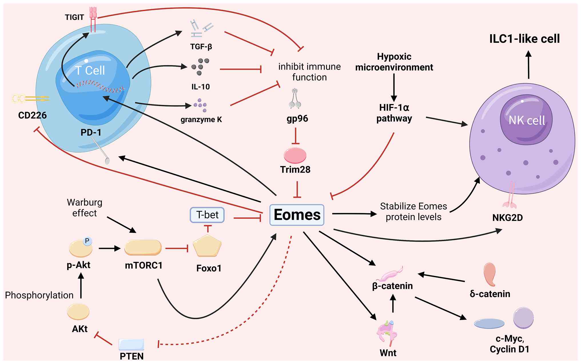 | Figure 1Compartment-specific framework of
Eomes in RCC. The figure separates Eomes-associated mechanisms into
three TME compartments: Infiltrating leukocytes, intrinsic RCC
tumor cells and stromal/endothelial components. In infiltrating
leukocytes, Eomes may support mature NK-cell cytotoxicity through
gp96/TRIM28-dependent protein stability and effector programs,
whereas persistent Eomes activity in exhausted CD8+ T
cells or Tr1-like cells may support PD-1/TIGIT expression,
IL-10/TGF-β secretion and immune suppression. In tumor cells,
possible Eomes links with the Wnt/β-catenin and PI3K/Akt/mTOR axes,
cell stemness and apoptosis resistance are shown as
hypothesis-generating mechanisms. In stromal/endothelial
compartments, Eomes-associated angiogenic effects are shown mainly
as indirect immune-cytokine-mediated effects. Solid black arrows
represent activating regulatory relationships that are supported by
available research evidence. Solid red lines denote inhibitory or
degradative negative regulation with supporting experimental
evidence. The dashed red line indicates an indirect, putative
negative regulatory relationship. Eomes, eomesodermin; RCC, renal
cell carcinoma; TME, tumor microenvironment; NK, natural killer;
gp96, heat shock protein glycoprotein 96; TRIM28, tripartite
motif-containing 28; TIGIT, T cell immunoglobulin and ITIM domain;
HIF-1α, hypoxia-inducible factor-1α; NKG2D, natural killer group
2D. |
Eomes has also been shown to mediate dual
ontogenetic roles. It is able to coordinate morphogenetic processes
during embryonic development. By contrast, it can also
simultaneously regulate immune cell dynamics through the modulation
of differentiation trajectories, functional sustainability and
memory programming in NK cells, CD8+ T cells and ILCs
(6,7,30).
In immune cells, both the expression patterns and the functions of
Eomes serve as cell markers. Together with T-bet, Eomes sustains NK
cytotoxicity function. NK cells with high Eomes expression have
been typically shown to exhibit stronger cytotoxicity and a
stronger IFN-γ secretion capability, whereas NK cells with lower
Eomes expression were shown to exhibit a tendency to differentiate
into ILC1 cell types, which resulted in markedly reduced antitumor
activity (31). In
CD8+ T cells, Eomes was shown to enhance interleukin
(IL)-2 signaling via increasing the expression of CD25, thereby
promoting the proliferation and survival of effector T cells, and
contributing to the formation and maintenance of memory T cells
(32). In addition, Eomes was
shown to directly bind to the promoter regions of immune checkpoint
molecules, such as T cell immunoglobulin and ITIM domain (TIGIT)
and programmed cell death protein 1 (PD-1), thereby promoting their
expression and inducing T-cell exhaustion (33,34).
Eomes expression and activity have been demonstrated
to be subject to various modes of epigenetic control. In γ-δ T
cells, PRF1 and GZMB production was found to be suppressed as a
result of a reduction in Eomes expression induced by histone
deacetylase (HDAC) inhibitors (35). Furthermore, the phosphorylated
form of lysine-specific demethylase 1 (nLSD1p) was found to
regulate the nuclear localization and transcriptional activity of
Eomes through its interaction with Eomes, inducing CD8+
T-cell exhaustion (18,36). Additionally, the process of
ubiquitination has been shown to regulate the stability of Eomes. A
study by Xu et al (21)
revealed that the heat-shock protein glycoprotein 96 (gp96)
inhibits tripartite motif-containing 28 (TRIM28)-mediated Eomes
ubiquitination and degradation, thereby enhancing NK maturation and
antitumor effects.
Cell-compartment-specific framework of
Eomes in RCC
To integrate the operation of the mechanisms
described below, Eomes should be interpreted in three major RCC TME
compartments. First, with respect to infiltrating leukocytes,
divergent functions have been identified for Eomes in
CD8+ T cells, NK cells, ILC1-like cells and type 1 Tregs
(Tr1-like) cells. Secondly, in intrinsic RCC tumor cells, possible
links with cell stemness, EMT, apoptosis resistance and
proliferative signaling remain incompletely validated, and the
links should therefore be interpreted with caution. Thirdly, in
stromal and endothelial compartments, the majority of the
Eomes-associated effects that have been proposed are probably
indirect, mediated through immune cytokines, angiogenic signaling
or tumor-stroma crosstalk. This compartment-based framework also
explains the 'double-edged' character of Eomes: Restoring Eomes may
support NK-cell cytotoxicity, whereas persistent Eomes in exhausted
or regulatory lymphocyte populations may reinforce immune
suppression. This evidence hierarchy, spanning direct RCC
clinical/single-cell data, RCC-associated model evidence, and
indirect or cross-model inferences, is summarized in Table I.
 | Table IEvidence hierarchy for Eomes-related
mechanisms in RCC. |
Table I
Evidence hierarchy for Eomes-related
mechanisms in RCC.
| Evidence
source | Eomes-related
mechanism | Main
compartment | Interpretation in
RCC | Evidence
strength |
|---|
| Human RCC tissues
and single-cell studies | Eomes-positive
CD8+ T-cell exhaustion, PD-1/TIGIT expression and
Eomes-positive Tr1-like immunosuppressive cells | Infiltrating
leukocytes | Supports an
association with immune dysfunction, tumor progression and response
to immunotherapy; causality still requires functional
validation. | Direct RCC
clinical/single-cell evidence |
| RCC-associated
NK-cell observations and immune-cell models | Eomes maintenance
of NK-cell cytotoxicity, gp96/ TRIM28-dependent Eomes stability and
NK-to-ILC1-like plasticity | Infiltrating
leukocytes | Supports a
protective role of Eomes in mature NK cells, whereas TGF-β and
HIF-1α may suppress Eomes in NK-cell contexts. | RCC-associated plus
immune-model evidence |
| RCC pathway studies
and non-RCC tumor models | PI3K/Akt/mTOR,
PTEN, Wnt/β-catenin, apoptosis, EMT and stemness-related
mechanisms | Tumor cells |
Tumor-cell-intrinsic effects remain
incompletely validated in RCC and should be interpreted as
hypotheses requiring gain- and loss-of-function experiments. | Indirect or
hypothesis-generating evidence |
| RCC angiogenesis
studies and IFN-γ biology | IFN-γ, VEGF/bFGF,
CXCL9/CXCL10 and PinX1/miR-125a-3p/ VEGF signaling | Stroma/
endothelium | Eomes may affect
angiogenesis indirectly through immune-cell-derived IFN-γ; direct
endothelial or stromal Eomes evidence in RCC is limited. | Indirect
RCC-related evidence |
| Developmental
biology, chronic infection, CAR-T and other cancer studies | BAF interaction,
β-catenin coactivation, LSD1/G9a modulation and engineered T-cell
approaches | Cross-model/
translational hypotheses | These findings
provide mechanistic leads but should not be presented as
established RCC mechanisms or clinical strategies. | Cross-model
evidence |
Role of Eomes in the immune microenvironment
of RCC
Role of Eomes in infiltrating
CD8+ T cells
The TME of RCC harbors a diverse array of
dysfunctional immune cells. Among them, CD8+ T-cell
exhaustion is central to immune evasion (36,37). Eomes has been shown to modulate
the efficacy of antitumor immune responses through alterations in
CD8+ T-cell differentiation and functional status
(6,38). In addition, previous studies have
shown that, in advanced or metastatic RCC, tumor-infiltrating
CD8+ T cells exhibit heterogeneous Eomes expression
patterns. In particular, the population of CD8+ T cells
of the Eomes+ T-betlow subtype has been
observed to be increased, which represents the dysfunctional
phenotype with diminished secretion of key cytokines (including
IFN-γ and TNF-α) and decreased cytotoxicity (namely, reduced
expression of PRF1 and GZMB) (14,36,39). In terms of the underlying
mechanism, Eomes engages the promoter locus of TIGIT, thereby
promoting its expression. TIGIT is a key immune checkpoint receptor
that mediates its function via binding to CD155, thereby inhibiting
T-cell activity; this forms an Eomes-TIGIT regulatory axis that may
exacerbate T-cell exhaustion (33).
The balance between the nuclear and cytoplasmic
localization of Eomes is critical for CD8+ T-cell
function (28). In chronic viral
infection models, exhausted CD8+ T cells have been shown
to exhibit high nuclear Eomes/T-bet ratios, leading to the
transcriptional activation of inhibitory receptors, such as PD-1
(28,40). In patients with RCC, nuclear Eomes
in tumor-infiltrating CD8+ T cells may competitively
bind to T-box elements in the promoter of the PDCD1 gene
(which encodes PD-1), thereby both weakening the transcriptional
repression mediated by T-bet and sustaining high expression levels
of PD-1 (14,28). Eomes has also been shown to
downregulate the co-stimulatory molecule CD226, which further
limits the activation and proliferation of CD8+ T cells.
Bhat et al (35)
demonstrated that CD226 expression is reduced in Eomes+
CD8+ T cells, where the loss of CD226 was found to be
associated with resistance to anti-PD-1 therapy. However, since
several of the mechanistic details have been determined from cases
of chronic infection or non-RCC immune models, these findings
should be interpreted as supportive evidence, rather than as
definitive proof of RCC-specific causality.
Role of Eomes in NK cell and ILC1
differentiation
NK cells perform a vital role in immune surveillance
of RCC (41,42). Eomes is a critical transcription
factor for NK cell maturation and functional maintenance. Its
expression has been shown to be closely correlated with NK cell
cytotoxicity and cytokine production (43) (Fig.
2). Although NK cells and ILC1 cells share a similar lineage
and phenotypic markers, they differ significantly in terms of their
function. High expression levels of Eomes support the maturation of
NK cells with cytotoxicity and immune surveillance, whereas the
loss or downregulation of Eomes induces a shift toward an
'ILC1-like' phenotype (43-45). Within the RCC TME, both activation
of the TGF-β signaling pathway and other inhibitory signals may
suppress Eomes expression, thereby contributing to NK cell
dysfunction (12,46,47). Under physiological conditions,
Eomes maintains NK cell cytotoxicity by transcriptionally
activating effectors, such as PRF1, GZMB, IFN-γ and
granulocyte-macrophage colony-stimulating factor (GM-CSF), and also
by supporting activating receptors and chemokine receptors that are
involved in tumor recognition and tissue homing (44,48-51). Hypoxia-inducible factor-1α
(HIF-1α) in the hypoxic TME may inhibit Eomes expression in the
NK-cell context, thereby promoting ILC1-like markers such as CD49a
and C-X-C motif chemokine receptor 6 (CXCR6) (52-55). Therefore, selectively modulating
the HIF-1α-Eomes axis in NK cells should be viewed as an
experimental strategy, rather than as an established RCC therapy.
In addition, decreased expression levels of gp96 in RCC have been
documented to relieve its suppression of the ubiquitin ligase
Trim28, thereby promoting Eomes ubiquitination and degradation, as
well as contributing to NK cell dysfunction (21). Dysregulated cytokines, such as
TGF-β and IL-15, have been shown to activate ILC1
differentiation-associated transcription factors, including homolog
of Blimp-1 in T cells (Hobit) and runt-related transcription factor
3 (Runx3), thereby reprogramming Eomes-deficient NK cells into an
ILC1-like state (7,56,57). This remodeling is characterized by
the downregulation of cytotoxic factors and an increased expression
both of inhibitory receptors, such as TIGIT and CD96, and of
immune-regulating factors, such as IL-10 and TGF-β (58,59).
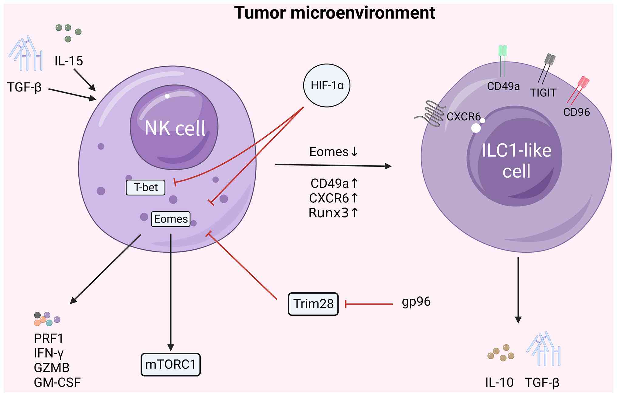 | Figure 2Proposed mechanism via which Eomes
regulates NK cells and their antitumor functions in the hypoxic TME
of RCC. NK cells with high expression exhibit cytotoxic activity,
and secrete effector molecules, including PRF1, IFN-γ, GZMB and
GM-CSF. gp96 may maintain Eomes stability by limiting
TRIM28-mediated degradation. IL-15 and TGF-β can promote the
transformation of NK cells into ILC1-like cells by activating
transcription factors associated with ILC1 differentiation, such as
RUNX3. Sustained HIF-1α activation may inhibit Eomes in NK-cell
contexts, promoting ILC1-like markers such as CD49a and CXCR6, and
reducing cytotoxic capacity. These mechanisms are shown as
RCC-associated or immune-model-supported pathways, rather than as
fully validated RCC-specific causal chains. Solid black arrows
represent stimulatory/activating regulation, cellular phenotypic
transition, and the secretion of effector molecules; and solid red
lines represent inhibitory or degradative regulatory effects.
Eomes, eomesodermin; RCC, renal carcinoma; NK, natural killer; TME,
tumor microenvironment; PRF1, perforin; GZMB, granzyme B; GM-CSF,
granulocyte-macrophage colony-stimulating factor; gp96, heat shock
protein glycoprotein 96; TRIM28, tripartite motif-containing 28;
ILC1, innate lymphoid cell 1; RUNX3, runt-related transcription
factor 3; HIF-1α, hypoxia-inducible factor-1α; CXCR6, CXC motif
chemokine receptor 6; TIGIT, T cell immunoglobulin and ITIM
domain. |
Accumulating evidence suggests that
Eomeslow or Eomes-ILC1-like cells can be enriched in the
RCC TME, where their heightened presence has been associated with
tumor progression and resistance to immunotherapy (15,60).
In conclusion, Eomes has been demonstrated to act as
a core controller of NK cell function, although the therapeutic
implications are cell-type-specific. Restoring or stabilizing Eomes
may be useful for mature NK cells when its loss drives the
conversion of NK cells into the ILC1-like state, and cytotoxic
failure. This does not imply that a global activation of Eomes
would be beneficial, however, since other Eomes-positive lymphocyte
states may contribute to exhaustion or immunosuppression.
Eomes modulates regulatory T cells
(Tregs) and the immuno-suppressive microenvironment
Eomes reshapes the immune microenvironment of RCC.
It can promote immune escape during tumor progression by modulating
the recruitment, expansion and functional polarization of
immunosuppressive populations (Fig.
3). These immunosuppressive cells include Tregs and Tr1-like
cells (61,62). A single-cell transcriptomic study
previously conducted by Bonnal et al (62) identified a subset of
Eomes+ Tr1-like cells within the TME. These cells
underwent clonal expansion, expressed high levels of IL-10 and
granzyme K, and exhibited potent immunosuppressive functions.
Unlike conventional Forkhead box (FOX)P3+ Tregs,
however, Tr1-like cells primarily inhibited effector T-cell
responses through cytokine secretion, instead of via direct
cell-cell contact. In particular, their abundance was shown to be
positively correlated with cancer progression (62). A study by Jian et al
(63) revealed that tumor cells
with high Eomes expression promoted the secretion of C-C motif
chemokine ligand 20 (CCL20), also recruiting C-C chemokine receptor
type 6 (CCR6)+ Tregs into the TME, thereby enhancing
immunosuppression. Collectively, these studies suggested that Eomes
may function as a vital regulator in remodeling the immune
microenvironment of RCC by enhancing the function of Tregs and
other immunosuppressive cells.
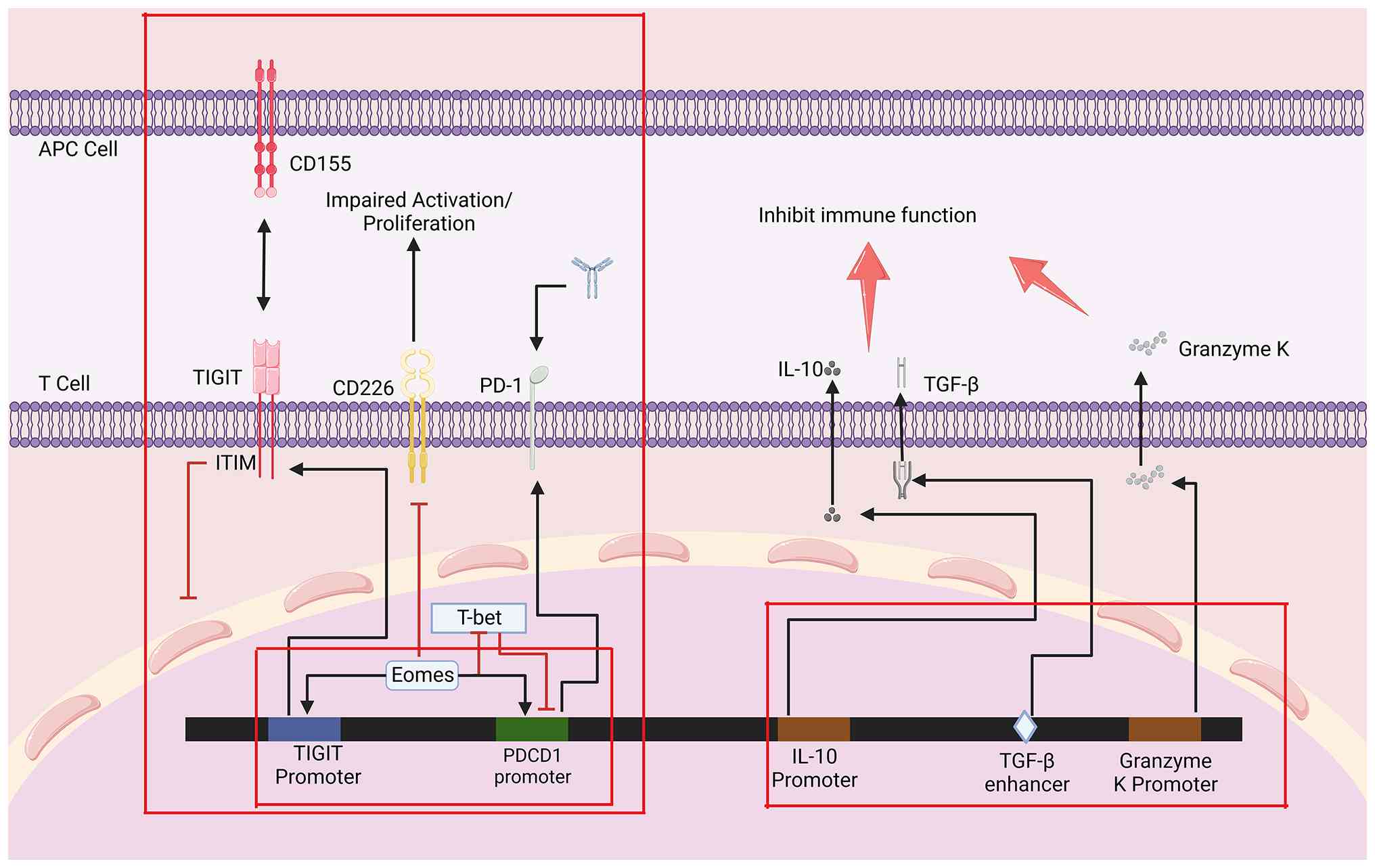 | Figure 3Eomes-associated transcriptional
programs in exhausted or regulatory lymphocyte states. Eomes can
regulate or correlate with immune checkpoints and regulatory
molecules, including TIGIT, CD226, PD-1, IL-10, TGF-β and granzyme
K, depending on cell state. By inhibiting T-bet, an inhibitor of
PD-1 expression, Eomes may support TIGIT and PD-1 expression and
reduce co-stimulatory signaling, whereas in Tr1-like cells it may
contribute to IL-10/TGF-β-associated immunosuppression. Mechanistic
links validated outside RCC are shown as indirect evidence, and
these require RCC-specific confirmation. Solid black arrows
represent stimulatory/activating regulation, cellular phenotypic
transition, and the secretion of effector molecules; solid red
lines represent inhibitory or degradative regulatory effects. Red
arrows represent IL-10, TGF-β and granzyme K, which all play a role
in inhibiting immune function. Red boxes demarcate two independent
functional regulatory modules: The left box outlines the
transcriptional regulatory module of immune checkpoint receptors on
T cells, and the right box outlines the transcriptional regulatory
module of immunosuppressive effector molecules. Eomes,
eomesodermin; TIGIT, T cell immunoglobulin and ITIM domain; PD-1,
programmed cell death protein 1; RCC, renal cell carcinoma. |
The accumulation of Eomes+ Tr1-like cells
has also been associated with resistance to immunotherapy in RCC.
An increase in their numbers was negatively correlated with
progression-free survival in patients receiving anti-PD-1 therapy.
This may be due to Eomes+ Tr1-like cells producing high
levels of IL-10 and TGF-β, which has the effect of inhibiting both
the infiltration and activation of effector T cells (62,64). In particular, Eomes+
Tr1-like cells do not produce the same specific effector cytokines
as are produced by Th1 or Th2 cells, such as IFN-γ or IL-4
respectively (65-67). Furthermore, Eomes may sculpt the
immune microenvironment through regulation of macrophage
polarization. For example, in hepatocellular carcinoma,
macrophage-derived itaconic acid was shown to upregulate the
expression of Eomes via an epigenetic mechanism (namely, H3K4me3
modification) to promote CD8+ T-cell exhaustion
(68). Given the abundance of M2
macrophages in the RCC microenvironment, a similar mechanism is
plausible, although the evidence remains indirect and requires
RCC-specific validation.
Regulation of RCC progression by Eomes
Role of Eomes in cancer stemness
Cancer stem cells (CSCs) perform a key role in
progression, distant metastasis and treatment resistance in RCC.
CSCs are characterized by self-renewal and multipotent
differentiation potential (69-71). In solid tumors, including RCC, the
presence of CSCs is closely associated with poor clinical outcomes.
Previous studies have suggested that the concurrent expression of
stemness markers (such as Oct4 and Nanog) in RCC tissues is
associated with poor patient prognosis (72,73). In the embryonic hematopoietic
system, Eomes operates upstream of runt-related transcription
factor 1 (Runx1) in the hemogenic endothelium lineage, serving to
regulate lineage differentiation and the self-renewal of
hematopoietic stem cells (74).
On the basis of this developmental role, Eomes may regulate genetic
networks that are involved in CSC-like properties in RCC; however,
this tumor-cell-intrinsic mechanism requires further experimental
validation in RCC models.
The long non-coding RNA (lncRNA) HOX transcript
antisense RNA has been shown to promote RCC stemness and
angiogenesis both through formation of a feedback loop with the
androgen receptor, and by synergistically activating the
transcription of glioma-associated oncogene family zinc finger 2
(GLI2) (75). Similar regulatory
mechanisms governing RCC stemness may also be observed in the
Wnt/β-catenin cascade. In RCC, δ-catenin has been documented to
induce the transcription and expression of downstream target genes,
including those encoding c-Myc, cyclin D1, membrane type 1 matrix
metalloproteinase (MT1-MMP) and Bcl-2, by enhancing the nuclear
translocation of β-catenin and driving signaling pathways that are
associated with stemness maintenance (76). Eomes may act as a transcriptional
co-activator of β-catenin, although the direct contribution of this
proposed Eomes-β-catenin mechanism to RCC CSC biology has yet to be
validated (76).
Role of Eomes on cell proliferation,
apoptosis and metastasis of RCC
Although Eomes is conventionally recognized for
immune regulation, abnormal expression in tumor cells may influence
malignant biological behavior. Research on human protein expression
patterns has suggested that Eomes exhibits high expression levels
in renal cancer tissues (77). A
regulatory link between aberrant PI3K/Akt/mTOR signaling and Eomes
expression in RCC is plausible, but direct tumor-cell causality
remains incompletely established. Phosphorylation of Akt by
mechanistic target of rapamycin complex 1 (mTORC1) inhibits FOXO1,
and suppressed FOXO1 activity upregulates T-bet and inhibits Eomes
expression. By contrast, activated FOXO1 can promote Eomes
transcription, suggesting a potential Akt/FOXO1/T-bet/Eomes
feedback axis. In parallel, Wong et al (15) found that deletion of Eomes
impaired IL-15 receptor signaling and downstream PI3K/Akt
activation in immune cells. Collectively, these findings suggested
that Eomes may influence RCC progression directly in tumor cells,
or indirectly through antitumor immune cells, although these two
contexts should not be conflated. In another tumor model, Eomes
promoted G2/M phase cell cycle progression by promoting
CCL20 and CCR6 expression; its relevance to RCC proliferation
remains hypothesis-generating (63) (Fig.
4).
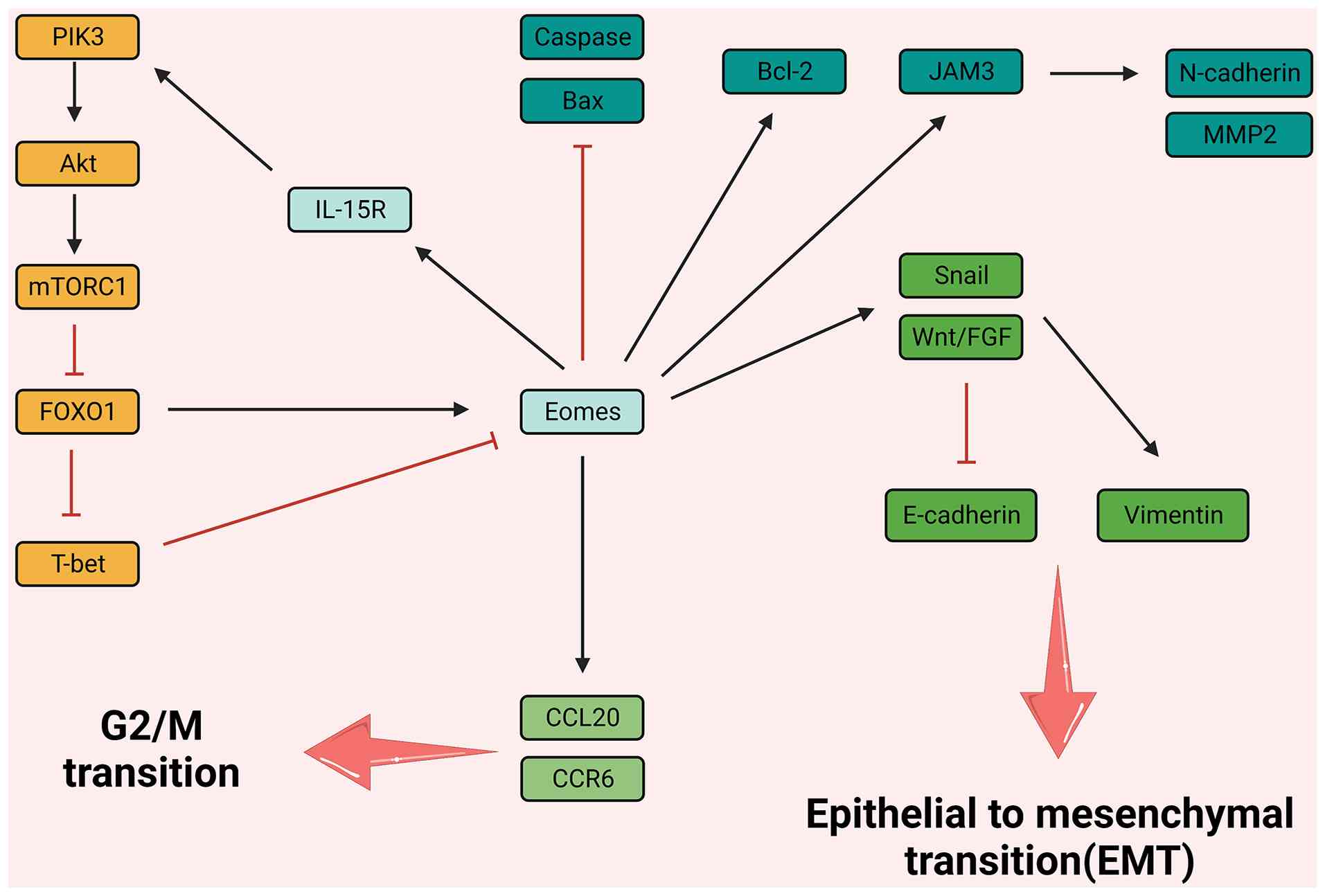 | Figure 4Proposed mechanisms linking Eomes
with RCC proliferation, apoptosis and EMT. PI3K/Akt/mTOR, FOXO1,
CCL20/CCR6, JAM3, Bax/caspase-3, Bcl-2 and Wnt/FGF-related EMT
markers are shown as candidate pathways. Solid black arrows
represent stimulatory/activating regulatory relationships. Solid
red lines denote inhibitory or suppressive regulatory effects. Red
arrows indicate the overall functional outcomes of the
corresponding pathways. Eomes, eomesodermin; RCC, renal cell
carcinoma; FOXO, Forkhead box; CCL20, C-C motif chemokine ligand
20; CCR6, C-C chemokine receptor type 6; JAM3, junctional adhesion
molecule 3; FGF, fibroblast growth factor; EMT,
epithelial-mesenchymal transition. |
In terms of apoptosis regulation, Eomes may affect
cell survival through pathways involving Bax, caspase-3 and Bcl-2
(16,78,79). In a study by Wagner et al
(16), inducible knockout of
Eomes led to rapid NK cell loss, accompanied by increased caspase-3
expression and upregulation of the Bax pathway. These findings
support a survival role for Eomes in NK cells. Whether a similar
mechanism operates in RCC cells remains uncertain. Concomitantly,
Junction adhesion molecule 3 (JAM3) has been demonstrated to
facilitate RCC cell migration, whilst inhibiting apoptosis by
upregulating N-cadherin and MMP2. Considering that Eomes is a T-box
transcription factor, Eomes may contribute to JAM3-associated RCC
migration by transcriptional regulation, although direct promoter
binding requires validation (80). In addition, Eomes has been linked
to EMT-associated transcriptional programs in other contexts.
Therefore, its proposed role in regulating E-cadherin, N-cadherin,
vimentin and Twist in RCC should be regarded as an experimental
hypothesis (61,81).
Role of Eomes in stromal/endothelial
regulation and RCC angiogenesis
Through indirect pathways, Eomes profoundly affects
the function and phenotype of stromal cells, thereby reshaping the
structure and function of the entire TME at the macro level. The
clearest indirect regulation occurs in angiogenesis. Angiogenesis
is a hallmark of RCC progression that is primarily driven by the
dysregulated activation of the VEGF/VEGFR pathway (75). Eomes was found to both enhance
IFN-γ production and bind to the Ifng promoter and
cis-regulatory non-coding sequences in BW5147 cells (a
murine T-cell lymphoma cell line), with the caveat that this cell
line should not be interpreted as an RCC TME component (82). IFN-γ is a classical
anti-angiogenic cytokine that blocks neovascularization by inducing
CXC-motif ligand (CXCL)9/10, thereby inhibiting endothelial cell
proliferation and downregulating VEGF and fibroblast growth factor
(FGF) signalling (83,84) (Fig.
5). In RCC cell lines, exogenously added IFN-γ has been shown
to reduce the mRNA transcript levels of VEGF and basic (b)FGF.
Therefore, the most cautious interpretation one may offer is that
Eomes influences endothelial behavior indirectly through
immune-cell-derived IFN-γ, rather than through a proven
endothelial-cell-intrinsic Eomes pathway.
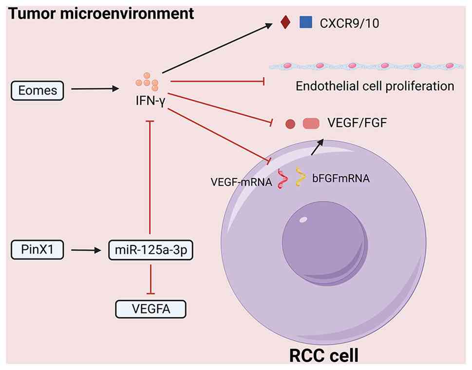 | Figure 5Proposed association between
Eomes-associated immune signaling and RCC angiogenesis. RCC tumor
cells and stromal/endothelial cells regulate angiogenesis through
VEGF, bFGF and VEGFR signaling. IFN-γ may indirectly inhibit
endothelial proliferation and angiogenic signaling by inducing
CXCL9/CXCL10, and reducing VEGF/bFGF, expression. The
PinX1/miR-125a3p/VEGF axis represents an RCC-associated
angiogenesis pathway that may interact with immune cytokine
signaling. Solid black arrows represent stimulatory/activating
regulatory effects. Solid red lines denote inhibitory regulatory
effects. The red triangle represents CXCR9, and the blue square
represents CXCR10. The red circle represents VEGF, while the pink
rectangle represents FGF. Eomes, eomesodermin; RCC, renal cell
carcinoma; bFGF, basic fibroblast growth factor; CXCL, C-X-C motif
chemokine ligand; PinX1, PIN2/TERF1-interacting telomerase
inhibitor 1; miR, microRNA. |
PIN2/TERF1-interacting telomerase inhibitor 1
(PinX1) is a tumor suppressor in RCC that is able to
transcriptionally activate microRNA (miR or miRNA)-125a-3p in a
specificity protein 1 (Sp1)-dependent manner. This miRNA binds
directly to the 3′-untranslated region (3′-UTR) of VEGFA, thereby
suppressing its translation (85,86). However, high miR-125a-3p
expression in Tr1-like T cells was shown to negatively regulate
IFN-γ, suggesting that a dynamic balance exists between the
PinX1/miR-125a-3p/VEGF and the Eomes/IFN-γ signaling axes (87); in spite of this, this association
should be regarded as a mechanistic model, rather than as a
validated therapeutic basis for combined Eomes-directed and
anti-VEGF treatment.
Regulation of Eomes-associated epigenetic
regulation mechanisms and signaling pathways
Epigenetic regulation of Eomes
expression
Eomes expression is tightly controlled by various
epigenetic mechanisms. HDAC inhibitors, such as trichostatin A,
have been reported to reduce Eomes expression and to suppress the
production of PRF1 and GZMB in γ-δ T cells (35,88,89). This suggests that HDAC activity
can affect Eomes-associated cytotoxic programs in immune cells.
Additionally, nLSD1p has been shown to interact with Eomes,
regulating its nuclear localization and transcriptional activity
and thereby influencing CD8+ T-cell function (18). In patients with immune-resistant
melanoma, nLSD1p was found to be overexpressed in
PD-1+CD8+ T cells, and it co-localized with
Eomes in the nucleus. Collectively, these findings have provided a
useful immune-oncology model, although direct validation in RCC
remains necessary (18,90).
DNA methylation is a key epigenetic regulatory
mechanism. In RCC, the tumor suppressor gene serine protease
inhibitor Kazal-type 5 (SPINK5) is silenced by H3K9me2 methylation
at its promoter. Euchromatic histone-lysine N-methyltransferase 2
(EHMT2; also commonly known as G9a) is the main enzyme that
catalyzes H3K9me2 methylation, and its high expression has been
associated with RCC progression (91,92). If H3K9me2 modifications occur at
the Eomes promoter, G9a may inhibit EOMES transcription by
increasing chromatin compaction, although this specific axis has
not yet been fully established in RCC. Additionally, Eomes
expression is regulated by lncRNAs and miRNAs. In CD4+ T
cell-based competing endogenous RNA (ceRNA) networks, Eomes has
been shown to be modulated by the small, non-coding miRNA
has-miR-23b-3p, which is, in turn, modulated by the lncRNA
AC104820.2. By contrast, in the CD8+ T-cell ceRNA
network, Eomes is regulated by the small, non-coding miRNA
has-miR-23a-3p, which is itself regulated by the lncRNA AC000476.1.
At the present time, these axes should be regarded as candidate
regulatory mechanisms, pending RCC-specific functional verification
(93,94).
Eomes and the PI3K/Akt/mTOR signaling
pathway
Aberrant activation of the PI3K/Akt/mTOR signaling
pathway is a hallmark of RCC, which causes enhanced tumor cell
proliferation and survival, and metabolic reprogramming (95). In RCC, the Ras-related protein
RAP2a has been shown to promote metastasis by upregulating
phosphorylated Akt (96). A
bidirectional regulatory association between Eomes and this
signaling pathway is plausible; however, the direction may differ
between immune cells and tumor cells. In immune cells, Eomes may be
associated with IL-15 receptor signaling and Akt activation,
whereas in tumor cells, Eomes may either be downstream of, or
interact with, PI3K/Akt/mTOR-associated transcriptional programs.
However, the direct binding of Eomes to PI3K/Akt target gene
promoters in RCC has yet to be firmly established, and additional
evidence is required.
In NK cells, a positive feedback loop involving
IL-15, phosphoinositide-dependent kinase 1 (PDK1), mTOR, nuclear
factor IL-3-regulated protein, Eomes and CD122 serves a vital role
in primary stages of NK cell development. Within this loop, Eomes
enhances the responsiveness of NK cells to IL-15 by upregulating
CD122, thereby promoting its own expression in a self-reinforcing
circuit (96,97). Furthermore, Zeng et al
(98) suggested that in RCC, the
lncRNA distal-less homeo box 6-antisense 1 (DLX6-AS1) sequesters
miR-26a (functioning as a ceRNA) to upregulate the tumor suppressor
gene PTEN, a negative regulator of the PI3K/Akt pathway, thereby
limiting tumor progression; this study did not examine Eomes.
Whether Eomes intersects with this DLX6-AS1/miR-26a/PTEN axis in
RCC has not been directly investigated and remains an open question
for future study. Given that Eomes is a key regulator of Akt
activation, and PTEN is a critical negative modulator of the
PI3K/Akt pathway, Eomes may indirectly regulate PTEN expression and
downstream Akt activation by interacting with DLX6-AS1 or miR-26a;
for example, by transcriptionally regulating DLX6-AS1 expression or
modulating miR-26a maturation (98,99).
The regulation of mTORC1 signaling has also been
known to intersect with metabolic reprogramming. In NK cells,
inhibiting mTORC1 activity may suppress ubiquitin-mediated Eomes
degradation, thereby enhancing the antitumor function of NK cells
(100). In RCC, the Warburg
effect has been demonstrated to activate mTORC1 signaling, and may
alter Eomes-associated transcriptional programs; however,
supporting evidence for a direct mTORC1-Eomes oncogenic axis in RCC
remains scarce (101,102). Therefore, targeting the
mTORC1-Eomes axis should currently be viewed as a preclinical
hypothesis, rather than as a validated treatment strategy.
Eomes and the Wnt/β-catenin pathway
The pathogenesis and progression of RCC are also
impacted by the Wnt/β-catenin signaling pathway (Fig. 6). As discussed above, RCC-specific
evidence supports a role for δ-catenin- and β-catenin-associated
signaling in cell proliferation and apoptosis resistance (76). However, direct Eomes-β-catenin
co-activation is supported mainly by developmental models, and
requires further validation in RCC. Therefore, the
Eomes-Wnt/β-catenin signaling axis is best interpreted as a
hypothesis-generating bridge between developmental transcriptional
programs and RCC stemness, rather than as a proven RCC driver
(29,76,103). Beyond a direct protein-protein
interaction with β-catenin, Eomes also cooperates with chromatin
remodelers to amplify Wnt-dependent transcription, and the BAF
complex (a large ATP-dependent protein group that moves and changes
nucleosomes to control gene activity) represents a critical
epigenetic cofactor in this process. During embryonic development,
Eomes interacts with the BAF chromatin remodeling complex, which
serves to maintain chromatin accessibility in the trophoblast
ectoderm (TE) and to regulate TE-specific gene expression (104). Given that Eomes often functions
as a transcriptional activator, and the BAF complex facilitates
chromatin accessibility for target gene binding, it is plausible
that the Eomes-BAF complex may target and activate c-Myc expression
in RCC cells, a process that would further enhance RCC cell
proliferation, given that c-Myc is a well-established driver of RCC
cell proliferation. However, this regulatory axis (Eomes-BAF→c-Myc)
requires further validation via chromatin immunoprecipitation
sequencing, co-immunoprecipitation analysis and functional assays
in RCC models.
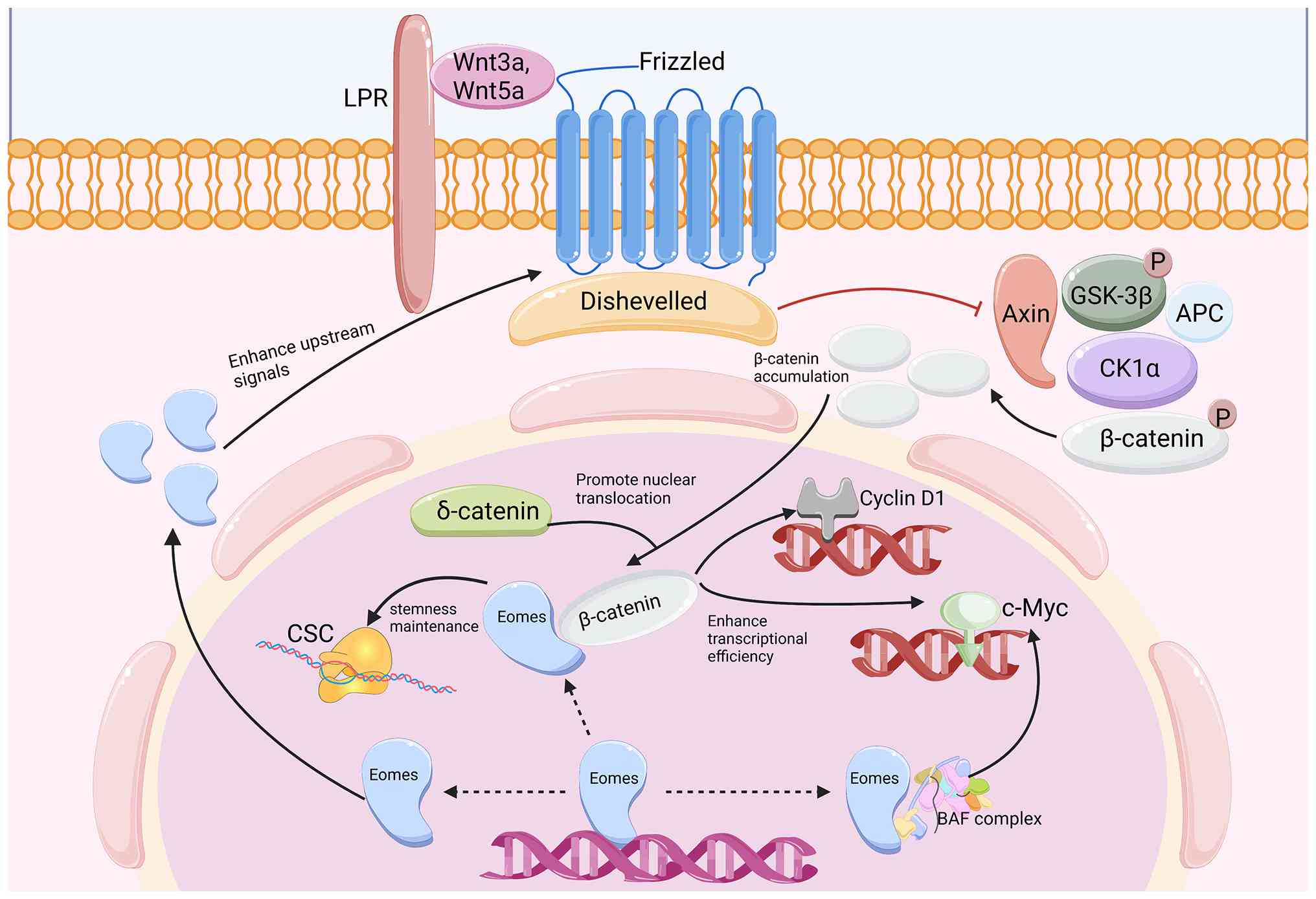 | Figure 6Eomes-Wnt/β-catenin signaling model.
Wnt ligands (Wnt3a/Wnt5a) can bind to FZD receptors and promote
β-catenin accumulation and nuclear translocation. In RCC,
δ-catenin- and β-catenin-associated pathways have been associated
with cell proliferation and apoptosis resistance. Eomes-β-catenin
cooperation and BAF-dependent chromatin remodeling are mainly
inferred from developmental models, although these require direct
RCC validation. Solid black arrows represent stimulatory/activating
regulatory relationships. Solid red lines denote inhibitory
regulatory effects. Black dashed arrows indicate indirect, putative
regulatory interactions and functional cooperativity that lack
direct experimental validation in RCC. Eomes, eomesodermin; FZD,
frizzled; RCC, renal cell carcinoma; CK1α, casein kinase 1; GSK-3β,
glycogen synthase kinase 3β; CSCs, cancer stem cells; BAF,
SWItch/sucrose non-fermentable. |
Potential of Eomes as an exploratory
biomarker, and cell-type-specific therapeutic targets for RCC
Role of Eomes in the prognosis evaluation
of RCC
Single-cell transcriptomic data from a previous
study showed that CD8+ T cells within RCC tissues
express Eomes at high levels, and the clonal expansion of
Eomes+ Tr1-like cells is associated with RCC disease
progression (61,62). These findings suggest that either
the expression level of Eomes or the proportion of Eomes-positive
immune-cell subsets could be explored as prognostic biomarkers.
However, Eomes should not currently be considered as a standalone
diagnostic marker without independent validation in large RCC
cohorts and in matched protein-level assays.
The expression levels of Eomes may vary among
different RCC subtypes; however, the present evidence is
insufficient to conclude that Eomes is able to reliably distinguish
ccRCC from papillary RCC, or from other RCC subtypes. ccRCC is
characterized by activation of the HIF pathway, whereas HIF-1α may
suppress Eomes in the context of NK cells. This apparent tension
can be reconciled by considering the cell types separately: In
tumor-infiltrating NK cells, hypoxic HIF-1α signaling may suppress
Eomes and promote NK dysfunction, whereas in intrinsic tumor cells,
HIF-associated metabolic and epigenetic programs may be correlated
with different Eomes expression patterns. Therefore, any
subtype-differentiation claim requires direct analysis of TCGA,
CPTAC, Human Protein Atlas and RCC single-cell datasets before any
sort of clinical use can be proposed (12,54,55).
Application prospects of Eomes-targeted
therapy in RCC Eomes regulation and immunotherapy
ICIs represent a significant therapeutic approach
for RCC, although their efficacy is influenced by immune-cell
functional status in the TME. As a key regulator of immune cell
function, Eomes may be explored as a target for improving ICI
responses; however, any intervention would require
cell-type-specific modulation experiments to be performed.
Enhancing the activity of Eomes may be beneficial in mature NK
cells or selected memory CD8+ T-cell states, whereas
reducing persistent Eomes activity may be considered in exhausted
or Tr1-like immunosuppressive populations. HDAC inhibitors, PD-1
inhibitors and CD226-associated strategies should therefore be
discussed as experimental combinations, rather than as established
Eomes-directed RCC therapies (11,35).
Chimeric antigen receptor T-cell (CAR-T) therapy has
shown promising results in hematological malignancies, although its
efficacy in solid tumors is limited by T-cell exhaustion. Eomes
expression has been shown to promote both the proliferation and
survival of engineered T cells, and their IL-2 responsiveness, in
certain contexts. However, Eomes-engineered CAR-T cells have not
been validated as an RCC therapy, and the overexpression of Eomes
may also carry exhaustion-associated risks, depending on the T-cell
state. Therefore, Eomes modulation in CAR-T cells should be
presented as a preclinical hypothesis that requires further
RCC-specific testing (105,106).
Epigenetic regulation and
Eomes-targeted drug development
As an alternative approach, targeting the
epigenetic regulatory mechanisms of Eomes may provide experimental
strategies for RCC research. Through the disruption of the
interaction between nLSD1p and Eomes, LSD1 inhibitors are capable
of restoring the transcriptional activity of Eomes, and enhancing
CD8+ T-cell function in selected immune contexts
(18,90). In RCC, G9a has been shown to
suppress SPINK5 expression through H3K9me2 methylation, but whether
G9a inhibitors modulate Eomes in RCC has yet to be fully
determined. Similarly, non-coding RNA axes associated with Eomes
should be regarded as candidate mechanisms only, until their direct
role in RCC has been validated (93,94).
Eomes-targeted application of
Eomes-associated signaling pathway inhibitors
The interaction between Eomes and the PI3K/Akt/mTOR
and Wnt/β-catenin signaling pathways offers possible experimental
combinations for RCC. mTOR inhibitors such as everolimus are
clinically used in RCC, although their Eomes-dependent effects have
yet to be established. At the present time, combining mTOR
inhibitors, PD-1 inhibitors or β-catenin inhibitors with
Eomes-directed modulation should be considered as a research
hypothesis, not as a near-clinical strategy. At present, no
Eomes-specific drug, validated predictive assay or completed RCC
clinical trial supports routine Eomes-targeted treatment (100).
Conclusion
Although progress has been made in studying the
role of Eomes in RCC, its clinical translation faces significant
challenges. The dual role of Eomes in immune and tumor cells may
result in off-target effects during targeted therapies.
Upregulation of Eomes enhances effector T-cell function, but may
concurrently promote tumor progression. Therefore, developing
cell-type-specific Eomes modulation strategies (using immune
cell-specific promoters to drive Eomes expression) is crucial
(12,63). The functional overlap and
compensatory mechanisms between Eomes and other transcription
factors (such as T-bet) may compromise the efficacy of targeted
therapies. Previous studies have shown that T-bet and Eomes can
compete for binding to the PDCD1 promoter in CD8+
T cells; therefore, regulating the ratio of the two may be more
effective, instead of targeting them individually (2,14,28).
Eomes may have exploratory clinical value; however,
the assessment of its use as a biomarker or therapeutic target
remains preliminary. A biomarker model incorporating Eomes should
be validated with other markers, such as Oct4, Nanog and CD226, and
should distinguish Eomes-positive cell types, rather than measuring
bulk expression alone. Future research directions should prioritize
the following: i) Characterizing the heterogeneity and functional
subsets of Eomes+ cells within the RCC microenvironment
using single-cell sequencing technologies; ii) testing
cell-type-specific Eomes modulation in RCC models before
considering clinical trials; iii) exploring the combinatorial
detection of Eomes with other biomarkers (such as Oct4, Nanog and
CD226) to optimize the RCC prognosis assessment model; and iv)
defining how Eomes interacts with T-bet, HIF signaling, and the
PI3K/Akt/mTOR and Wnt/β-catenin signaling pathways in each cellular
compartment.
Availability of data and materials
Not applicable.
Authors' contributions
ZC and XW designed the scope and overall structure
of this review. DH, JL and RF performed the structured literature
searches, and critically synthesized and interpreted the included
research findings. ZC, DH and JL contributed to the visual
presentation of the synthesized evidence. DH and JL drafted the
major sections of the manuscript, while ZC, YW and XC were
responsible for critical revision and editing of the full
manuscript. All authors read and approved the final manuscript.
Data authentication is not applicable.
Ethics approval and consent to
participate
Not applicable.
Patient consent for publication
Not applicable.
Competing interests
The authors declare that they have no competing
interests.
Acknowledgments
Not applicable.
Funding
This study was supported by the National Natural Science
Foundation of China (grant no. 82560531), the Academic Enhancement
Support Program of Hainan Medical University (grant no.
XSTS2025005), the College Students' Innovation and Entrepreneurship
Training Program of the Education Department of Hainan province
(grant no. S202511810006), the College Students' Research
Innovation Training Program of Hainan Medical University (grant no.
RZ2500002143), the Hainan Provincial Joint Project for Health
Science and Technology Innovation (grant no. WSJK2026QNO82), the
Natural Science Foundation of Shanxi Province (grant no.
202103021224375), the Fund Program for the Scientific Activities of
Selected Returned Overseas Professionals in Shanxi Province (grant
no. 2022047) and the Research Project Supported by Shanxi
Scholarship Council of China (grant no. 2022-2024).
References
|
1
|
Bukavina L, Bensalah K, Bray F, Carlo M,
Challacombe B, Karam JA, Kassouf W, Mitchell T, Montironi R,
O'Brien T, et al: Epidemiology of renal cell carcinoma: 2022
update. Eur Urol. 82:529–542. 2022. View Article : Google Scholar : PubMed/NCBI
|
|
2
|
Sung H, Ferlay J, Siegel RL, Laversanne M,
Soerjomataram I, Jemal A and Bray F: Global cancer statistics 2020:
GLOBOCAN estimates of incidence and mortality worldwide for 36
cancers in 185 countries. CA Cancer J Clin. 71:209–249.
2021.PubMed/NCBI
|
|
3
|
Badoiu SC, Greabu M, Miricescu D,
Stanescu-Spinu II, Ilinca R, Balan DG, Balcangiu-Stroescu AE, Mihai
DA, Vacaroiu IA, Stefani C, et al: PI3K/AKT/mTOR dysregulation and
reprogramming metabolic pathways in renal cancer: Crosstalk with
the VHL/HIF axis. Int J Mol Sci. 24:83912023. View Article : Google Scholar : PubMed/NCBI
|
|
4
|
Braun DA, Bakouny Z, Hirsch L, Flippot R,
Allen EMV, Wu CJ and Choueiri TK: Beyond conventional
immune-checkpoint inhibition-novel immunotherapies for renal cell
carcinoma. Nat Rev Clin Oncol. 18:199–214. 2021. View Article : Google Scholar : PubMed/NCBI
|
|
5
|
Talbot CD, Walsh MD, Cutty SJ, Elsayed R,
Vlachaki E, Bruce AEE, Wardle FC and Nelson AC: Eomes function is
conserved between zebrafish and mouse and controls left-right
organiser progenitor gene expression via interlocking feedforward
loops. Front Cell Dev Biol. 10:9824772022. View Article : Google Scholar : PubMed/NCBI
|
|
6
|
Lin YH, Duong HG, Limary AE, Kim ES, Hsu
P, Patel SA, Wong WH, Indralingam CS, Liu YC, Yao P, et al: Small
intestine and colon tissue-resident memory CD8+ T cells exhibit
molecular heterogeneity and differential dependence on Eomes.
Immunity. 56:207–223.e8. 2023. View Article : Google Scholar
|
|
7
|
Liang Z, Anderson HD, Locher V, O'Leary C,
Riesenfeld SJ, Jabri B, McDonald BD and Bendelac A: Eomes
expression identifies the early bone marrow precursor to classical
NK cells. Nat Immunol. 25:1172–1182. 2024. View Article : Google Scholar : PubMed/NCBI
|
|
8
|
Zhang J, Marotel M, Fauteux-Daniel S,
Mathieu AL, Viel S, Marçais A and Walzer T: T-bet and Eomes govern
differentiation and function of mouse and human NK cells and ILC1.
Eur J Immunol. 48:738–750. 2018. View Article : Google Scholar : PubMed/NCBI
|
|
9
|
Pearce EL, Mullen AC, Martins GA, Krawczyk
CM, Hutchins AS, Zediak VP, Banica M, DiCioccio CB, Gross DA, Mao
CA, et al: Control of effector CD8+ T cell function by the
transcription factor Eomesodermin. Science. 302:1041–1043. 2003.
View Article : Google Scholar : PubMed/NCBI
|
|
10
|
He J, Chen D, Xiong W, Hou X, Quan Y, Yang
M and Dong Z: Eomesodermin spatiotemporally orchestrates the early
and late stages of NK cell development by targeting KLF2 and T-bet,
respectively. Cell Mol Immunol. 21:662–673. 2024. View Article : Google Scholar : PubMed/NCBI
|
|
11
|
Weulersse M, Asrir A, Pichler AC, Lemaitre
L, Braun M, Carrié N, Joubert MV, Le Moine M, Do Souto L, Gaud G,
et al: Eomes-Dependent loss of the Co-activating Receptor CD226
Restrains CD8+ T Cell Anti-tumor functions and limits the efficacy
of cancer immunotherapy. Immunity. 53:824–839.e10. 2020. View Article : Google Scholar
|
|
12
|
Gao Y, Souza-Fonseca-Guimaraes F, Bald T,
Ng SS, Young A, Ngiow SF, Rautela J, Straube J, Waddell N, Blake
SJ, et al: Tumor immunoevasion by the conversion of effector NK
cells into type 1 innate lymphoid cells. Nat Immunol. 18:1004–1015.
2017. View Article : Google Scholar : PubMed/NCBI
|
|
13
|
Long SA, Thorpe J, DeBerg HA, Gersuk V,
Eddy J, Harris KM, Ehlers M, Herold KC, Nepom GT and Linsley PS:
Partial exhaustion of CD8 T cells and clinical response to
teplizumab in new-onset type 1 diabetes. Sci Immunol.
1:eaai77932016. View Article : Google Scholar
|
|
14
|
Li J, He Y, Hao J, Ni L and Dong C: High
levels of eomes promote exhaustion of Anti-tumor CD8+ T cells.
Front Immunol. 9:29812018. View Article : Google Scholar
|
|
15
|
Wong P, Foltz JA, Chang L, Neal CC, Yao T,
Cubitt CC, Tran J, Kersting-Schadek S, Palakurty S, Jaeger N, et
al: T-BET and EOMES sustain mature human NK cell identity and
antitumor function. J Clin Invest. 133:e1625302023. View Article : Google Scholar : PubMed/NCBI
|
|
16
|
Wagner JA, Wong P, Schappe T,
Berrien-Elliott MM, Cubitt C, Jaeger N, Lee M, Keppel CR, Marin ND,
Foltz JA, et al: Stage-specific requirement for eomes in mature NK
cell homeostasis and cytotoxicity. Cell Rep. 31:1077202020.
View Article : Google Scholar : PubMed/NCBI
|
|
17
|
Arnold SJ, Hofmann UK, Bikoff EK and
Robertson EJ: Pivotal roles for eomesodermin during axis formation,
epithelium-to-mesenchyme transition and endoderm specification in
the mouse. Development. 135:501–511. 2008. View Article : Google Scholar : PubMed/NCBI
|
|
18
|
Tu WJ, McCuaig RD, Tan AHY, Hardy K,
Seddiki N, Ali S, Dahlstrom JE, Bean EG, Dunn J, Forwood J, et al:
Targeting nuclear LSD1 to reprogram cancer cells and reinvigorate
exhausted T cells via a novel LSD1-EOMES switch. Front Immunol.
11:12282020. View Article : Google Scholar : PubMed/NCBI
|
|
19
|
Matinzadeh M, Ebrahimi E, Saffari M and
Shirkoohi R: Type I EMT Inducer Eomesodermin has a significant
expression in breast cancer. Basic Clin Cancer Res. 8:17–22.
2016.
|
|
20
|
Wang R, Kang Y, Löhr CV, Fischer KA,
Bradford CS, Johnson G, Dashwood WM, Williams DE, Ho E and Dashwood
RH: Reciprocal regulation of BMF and BIRC5 (Survivin) linked to
Eomes overexpression in colorectal cancer. Cancer Lett.
381:341–348. 2016. View Article : Google Scholar : PubMed/NCBI
|
|
21
|
Xu Y, Li X, Cheng F, Zhao B, Fang M, Li Z
and Meng S: Heat shock protein gp96 drives natural killer cell
maturation and anti-tumor immunity by counteracting Trim28 to
stabilize Eomes. Nat Commun. 15:11062024. View Article : Google Scholar : PubMed/NCBI
|
|
22
|
Lyu Y, Jia W, Wu Y, Zhao X, Xia Y, Guo X
and Kang J: Cpmer: A new conserved eEF1A2-binding partner that
regulates Eomes translation and cardiomyocyte differentiation. Stem
Cell Reports. 17:1154–1169. 2022. View Article : Google Scholar : PubMed/NCBI
|
|
23
|
Araki Y, Fann M, Wersto R and Weng NP:
Histone acetylation facilitates rapid and robust memory CD8 T cell
response through differential expression of effector molecules
(eomesodermin and its targets: Perforin and granzyme B). J Immunol.
180:8102–8108. 2008. View Article : Google Scholar : PubMed/NCBI
|
|
24
|
Russ AP, Wattler S, Colledge WH, Aparicio
SA, Carlton MB, Pearce JJ, Barton SC, Surani MA, Ryan K, Nehls MC,
et al: Eomesodermin is required for mouse trophoblast development
and mesoderm formation. Nature. 404:95–99. 2000. View Article : Google Scholar : PubMed/NCBI
|
|
25
|
Shimojo H, Masaki T and Kageyama R: The
Neurog2-Tbr2 axis forms a continuous transition to the neurogenic
gene expression state in neural stem cells. Dev Cell.
59:1913–1923.e6. 2024. View Article : Google Scholar : PubMed/NCBI
|
|
26
|
Müller CW and Herrmann BG:
Crystallographic structure of the T domain-DNA complex of the
Brachyury transcription factor. Nature. 389:884–888. 1997.
View Article : Google Scholar
|
|
27
|
Naiche LA, Harrelson Z, Kelly RG and
Papaioannou VE: T-box genes in vertebrate development. Annu Rev
Genet. 39:219–239. 2005. View Article : Google Scholar : PubMed/NCBI
|
|
28
|
McLane LM, Ngiow SF, Chen Z, Attanasio J,
Manne S, Ruthel G, Wu JE, Staupe RP, Xu W, Amaravadi RK, et al:
Role of nuclear localization in the regulation and function of
T-bet and Eomes in exhausted CD8 T cells. Cell Rep. 35:1091202021.
View Article : Google Scholar
|
|
29
|
Belinson H, Nakatani J, Babineau BA,
Birnbaum RY, Ellegood J, Bershteyn M, McEvilly RJ, Long JM, Willert
K, Klein OD, et al: Prenatal β-catenin/Brn2/Tbr2 transcriptional
cascade regulates adult social and stereotypic behaviors. Mol
Psychiatry. 21:1417–1433. 2016. View Article : Google Scholar : PubMed/NCBI
|
|
30
|
Barbarin A, Cayssials E, Jacomet F, Nunez
NG, Basbous S, Lefèvre L, Abdallah M, Piccirilli N, Morin B, Lavoue
V, et al: Phenotype of NK-Like CD8(+) T cells with innate features
in humans and their relevance in cancer diseases. Front Immunol.
8:3162017. View Article : Google Scholar : PubMed/NCBI
|
|
31
|
Verma R, Er JZ, Pu RW, Sheik Mohamed J,
Soo RA, Muthiah HM, Tam JKC and Ding JL: Eomes expression defines
group 1 innate lymphoid cells during metastasis in human and mouse.
Front Immunol. 11:11902020. View Article : Google Scholar : PubMed/NCBI
|
|
32
|
Furusawa A, Reiser J, Sadashivaiah K,
Simpson H and Banerjee A: Eomesodermin Increases Survival and IL-2
Responsiveness of Tumor-specific CD8+ T cells in an adoptive
transfer model of cancer immunotherapy. J Immunother. 41:53–63.
2018. View Article : Google Scholar
|
|
33
|
Jia B, Zhao C, Rakszawski KL, Claxton DF,
Ehmann WC, Rybka WB, Mineishi S, Wang M, Shike H, Bayerl MG, et al:
Eomes+T-betlow CD8+ T cells are functionally impaired and are
associated with poor clinical outcome in patients with acute
myeloid leukemia. Cancer Res. 79:1635–1645. 2019. View Article : Google Scholar : PubMed/NCBI
|
|
34
|
He H, Yi Y, Cai X, Wang J, Ni X, Fu Y and
Qiu S: Down-regulation of EOMES drives T-cell exhaustion via
abolishing EOMES-mediated repression of inhibitory receptors of T
cells in liver cancer. J Cell Mol Med. 25:161–169. 2021. View Article : Google Scholar
|
|
35
|
Bhat SA, Vedpathak DM and Chiplunkar SV:
Checkpoint blockade rescues the repressive effect of histone
deacetylases inhibitors on γδ T cell function. Front Immunol.
9:16152018. View Article : Google Scholar
|
|
36
|
Braun DA, Street K, Burke KP, Cookmeyer
DL, Denize T, Pedersen CB, Gohil SH, Schindler N, Pomerance L,
Hirsch L, et al: Progressive immune dysfunction with advancing
disease stage in renal cell carcinoma. Cancer Cell. 39:632–648.e8.
2021. View Article : Google Scholar : PubMed/NCBI
|
|
37
|
Pichler R, Siska PJ, Tymoszuk P, Martowicz
A, Untergasser G, Mayr R, Weber F, Seeber A, Kocher F, Barth DA, et
al: A chemokine network of T cell exhaustion and metabolic
reprogramming in renal cell carcinoma. Front Immunol.
14:10951952023. View Article : Google Scholar : PubMed/NCBI
|
|
38
|
Ruan X, Wu L, Tang Z, Li Y, Wang J, Jiang
H, Zhang L, Wang S, Chen Z, Yuan C, et al: Two chemotherapeutic
agents expand stem-like CD62L+CD8+ T cells in antitumor immune
responses. Front Immunol. 16:15338572025. View Article : Google Scholar
|
|
39
|
Chevrier S, Levine JH, Zanotelli VRT,
Silina K, Schulz D, Bacac M, Ries CH, Ailles L, Jewett MAS, Moch H,
et al: An immune atlas of clear cell renal cell carcinoma. Cell.
169:736–749.e18. 2017. View Article : Google Scholar : PubMed/NCBI
|
|
40
|
Kao C, Oestreich KJ, Paley MA, Crawford A,
Angelosanto JM, Ali MA, Intlekofer AM, Boss JM, Reiner SL, Weinmann
AS and Wherry EJ: Transcription factor T-bet represses expression
of the inhibitory receptor PD-1 and sustains virus-specific CD8+ T
cell responses during chronic infection. Nat Immunol. 12:663–671.
2011. View Article : Google Scholar : PubMed/NCBI
|
|
41
|
Terrén I, Orrantia A, Mikelez-Alonso I,
Vitallé J, Zenarruzabeitia O and Borrego F: NK Cell-Based
immunotherapy in renal cell carcinoma. Cancers (Basel). 12:3162020.
View Article : Google Scholar : PubMed/NCBI
|
|
42
|
Ziblat A, Iraolagoitia XLR, Nuñez SY,
Torres NI, Secchiari F, Sierra JM, Spallanzani RG, Rovegno A, Secin
FP, Fuertes MB, et al: Circulating and Tumor-Infiltrating NK cells
from clear cell renal cell carcinoma patients exhibit a
predominantly inhibitory phenotype characterized by overexpression
of CD85j, CD45, CD48 and PD-1. Front Immunol. 12:6816152021.
View Article : Google Scholar : PubMed/NCBI
|
|
43
|
Zhang J, Le Gras S, Pouxvielh K, Faure F,
Fallone L, Kern N, Moreews M, Mathieu AL, Schneider R, Marliac Q,
et al: Sequential actions of EOMES and T-BET promote stepwise
maturation of natural killer cells. Nat Commun. 12:54462021.
View Article : Google Scholar : PubMed/NCBI
|
|
44
|
Kiekens L, Van Loocke W, Taveirne S,
Wahlen S, Persyn E, Van Ammel E, Van Ammel E, De Vos Z, Matthys P,
Van Nieuwerburgh F, et al: T-BET and EOMES accelerate and enhance
functional differentiation of human natural killer cells. Front
Immunol. 12:7325112021. View Article : Google Scholar : PubMed/NCBI
|
|
45
|
Daussy C, Faure F, Mayol K, Viel S,
Gasteiger G, Charrier E, Bienvenu J, Henry T, Debien E, Hasan UA,
et al: T-bet and Eomes instruct the development of two distinct
natural killer cell lineages in the liver and in the bone marrow. J
Exp Med. 211:563–577. 2014. View Article : Google Scholar : PubMed/NCBI
|
|
46
|
Kajdaniuk D, Hudy D, Strzelczyk JK,
Młynarek K, Słomian S, Potyka A, Szymonik E, Strzelczyk J, Foltyn
W, Kos-Kudła B and Marek B: Transforming growth factors β and their
signaling pathway in renal cell carcinoma and peritumoral
spacetranscriptome analysis. Clin Transl Oncol. 26:1229–1239. 2024.
View Article : Google Scholar :
|
|
47
|
Regis S, Dondero A, Caliendo F, Bottino C
and Castriconi R: NK cell function regulation by TGF-β-Induced
epigenetic mechanisms. Front Immunol. 11:3112020. View Article : Google Scholar
|
|
48
|
Ma S, Yan J, Barr T, Zhang J, Chen Z, Wang
LS, Sun JC, Chen J, Caligiuri MA and Yu J: The RNA m6A reader
YTHDF2 controls NK cell antitumor and antiviral immunity. J Exp
Med. 218:e202102792021. View Article : Google Scholar : PubMed/NCBI
|
|
49
|
Thelen B, Schipperges V, Knörlein P,
Hummel JF, Arnold F, Kupferschmid L, Klose CSN, Arnold SJ, Boerries
M and Tanriver Y: Eomes is sufficient to regulate IL-10 expression
and cytotoxic effector molecules in murine CD4+ T cells. Front
Immunol. 14:10582672023. View Article : Google Scholar
|
|
50
|
Gordon SM, Chaix J, Rupp LJ, Wu J, Madera
S, Sun JC, Lindsten T and Reiner SL: The transcription factors
T-bet and Eomes control key checkpoints of natural killer cell
maturation. Immunity. 36:55–67. 2012. View Article : Google Scholar : PubMed/NCBI
|
|
51
|
Russo E, D'Aquino C, Di Censo C,
Laffranchi M, Tomaipitinca L, Licursi V, Garofalo S, Promeuschel J,
Peruzzi G, Sozio F, et al: Cxcr3 promotes protection from
colorectal cancer liver metastasis by driving NK cell infiltration
and plasticity. J Clin Invest. 135:e1840362025. View Article : Google Scholar : PubMed/NCBI
|
|
52
|
Liikanen I, Lauhan C, Quon S, Omilusik K,
Phan AT, Bartrolí LB, Ferry A, Goulding J, Chen J, Scott-Browne JP,
et al: Hypoxia-inducible factor activity promotes antitumor
effector function and tissue residency by CD8+ T cells. J Clin
Invest. 131:e1437292021. View Article : Google Scholar : PubMed/NCBI
|
|
53
|
Doedens AL, Phan AT, Stradner MH, Fujimoto
JK, Nguyen JV, Yang E, Johnson RS and Goldrath AW:
Hypoxia-inducible factors enhance the effector responses of CD8(+)
T cells to persistent antigen. Nat Immunol. 14:1173–1182. 2013.
View Article : Google Scholar : PubMed/NCBI
|
|
54
|
Sobecki M, Krzywinska E, Nagarajan S,
Audigé A, Huỳnh K, Zacharjasz J, Debbache J, Kerdiles Y, Gotthardt
D, Takeda N, et al: NK cells in hypoxic skin mediate a trade-off
between wound healing and antibacterial defence. Nat Commun.
12:47002021. View Article : Google Scholar : PubMed/NCBI
|
|
55
|
Ni J, Wang X, Stojanovic A, Zhang Q,
Wincher M, Bühler L, Arnold A, Correia MP, Winkler M, Koch PS, et
al: Single-Cell RNA Sequencing of Tumor-Infiltrating NK cells
reveals that inhibition of transcription factor HIF-1α unleashes NK
cell activity. Immunity. 52:1075–1087.e8. 2020. View Article : Google Scholar
|
|
56
|
Yomogida K, Bigley TM, Trsan T, Gilfillan
S, Cella M, Yokoyama WM, Egawa T and Colonna M: Hobit confers
tissue-dependent programs to type 1 innate lymphoid cells. Proc
Natl Acad Sci USA. 118:e21179651182021. View Article : Google Scholar : PubMed/NCBI
|
|
57
|
Levanon D, Negreanu V, Lotem J, Bone KR,
Brenner O, Leshkowitz D and Groner Y: Transcription factor Runx3
regulates interleukin-15-dependent natural killer cell activation.
Mol Cell Biol. 34:1158–1169. 2014. View Article : Google Scholar : PubMed/NCBI
|
|
58
|
Sun H, Huang Q, Huang M, Wen H, Lin R,
Zheng M, Qu K, Li K, Wei H, Xiao W, et al: Human CD96 correlates to
natural killer cell exhaustion and predicts the prognosis of human
hepatocellular carcinoma. Hepatology. 70:168–183. 2019. View Article : Google Scholar
|
|
59
|
Chen Y, Zhang W, Cheng M, Hao X, Wei H,
Sun R and Tian Z: Galectin-3-ITGB1 signaling mediates interleukin
10 production of hepatic conventional natural killer cells in
hepatitis B virus transgenic mice and correlates with
hepatocellular carcinoma progression in patients. Viruses.
16:7372024. View Article : Google Scholar : PubMed/NCBI
|
|
60
|
Park E, Patel S, Wang Q, Andhey P, Zaitsev
K, Porter S, Hershey M, Bern M, Plougastel-Douglas B, Collins P, et
al: Toxoplasma gondii infection drives conversion of NK cells into
ILC1-like cells. Elife. 8:e476052019. View Article : Google Scholar : PubMed/NCBI
|
|
61
|
Dielmann A, Letsch A, Nonnenmacher A,
Miller K, Keilholz U and Busse A: Favorable prognostic influence of
T-box transcription factor Eomesodermin in metastatic renal cell
cancer patients. Cancer Immunol Immunother. 65:181–192. 2016.
View Article : Google Scholar : PubMed/NCBI
|
|
62
|
Bonnal RJP, Rossetti G, Lugli E, De Simone
M, Gruarin P, Brummelman J, Drufuca L, Passaro M, Bason R,
Gervasoni F, et al: Clonally expanded EOMES+ Tr1-like cells in
primary and metastatic tumors are associated with disease
progression. Nat Immunol. 22:735–745. 2021. View Article : Google Scholar : PubMed/NCBI
|
|
63
|
Lian J, Liu S, Yue Y, Yang Q, Zhang Z,
Yang S and Zhang Y: Eomes promotes esophageal carcinoma progression
by recruiting Treg cells through the CCL20-CCR6 pathway. Cancer
Sci. 112:144–154. 2021. View Article : Google Scholar :
|
|
64
|
Roncarolo MG, Gregori S, Bacchetta R,
Battaglia M and Gagliani N: The biology of T regulatory type 1
cells and their therapeutic application in Immune-mediated
diseases. Immunity. 49:1004–1019. 2018. View Article : Google Scholar : PubMed/NCBI
|
|
65
|
Groux H, O'Garra A, Bigler M, Rouleau M,
Antonenko S, de Vries JE and Roncarolo MG: A CD4+ T-cell subset
inhibits antigen-specific T-cell responses and prevents colitis.
Nature. 389:737–742. 1997. View
Article : Google Scholar : PubMed/NCBI
|
|
66
|
Geginat J, Vasco C, Gruarin P, Bonnal R,
Rossetti G, Silvestri Y, Carelli E, Pulvirenti N, Scantamburlo M,
Moschetti G, et al: Eomesodermin-expressing type 1 regulatory
(EOMES+ Tr1)-like T cells: Basic biology and role in
immune-mediated diseases. Eur J Immunol. 53:e21497752023.
View Article : Google Scholar
|
|
67
|
Bi K, He MX, Bakouny Z, Kanodia A,
Napolitano S, Wu J, Grimaldi G, Braun DA, Cuoco MS, Mayorga A, et
al: Tumor and immune reprogramming during immunotherapy in advanced
renal cell carcinoma. Cancer Cell. 39:649–661.e5. 2021. View Article : Google Scholar : PubMed/NCBI
|
|
68
|
Gu X, Wei H, Suo C, Shen S, Zhu C, Chen L,
Yan K, Li Z, Bian Z, Zhang P, et al: Itaconate promotes
hepatocellular carcinoma progression by epigenetic induction of
CD8+ T-cell exhaustion. Nat Commun. 14:81542023. View Article : Google Scholar
|
|
69
|
Chu X, Tian W, Ning J, Xiao G, Zhou Y,
Wang Z, Zhai Z, Tanzhu G, Yang J and Zhou R: Cancer stem cells:
Advances in knowledge and implications for cancer therapy. Signal
Transduct Target Ther. 9:1702024. View Article : Google Scholar : PubMed/NCBI
|
|
70
|
Murphy N, Shah P, Shih A, Khalili H, Liew
A, Zhu X and Lee A: Single-cell sequencing in genitourinary
malignancies. Adv Exp Med Biol. 1255:153–164. 2020. View Article : Google Scholar : PubMed/NCBI
|
|
71
|
Lasorsa F, Rutigliano M, Milella M, Ferro
M, Pandolfo SD, Crocetto F, Autorino R, Battaglia M, Ditonno P and
Lucarelli G: Cancer stem cells in renal cell carcinoma: Origins and
biomarkers. Int J Mol Sci. 24:131792023. View Article : Google Scholar : PubMed/NCBI
|
|
72
|
Rasti A, Mehrazma M, Madjd Z, Abolhasani
M, Saeednejad Zanjani L and Asgari M: Co-expression of cancer stem
cell markers OCT4 and NANOG predicts poor prognosis in renal cell
carcinomas. Sci Rep. 8:117392018. View Article : Google Scholar : PubMed/NCBI
|
|
73
|
Zhang Z and Zhang Y: Transcriptional
regulation of cancer stem cell: Regulatory factors elucidation and
cancer treatment strategies. J Exp Clin Cancer Res. 43:992024.
View Article : Google Scholar : PubMed/NCBI
|
|
74
|
Harland LTG, Simon CS, Senft AD, Costello
I, Greder L, Imaz-Rosshandler I, Göttgens B, Marioni JC, Bikoff EK,
Porcher C, et al: The T-box transcription factor Eomesodermin
governs haemogenic competence of yolk sac mesodermal progenitors.
Nat Cell Biol. 23:61–74. 2021. View Article : Google Scholar : PubMed/NCBI
|
|
75
|
Bai JY, Jin B, Ma JB, Liu TJ, Yang C,
Chong Y, Wang X, He D and Guo P: HOTAIR and androgen receptor
synergistically increase GLI2 transcription to promote tumor
angiogenesis and cancer stemness in renal cell carcinoma. Cancer
Lett. 498:70–79. 2021. View Article : Google Scholar
|
|
76
|
Ju L, Shan L, Yin B and Song Y: δ-Catenin
regulates proliferation and apoptosis in renal cell carcinoma via
promoting β-catenin nuclear localization and activating its
downstream target genes. Cancer Med. 9:2201–2212. 2020. View Article : Google Scholar : PubMed/NCBI
|
|
77
|
Miricescu D, Balan DG, Tulin A, Stiru O,
Vacaroiu IA, Mihai DA, Popa CC, Papacocea RI, Enyedi M, Sorin NA,
et al: PI3K/AKT/mTOR signalling pathway involvement in renal cell
carcinoma pathogenesis (Review). Exp Ther Med. 21:5402021.
View Article : Google Scholar : PubMed/NCBI
|
|
78
|
Chaudhry MZ, Chen E, Man HO, Jones A,
Denman R, Yu H, Huang Q, Ilich A, Schreuder J, Navarro S, et al:
GFI1-driven transcriptional and epigenetic programs maintain CD8+ T
cell stemness and persistence. Nat Immunol. 26:894–907. 2025.
View Article : Google Scholar : PubMed/NCBI
|
|
79
|
Gobé G, Rubin M, Williams G, Sawczuk I and
Buttyan R: Apoptosis and expression of Bcl-2, Bcl-XL, and Bax in
renal cell carcinomas. Cancer Invest. 20:324–332. 2002. View Article : Google Scholar : PubMed/NCBI
|
|
80
|
Li X, Yin A, Zhang W, Zhao F, Lv J, Lv J
and Sun J: Jam3 promotes migration and suppresses apoptosis of
renal carcinoma cell lines. Int J Mol Med. 42:2923–2929.
2018.PubMed/NCBI
|
|
81
|
Marconi GD, Fonticoli L, Rajan TS,
Pierdomenico SD, Trubiani O, Pizzicannella J and Diomede F:
Epithelial-mesenchymal transition (EMT): The Type-2 EMT in wound
healing, tissue regeneration and organ fibrosis. Cells.
10:15872021. View Article : Google Scholar : PubMed/NCBI
|
|
82
|
Fukuoka N, Harada M, Nishida A, Ito Y,
Shiota H and Kataoka T: Eomesodermin promotes interferon-γ
expression and binds to multiple conserved noncoding sequences
across the Ifng locus in mouse thymoma cell lines. Genes Cells.
21:146–162. 2016. View Article : Google Scholar : PubMed/NCBI
|
|
83
|
Tokunaga R, Zhang W, Naseem M, Puccini A,
Berger MD, Soni S, McSkane M, Baba H and Lenz HJ: CXCL9, CXCL10,
CXCL11/CXCR3 axis for immune activation-A target for novel cancer
therapy. Cancer Treat Rev. 63:40–47. 2018. View Article : Google Scholar
|
|
84
|
Sasamura H, Takahashi A, Miyao N, Yanase
M, Masumori N, Kitamura H, Itoh N and Tsukamoto T: Inhibitory
effect on expression of angiogenic factors by antiangiogenic agents
in renal cell carcinoma. Br J Cancer. 86:768–773. 2002. View Article : Google Scholar : PubMed/NCBI
|
|
85
|
Hou P, Li H, Yong H, Chen F, Chu S, Zheng
J and Bai J: PinX1 represses renal cancer angiogenesis via the
mir-125a-3p/VEGF signaling pathway. Angiogenesis. 22:507–519. 2019.
View Article : Google Scholar : PubMed/NCBI
|
|
86
|
You D, Tong K, Li Y, Zhang T, Wu Y, Wang
L, Chen G and Zhang X: PinX1 plays multifaceted roles in human
cancers: A review and perspectives. Mol Biol Rep. 51:11632024.
View Article : Google Scholar : PubMed/NCBI
|
|
87
|
De Simone M, Chirichella M, Emming S,
Mazzara S, Ranzani V, Gruarin P, Moschetti G, Pulvirenti N, Maglie
S, Vasco C, et al: Ex vivo microRNA and gene expression profiling
of human Tr1-like cells suggests a role for miR-92a and -125a in
the regulation of EOMES and IL-10R. Eur J Immunol. 51:3243–3246.
2021. View Article : Google Scholar : PubMed/NCBI
|
|
88
|
Cai S, Xue B, Li S, Wang X and Zeng X, Zhu
Z, Fan X, Zou Y, Yu H, Qiao S and Zeng X: Methionine regulates
maternal-fetal immune tolerance and endometrial receptivity by
enhancing embryonic IL-5 secretion. Cell Rep. 44:1152912025.
View Article : Google Scholar : PubMed/NCBI
|
|
89
|
Gil N, Perry RB, Mukamel Z, Tuck A, Bühler
M and Ulitsky I: Complex regulation of Eomes levels mediated
through distinct functional features of the Meteor long non-coding
RNA locus. Cell Rep. 42:1125692023. View Article : Google Scholar : PubMed/NCBI
|
|
90
|
Splendiani E, Besharat ZM, Covre A, Maio
M, Di Giacomo AM and Ferretti E: Immunotherapy in melanoma: Can we
predict response to treatment with circulating biomarkers?
Pharmacol Ther. 256:1086132024. View Article : Google Scholar : PubMed/NCBI
|
|
91
|
Li RG, Deng H, Liu XH, Chen ZY, Wan SS and
Wang L: Histone methyltransferase G9a promotes the development of
renal cancer through epigenetic silencing of tumor suppressor gene
SPINK5. Oxid Med Cell Longev. 2021:66507812021. View Article : Google Scholar : PubMed/NCBI
|
|
92
|
Liao C, Wang Q, An J, Zhang M, Chen J, Li
X, Xiao L, Wang J, Long Q, Liu J and Guan X: SPINKs in tumors:
Potential therapeutic targets. Front Oncol. 12:8337412022.
View Article : Google Scholar : PubMed/NCBI
|
|
93
|
Yan Z, Lijuan Y, Yinhang W, Yin J, Jiamin
X, Wei W, Yuefen P and Shuwen H: Screening and analysis of RNAs
associated with activated memory CD4 and CD8 T cells in liver
cancer. World J Surg Oncol. 20:22022. View Article : Google Scholar : PubMed/NCBI
|
|
94
|
Duan J, Pan Y, Yang X, Zhong L, Jin Y, Xu
J, Zhuang J and Han S: Screening of T Cell-Related Long Noncoding
RNA-MicroRNA-mRNA regulatory networks in Non-Small-cell lung
cancer. Biomed Res Int. 2020:58167632020. View Article : Google Scholar : PubMed/NCBI
|
|
95
|
Pantuck AJ, Seligson DB, Klatte T, Yu H,
Leppert JT, Moore L, O'Toole T, Gibbons J, Belldegrun AS and Figlin
RA: Prognostic relevance of the mTOR pathway in renal cell
carcinoma: Implications for molecular patient selection for
targeted therapy. Cancer. 109:2257–2267. 2007. View Article : Google Scholar : PubMed/NCBI
|
|
96
|
Wu JX, Du WQ, Wang XC, Wei LL, Huo FC, Pan
YJ, Wu XJ and Pei DS: Rap2a serves as a potential prognostic
indicator of renal cell carcinoma and promotes its migration and
invasion through up-regulating p-Akt. Sci Rep. 7:66232017.
View Article : Google Scholar : PubMed/NCBI
|
|
97
|
Yang M, Chen S, Du J, He J, Wang Y, Li Z,
Liu G, Peng W, Zeng X, Li D, et al: NK cell development requires
Tsc1-dependent negative regulation of IL-15-triggered mTORC1
activation. Nat Commun. 7:127302016. View Article : Google Scholar : PubMed/NCBI
|
|
98
|
Zeng X, Hu Z, Ke X, Tang H, Wu B, Wei X
and Liu Z: Long noncoding RNA DLX6-AS1 promotes renal cell
carcinoma progression via miR-26a/PTEN axis. Cell Cycle.
16:2212–2219. 2017. View Article : Google Scholar : PubMed/NCBI
|
|
99
|
Parsons R: Discovery of the PTEN tumor
suppressor and its connection to the PI3K and AKT oncogenes. Cold
Spring Harb Perspect Med. 10:a0361292020. View Article : Google Scholar : PubMed/NCBI
|
|
100
|
He J, Chen D, Xiong W, Wang Y, Chen S,
Yang M and Dong Z: A Single-cell analysis of the NK-cell landscape
reveals that dietary restriction boosts NK-cell antitumor immunity
via eomesodermin. Cancer Immunol Res. 12:1508–1524. 2024.
View Article : Google Scholar : PubMed/NCBI
|
|
101
|
Lu J: The Warburg metabolism fuels tumor
metastasis. Cancer Metastasis Rev. 38:157–164. 2019. View Article : Google Scholar : PubMed/NCBI
|
|
102
|
Kasprzak A: Insulin-like growth factor 1
(IGF-1) signaling in glucose metabolism in colorectal cancer. Int J
Mol Sci. 22:64342021. View Article : Google Scholar : PubMed/NCBI
|
|
103
|
Xu T, Su P, Wu L, Li D, Qin W, Li Q, Zhou
J and Miao YL: OCT4 regulates WNT/β-catenin signaling and prevents
mesoendoderm differentiation by repressing EOMES in porcine
pluripotent stem cells. J Cell Physiol. 238:2855–2866. 2023.
View Article : Google Scholar : PubMed/NCBI
|
|
104
|
Bisia AM, Xypolita ME, Bikoff EK,
Robertson EJ and Costello I: Eomesodermin in conjunction with the
BAF complex promotes expansion and invasion of the trophectoderm
lineage. Nat Commun. 16:50792025. View Article : Google Scholar : PubMed/NCBI
|
|
105
|
Zhang H, Yao J, Ajmal I, Farooq MA and
Jiang W: shRNA-mediated gene silencing of HDAC11 empowers CAR-T
cells against prostate cancer. Front Immunol. 15:13694062024.
View Article : Google Scholar : PubMed/NCBI
|
|
106
|
Albelda SM: CAR T cell therapy for
patients with solid tumours: Key lessons to learn and unlearn. Nat
Rev Clin Oncol. 21:47–66. 2024. View Article : Google Scholar
|

















