Introduction
Lung cancer has the highest morbidity and mortality
rates among malignant tumors worldwide, primarily comprising
non-small cell lung cancer (NSCLC) and small cell lung cancer
(1). Lung adenocarcinoma (LUAD)
serves as the most prevalent histological type of NSCLC (2). For decades, surgery, radiotherapy
and chemotherapy have remained the principal treatment strategies
for lung cancer. Nevertheless, the median overall survival for this
disease remains disappointingly low, often limited to a few months.
LUAD progresses through stages, starting from atypical adenomatous
hyperplasia to adenocarcinoma in situ, then to minimally
invasive adenocarcinoma, ultimately leading to dominant invasive
adenocarcinoma (3). Attention to
early surgical intervention for lung cancer has improved survival
outcomes in patients diagnosed at early stages (4). Despite advances in understanding
LUAD, its aggressive nature and associated risks continue to impact
patient survival and prognosis. Recent years have witnessed
promising results from targeted therapies based on biomarkers,
markedly enhancing survival rates among LUAD patients (5). However, LUAD remains one of the most
invasive and lethal tumor types, often diagnosed at advanced stages
involving metastatic tumors. This is primarily due to the challenge
of distinguishing benign from malignant solid lung nodules at an
early stage, resulting in a 5-year survival rate of <20%
(6,7). The rapid progress in targeted
therapy for advanced LUAD has paved the way for personalized
treatment approaches (8). Most
LUAD patients present with advanced stages at diagnosis due to
inadequate screening and the late onset of symptoms (9). Immunotherapy has emerged as a key
strategy for treating advanced LUAD, primarily relying on immune
checkpoint inhibitors (ICIs), which have demonstrated substantial
antitumor activity across a number of solid tumors (10-12). However, despite the introduction
of immunotherapy, the 5-year overall survival (OS) rate for LUAD
remains only 25% (13). Current
precision medicine initiatives emphasize LUAD targeted therapies
and immunotherapies informed by a spectrum of biomarkers, including
genetic alterations and changes at the transcriptional or protein
levels. The development of stratified biomarkers has markedly
enhanced the implementation of personalized treatment strategies in
clinical settings. Studies have underscored the critical role of
the immune microenvironment in tumor progression, highlighting its
potential as a predictor of cancer prognosis and treatment
response. Thus, a comprehensive understanding of the
characteristics and regulatory mechanisms of the LUAD immune
microenvironment is essential for the development of novel
immunotherapy strategies.
Immunological features and immunotherapy of
LUAD
The tumor microenvironment (TME) represents a
multicellular community composed of malignant epithelial cells,
infiltrating immune cells, stromal cells and various other cell
types, which collectively influence disease progression (PD) and
treatment response (14). Immune
cells present in the TME include macrophages, mast cells, natural
killer (NK) cells, dendritic cells (DCs) and T and B lymphocytes.
Recruitment of different immune cell subsets to the TME occurs
through interactions between chemokines and their receptors,
shaping the TME specific to LUAD (15,16). The immune system can establish
immunosuppressive conditions within the TME, facilitating tumor
growth (17,18). Considerable attention has been
directed toward the TME and its implications for prognosis and
treatment response of lung cancer patients, marking a significant
focus in cancer research in recent years. Interactions between
tumor cells, immune cells and various regulatory factors are
considered to collectively influence lung cancer development and
treatment outcomes (19).
Targeted therapy and immunotherapy have both demonstrated promising
efficacy in selected patients with advanced or locally advanced
lung cancer (20). However, the
combination of targeted therapy and immunotherapy has not exhibited
synergistic antitumor effects in vitro or in clinical trials
(8). Further characterization of
the TME in oncogene-driven LUAD has revealed generally weak
immunogenicity. Moreover, dynamic interactions among multiple
immune cells regulate the TME; thus, evaluating the effects of ICIs
requires consideration beyond just interactions between T cells and
tumor cells (21,22).
In recent years, the advent of ICIs has
revolutionized the treatment landscape for LUAD. Results from
multiple clinical trials demonstrate that ICIs targeting programmed
death-ligand 1 (PD-L1) and cytotoxic T-lymphocyte-associated
protein 4 (CTLA-4) now constitute the first-line standard treatment
for lung cancer, whether as monotherapy or in conjunction with
platinum-based chemotherapy (10,23-26). Current targeted ICIs have shown
encouraging therapeutic effects; for instance, durvalumab, an
anti-PD-L1 antibody, has confirmed significant improvements in
progression-free survival compared with placebo in patients with
locally advanced unresectable stage 3 lung cancer, with a sustained
OS of 47.5 months in the treatment group vs. 29.1 months in the
placebo group at the 5-year follow-up (27). Consequently, durvalumab received
approval from the European Medicines Agency for patients with
unresectable stage 3 PD-L1-positive lung cancer who do not
experience PD after concurrent/sequential chemoradiotherapy.
Recently, ICIs have also gained approval for use as adjuvant
therapy following chemotherapy in specific patient populations. The
Impower010 trial evaluated atezolizumab after adjuvant
platinum-based chemotherapy in patients with completely resected
stage IB-IIIA NSCLC, showing improved disease-free survival (DFS)
compared with best supportive care in PD-L1 ≥1% tumors. Notably, in
the PD-L1 ≥50% population, a 57% reduction in the risk of PD was
observed (28). The improvements
in DFS and early immune responses offer hope for improved treatment
outcomes for patients. A meta-analysis by Guven et al
(29) comparing neoadjuvant
chemoimmunotherapy to standard neoadjuvant chemotherapy
demonstrated a 41% reduction in the risk of PD or death. As ICIs
become increasingly prevalent in lung cancer treatment, redefining
the therapeutic approach for metastatic lung cancer recurrence
becomes essential.
Despite significant advances in the treatment of
lung cancer using ICIs, the OS rate remains low, with lung cancer
continuing to be the leading cause of cancer-related mortality.
Recent studies indicate that regulatory T cells (Tregs) serve as
key regulators of immune function in lung cancer, capable of
suppressing antitumor immunity and contributing to the clinical
failure of ICIs (30). Evidence
suggests that various resident immune cell subsets within the lung
cancer TME may facilitate immune escape, thereby diminishing the
efficacy of ICI-based immunotherapy (31). Among these, Tregs, a specialized
subset of T cells, suppress antitumor immune responses and promote
self-tolerance (32). The
presence of Tregs may also act as a predictor of response to ICI
immunotherapy. A study involving patients with NSCLC, gastric
cancer and malignant melanoma treated with nivolumab,
pembrolizumab, or the anti-PD-L1 antibody atezolizumab found
elevated PD-1 levels on Treg surfaces in non-responders. Notably,
the ratio of tumor-infiltrating PD-1+CD8+ T
cells to Tregs proved to be a more reliable predictor of treatment
response than other factors, including PD-L1 expression and tumor
mutational burden. Consequently, the authors concluded that
PD-1+ Tregs could serve as predictive biomarkers and
should be targeted therapeutically to enhance the clinical efficacy
of ICIs in lung cancer (33).
These findings support the association between circulating and
tumor-infiltrating Treg levels with lung cancer prognosis and
underscore the immunosuppressive role of Tregs in this context.
Thus, Tregs and their related cellular processes present a
promising therapeutic target for lung cancer treatment, especially
in conjunction with ICIs. However, despite the potential for
targeting Tregs in lung cancer therapy, several limitations must be
addressed before clinical implementation. While a number of studies
support Tregs as potential prognostic biomarkers and/or predictors
of treatment failure, their true utility as predictive biomarkers
remains ambiguous. Additionally, targeting Tregs necessitates
extreme caution. For instance, in a mouse model of pancreatic
cancer, the genetic ablation of Tregs resulted in substantial
remodeling of the TME and accelerated tumor formation (34). Therefore, the combination of
Treg-targeted therapies and ICIs warrants thorough investigation
prior to clinical application.
Overview of immunosuppression of regulatory
T cells in the tumor microenvironment
Several resident immune cell types within the LUAD
TME may contribute to immune escape, thus diminishing the
effectiveness of immunotherapy based on ICIs. Among these cell
types are Tregs, a specialized subset of T cells that suppress
immune responses to promote self-tolerance (31). Tregs are a class of
immunoregulatory suppressor cells characterized by the expression
of CD4, CD25 and the transcription factor forkhead box protein P3
(Foxp3). They play a critical role in maintaining immune
homeostasis and inducing immune tolerance (35). Initially identified for their
function in sustaining autoimmune tolerance and homeostasis,
tumor-infiltrating Tregs comprise 10-50% of tumor CD4+ T
cells, compared with only 2-5% in the peripheral blood of healthy
individuals. Notably, these Tregs demonstrate distinct
immunological profiles and activation phenotypes (36,37). Accumulating evidence has further
elucidated their roles in both autoimmune diseases and cancer
(38-40).
Tregs can be classified based on their developmental
origin into thymus-derived Tregs (tTregs), peripheral Tregs
(pTregs) and in vitro induced Tregs (iTregs) (37,41,42). tTregs, generated in the thymus,
are activated by autoantigens presented by thymic epithelial cells
and subsequently migrate to the periphery, where they primarily
inhibit specific antigens. Conversely, iTregs are differentiated
from naïve CD4+ T cells in the thymus following TCR
signaling, comprising various subsets such as Foxp3+
iTregs, type 1 Tregs (identified by the phenotype CD4+
CD25low CD45RBlow) and Th3 cells (identified
by the phenotype CD4+ CD25low) (43). iTregs are marked by incomplete
epigenetic regulation and functional instability, which have strong
associations with autoimmune diseases, cancer and PD (44). Functionally and phenotypically,
Tregs can be divided into naïve Tregs (or resting Tregs), effector
Tregs (eTregs) and non-Tregs. Naïve Tregs are characterized as
CD4+Foxp3low CD25hi, eTregs
exhibit a CD4+Foxp3hi CD25hi
phenotype, while non-Tregs are identified as
CD4+Foxp3low (45,46). Tregs possess a powerful capacity
to suppress the host immune response, thereby preventing autoimmune
diseases. However, tumor cells can recruit Tregs into the TME to
inhibit antitumor immunity, thus diminishing the efficacy of tumor
immunotherapy (47). Therefore,
inhibiting the immunosuppressive function of Tregs represents a key
strategy in contemporary immunotherapy. Additionally, Tregs not
only regulate T cells but also modulate B cells, NK cells, DCs and
macrophages through both humoral and cell-cell contact mechanisms
(48). Multiple molecules are
involved in the Treg-mediated inhibition mechanism, including
CTLA-4, IL-2, IL-10, TGF-β, IL-35, glucocorticoid-induced TNF
receptor, lymphocyte activation gene 3, granzyme B, adenosine and
cAMP (47).
Considering the various Treg subsets with differing
phenotypes and functions, the application of FoxP3 or other markers
to distinguish these subsets may lead to confounding results due to
the presence of non-Treg and other functional Treg subtypes
(49). Single-cell RNA sequencing
(scRNA-seq) has gained prominence in immunological research,
substantially advancing the understanding of different Treg subsets
by analyzing the heterogeneity of immune cell subsets at the
single-cell level (50).
Moreover, the integration of spatial genomics offers a
comprehensive perspective on the distribution of various Treg
subsets within tumors. Combining single-cell sequencing data from
patients at different tumor stages can further elucidate the
spatiotemporal heterogeneity of Treg populations.
Research progress of Tregs in LUAD
The prognostic significance of Treg
infiltration in LUAD
Treg infiltration and clinical
outcomes in LUAD
Tregs play a pivotal role in shaping the
immunosuppressive TME and are closely linked to tumor progression,
metastasis, treatment response and clinical outcomes in LUAD
(47). Tumor-associated Treg
populations can be categorized into tumor-resident Tregs,
tissue-resident Tregs and circulating-derived Treg subsets, each
exhibiting distinct functional characteristics within the TME
(51). Accumulating evidence
indicates that Treg infiltration is typically increased in LUAD
tissues, metastatic lymph nodes and peripheral blood and is
associated with advanced disease stages, tumor recurrence and
metastatic progression (52,53). Compared with normal lung tissues
and peripheral blood, LUAD tumors demonstrate markedly higher Treg
accumulation, suggesting that Treg recruitment and expansion
contribute to the establishment of an immunosuppressive niche that
facilitates tumor growth (54).
The association between Treg infiltration and
clinical outcome has been extensively investigated, with most
studies supporting an unfavorable prognostic association,
particularly when Tregs are enriched within the tumor. Increased
intratumoral Treg abundance has been associated with shorter OS and
DFS, although the strength of this association varies according to
tumor stage, Treg phenotype, tissue compartment and detection
method (55). Xiong et al
(55), for example, identified a
close relationship between hypoxia-associated prognostic signatures
and immune-cell infiltration, including Treg accumulation,
suggesting that Treg-associated immune remodeling may contribute to
adverse clinical outcomes. Xiao et al (56) provided more direct evidence
linking a Treg-regulatory factor with patient survival by showing
that Galectin-9 is upregulated in LUAD, suppresses CD8+
T-cell cytotoxicity, promotes Treg expansion and is associated with
increased Treg infiltration and poor survival. Importantly, the
latter findings support a biologically plausible link between Treg
accumulation and unfavorable outcome rather than merely describing
a statistical correlation.
However, the prognostic role of Tregs should not be
interpreted as uniformly detrimental in all LUAD patients. Tregs
comprise functionally heterogeneous populations and their
prognostic significance may depend on their phenotype, localization
and interaction with other immune-cell populations. Spatial immune
profiling has shown that the relative distribution of conventional
CD4+ T cells, inducible Tregs, natural Tregs and CD8+ T cells can
independently predict DFS, emphasizing that the composition and
spatial organization of the T-cell compartment may provide more
prognostic information than total Treg abundance alone (57). Thus, current evidence supports
Treg infiltration as an important component of the LUAD prognostic
landscape, but does not establish a simple linear relationship in
which increasing Treg numbers invariably predict poor outcome.
Molecular and cellular determinants
associated with Treg-related prognosis
Beyond the direct assessment of Treg abundance,
several studies have investigated tumor-associated molecules and
cellular interactions that may influence Treg infiltration and
thereby contribute to LUAD prognosis. These studies provide
complementary evidence, but their ability to establish Tregs as the
principal determinant of outcome varies considerably.
At the molecular level, GPD1L expression is reduced
in LUAD and negatively correlates with Treg infiltration, while low
GPD1L expression is associated with tumor progression and poor
prognosis (58). Similarly, Zinc
Finger CCCH-Type Containing 12D (ZC3H12D), a tumor suppressor
involved in the regulation of inflammatory transcripts, is
downregulated in LUAD and associated with shorter OS. Single-cell
RNA sequencing further showed that ZC3H12D is highly expressed in
immune-cell populations, including Tregs, suggesting a potential
role in shaping the immune microenvironment (59-61). By contrast, SKA3 and TUBA1C are
overexpressed in LUAD and associated with poor survival, together
with increased infiltration of immunosuppressive populations
including Tregs (61-63). Collectively, these findings
support the existence of a Treg-associated molecular phenotype
linked to unfavorable prognosis. Nevertheless, because these
studies primarily establish associations among gene expression,
Treg infiltration and survival, they do not by themselves
demonstrate that altered Treg function is the causal mechanism
through which these genes determine patient outcome.
The relationship between Tregs and the broader TME
provides stronger biological context for these associations. The
leukocyte-associated immunoglobulin-like receptor (LAIR) family
functions as a collagen receptor and mediates interactions between
immune cells and the extracellular matrix (64). LAIR2 expression is particularly
enriched in FoxP3-expressing Tregs and the CD4+LAIR2+ Treg
signature has prognostic significance in LUAD patients (65). Similarly, EPHA5 mutations are
associated with increased CD8+ T-cell and M1 macrophage
infiltration, reduced recruitment of immunosuppressive Tregs and
prolonged survival in patients receiving immunotherapy (66). These findings suggest that the
prognostic relevance of Tregs may depend not only on their absolute
abundance but also on the balance between immunosuppressive and
antitumor immune populations.
Other studies provide additional evidence that Tregs
participate in a multicellular network within the LUAD TME. ILT4 is
an immunosuppressive molecule predominantly expressed in myeloid
innate cells and has been implicated in tumor proliferation,
invasion and metastasis (67).
Cancer-associated fibroblasts (CAFs) can cooperate with Tregs to
establish an immunosuppressive niche and the coexistence of CAFs
and FoxP3+ Tregs is associated with poor clinical outcomes in LUAD
(68). In addition, immune
regulatory molecules including ILT4, IκB kinase α (IKKα),
5-hydroxytryptamine and GRP94 have been reported to promote Treg
recruitment or activation, thereby contributing to immune escape
and unfavorable prognosis (69-72).
Among these studies, the findings of Song et
al (70) provide particularly
important mechanistic support for the functional relevance of
Tregs. Loss of IKKα in LUAD cells stimulates the production of
Treg-inducing cytokines, thereby enhancing Treg recruitment and
expansion within the TME. The resulting increase in Treg
accumulation impairs antitumor immune responses and facilitates
tumor immune escape. This study indicates that tumor-intrinsic
alterations can actively reshape Treg-mediated immunosuppression
through tumor-immune-cell communication. Therefore, Tregs may
function not simply as a prognostic correlate, but as an
intermediary linking tumor-cell alterations to immune suppression
and disease progression.
Taken together, these molecular and cellular studies
support a model in which Treg-associated prognosis is generated by
interactions among tumor-intrinsic factors, stromal cells, immune
regulatory molecules and effector immune populations rather than by
Treg abundance alone. However, because a number of these studies
rely on correlative analyses, their findings should be interpreted
as evidence for a Treg-associated prognostic network rather than
definitive evidence that Tregs independently determine patient
survival. Biomarkers affecting Treg infiltration and LUAD prognosis
are illustrated in Fig. 1 and
summarized in Table I.
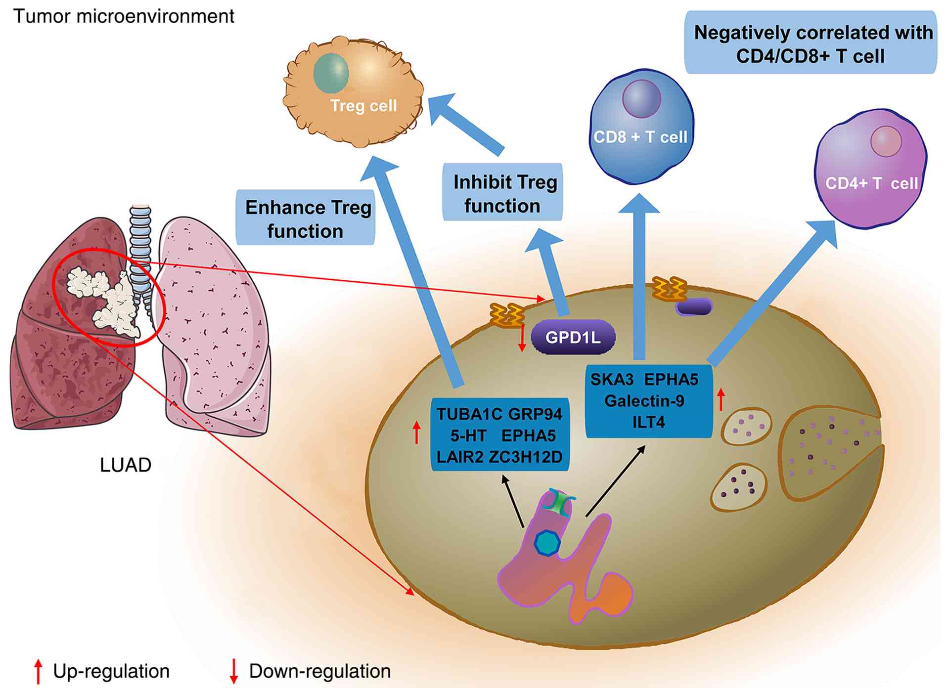 | Figure 1Biomarkers associated with Treg
infiltration and prognosis in LUAD. This schematic summarizes key
biomarkers involved in regulating Treg accumulation, activation and
immunosuppressive function within the LUAD tumor microenvironment.
It highlights their contributions to tumor immune regulation and
clinical outcomes. Notably, Galectin-9, ZC3H12D, SKA3, LAIR2,
TUBA1C, ILT4 and GRP94 are identified as dysregulated in LUAD,
correlating with altered Treg infiltration and function. These
molecules promote Treg proliferation and/or immunosuppressive
activity while suppressing T cell-mediated antitumor immunity and
are closely associated with unfavorable prognosis in LUAD patients.
Treg, regulatory T cell; LUAD, lung adenocarcinoma; GRP94,
glucose-regulated protein 94; ILT4, immunoglobulin-like transcript
4; LAIR2, leukocyte-associated immunoglobulin-like receptor 2;
SKA3, spindle and kinetochore associated complex subunit 3; TUBA1C,
tubulin alpha 1c; ZC3H12D, zinc finger CCCH-type containing
12D. |
 | Table IBiomarkers for regulating Treg
infiltration and LUAD prognosis. |
Table I
Biomarkers for regulating Treg
infiltration and LUAD prognosis.
| First author/s,
year | Number | Target genes | Expression
level | Location | Function | Mechanism | (Refs.) |
|---|
| Xiao et al,
2024 | 1 | Galectin-9 | Upregulation | LUAD | 1. Inhibited
CD8+ T cells
2. Induced Treg proliferation | Galectin-9 is
regulated by NFATc2 | (56) |
| Fan et al,
2023 | 2 | GPD1L | Downregulation | LUAD | 1. Correlated with
N stage and clinical stage
2. Negatively correlated with Treg and neutrophil
infiltration
3. Inhibits the proliferation of LUAD | Not fully
elucidated | (58) |
| Chen et al,
2022 | 3 | ZC3H12D | 1. Downregulation
in LUAD 2. Upregulation in Tregs | LUAD, Tregs and
Immune cells | 1. Negatively
correlated with OS
2. Regulation of the ceRNA network | Regulates mRNA,
miRNA, lncRNA, immune cells and immune molecules to affect the
prognosis of LUAD | (60) |
| Lin et al,
2022 | 4 | SKA3 | Upregulation | LUAD, Tregs and
Immune cells | 1. Correlated with
poor survival
2. Negatively correlated with CD4/CD8+ T cells | SKA3 regulated the
mitotic cell cycle, mitosis, chromosome segregation and cell
division via pathways | (62) |
| Ly et al,
2021 | 5 | LAIR2 | Upregulation | LUAD, Tregs and
Immune cells | 1. Correlated with
poor survival
2. Enhanced in vitro tumor invasion into collagen | 1. LAIR2 expression
co-localized with Foxp3
2. LAIR2 was preferentially produced by activated CD4+ T
cells | (65) |
| Bian et al,
2021 | 6 | TUBA1C | Upregulation | LUAD | 1. Correlated with
poor survival
2. Correlated with age, stage and lymph node metastasis
3. Activate and enhance the function of Tregs | TUBA1C regulated
the cell cycle, p53 signaling pathway, glycolysis and
gluconeogenesis | (63) |
| Chen et al,
2020 | 7 | EPHA5 | EPHA5-mutant
(Mut) | LUAD | 1. Mut enhanced
infiltration of CD8+ T cells and M1
macrophages
2. Mut decreased infiltration of Tregs | EPHA5 mutation
cooccurred with homologous recombination (HR) or mismatch repair
(MMR) gene mutations | (66) |
| Li et al,
2020 | 8 | ILT4 | Upregulation | LUAD | 1. Correlated with
poor survival
2. Negatively correlated with CD8+ T cells
3. Enhance infiltration of Tregs | Not fully
elucidated | (69) |
| Duan et al,
2020 | 9 | GRP94 | Upregulation | LUAD | 1. Correlated with
poor survival
2. Enhance infiltration of Tregs | 1. Inhibited the
expression of caspase-7 and CHOP
2. Promoted the expression of TGF-beta and SMAD2 | (71) |
| Liu et al,
2019 | 10 | 5-HT | Upregulation | Serum | 1. Correlated with
poor survival
2. Enhance infiltration of Tregs
3. decreased the ratio of Th1/Th2 cells | 5-HT1aR activates
p-signal transducer and activator of transcription 3 (STAT3) and
autophagy | (72) |
| Bahabayi et
al, 2025 | 11 | TCF1 | Downregulation in
Tregs | Tregs | TCF1-related
indicators showed good auxiliary diagnostic significance for early
LUAD | Downregulation of
Notch signaling contributes to the decrease in TCF1+
Treg subsets | (127) |
| Wang et al,
2026 | 12 | CCL20 | Upregulation in
Tregs | Tregs | 1. High CCL20
expression correlated with increased Treg infiltration and served
as an independent prognostic biomarker
2. CCL20 promoted the proliferation of LUAD | 1. NF-κB/STAT
signaling
2. CCL20-CCR6 ligand-receptor pair | (128) |
Treg-associated prognostic signatures
and predictive models
Recent advances in transcriptomic analysis and
computational biology have expanded the assessment of Treg-related
prognostic features from single biomarkers to multidimensional
molecular and immune signatures. Several studies have developed
prognostic models incorporating immune infiltration, tumor
metabolism, genetic alterations, or immune-related regulatory
networks, consistently demonstrating that the composition of the
LUAD immune microenvironment contains clinically relevant
prognostic information. Shao et al (73) identified an alternative
splicing-based prognostic signature associated with B-cell and
immune infiltration, while Wang et al (74) developed an immune
infiltration-related model that identified immune-cell populations
associated with patient survival. Similarly, Li et al
(75) established a
mitochondrial-related gene signature associated with both immune
infiltration and survival, suggesting an interplay between tumor
metabolic status and immune regulation, whereas Gao et al
(76) developed an
immunophenotype-based risk score that effectively stratified
patients according to distinct immune characteristics. Liu et
al (77) further demonstrated
that competing endogenous RNA networks interact with
tumor-infiltrating immune cells to form prognostically relevant
regulatory networks and Song et al (78) showed that an immune-related risk
model incorporating TP53 status could predict both prognosis and
immunotherapy response. Wu et al (79) likewise identified multi-gene
immune-related signatures associated with prognosis and potential
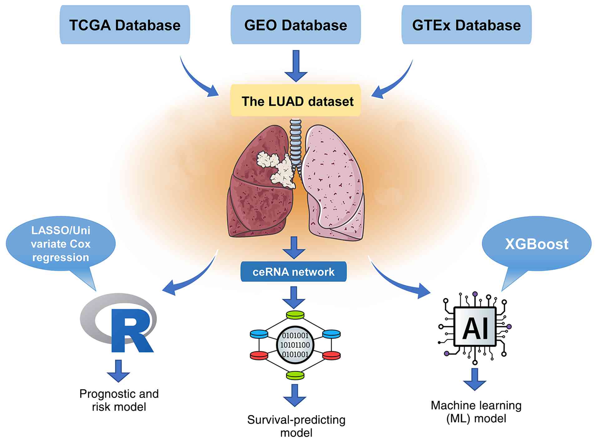
immunotherapy responsiveness. Fig.
2 and Table II show LUAD
prognosis, survival and machine learning models derived from The
Cancer Genome Atlas (TCGA; https://portal.gdc.cancer.gov/), Gene Expression
Omnibus (GEO; https://www.ncbi.nlm.nih.gov/geo/), and
Genotype-Tissue Expression (GTEx; https://gtexportal.org/home/) databases.
| Table IITargeting Treg infiltration
prognostic model of LUAD. |
Table II
Targeting Treg infiltration
prognostic model of LUAD.
| First author/s,
year | Number | Prognostic
model | Database | Model
algorithm | Function | (Refs.) |
|---|
| Shao et al,
2023 | 1 | Risk model
(smRNA-seq) | TCGA | Cox regression
analyses and LASSO regression | 1. B cells were
most markedly correlated with the risk score
2. Promotion of regulatory T cell differentiation | (73) |
| Wang et al,
2024 | 2 | Machine learning
(ML) model | TCGA, GTEx | The algorithm
eXtreme Gradient Boosting (XGBoost) | 1. Treg
infiltration was negatively correlated with prognosis
2. PCBP1-AS1 was negatively correlated with Treg infiltration and
TGF-β expression | (74) |
| Li et al,
2022 | 3 | Prognostic model
based on MTRGs | TCGA, GEO | 1. Univariate Cox
regression analysis
2. LASSO Cox regression analysis | 1. The high-risk
group was correlated with poor survival
2. Risk score was an independent predictor of LUAD
prognosis
3. Treg infiltration was higher in the low-risk group | (75) |
| Gao et al,
2022 | 4 | Prognostic risk
score (PRS) model | TCGA, GEO | 1. Univariate Cox
regression analysis
2. LASSO Cox regression analysis | 1. Tregs and PD-L1
were correlated with poor survival
2. TIICs and IPSs are higher in the low-risk group | (76) |
| Liu et al,
2022 | 6 | Survival-predicting
model | TCGA, GEO | 1. CIBERSORT
algorithm
2. ceRNA network | Monocytes and Tregs
were correlated with poor survival | (77) |
| Song et al,
2022 | 7 | Immune-related risk
model (IRM) | TCGA | LASSO
algorithm | 1. High-index group
had lower relative infiltration of Tregs
2. The IRM index is positively correlated with PD-L1
expression | (78) |
| Wang et al,
2021 | 8 | Prognostic nomogram
model | TCGA, GEO | 1. Nomogram
algorithm
2. Cox regression analysis | High-index group
was correlated with poor survival | (80) |
| Wu et al,
2020 | 9 | Risk score
model | TCGA, GEO | LASSO Cox
regression analysis | Tregs enhanced
activation and immunologic checkpoint molecular expression increase
in the high-risk group | (79) |
| Szentkereszty et
al, 2024 | 10 | Retrospective
study | A single-center
case database | 1.
Immunohistochemical detection
2. Kaplan-Meier analysis
3. Multivariate Cox regression analysis | Detection of NKp46
and FoxP3 positivity to identify NK and Tregs | (129) |
| Zhao et al,
2025 | 11. | Prognostic risk
score (PRS) model | GEO | 1. CIBERSORT and
weighted correlation network analysis (WGCNA)
2. LASSO and univariate and multivariate Cox regression
analyses | 1. The expression
of Treg surface markers in LUAD cells was increased
2. The expression of SCN9A was decreased compared with that in
normal lung epithelial cells. | (130) |
| He et al,
2026 | 12 | Single-cell
Prognostic risk score (PRS) model | GEO | 1. Univariate Cox
and LASSO regression analyses
2. Kaplan-Meier and ROC curves
3. TMB, Spearman and functional enrichment analysis | 1. The risk score
could be an independent prognostic factor
2. CENPM, ZNF101, MAGEH1 and IKZF4 were up-regulated in LUAD | (131) |
| Xue et al,
2025 | 13 | An anoikis-related
prognostic model | GEO | 1. Univariate Cox
regression analysis, LASSO regression and random forest
algorithms
2. Machine learning | 1. This model can
accurately predict the survival rate of LUAD patients
2. LUAD patients exhibited an immunosuppressive TME phenotype | (132) |
Taken together, these studies provide relatively
consistent evidence that the immune landscape of LUAD, and the
molecular programs associated with immune-cell infiltration, has
substantial prognostic value. However, most of these models were
not specifically designed around Tregs and their predictive
performance reflects the combined contribution of multiple immune
and tumor-related features. Therefore, they should be interpreted
as indirect evidence supporting the prognostic relevance of
Treg-associated immune states rather than as evidence that Tregs
independently determine patient outcome. Among these studies, the
work of Wang et al (80)
provides more direct evidence for the clinical relevance of Treg
biology, as a Treg-related prognostic nomogram was specifically
developed for LUAD, demonstrating that Treg-associated genes can
contribute to survival prediction. Nevertheless, even this
Treg-focused model supports the prognostic utility of a
Treg-associated molecular signature rather than establishing total
Treg infiltration as a standalone biomarker. Thus, the current
computational literature supports the concept that Treg-related
molecular characteristics can improve prognostic risk
stratification, but the incremental value of Treg-specific features
over broader immune signatures remains to be systematically
determined.
An important limitation is that most existing models
are derived from bulk transcriptomic datasets, in which
Treg-associated signals may be confounded by other immune-cell
populations, tumor purity, or differences in Treg abundance and
phenotype. Moreover, the predictive performance of these models
does not necessarily establish a biological or causal role for
Tregs in determining survival. Future studies should therefore
integrate Treg-specific single-cell and spatial features with
clinical variables and longitudinal treatment information to
determine whether specific tumor-promoting Treg subsets can provide
more robust and reproducible prognostic information than bulk
Treg-related signatures.
Tregs as prognostic biomarkers:
Current evidence and future challenges
Overall, the available evidence supports several
relatively robust conclusions. First, Tregs are consistently
enriched in the LUAD TME and are associated with an
immunosuppressive immune contexture (47,52-54). Second, a substantial proportion of
clinical and molecular studies associate increased Treg
infiltration or Treg-related signatures with advanced disease and
unfavorable survival outcomes (55,56,58-65). Third, mechanistic studies such as
the IKKα study provide biological support for the concept that
tumor cells can actively promote Treg recruitment and expansion,
thereby linking Treg accumulation to immune suppression and tumor
progression (70). Finally,
computational studies indicate that Treg-associated molecular
characteristics can contribute to prognostic risk stratification
and may have potential value in predicting immunotherapy response
(73-78).
Nevertheless, the current evidence does not justify
concluding that Treg infiltration alone is a universally applicable
or independent prognostic biomarker for LUAD. The prognostic effect
may differ according to Treg subset, spatial localization, disease
stage, detection methodology and the relative abundance of other
immune populations. Furthermore, a number of molecular and
machine-learning studies demonstrate associations rather than
causality and their predictive performance may reflect the combined
effects of multiple biological processes rather than Treg biology
alone. Importantly, even when high Treg infiltration is associated
with poor survival, this does not necessarily establish that Tregs
are the primary drivers of tumor progression; Treg accumulation may
also reflect an already established immunosuppressive or
tumor-promoting microenvironment.
Therefore, the most appropriate interpretation of
the current literature is that Treg infiltration and
Treg-associated molecular signatures represent promising components
of a multidimensional prognostic framework for LUAD, rather than
definitive standalone biomarkers. Future studies should determine
whether specific tumor-promoting Treg subsets, rather than total
Treg abundance, provide stronger and more reproducible prognostic
information. Integrating single-cell sequencing, spatial
transcriptomics, longitudinal clinical sampling and functional
validation may help distinguish Tregs that actively drive tumor
progression from those that primarily reflect the existing immune
state of the tumor.
Mechanisms underlying Treg-mediated
immune escape in LUAD
Tumor immune escape serves as a hallmark of cancer
progression, with Tregs constituting one of the most significant
immunosuppressive components within the LUAD TME. Unlike
conventional immune cells that directly eliminate tumor cells,
Tregs suppress antitumor immune responses through multiple
mechanisms, including immune checkpoint regulation, secretion of
inhibitory cytokines, metabolic remodeling and interactions with
other stromal and immune populations. Increasing evidence indicates
that Treg-mediated immune escape is not driven by a single pathway,
but rather arises from coordinated regulation among tumor cells,
immune cells and metabolic networks.
Immune inhibitory molecules mediated
by Tregs
A primary mechanism through which Tregs facilitate
immune escape is the expression of inhibitory receptors and
immunosuppressive molecules that directly impair effector immune
responses. Tregs are known to highly express immune checkpoint
molecules such as CTLA-4, PD-1, TIGIT and LAG-3, which suppress the
activation and cytotoxic functions of CD8+ T cells and
NK cells, thereby enabling tumor cells to evade immune surveillance
(81). Among these molecules,
CTLA-4 expressed by Tregs competitively binds to CD80/CD86 on
antigen-presenting cells, reducing costimulatory signaling and
limiting T cell activation. Studies indicate that blocking CTLA-4
signaling can diminish Treg-mediated immunosuppression and enhance
antitumor immune responses, underscoring the significance of
Treg-associated checkpoint regulation in cancer immunotherapy
(82). In LUAD, tumor cells can
further amplify Treg-mediated immune suppression by inducing
immunoregulatory molecules. Tumor-derived factors, for example,
promote Treg recruitment and activation through the CCL22/CCR4
axis, resulting in increased Treg accumulation at the tumor site
and suppression of CD8+ T cell-mediated tumor killing
(83,84). Additionally, tumor-associated
immune checkpoint molecules and soluble immunosuppressive mediators
contribute to sustaining Treg suppressive activity, facilitating
immune escape (83,84).
Metabolic regulation contributes to
Treg-mediated immune tolerance
Beyond immune checkpoint pathways, metabolic
reprogramming within the TME plays a key role in maintaining Treg
function and promoting immune escape. Tumor cells commonly generate
a metabolically unfavorable environment characterized by glucose
depletion, heightened lactate accumulation, hypoxia and altered
amino acid metabolism. Tregs exhibit significant metabolic
adaptability, allowing them to survive under these challenging
conditions while maintaining suppressive activity as effector T
cells become functionally exhausted (85). Glucose competition is a critical
metabolic mechanism regulating Treg-mediated immune suppression.
Activated Tregs efficiently utilize available metabolic substrates
and resist metabolic stress, while glucose deprivation impairs
effector T cell proliferation and cytokine production, conferring a
selective advantage to Tregs in the TME (85). Furthermore, metabolic pathways
related to lipid metabolism, mitochondrial activity and oxidative
phosphorylation contribute to Treg stability and functional
maintenance. Consequently, alterations in tumor metabolism may
indirectly enhance Treg-mediated immune suppression and promote
immune escape in LUAD (86).
Glutamine (Gln) metabolism plays a vital role in LUAD. Liu et
al (87) constructed a risk
model for LUAD using Cox regression and Lasso regression analysis
based on genes associated with Gln metabolism, categorizing
patients into low-risk and high-risk groups. The low-risk group was
characterized by low Gln metabolism, a survival advantage, a 'hot'
immunophenotype and benefits from immunotherapy. Among immune
cells, tumor-infiltrating T cells exhibited the most active levels
of Gln metabolism, particularly highlighting CD8 T cell depletion
and Treg suppression. These findings suggest that targeting the
metabolic function of Tregs may present a promising strategy for
restoring antitumor immunity.
Interaction between Tregs and other
components of the LUAD microenvironment
Treg-mediated immune escape is a complex process
that relies on extensive interactions between Tregs and various
cellular components within the TME. Tumor cells, tumor-associated
macrophages (TAMs), CAFs and other immune populations work together
to regulate Treg recruitment, expansion and suppressive function.
Currently, advancements in single-cell technology have markedly
contributed to the analysis of TME-infiltrating Tregs and their
roles in immune escape. For instance, by utilizing scRNA-seq to
identify molecular subtypes and homologous immune metabolism
characteristics of LUAD immune cell marker genes, researchers
developed the LID-score model. Patients with low LID scores
demonstrate higher survival rates due to abundant immune cell
infiltration, immune activation and low genetic variation. However,
elevated levels of Tregs in the immune microenvironment can lead to
immune cell dysfunction, promoting tumor immune escape and reducing
responsiveness to immunotherapy (88). Furthermore, fibroblast growth
factor 11 (FGF11) has been observed to accelerate tumor
proliferation in various cancer types. Notably, FGF11 gene
expression is negatively correlated with most immune cell
populations, particularly with characteristic genes of functional T
cells such as Tregs and resting Tregs. This suggests that FGF11
could emerge as a new biomarker in LUAD patients, as it may enhance
tumor cell immune escape by promoting T-cell depletion within the
TME, leading to poorer prognoses (89). Chemokines also play a critical
role in tumor immunotherapy, with studies showing that CCL28
secreted by LUAD can induce Treg infiltration, further supporting
immune escape (90). Animal
studies have demonstrated that CCR2 knockout mice exhibit fewer
CD4+ T cells and Tregs in tumors, alongside increased
infiltration of CD8 T cells producing IFN-γ and granzyme B
(91). Additionally,
TNF-α-dependent inflammation related to LUAD has been shown to
upregulate MHC-II in alveolar type II cells via CXCR-2, promoting
Treg expansion (92).
Treg-related molecular signatures and
implications for immunotherapy
With the advancement of transcriptomic sequencing
and bioinformatics, there is growing interest in Treg-associated
molecular signatures as potential biomarkers for immune escape and
treatment response. Various studies have identified immune-related
gene signatures linked with Treg infiltration, tumor progression
and LUAD prognosis (93-95). These findings suggest that
Treg-related characteristics may reflect tumor immune status and
hold predictive value for immunotherapy.
Nonetheless, Treg-targeted therapeutic strategies
pose challenges. While the depletion or functional inhibition of
Tregs may enhance antitumor immunity, systemic elimination may
induce autoimmune toxicity due to Tregs' critical role in
maintaining immune homeostasis. Therefore, selectively targeting
tumor-associated Tregs or their specific suppressive pathways is
seen as a more promising therapeutic approach (96-98).
In summary, Tregs facilitate immune escape in LUAD
through a multifaceted regulatory network involving inhibitory
molecules, metabolic adaptations and interactions with other TME
components. Tregs influence tumor characteristics through cytokines
and ligands, activating multiple signaling pathways. Additionally,
tumor cells recruit Tregs via chemokines, cytokines and metabolic
regulations to enhance immune escape. Thus, understanding the
functional heterogeneity of tumor-associated Tregs and identifying
tumor-specific regulatory mechanisms are critical for developing
targeted immunotherapeutic strategies. Biomarkers that promote
immune escape in LUAD are illustrated in Fig. 3 and summarized in Table III.
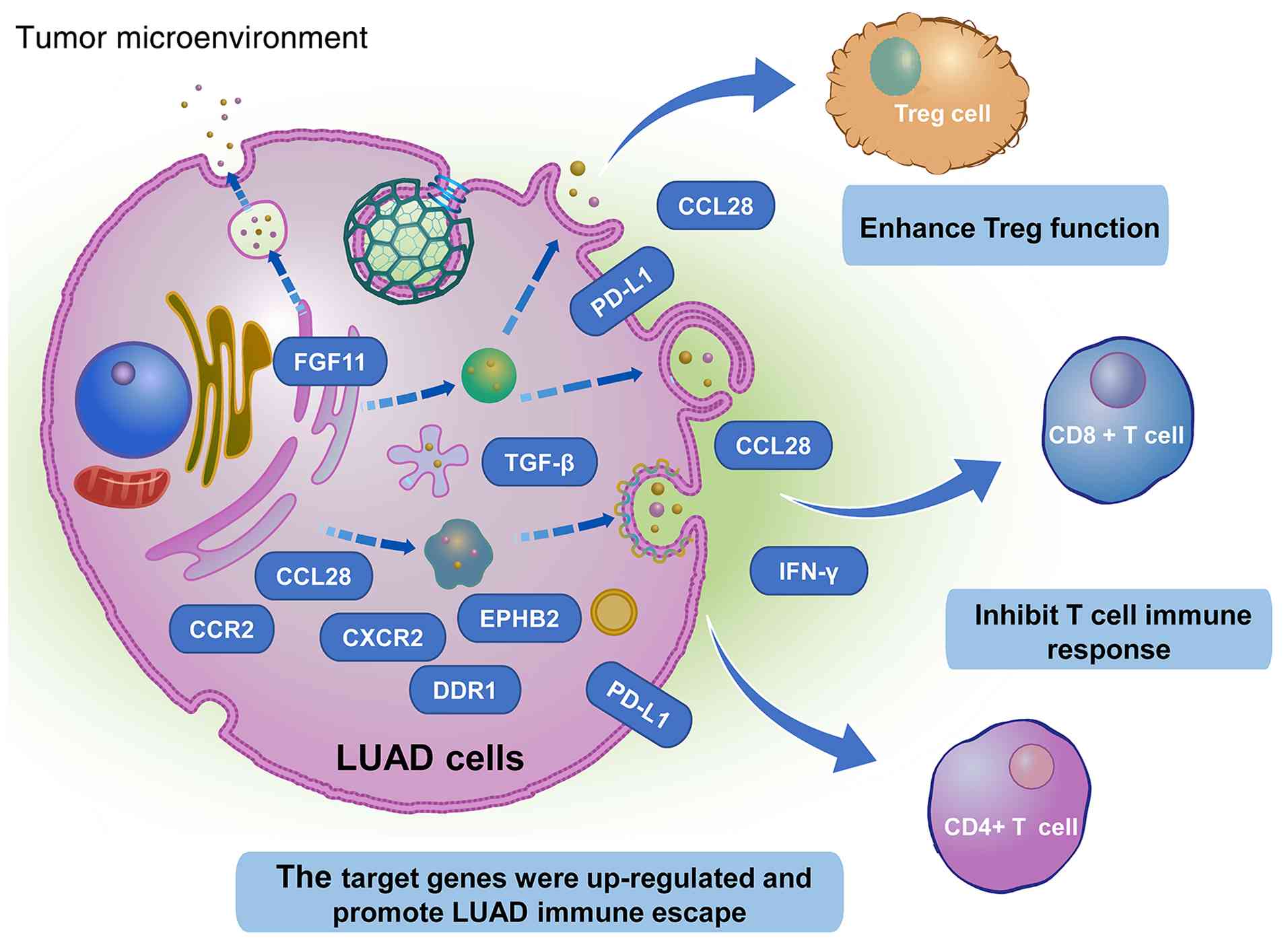 | Figure 3Biomarkers regulating Treg and T cell
infiltration and promoting immune escape in LUAD. This schematic
illustrates the major molecular pathways regulating the balance
between immunosuppressive Tregs and antitumor effector T cells
within the LUAD tumor microenvironment. Biomarkers such as FGF11,
CCL28, CCR2, CXCR2, EPHB2, PD-L1, TGF-β and DDR1 are upregulated in
LUAD and contribute to immune escape by promoting Treg recruitment,
proliferation, or suppressive activity while impairing T
cell-mediated antitumor immunity. These findings underscore the
importance of targeting Treg-associated regulatory networks as a
potential therapeutic strategy for LUAD. Treg, regulatory T cell;
LUAD, lung adenocarcinoma; CCL28, C-C motif chemokine ligand 28;
CCR2, C-C motif chemokine receptor 2; CXCR2, C-X-C motif chemokine
receptor 2; DDR1, discoidin domain receptor tyrosine kinase 1;
EPHB2, EPH receptor B2; FGF11, fibroblast growth factor 11; PD-L1,
programmed death-ligand 1; TGF-β, transforming growth factor
beta. |
| Table IIIBiomarkers for regulating Treg
infiltration and LUAD immune escape. |
Table III
Biomarkers for regulating Treg
infiltration and LUAD immune escape.
| First author/s,
year | Number | Target Genes | Expression
level | Location | Function | (Refs.) |
|---|
| Zhai et al,
2023 | 1 | FGF11 | Upregulation | LUAD | 1. Correlated with
poor survival
2. Enhance infiltration of Tregs | (89) |
| Su et al,
2023 | 2 | CCL28 | Upregulation | LUAD | 1. Enhance
infiltration of Tregs
2. Inhibited the immune response to treatment | (90) |
| Mittal et
al, 2020 | 3 | CCR2 MCP-1 | Upregulation | LUAD | 1. Promote MDSC
recruitment to TME
2. Inhibition of the host T cell immune response | (91) |
| Guo et al,
2020 | 4 | CXCR-2 TNF-α | Upregulation | LUAD | Enhance the
proliferation of Tregs | (92) |
| Liu et al,
2022 | 6 | EPHB2 | Upregulation | LUAD | 1. Promote LUAD
proliferation, invasion and migration
2. Enhance the suppressive function of Tregs | (87) |
| Liu et al,
2021 | 7 | CCL28 | Upregulation | LUAD | 1. Enhance the
angiogenesis and metastasis of LUAD
2. Enhance the proliferation of Tregs | (95) |
| Shima et al,
2020 | 8 | PD-L1 | Upregulation | LUAD | Enhance the
proliferation of Tregs | (96) |
| Islas-Vazquez et
al, 2015 | 9 | TGF-β (LAP) | Upregulation | Tregs | Enhance the
suppressive function of Tregs | (97) |
| Maitz et al,
2023 | 10 | DDR1 | Upregulation | LUAD | Knockout of DDR1
increased the tumor burden, decreased CD8+ cytotoxic T
cells, increased CD4+ helper T cells and Tregs. | (98) |
| Huang et al,
2024 | 11. | Ku70 | Upregulation | Tregs | 1. Repair DNA
double-strand breaks
2. Enhance the suppressive function of Tregs | (133) |
Diagnosis of Tregs and LUAD
The prognostic value of tumor-infiltrating
lymphocytes is critical in solid tumors, reflecting the immune
response's ability to eliminate tumor cells and predicting the
clinical efficacy of specific cancer therapies. The presence of
Tregs is associated with patient survival across various tumor
types (99). Numerous studies
have reported correlations between Treg infiltration and disease
prognosis in different tumors. However, methodologies to assess
Treg levels in tumor tissue vary among studies; some have utilized
immunohistochemical sections, while others have employed tissue
microarrays (100). Recent
analyses have highlighted the role of tumor-infiltrating Tregs in
prognosis and diagnosis through bioinformatics, demonstrating their
potential as markers for early tumor diagnosis and prognosis. Some
studies have used FoxP3 as the sole marker for Tregs, while others
have included FoxP3, CD25 and CCR4 (101). Furthermore, Treg prognostic
significance is influenced by tumor site, molecular subtype and
tumor stage (100). To
accurately determine Treg levels in tumor tissues and assess their
phenotypic characteristics and prognostic significance across
various tumor types, stages and histological or molecular subtypes,
consistency in marker selection and meticulous analysis are
essential. Such approaches will enhance the design of more
effective immunotherapies and aid in identifying appropriate
targets for Treg treatment in select solid tumor patients.
Recently, tumor mutation burden (TMB) has emerged as
a promising biomarker for predicting immunotherapy efficacy across
different tumor types (102).
Tumor protein D52-like 2 (TPD52L2), a member of the TPD52 family,
regulates tumor cell proliferation and apoptosis. Zhong et
al (103) evaluated the
correlation between TPD52L2 and levels of immune cell infiltration,
immunosuppressive genes and TMB in LUAD. Results indicated that
TPD52L2 was highly expressed in LUAD and predicted poorer survival
outcomes, with a positive correlation identified between TPD52L2
expression and Treg infiltration levels, suggesting that TPD52L2
may serve as a potential prognostic biomarker. Additionally, Tregs
play a role in the early development of lesions associated with
LUAD. An independent study of resected lung nodules, including
adenocarcinomas and associated precancerous lesions, confirmed
increased Treg levels in these precancerous conditions (104). Zhao et al (105) collected blood samples from 35
healthy controls and 34 early-stage LUAD patients, with flow
cytometry analysis revealing upregulated proportions and absolute
numbers of Tregs in the peripheral blood of LUAD patients.
Moreover, INTS7, a subunit of the integron complex, is found to be
upregulated in various tumors (106). INTS7 expression negatively
correlates with the infiltration of memory B cells, Tregs,
monocytes and activated mast cells. Consequently, INTS7 may serve
as a potential diagnostic marker for LUAD (106). Thus, Treg upregulation
contributes to the early diagnosis of LUAD.
Therapeutic strategies and research
progress of targeting Tregs
Due to the role of Tregs in tumor immunity, various
therapeutic approaches targeting Tregs have emerged in tumor
therapy, including the disruption of their immunosuppressive
function, blockade of their transport to tumors and inhibition of
their development and activation (107,108). Currently, targeting Tregs in
LUAD therapeutic strategies often involves two or more of these
approaches to enhance antitumor immunity (109). Research by Zhang et al
(110) demonstrated that
Pexidartinib reduces the CCL22 source from TAMs in LUAD, thereby
suppressing Treg infiltration and enhancing the efficacy of PD-1
antibody therapy. Following Pexidartinib treatment, the total
number of T cells, CD8+ T cells and Tregs decreased,
while the CD8+/Treg ratio markedly increased. When
Pexidartinib was combined with PD-1 antibody in LUAD subcutaneous
tumor models, this combined treatment improved tumor treatment
efficacy compared with monotherapy (111). Monocarboxylate transporter 4
(MCT4), responsible for lactate efflux, has been associated with
poor prognosis in various tumors (112). TMPs coated with Fluvastatin
(TMP-F) were found to slow LUAD progression by inhibiting MCT4
expression and blocking lactate outflow. TMP-F induced infiltration
of NK cells and CD8+ T cells while polarizing M2
macrophages to M1 macrophages. The depletion of Tregs and
myeloid-derived suppressor cells (MDSCs) reversed the
immunosuppressive TME. Additionally, TMP-F enhanced the antitumor
effects of cisplatin and inhibited lung metastasis. Overall, TMP-F
treatment, alone or in combination with cisplatin, represents a
promising strategy to improve efficacy in LUAD (112). Fu-Zheng-Qu-Xie decoction, a
traditional Chinese medicine, has shown potential in improving
immune function and survival in early lung cancer patients
(113). Zhang et al
(113) evaluated the clinical
efficacy of this herbal prescription in preventing postoperative
recurrence and metastasis of early-stage LUAD. Following
administration of FZQX, positive immune indicators (CD4+
T/CD8+ T) increased, while negative immune indicators
(CD8+ T, MDSCs, Tregs) decreased. Furthermore, FZQX
prescription regulated MDSCs to remodel the immunosuppressive TME,
thereby contributing to the prevention of lung cancer recurrence.
Ganesan et al (114)
utilized CD25-depleting mAb monotherapy to preferentially deplete
Tregs, thereby enhancing CD8+ T-cell-mediated antitumor
immunity. The combination with Carboplatin therapy markedly
improved survival in LUAD patients, indicating that Treg depletion
coupled with cytotoxic therapy may serve as an advantageous
treatment strategy for advanced LUAD.
Prospects
Tregs play a pivotal role in maintaining immune
homeostasis and modulating antitumor immune responses. In LUAD,
increasing evidence indicates that tumor-associated Tregs
contribute to tumor progression, immune escape and resistance to
immunotherapy by suppressing effector immune cells and remodeling
the TME. Consequently, targeting Tregs has emerged as a promising
therapeutic strategy to enhance treatment outcomes in LUAD.
However, despite significant advances in understanding Treg
biology, challenges persist regarding the precise identification,
selective targeting and clinical translation of Treg-based
therapies.
While Treg depletion or functional inhibition can
enhance antitumor immunity, the systemic elimination of Tregs
carries a risk of autoimmune toxicity due to their vital role in
maintaining immune tolerance. Future studies should therefore
concentrate on developing strategies that selectively target
tumor-infiltrating Tregs while preserving peripheral immune
homeostasis. Potential approaches may involve targeting
tumor-enriched Treg-associated molecules, chemokine receptors,
immune checkpoints, or signaling pathways that are preferentially
activated in tumor-associated Tregs. For instance, identifying
LUAD-specific Treg surface markers and developing antibody-based
therapies, small-molecule inhibitors, or engineered immune cells
aimed at these molecules could lead to more precise therapeutic
strategies compared with global Treg depletion (115).
Another significant challenge is the functional
heterogeneity of Tregs within LUAD. Current evidence suggests that
Tregs constitute a diverse population comprising multiple subsets
with distinct transcriptional profiles, spatial distributions and
immunosuppressive functions. Traditional bulk transcriptomic
approaches cannot accurately differentiate these functional
subsets. Therefore, techniques such as scRNA-seq, single-cell
epigenomic analysis and spatial transcriptomics offer powerful
methodologies for identifying tumor-specific Treg populations while
revealing their developmental trajectories, cellular interactions
and regulatory mechanisms. Future integration of single-cell
multi-omics and spatial analysis may facilitate the identification
of highly suppressive Treg subsets and unveil new targets for
precision immunotherapy (116).
In addition to directly targeting Tregs, combination
therapeutic strategies may represent a more effective means of
overcoming Treg-mediated immune suppression. Given that Tregs
extensively interact with metabolic pathways and other TME
components, combining Treg-targeted therapies with metabolic
inhibitors may disrupt the metabolic advantages that sustain Treg
survival and function. Furthermore, combining these therapies with
ICIs, tumor vaccines, or other immunotherapeutic approaches could
enhance effector immune responses and improve treatment efficacy.
Such strategies may help overcome the limitations of single-agent
immunotherapy and achieve more durable antitumor responses in LUAD
patients (117,118).
Studies have demonstrated that immune-related gene
signatures and Treg-associated molecular characteristics may serve
as potential biomarkers for predicting LUAD prognosis and responses
to immunotherapy (119-125). However, most current prediction
models rely on bulk sequencing data, which may not fully capture
the complex spatial and functional heterogeneity of Tregs within
tumors. The rapid advancement of artificial intelligence (AI) and
machine learning approaches offers new opportunities for
integrating transcriptomic, spatial, clinical and immunological
data, potentially enhancing patient stratification and treatment
selection. AI-based models may assist in identifying patients with
Treg-dominant immunosuppressive microenvironments and predicting
their responses to Treg-targeted therapies or combination
immunotherapies (126).
Moreover, the translation of Treg-related findings
into clinical applications necessitates further investigation into
treatment safety, optimal therapeutic timing and patient selection
criteria. Dynamic changes in Tregs during tumor progression and
therapeutic intervention remain inadequately understood.
Longitudinal studies that combine clinical samples, single-cell
sequencing and functional validation will be essential in
elucidating how Treg populations evolve throughout PD and treatment
resistance. In conclusion, Tregs represent a key regulatory
component of the LUAD immune microenvironment, markedly influencing
tumor immune escape and therapeutic resistance. Future advancements
in selective Treg targeting strategies, single-cell and spatial
technologies, combination immunotherapy and AI-driven precision
medicine will create new avenues for understanding and manipulating
Treg biology, ultimately enhancing the clinical management of LUAD
patients.
Availability of data and materials
Not applicable.
Authors' contributions
LJW was responsible for conceptualization and
supervision and writing-review and funding acquisition and editing.
CJR was responsible for conceptualization and supervision and
writing-review and editing. Data authentication is not applicable.
All authors read and approved the final manuscript.
Ethics approval and consent to
participate
Not applicable.
Patient consent for publication
Not applicable.
Competing interests
The authors declare that they have no competing
interests.
Acknowledgments
Not applicable.
Funding
The present study was supported by The Science and Technology
Plan Project of Taizhou (grant no. 24ywa19) and Science and
Technology Program of Traditional Chinese Medicine of Zhejiang
Province (grant no. 2024ZL1239).
References
|
1
|
Wei X, Li X, Hu S, Cheng J and Cai R:
Regulation of ferroptosis in lung adenocarcinoma. Int J Mol Sci.
24:146142023. View Article : Google Scholar : PubMed/NCBI
|
|
2
|
Spella M and Stathopoulos GT: Immune
resistance in lung adenocarcinoma. Cancers (Basel). 13:3842021.
View Article : Google Scholar : PubMed/NCBI
|
|
3
|
Inamura K: Clinicopathological
characteristics and mutations driving development of early lung
adenocarcinoma: Tumor initiation and progression. Int J Mol Sci.
19:12592018. View Article : Google Scholar : PubMed/NCBI
|
|
4
|
Zhang P, Zhang X, Cui Y, Gong Z, Wang W
and Lin S: Revealing the role of regulatory T cells in the tumor
microenvironment of lung adenocarcinoma: A novel prognostic and
immunotherapeutic signature. Front Immunol. 14:12441442023.
View Article : Google Scholar : PubMed/NCBI
|
|
5
|
The L: Lung cancer: Some progress, but
still a lot more to do. Lancet. 394:18802019. View Article : Google Scholar
|
|
6
|
Huang S, Yang J, Shen N, Xu Q and Zhao Q:
Artificial intelligence in lung cancer diagnosis and prognosis:
Current application and future perspective. Semin Cancer Biol.
89:30–37. 2023. View Article : Google Scholar : PubMed/NCBI
|
|
7
|
Wang Y, Xu M, Wei X, Huang H, Chen Q, Chen
B, Bao X and Li J: Prospective proteomics for discovering
biomarkers in lung adenocarcinoma: A literature review. Transl
Cancer Res. 14:6102–6117. 2025. View Article : Google Scholar : PubMed/NCBI
|
|
8
|
Kwok HH, Yang J and Lam DC: Breaking the
invisible barriers: Unleashing the full potential of immune
checkpoint inhibitors in Oncogene-driven lung adenocarcinoma.
Cancers (Basel). 15:27492023. View Article : Google Scholar : PubMed/NCBI
|
|
9
|
Mithoowani H and Febbraro M:
Non-Small-cell lung cancer in 2022: A review for general
practitioners in oncology. Curr Oncol. 29:1828–1839. 2022.
View Article : Google Scholar : PubMed/NCBI
|
|
10
|
Borghaei H, Paz-Ares L, Horn L, Spigel DR,
Steins M, Ready NE, Chow LQ, Vokes EE, Felip E, Holgado E, et al:
Nivolumab versus docetaxel in advanced nonsquamous Non-Small-cell
lung cancer. N Engl J Med. 373:1627–1639. 2015. View Article : Google Scholar : PubMed/NCBI
|
|
11
|
Garon EB, Rizvi NA, Hui R, Leighl N,
Balmanoukian AS, Eder JP, Patnaik A, Aggarwal C, Gubens M, Horn L,
et al: Pembrolizumab for the treatment of non-small-cell lung
cancer. N Engl J Med. 372:2018–2028. 2015. View Article : Google Scholar : PubMed/NCBI
|
|
12
|
Gibney GT, Weiner LM and Atkins MB:
Predictive biomarkers for checkpoint inhibitor-based immunotherapy.
Lancet Oncol. 17:e542–e551. 2016. View Article : Google Scholar : PubMed/NCBI
|
|
13
|
Siegel RL, Miller KD, Wagle NS and Jemal
A: Cancer statistics, 2023. CA Cancer J Clin. 73:17–48.
2023.PubMed/NCBI
|
|
14
|
Binnewies M, Roberts EW, Kersten K, Chan
V, Fearon DF, Merad M, Coussens LM, Gabrilovich DI,
Ostrand-Rosenberg S, Hedrick CC, et al: Understanding the tumor
immune microenvironment (TIME) for effective therapy. Nat Med.
24:541–550. 2018. View Article : Google Scholar : PubMed/NCBI
|
|
15
|
Giraldo NA, Sanchez-Salas R, Peske JD,
Vano Y, Becht E, Petitprez F, Validire P, Ingels A, Cathelineau X,
Fridman WH and Sautès-Fridman C: The clinical role of the TME in
solid cancer. Br J Cancer. 120:45–53. 2019. View Article : Google Scholar :
|
|
16
|
Nagarsheth N, Wicha MS and Zou W:
Chemokines in the cancer microenvironment and their relevance in
cancer immunotherapy. Nat Rev Immunol. 17:559–572. 2017. View Article : Google Scholar : PubMed/NCBI
|
|
17
|
Gonzalez H, Hagerling C and Werb Z: Roles
of the immune system in cancer: From tumor initiation to metastatic
progression. Genes Dev. 32:1267–1284. 2018. View Article : Google Scholar : PubMed/NCBI
|
|
18
|
Ollauri-Ibáñez C, Ayuso-Íñigo B, Solano-Sc
I, Díez P, Perez-Andres M, Muñoz-Félix JM, Rodríguez-Barbero A and
Pericacho M: Endoglin (CD105) overexpression is associated with an
immunosuppressive tumor microenvironment in murine lung cancer
models and human lung adenocarcinoma. Sci Rep. 16:268962026.
View Article : Google Scholar : PubMed/NCBI
|
|
19
|
Song L, Gong Y, Wang E, Huang J and Li Y:
Unraveling the tumor immune microenvironment of lung adenocarcinoma
using single-cell RNA sequencing. Ther Adv Med Oncol.
16:175883592312102742024. View Article : Google Scholar : PubMed/NCBI
|
|
20
|
Peng DH, Rodriguez BL, Diao L, Gaudreau
PO, Padhye A, Konen JM, Ochieng JK, Class CA, Fradette JJ, Gibson
L, et al: Th17 cells contribute to combination MEK inhibitor and
anti-PD-L1 therapy resistance in KRAS/p53 mutant lung cancers. Nat
Commun. 12:26062021. View Article : Google Scholar : PubMed/NCBI
|
|
21
|
Liu X, Hogg GD and DeNardo DG: Rethinking
immune checkpoint blockade: 'Beyond the T cell'. J Immunother
Cancer. 9:e0014602021. View Article : Google Scholar : PubMed/NCBI
|
|
22
|
Levitzki A and Klein S: My journey from
tyrosine phosphorylation inhibitors to targeted immune therapy as
strategies to combat cancer. Proc Natl Acad Sci USA.
116:11579–1186. 2019. View Article : Google Scholar : PubMed/NCBI
|
|
23
|
Rittmeyer A, Barlesi F, Waterkamp D, Park
K, Ciardiello F, von Pawel J, Gadgeel SM, Hida T, Kowalski DM, Dols
MC, et al: Atezolizumab versus docetaxel in patients with
previously treated non-small-cell lung cancer (OAK): A phase 3,
open-label, multicentre randomised controlled trial. Lancet.
389:255–265. 2017. View Article : Google Scholar
|
|
24
|
Reck M, Rodríguez-Abreu D, Robinson AG,
Hui R, Csőszi T, Fülöp A, Gottfried M, Peled N, Tafreshi A, Cuffe
S, et al: Pembrolizumab versus chemotherapy for PD-L1-positive
non-small-cell lung cancer. N Engl J Med. 375:1823–1833. 2016.
View Article : Google Scholar : PubMed/NCBI
|
|
25
|
Gandhi L, Rodríguez-Abreu D, Gadgeel S,
Esteban E, Felip E, De Angelis F, Domine M, Clingan P, Hochmair MJ,
Powell SF, et al: Pembrolizumab plus chemotherapy in metastatic
non-small-cell lung cancer. N Engl J Med. 378:2078–2092. 2018.
View Article : Google Scholar : PubMed/NCBI
|
|
26
|
Paz-Ares L, Luft A, Vicente D, Tafreshi A,
Gümüş M, Mazières J, Hermes B, Çay Şenler F, Csőszi T, Fülöp A, et
al: Pembrolizumab plus chemotherapy for squamous non-small-cell
lung cancer. N Engl J Med. 379:2040–2051. 2018. View Article : Google Scholar : PubMed/NCBI
|
|
27
|
Antonia SJ, Villegas A, Daniel D, Vicente
D, Murakami S, Hui R, Yokoi T, Chiappori A, Lee KH, de Wit M, et
al: Durvalumab after chemoradiotherapy in stage III non-small-cell
lung cancer. N Engl J Med. 377:1919–1929. 2017. View Article : Google Scholar : PubMed/NCBI
|
|
28
|
Felip E, Altorki N, Zhou C, Csőszi T,
Vynnychenko I, Goloborodko O, Luft A, Akopov A, Martinez-Marti A,
Kenmotsu H, et al: Adjuvant atezolizumab after adjuvant
chemotherapy in resected stage IB-IIIA non-small-cell lung cancer
(IMpower010): A randomised, multicentre, open-label, phase 3 trial.
Lancet. 398:1344–1357. 2021. View Article : Google Scholar : PubMed/NCBI
|
|
29
|
Guven DC, Sahin TK and Kilickap S: The
efficacy and safety of neoadjuvant immunotherapy in patients with
non-small cell lung cancer. Cancers (Basel). 16:1562023. View Article : Google Scholar
|
|
30
|
Principe DR, Chiec L, Mohindra NA and
Munshi HG: Regulatory T-cells as an emerging barrier to immune
checkpoint inhibition in lung cancer. Front Oncol. 11:6840982021.
View Article : Google Scholar : PubMed/NCBI
|
|
31
|
Kondĕlková K, Vokurková D, Krejsek J,
Borská L, Fiala Z and Ctirad A: Regulatory T cells (TREG) and their
roles in immune system with respect to immunopathological
disorders. Acta Medica (Hradec Kralove). 53:73–77. 2010. View Article : Google Scholar
|
|
32
|
Duan W and Croft M: Control of regulatory
T cells and airway tolerance by lung macrophages and dendritic
cells. Ann Am Thorac Soc. 11(Suppl 5): S306–S313. 2014. View Article : Google Scholar : PubMed/NCBI
|
|
33
|
Kumagai S, Togashi Y, Kamada T, Sugiyama
E, Nishinakamura H, Takeuchi Y, Vitaly K, Itahashi K, Maeda Y,
Matsui S, et al: The PD-1 expression balance between effector and
regulatory T cells predicts the clinical efficacy of PD-1 blockade
therapies. Nat Immunol. 21:1346–1358. 2020. View Article : Google Scholar : PubMed/NCBI
|
|
34
|
Zhang Y, Lazarus J, Steele NG, Yan W, Lee
HJ, Nwosu ZC, Halbrook CJ, Menjivar RE, Kemp SB, Sirihorachai VR,
et al: Regulatory T-cell depletion alters the tumor
microenvironment and accelerates pancreatic carcinogenesis. Cancer
Discov. 10:422–439. 2020. View Article : Google Scholar : PubMed/NCBI
|
|
35
|
Sakaguchi S: Regulatory T cells: History
and perspective. Methods Mol Biol. 707:3–17. 2011. View Article : Google Scholar : PubMed/NCBI
|
|
36
|
Saito T, Nishikawa H, Wada H, Nagano Y,
Sugiyama D, Atarashi K, Maeda Y, Hamaguchi M, Ohkura N, Sato E, et
al: Two FOXP3(+)CD4(+) T cell subpopulations distinctly control the
prognosis of colorectal cancers. Nat Med. 22:679–684. 2016.
View Article : Google Scholar : PubMed/NCBI
|
|
37
|
Togashi Y and Nishikawa H: Regulatory T
cells: Molecular and cellular basis for immunoregulation. Curr Top
Microbiol Immunol. 410:3–27. 2017.PubMed/NCBI
|
|
38
|
Khattri R, Cox T, Yasayko SA and Ramsdell
F: An essential role for Scurfin in CD4+CD25+ T regulatory cells.
Nat Immunol. 4:337–342. 2003. View
Article : Google Scholar : PubMed/NCBI
|
|
39
|
Xiong Y, Wang L, Di Giorgio E, Akimova T,
Beier UH, Han R, Trevisanut M, Kalin JH, Cole PA and Hancock WW:
Inhibiting the coregulator CoREST impairs Foxp3+ Treg function and
promotes antitumor immunity. J Clin Invest. 130:1830–1842. 2020.
View Article : Google Scholar : PubMed/NCBI
|
|
40
|
Di Giorgio E, Wang L, Xiong Y, Akimova T,
Christensen LM, Han R, Samanta A, Trevisanut M, Bhatti TR, Beier UH
and Hancock WW: MEF2D sustains activation of effector Foxp3+ Tregs
during transplant survival and anticancer immunity. J Clin Invest.
130:6242–6260. 2020. View Article : Google Scholar : PubMed/NCBI
|
|
41
|
Abbas AK, Benoist C, Bluestone JA,
Campbell DJ, Ghosh S, Hori S, Jiang S, Kuchroo VK, Mathis D,
Roncarolo MG, et al: Regulatory T cells: Recommendations to
simplify the nomenclature. Nat Immunol. 14:307–308. 2013.
View Article : Google Scholar : PubMed/NCBI
|
|
42
|
Sakaguchi S, Miyara M, Costantino CM and
Hafler DA: FOXP3+ regulatory T cells in the human immune system.
Nat Rev Immunol. 10:490–500. 2010. View Article : Google Scholar : PubMed/NCBI
|
|
43
|
Roncarolo MG, Gregori S, Bacchetta R and
Battaglia M: Tr1 cells and the counter-regulation of immunity:
Natural mechanisms and therapeutic applications. Curr Top Microbiol
Immunol. 380:39–68. 2014.PubMed/NCBI
|
|
44
|
Stucchi A, Maspes F, Montee-Rodrigues E
and Fousteri G: Engineered Treg cells: The heir to the throne of
immunotherapy. J Autoimmun. 144:1029862024. View Article : Google Scholar
|
|
45
|
Tanaka A and Sakaguchi S: Targeting Treg
cells in cancer immunotherapy. Eur J Immunol. 49:1140–1146. 2019.
View Article : Google Scholar : PubMed/NCBI
|
|
46
|
Liu J, Zhang B, Zhang G and Shang D:
Reprogramming of regulatory T cells in inflammatory tumor
microenvironment: Can it become immunotherapy turning point? Front
Immunol. 15:13458382024. View Article : Google Scholar : PubMed/NCBI
|
|
47
|
Tanaka A and Sakaguchi S: Regulatory T
cells in cancer immunotherapy. Cell Res. 27:109–118. 2017.
View Article : Google Scholar
|
|
48
|
Sakaguchi S, Yamaguchi T, Nomura T and Ono
M: Regulatory T cells and immune tolerance. Cell. 133:775–787.
2008. View Article : Google Scholar : PubMed/NCBI
|
|
49
|
Nowatzky J, Stagnar C and Manches O:
OMIP-053: Identification, classification, and isolation of major
FoxP3 expressing human CD4+ treg subsets. Cytometry A. 95:264–267.
2019. View Article : Google Scholar
|
|
50
|
Aran D: Single-cell RNA sequencing for
studying human cancers. Annu Rev Biomed Data Sci. 6:1–22. 2023.
View Article : Google Scholar : PubMed/NCBI
|
|
51
|
Azizi E, Carr AJ, Plitas G, Cornish AE,
Konopacki C, Prabhakaran S, Nainys J, Wu K, Kiseliovas V, Setty M,
et al: Single-cell map of diverse immune phenotypes in the breast
tumor microenvironment. Cell. 174:1293–308.e36. 2018. View Article : Google Scholar : PubMed/NCBI
|
|
52
|
Haghshenas MR, Mojtahedi Z, Ghaderi A and
Amani D: Increase of regulatory T cells in metastatic stage and
CTLA-4 over expression in lymphocytes of patients with non-small
cell lung cancer (NSCLC). Lung Cancer. 77:306–311. 2012. View Article : Google Scholar : PubMed/NCBI
|
|
53
|
Schneider T, Kimpfler S, Warth A, Schnabel
PA, Dienemann H, Schadendorf D, Hoffmann H and Umansky V: Foxp3(+)
regulatory T cells and natural killer cells distinctly infiltrate
primary tumors and draining lymph nodes in pulmonary
adenocarcinoma. J Thorac Oncol. 6:432–438. 2011. View Article : Google Scholar : PubMed/NCBI
|
|
54
|
Xiao G, Li L, Tanzhu G, Liu Z, Gao X, Wan
X, Xiao D, Chen L, Xia X and Zhou R: Heterogeneity of tumor immune
microenvironment of EGFR/ALK-positive tumors versus
EGFR/ALK-negative tumors in resected brain metastases from lung
adenocarcinoma. J Immunother Cancer. 11:e0062432023. View Article : Google Scholar : PubMed/NCBI
|
|
55
|
Xiong L, He X, Wang L, Dai P, Zhao J, Zhou
X and Tang H: Hypoxia-associated prognostic markers and competing
endogenous RNA coexpression networks in lung adenocarcinoma. Sci
Rep. 12:213402022. View Article : Google Scholar : PubMed/NCBI
|
|
56
|
Xiao ZJ, Wang SQ, Chen JJ, Li Y, Jiang Y,
Tin VP, Liu J, Hu H, Wong MP, Pan Y and Yam JWP: The dual role of
the NFATc2/galectin-9 axis in modulating tumor-initiating cell
phenotypes and immune suppression in lung adenocarcinoma. Adv Sci
(Weinh). 11:e23060592024. View Article : Google Scholar : PubMed/NCBI
|
|
57
|
Zeng Z, Du W, Yang F, Hui Z, Wang Y, Zhang
P, Zhang X, Yu W, Ren X and Wei F: The spatial landscape of T cells
in the microenvironment of stage III lung adenocarcinoma. J Pathol.
262:517–528. 2024. View Article : Google Scholar : PubMed/NCBI
|
|
58
|
Fan Z, Wu S, Sang H, Li Q, Cheng S and Zhu
H: Identification of GPD1L as a potential prognosis biomarker and
associated with immune infiltrates in lung adenocarcinoma.
Mediators Inflamm. 2023:91622492023. View Article : Google Scholar : PubMed/NCBI
|
|
59
|
Wawro M, Kochan J, Krzanik S, Jura J and
Kasza A: Intact NYN/PIN-like domain is crucial for the degradation
of inflammationrelated transcripts by ZC3H12D. J Cell Biochem.
118:487–498. 2017. View Article : Google Scholar
|
|
60
|
Chen W, Guo Z, Wu J, Lin G, Chen S, Lin Q,
Yang J, Xu Y and Zeng Y: Identification of a ZC3H12D-regulated
competing endogenous RNA network for prognosis of lung
adenocarcinoma at single-cell level. BMC Cancer. 22:1152022.
View Article : Google Scholar : PubMed/NCBI
|
|
61
|
Sun RL, Liu FJ, Wu X, Wang LS, Wang PF and
Zhang CL: SKA3 Up-regulation promotes lung adenocarcinoma growth
and is a predictor of poor prognosis. Open Life Sci. 14:392–399.
2019. View Article : Google Scholar : PubMed/NCBI
|
|
62
|
Lin Y, An J, Zhuo X, Qiu Y, Xie W, Yao W,
Yin D, Wu L, Lei D, Li C, et al: Integrative multi-omics analysis
of identified SKA3 as a candidate oncogene correlates with poor
prognosis and immune infiltration in lung adenocarcinoma. Int J Gen
Med. 15:4635–4647. 2022. View Article : Google Scholar : PubMed/NCBI
|
|
63
|
Bian T, Zheng M, Jiang D, Liu J, Sun H, Li
X, Liu L, Zhang J and Liu Y: Prognostic biomarker TUBA1C is
correlated to immune cell infiltration in the tumor
microenvironment of lung adenocarcinoma. Cancer Cell Int.
21:1442021. View Article : Google Scholar : PubMed/NCBI
|
|
64
|
Peng DH, Rodriguez BL, Diao L, Chen L,
Wang J, Byers LA, Wei Y, Chapman HA, Yamauchi M, Behrens C, et al:
Collagen promotes anti-PD-1/PD-L1 resistance in cancer through
LAIR1-dependent CD8+ T cell exhaustion. Nat Commun. 11:45202020.
View Article : Google Scholar
|
|
65
|
Ly D, Li Q, Navab R, Zeltz C, Fang L,
Cabanero M, Zhu CQ, Tsao MS and Zhang L: Tumor-associated
regulatory T cell expression of LAIR2 is prognostic in lung
adenocarcinoma. Cancers (Basel). 14:2052021. View Article : Google Scholar
|
|
66
|
Chen Z, Chen J, Ren D, Zhang J, Yang Y,
Zhang H, Mao B and Ma H: EPHA5 mutations predict survival after
immunotherapy in lung adenocarcinoma. Aging (Albany NY).
13:598–618. 2020. View Article : Google Scholar : PubMed/NCBI
|
|
67
|
Baudhuin J, Migraine J, Faivre V, Loumagne
L, Lukaszewicz AC, Payen D and Favier B: Exocytosis acts as a
modulator of the ILT4-mediated inhibition of neutrophil functions.
Proc Natl Acad Sci USA. 110:17957–17962. 2013. View Article : Google Scholar : PubMed/NCBI
|
|
68
|
Kinoshita T, Ishii G, Hiraoka N, Hirayama
S, Yamauchi C, Aokage K, Hishida T, Yoshida J, Nagai K and Ochiai
A: Forkhead box P3 regulatory T cells coexisting with cancer
associated fibroblasts are correlated with a poor outcome in lung
adenocarcinoma. Cancer Sci. 104:409–415. 2013. View Article : Google Scholar : PubMed/NCBI
|
|
69
|
Li Q, Li J, Wang S, Wang J, Chen X, Zhou
D, Fang Y, Gao A and Sun Y: Overexpressed immunoglobulin-like
transcript (ILT) 4 in lung adenocarcinoma is correlated with
immunosuppressive T cell subset infiltration and poor patient
outcomes. Biomark Res. 8:112020. View Article : Google Scholar : PubMed/NCBI
|
|
70
|
Song NY, Li X, Ma B, Willette-Brown J, Zhu
F, Jiang C, Su L, Shetty J, Zhao Y, Shi G, et al: IKKα-deficient
lung adenocarcinomas generate an immunosuppressive microenvironment
by overproducing Treg-inducing cytokines. Proc Natl Acad Sci USA.
119:e21209561192022. View Article : Google Scholar
|
|
71
|
Duan XF and Xin YW: Overexpression of
molecule GRP94 favors tumor progression in lung adenocarcinoma by
interaction with regulatory T cells. Thorac Cancer. 11:704–712.
2020. View Article : Google Scholar : PubMed/NCBI
|
|
72
|
Liu Y, Zhang H, Wang Z, Wu P and Gong W:
5-Hydroxytryptamine1a receptors on tumour cells induce immune
evasion in lung adenocarcinoma patients with depression via
autophagy/pSTAT3. Eur J Cancer. 114:8–24. 2019. View Article : Google Scholar : PubMed/NCBI
|
|
73
|
Shao MM, Zhai K, Huang ZY, Yi FS, Zheng
SC, Liu YL, Qiao X, Chen QY, Wang Z and Shi HZ: Characterization of
the alternative splicing landscape in lung adenocarcinoma reveals
novel prognosis signature associated with B cells. PLoS One.
18:e02790182023. View Article : Google Scholar : PubMed/NCBI
|
|
74
|
Wang K, Yan T, Guo D, Sun S, Liu Y, Liu Q,
Wang G, Chen J and Du J: Identification of key immune cells
infiltrated in lung adenocarcinoma microenvironment and their
related long noncoding RNA. iScience. 27:1092202024. View Article : Google Scholar : PubMed/NCBI
|
|
75
|
Li YP, Liu GX, Wu ZL, Tu PH, Wei G, Yuan
M, Zhong MH and Deng KL: A novel Mitochondrial-related gene
signature for the tumor immune microenvironment evaluation and
prognosis prediction in lung adenocarcinoma. J Immunol Res.
2022:53661852022.PubMed/NCBI
|
|
76
|
Gao H, Liu Y, Hu Y, Ge M, Ding J and Ye Q:
Establishment and application of a prognostic risk score model
based on characteristics of different immunophenotypes for lung
adenocarcinoma. Front Genet. 13:8501012022. View Article : Google Scholar : PubMed/NCBI
|
|
77
|
Liu Y, Wu Q, Li J, Jia W, Zhai X, Yu J and
Zhu H: Comprehensive analysis and validation of competing
endogenous RNA network and tumor-infiltrating immune cells in lung
adenocarcinoma. Comb Chem High Throughput Screen. 25:2240–2254.
2022. View Article : Google Scholar : PubMed/NCBI
|
|
78
|
Song X, Chen Q, Wang J, Mao Q, Xia W, Xu
L, Jiang F and Dong G: Clinical and prognostic implications of an
immune-related risk model based on TP53 status in lung
adenocarcinoma. J Cell Mol Med. 26:436–448. 2022. View Article : Google Scholar :
|
|
79
|
Wu Q, Wang L, Wei H, Li B, Yang J, Wang Z,
Xu J, Zhou YL and Zhang B: Integration of multiple key molecules in
lung adenocarcinoma identifies prognostic and immunotherapeutic
relevant gene signatures. Int Immunopharmacol. 83:1064772020.
View Article : Google Scholar : PubMed/NCBI
|
|
80
|
Wang X, Xiao Z, Gong J, Liu Z, Zhang M and
Zhang Z: A prognostic nomogram for lung adenocarcinoma based on
immune-infiltrating Treg-related genes: From bench to bedside.
Transl Lung Cancer Res. 10:167–182. 2021. View Article : Google Scholar : PubMed/NCBI
|
|
81
|
Qiu Y, Ke S, Chen J, Qin Z, Zhang W, Yuan
Y, Meng D, Zhao G, Wu K, Li B and Li D: FOXP3+ regulatory T cells
and the immune escape in solid tumours. Front Immunol.
13:9829862022. View Article : Google Scholar : PubMed/NCBI
|
|
82
|
Li Z, Li D, Tsun A and Li B: FOXP3+
regulatory T cells and their functional regulation. Cell Mol
Immunol. 12:558–565. 2015. View Article : Google Scholar : PubMed/NCBI
|
|
83
|
Chen BJ, Zhao JW, Zhang DH, Zheng AH and
Wu GQ: Immunotherapy of cancer by targeting regulatory T cells. Int
Immunopharmacol. 104:1084692022. View Article : Google Scholar : PubMed/NCBI
|
|
84
|
Black CC, Turk MJ, Dragnev K and Rigas JR:
Adenocarcinoma contains more immune tolerance regulatory t-cell
lymphocytes (versus squamous carcinoma) in non-small-cell lung
cancer. Lung. 191:265–270. 2013. View Article : Google Scholar : PubMed/NCBI
|
|
85
|
Li C, Jiang P, Wei S, Xu X and Wang J:
Regulatory T cells in tumor microenvironment: New mechanisms,
potential therapeutic strategies and future prospects. Mol Cancer.
19:1162020. View Article : Google Scholar : PubMed/NCBI
|
|
86
|
Josefowicz SZ, Lu LF and Rudensky AY:
Regulatory T cells: Mechanisms of differentiation and function.
Annu Rev Immunol. 30:531–564. 2012. View Article : Google Scholar : PubMed/NCBI
|
|
87
|
Liu J, Shen H, Gu W, Zheng H, Wang Y, Ma G
and Du J: Prediction of prognosis, immunogenicity and efficacy of
immunotherapy based on glutamine metabolism in lung adenocarcinoma.
Front Immunol. 13:9607382022. View Article : Google Scholar : PubMed/NCBI
|
|
88
|
Liu ZT, Shen JT, Lei YJ, Huang YC, Zhao
GQ, Zheng CH, Wang X, Wang YT, Chen L, Li ZX, et al: Molecular
subtyping based on immune cell marker genes predicts prognosis and
therapeutic response in patients with lung adenocarcinoma. BMC
Cancer. 23:11412023. View Article : Google Scholar : PubMed/NCBI
|
|
89
|
Zhai B, Wu J and Li T: Fibroblast growth
factor 11 enables tumor cell immune escape by promoting T cell
exhaustion and predicts poor prognosis in patients with lung
adenocarcinoma. J Oncol. 2023:93036322023. View Article : Google Scholar : PubMed/NCBI
|
|
90
|
Su X, Wang G, Zheng S, Ge C, Kong F and
Wang C: Comprehensive explorations of CCL28 in lung adenocarcinoma
immunotherapy and experimental validation. J Inflamm Res.
16:1325–1342. 2023. View Article : Google Scholar : PubMed/NCBI
|
|
91
|
Mittal P, Wang L, Akimova T, Leach CA,
Clemente JC, Sender MR, Chen Y, Turunen BJ and Hancock WW: The
CCR2/MCP-1 chemokine pathway and lung adenocarcinoma. Cancers
(Basel). 12:37232020. View Article : Google Scholar : PubMed/NCBI
|
|
92
|
Guo N, Wen Y, Wang C, Kang L, Wang X, Liu
X, Soulika AM, Liu B, Zhao M, Han X, et al: Lung
adenocarcinoma-related TNF-α-dependent inflammation upregulates
MHC-II on alveolar type II cells through CXCR-2 to contribute to
Treg expansion. FASEB J. 34:12197–12213. 2020. View Article : Google Scholar : PubMed/NCBI
|
|
93
|
Dejima H, Hu X, Chen R, Zhang J, Fujimoto
J, Parra ER, Haymaker C, Hubert SM, Duose D, Solis LM, et al:
Immune evolution from preneoplasia to invasive lung adenocarcinomas
and underlying molecular features. Nat Commun. 12:27222021.
View Article : Google Scholar : PubMed/NCBI
|
|
94
|
Hu X, Estecio MR, Chen R, Reuben A, Wang
L, Fujimoto J, Carrot-Zhang J, McGranahan N, Ying L, Fukuoka J, et
al: Evolution of DNA methylome from precancerous lesions to
invasive lung adenocarcinomas. Nat Commun. 12:6872021. View Article : Google Scholar : PubMed/NCBI
|
|
95
|
Liu B and Wei C: Hypoxia induces
overexpression of CCL28 to recruit treg cells to enhance
angiogenesis in lung adenocarcinoma. J Environ Pathol Toxicol
Oncol. 40:65–74. 2021. View Article : Google Scholar : PubMed/NCBI
|
|
96
|
Shima T, Shimoda M, Shigenobu T, Ohtsuka
T, Nishimura T, Emoto K, Hayashi Y, Iwasaki T, Abe T, Asamura H and
Kanai Y: Infiltration of tumor-associated macrophages is involved
in tumor programmed death-ligand 1 expression in early lung
adenocarcinoma. Cancer Sci. 111:727–738. 2020. View Article : Google Scholar :
|
|
97
|
Islas-Vazquez L, Prado-Garcia H,
Aguilar-Cazares D, Meneses-Flores M, Galicia-Velasco M,
Romero-Garcia S, Camacho-Mendoza C and Lopez-Gonzalez JS: LAP
TGF-Beta Subset of CD4(+)CD25(+)CD127(-) treg cells is increased
and overexpresses LAP TGF-Beta in lung adenocarcinoma patients.
Biomed Res Int. 2015:4309432015. View Article : Google Scholar : PubMed/NCBI
|
|
98
|
Maitz K, Valadez-Cosmes P, Raftopoulou S,
Kindler O, Kienzl M, Bolouri H, Houghton AM, Schicho R, Heinemann A
and Kargl J: Altered treg infiltration after discoidin domain
receptor 1 (DDR1) inhibition and knockout promotes tumor growth in
lung adenocarcinoma. Cancers (Basel). 15:57672023. View Article : Google Scholar : PubMed/NCBI
|
|
99
|
Kong F, Ye Q and Xiong Y: Comprehensive
analysis of prognosis and immune function of CD70-CD27 signaling
axis in pan-cancer. Funct Integr Genomics. 23:482023. View Article : Google Scholar : PubMed/NCBI
|
|
100
|
Shang B and Liu Y, Jiang SJ and Liu Y:
Prognostic value of tumor-infiltrating FoxP3+ regulatory T cells in
cancers: A systematic review and meta-analysis. Sci Rep.
5:151792015. View Article : Google Scholar : PubMed/NCBI
|
|
101
|
deLeeuw RJ, Kost SE, Kakal JA and Nelson
BH: The prognostic value of FoxP3+ tumor-infiltrating lymphocytes
in cancer: A critical review of the literature. Clin Cancer Res.
18:3022–3029. 2012. View Article : Google Scholar : PubMed/NCBI
|
|
102
|
Choucair K, Morand S, Stanbery L, Edelman
G, Dworkin L and Nemunaitis J: TMB: A promising immune-response
biomarker, and potential spearhead in advancing targeted therapy
trials. Cancer Gene Ther. 27:841–853. 2020. View Article : Google Scholar : PubMed/NCBI
|
|
103
|
Zhong A, Chen T, Zhou T, Zhang Z and Shi
M: TPD52L2 is a prognostic biomarker and correlated with immune
infiltration in lung adenocarcinoma. Front Pharmacol.
12:7284202021. View Article : Google Scholar : PubMed/NCBI
|
|
104
|
Yanagawa J, Tran LM, Salehi-Rad R, Lim RJ,
Dumitras C, Fung E, Wallace WD, Prosper AE, Fishbein G, Shea C, et
al: Single-cell characterization of pulmonary nodules implicates
suppression of immunosurveillance across early stages of lung
adenocarcinoma. Cancer Res. 83:3305–3319. 2023. View Article : Google Scholar : PubMed/NCBI
|
|
105
|
Zhao Y, Zhao M, Li M, Ma X, Zheng M, Nie
Y, Zhu Y, Ren J, Hasimu A, Yuan Z, et al: Alterations in Helios+ T
cell subsets in peripheral blood of early-stage lung adenocarcinoma
patients: Implications for early diagnosis. Immunobiology.
228:1527492023. View Article : Google Scholar
|
|
106
|
Li X, Yao Y, Qian J, Jin G, Zeng G and
Zhao H: Overexpression and diagnostic significance of INTS7 in lung
adenocarcinoma and its effects on tumor microenvironment. Int
Immunopharmacol. 101:1083462021. View Article : Google Scholar : PubMed/NCBI
|
|
107
|
Zhu X, Tang W, Fan Z, Sun S and Tan X:
Bilirubin nanoparticles modulate Treg/Th17 cells and functional
metabolism of gut microbiota to inhibit lung adenocarcinoma.
Biochim Biophys Acta Mol Basis Dis. 1871:1676412025. View Article : Google Scholar
|
|
108
|
Shanahan SL, Kunder N, Inaku C, Hagan NB,
Gibbons G, Mathey-Andrews N, Anandappa G, Soares S, Pauken KE,
Jacks T and Schenkel JM: Longitudinal intravascular antibody
labeling identified regulatory T cell recruitment as a therapeutic
target in a mouse model of lung cancer. J Immunol. 213:906–918.
2024. View Article : Google Scholar : PubMed/NCBI
|
|
109
|
Lin S, Liu D, Liang T, Zhuang Y, Wang X,
Ma S, Li Q and Hu K: Cryoablation-induced modulation of Treg cells
and the TGF-β pathway in lung adenocarcinoma: Implications for
increased antitumor immunity. BMC Med. 23:892025. View Article : Google Scholar
|
|
110
|
Zhang W, Jiang X, Zou Y, Yang MM, Chen Y
and He M: Pexidartinib synergize PD-1 antibody through inhibiting
treg infiltration by reducing TAM-derived CCL22 in lung
adenocarcinoma. Front Pharmacol. 14:10927672023. View Article : Google Scholar : PubMed/NCBI
|
|
111
|
Xie Y, Lv Z, Wang Y, Ma J, Wei X, Zheng G
and Wu J: Study on the efficacy of IFN-γ- and sPD-1-overexpressing
BMSCs in enhancing immune effects for the treatment of lung
adenocarcinoma. Front Immunol. 16:15544672025. View Article : Google Scholar
|
|
112
|
Chen WJ, Tan Q, Guo MF, Liao T, Li Y, Yin
Z, Zhou E, Deng J, Li M, Yang Z, et al: Tumor cell-derived
microparticles packaging monocarboxylate transporter4 inhibitor
fluvastatin suppress lung adenocarcinoma via tumor microenvironment
remodeling and improve chemotherapy. Chem Engineering J.
451:1389722023. View Article : Google Scholar
|
|
113
|
Zhang S, Chen W, Wang Y, Wu J, Xu L, Yu Y,
Tian J, Xu R, Fang Z, Jiang L, et al: Chinese herbal prescription
Fu-Zheng-Qu-Xie prevents recurrence and metastasis of postoperative
Early-Stage lung adenocarcinoma: A prospective cohort study
followed with potential mechanism exploration. Oxid Med Cell
Longev. 2021:66738282021. View Article : Google Scholar : PubMed/NCBI
|
|
114
|
Ganesan AP, Johansson M, Ruffell B,
Yagui-Beltrán A, Lau J, Jablons DM and Coussens LM:
Tumor-infiltrating regulatory T cells inhibit endogenous cytotoxic
T cell responses to lung adenocarcinoma. J Immunol. 191:2009–2017.
2013. View Article : Google Scholar : PubMed/NCBI
|
|
115
|
Li N and Tian D: Targeting
tumor-infiltrating regulatory T cells based on immunometabolism.
Cancer Biol Med. 23:186–200. 2026. View Article : Google Scholar : PubMed/NCBI
|
|
116
|
Song X, Zhu Y, Geng W, Jiao J, Liu H, Chen
R, He Q, Wang L, Sun X, Qin W, et al: Spatial and single-cell
transcriptomics reveal cellular heterogeneity and a novel
cancer-promoting Treg cell subset in human clear-cell renal cell
carcinoma. J Immunother Cancer. 13:e0101832025. View Article : Google Scholar : PubMed/NCBI
|
|
117
|
Fukushima H, Furusawa A, Takao S,
Thankarajan E, Luciano MP, Usama SM, Kano M, Okuyama S, Yamamoto H,
Suzuki M, et al: Near-infrared duocarmycin photorelease from a
Treg-targeted antibody-drug conjugate improves efficacy of PD-1
blockade in syngeneic murine tumor models. Oncoimmunol.
13:23705442024. View Article : Google Scholar
|
|
118
|
Isoyama S, Mori S, Sugiyama D, Kojima Y,
Tada Y, Shitara K, Hinohara K, Dan S and Nishikawa H: Cancer
immunotherapy with PI3K and PD-1 dual-blockade via optimal
modulation of T cell activation signal. J Immunother Cancer.
9:e0022792021. View Article : Google Scholar : PubMed/NCBI
|
|
119
|
Togashi Y, Shitara K and Nishikawa H:
Regulatory T cells in cancer immunosuppression-implications for
anticancer therapy. Nat Rev Clin Oncol. 16:356–371. 2019.
View Article : Google Scholar : PubMed/NCBI
|
|
120
|
Dees S, Ganesan R, Singh S and Grewal IS:
Regulatory T cell targeting in cancer: Emerging strategies in
immunotherapy. Eur J Immunol. 51:280–291. 2021. View Article : Google Scholar
|
|
121
|
Romano M, Fanelli G, Albany CJ, Giganti G
and Lombardi G: Past, present, and future of regulatory T cell
therapy in transplantation and autoimmunity. Front Immunol.
10:432019. View Article : Google Scholar : PubMed/NCBI
|
|
122
|
Lucca LE and Dominguez-Villar M:
Modulation of regulatory T cell function and stability by
co-inhibitory receptors. Nat Rev Immunol. 20:680–693. 2020.
View Article : Google Scholar : PubMed/NCBI
|
|
123
|
Alissafi T, Hatzioannou A, Legaki AI,
Varveri A and Verginis P: Balancing cancer immunotherapy and
immune-related adverse events: The emerging role of regulatory T
cells. J Autoimmun. 104:1023102019. View Article : Google Scholar : PubMed/NCBI
|
|
124
|
Shan Y, Xie T, Sun Y, Lu Z, Topatana W,
Juengpanich S, Chen T, Han Y, Cao J, Hu J, et al: Lipid metabolism
in tumor-infiltrating regulatory T cells: Perspective to precision
immunotherapy. Biomark Res. 12:412024. View Article : Google Scholar : PubMed/NCBI
|
|
125
|
Tay C, Tanaka A and Sakaguchi S:
Tumor-infiltrating regulatory T cells as targets of cancer
immunotherapy. Cancer Cell. 41:450–465. 2023. View Article : Google Scholar : PubMed/NCBI
|
|
126
|
Wang X, He J, Ding G, Tang Y and Wang Q:
Artificial Intelligence-Enabled Multi-omics for predicting immune
checkpoint inhibitor response and resistance. J Multidiscip
Healthc. 19:5720892026. View Article : Google Scholar : PubMed/NCBI
|
|
127
|
Bahabayi A, Zheng M, Hasimu A, Kang R, Li
Q, Xiong Z, Guan Z, Zhang Z, Liu T, Zeng X and Liu C: Attenuated
notch signaling decreases T-cell factor-1+ Treg subsets in lung
adenocarcinoma, assisting early patient screening. Immunol Lett.
276:1070342025. View Article : Google Scholar : PubMed/NCBI
|
|
128
|
Wang G, Wang G, Xu G, Wu Z, Wang G and
Dong Y: Integrating multi-omics and machine learning to uncover
CCL20 as a potential regulator of the immunosuppressive
microenvironment in lung adenocarcinoma. Front Immunol.
17:18705382026. View Article : Google Scholar : PubMed/NCBI
|
|
129
|
Szentkereszty M, Ladányi A, Gálffy G,
Tóvári J and Losonczy G: Density of tumor-infiltrating NK and Treg
cells is associated with 5 years progression-free and overall
survival in resected lung adenocarcinoma. Lung Cancer.
192:1078242024. View Article : Google Scholar
|
|
130
|
Zhao T, Yao Y, Sun Y, Lv Q, Sun C, Cheng
Y, Gao C and Zhuang J: Integrated bioinformatics screening and
experimental validation: Construction of a LUAD prediction model
based on Treg-related genes. PeerJ. 13:e202872025. View Article : Google Scholar : PubMed/NCBI
|
|
131
|
He H, Xiao Z, Xue J, Zhang Y, Qu J, Wu P
and He Q: Unveiling the prognostic and immunotherapeutic role of
Tregs in lung cancer using integrated analysis of single-cell and
bulk RNA-sequencing. Oncol Lett. 31:2332026. View Article : Google Scholar : PubMed/NCBI
|
|
132
|
Xue Y, Wang Y, Huang T, Dong Y and Tong X:
Construction and validation of an anoikis-related prognostic model
for lung adenocarcinoma based on bulk and single-cell
transcriptomic data. PLoS One. 20:e03357882025. View Article : Google Scholar : PubMed/NCBI
|
|
133
|
Huang Q, Tian N, Zhang J, Song S, Cheng H,
Liu X, Zhang W, Ye Y, Du Y, Dai X, et al: Nonclassical action of
Ku70 promotes Treg-suppressive function through a FOXP3-dependent
mechanism in lung adenocarcinoma. J Clin Invest. 134:e1780792024.
View Article : Google Scholar : PubMed/NCBI
|