Introduction
Nuclear alterations accompany a variety of cellular
processes, including cell differentiation (1), senescence (2), infection (3,4)
and diseases such as laminopathies (5) and cancer (6). In cancer, changes in nuclear
morphology are linked to carcinogenesis, as they facilitate tumour
development and may increase genetic diversity (7-9).
In addition to nuclear morphological changes, cancer frequently
shows dysregulated nuclear transport. Mutations, post-translational
modifications and changes in nuclear transport protein expression
contribute to malignancy by activating cancer-related signalling
pathways and promoting hallmarks of cancer, including cell
proliferation, invasiveness and resistance to therapy (10).
Gynaecological cancer comprises malignancies arising
in the female reproductive system. Uterine (endometrial), cervical
and ovarian cancers are the three most common types, although
vaginal and vulvar cancers are also included in this category
(11). Globally, gynaecological
cancers account for ~15% of all cancers affecting women (12). In the context of nuclear
alterations, gynaecological cancers are no exception. In the
Papanicolaou smear test, cervical cancer cells are distinguished
from healthy cells by nuclear irregularities (13). Nuclear alterations in size, shape,
chromatin condensation and/or nuclear protein expression have also
been identified in ovarian and endometrial cancer cells (14-16).
Inhibition of nucleocytoplasmic transport has
emerged as a promising therapeutic strategy for a range of cancers.
The first nuclear export inhibitor, Selinexor, received clinical
approval for multiple myeloma and diffuse large B-cell lymphoma in
2019 and 2020, respectively (17,18). However, a comprehensive summary of
recent findings in gynaecological cancers remains unavailable.
Therefore, the present review highlights the role of dysregulated
nuclear transport in gynaecological tumour progression and its
therapeutic potential.
Principles of nuclear transport in
eukaryotic cells, nuclear pore complex (NPC) structure and
function
A defining characteristic of eukaryotic cells that
distinguishes them from prokaryotic cells is the
compartmentalisation of genetic material within the nucleus. The
nuclear envelope (NE), a double lipid bilayer membrane, separates
gene transcription and translation into distinct subcellular
compartments, namely the nucleus and cytoplasm. Nuclear pores,
protein-lined channels within the NE, also known as NPCs, mediate
the regulated transport of macromolecules, including proteins and
micro (mi)RNAs, into and out of the nucleus, thereby maintaining
cellular homeostasis (19).
Dysregulation of nucleocytoplasmic transport, defined as the
regulated movement of macromolecules between the nucleus and
cytoplasm, contributes to oncogenesis, including in gynaecological
cancers, by altering the localisation of tumour suppressors,
oncogenes and cell-cycle regulators (20-22). NPCs are large aqueous channels
within the NE that serve as gateways for macromolecules entering
and leaving the nucleus by bridging the inner and outer nuclear
membranes and maintaining a selective and continuous exchange
between the nucleus and cytoplasm.
The overall structure of the NPC comprises
cytoplasmic filaments located on the cytosolic side of the NE, a
nuclear basket on the nucleoplasmic side and ring-like structures
surrounding a central transport channel (Fig. 1). These ring-like structures are
subdivided into four rings: The cytoplasmic ring on the cytoplasmic
side, the inner ring embedded within the NE, the nuclear ring
associated with the inner nuclear membrane and the luminal ring
situated within the NE lumen at the site of membrane fusion. The
cytoplasmic and nuclear rings are largely identical and both
contain characteristic Y-shaped assemblies known as Y-complexes.
The number of NPCs varies by cell type and cell-cycle stage, with
vertebrate cells typically containing ~1,000 NPCs. NPCs assemble
from multiple copies of ~30 distinct proteins, called nucleoporins
(Nups). In humans, the genome encodes 34 Nups, although the number
varies among species (20,23).
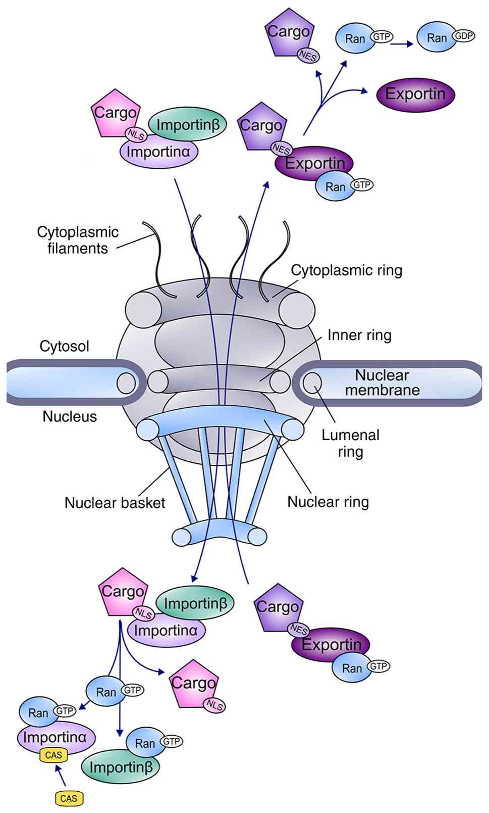 | Figure 1Primary structure of the NPC and a
schematic overview of Ran-mediated nucleocytoplasmic transport. The
NPC is embedded in the NE. The primary structure of the NPC
consists of cytoplasmic filaments, a nuclear basket and a central
core that is composed of four rings: the cytoplasmic ring on the
cytosolic side, the inner ring, the nuclear ring on the inner
nuclear membrane, and the lumenal ring residing in the lumen of the
NE. The assembly of a trimeric import complex in the cytoplasm is
required for the import of proteins bearing NLS. The nuclear
proteins importin α and importin β form a complex; importinα
engages the NLS of cargo proteins, and the complex travels through
the NPC into the nucleus, where RanGTP binding releases it. The
export of cargo proteins harbouring the NES sequence is initiated
by exportin binding and is facilitated by RanGTP. After exportin
binds RanGTP and a NES-harbouring cargo protein, the complex
travels into the cytosol, where GTP hydrolysis to GDP causes the
export complex to disassemble. NES, nuclear export signal; NLS,
nuclear localization signal; NPC, nuclear pore complex; NE, nuclear
envelope. |
The central Nups form a cylindrical structure with
eightfold rotational symmetry and twofold rotational symmetry along
the axis parallel to the nuclear envelope. This symmetrical core
encircles the central transport channel and serves as a scaffold to
which specific Nups attach on the cytoplasmic and nuclear sides of
the NE, forming the cytoplasmic filaments and nuclear basket,
respectively (23,24). The central scaffold contains Nups
that harbour intrinsically disordered phenylalanine (F) and glycine
(G) repeats. These molecules, known as FG-Nups, are relatively
hydrophilic and essential for the NPC permeability barrier
(19). Alterations in the
FG-Nup-mediated permeability barrier of the NPC may promote cancer
progression by enhancing oncogenic nuclear signalling, amplifying
hormone-dependent transcription and supporting cellular adaptation
to genomic stress and therapy (25-27). In several types of cancer,
including gynaecological cancers, FG-Nups are mutated or
overexpressed, altering transport selectivity and kinetics
(22,28).
FG-Nups, together with transport receptors and
components of the cytoplasmic filaments and nuclear basket, play
crucial roles in nucleocytoplasmic transport (29,30). Cytoplasmic filaments participate
in the recruitment of transport factors and are involved in the
final stages of protein and mRNA export (31). The nuclear basket serves as a
docking site for nuclear transport and is essential for processing
and sorting molecules as they pass through the NPC. The cytoplasmic
and nuclear rings function as anchors between the NPC and the NE.
These rings bind the nuclear membrane, connect to the inner ring
through Nup155 and provide docking sites for the recruitment of
asymmetrical Nups, such as Nup358, the principal component of the
cytoplasmic filaments (20,32). Studies have identified Nup155 as a
notable oncogenic biomarker associated with disease progression,
therapeutic response and immune evasion in various cancers,
including gynaecological malignancies, whereas upregulation of
Nup358 promotes tumour cell proliferation and invasion,
highlighting its role in cancer progression (33-36). Each Nup performs a distinct
function. FG-Nups form the diffusion barrier and/or interact with
transport factors; scaffold Nups provide structural support for the
NPC; transmembrane Nups anchor the complex within the membrane; and
linker Nups connect different scaffold elements. Individual Nups
may also perform multiple functions and occupy different locations
within the NPC. A detailed description of Nup organisation has been
reviewed elsewhere (19,20).
It is generally accepted that molecules <40 kDa
can freely diffuse through the NPC, whereas larger molecules cannot
cross the NE independently (24).
The two major components of the nuclear transport machinery, the
karyopherins and the Ran system, work together to transport cargo
through the NPC between the nucleus and the cytoplasm.
Karyopherins, comprising importins and exportins, are the principal
cargo receptors that interact with NPCs, recognise transport motifs
and bind cargo in a GTP/GDP-sensitive manner (37,38). The two classes of karyopherins,
karyopherin α and karyopherin β (importin α and importin β),
mediate cargo import into the nucleus. Members of the importin β
superfamily function in concert with the small Ras-like GTPase Ran.
They contain a RanGTP-binding domain at the N-terminus and multiple
short α-helical HEAT repeats that mediate interactions with
transport cargoes and NPCs (39).
GTP binding and hydrolysis, which generate RanGTP and RanGDP
complexes, direct cargo transport through NPCs (38). The asymmetric distribution of
RanGTP, which is abundant in the nucleus, and RanGDP, which is
abundant in the cytoplasm, drives the movement of cargo-receptor
complexes through the NPC. Importin β associates with import
cargoes either directly or through adaptor proteins such as
importin α. These adaptors are specialised cargoes that contain
N-terminal domains known as importin β-binding domains (40,41).
The Ran-mediated nuclear transport pathway is among
the most frequently dysregulated transport systems in cancer
(42) and underlies canonical
nucleocytoplasmic transport by regulating cargo interactions with
importins and exportins, as well as the directionality of nuclear
import and export. This pathway involves binding of the importin
β-importin α complex to the nuclear localisation signal (NLS) of
cargo proteins, facilitating passage through the central channel of
the NPC (Fig. 1). At the nuclear
side of the NPC, RanGTP binding promotes cargo release into the
nucleoplasm. Subsequently, RanGTP-CAS-importin α and
RanGTP-importin β complexes are exported to the cytoplasm, where
GTP hydrolysis leads to their dissociation. Similarly, exportin 1
(XPO1) mediates nuclear export by binding cargo proteins containing
a nuclear export signal (NES) in a RanGTP-dependent manner. The
resulting export complexes are transported to the cytoplasm, where
GTP hydrolysis triggers complex disassembly (23,32). Dysregulation of the classical Ran
pathway has been implicated in cancer, with elevated Ran expression
associating with aggressive tumour phenotypes and increased
cellular dependence on Ran for survival (43-45).
Beyond their established role in nucleocytoplasmic
transport, NPCs also perform transport-independent functions.
Increasing evidence indicates that NPCs contribute to gene
regulation and chromatin organisation through mechanisms unrelated
to molecular transport. Nups participate in genome architecture,
transcriptional complex assembly, chromatin structure regulation
and the coordination of transcription with nuclear transport
processes (46).
Alterations in nuclear transport in
gynaecological cancer cells
Nuclear transport can be dysregulated at several
levels in cancer cells, including modifications of cargo proteins
and transport receptors, altered expression of nuclear transport
components and changes in nuclear pore number.
The translocation of cargo from the cytoplasm to the
nucleus may be facilitated by direct post-translational
modification of the cargo itself or by modification of inhibitory
proteins that normally mask nuclear localisation sequences. An
example is the nuclear translocation of β-catenin in the canonical
Wnt signalling pathway. Wnt/β-catenin signalling promotes the
proliferation, migration and stem-like characteristics of
endometrial (47), ovarian
(48) and cervical cancer cells
(49). In the absence of Wnt
ligands, cytoplasmic β-catenin undergoes phosphorylation,
ubiquitination and subsequent proteasomal degradation. Wnt
signalling activation suppresses these modifications, allowing
β-catenin to accumulate and translocate to the nucleus, where it
activates target gene expression. In gynaecological cancers,
mutations in the β-catenin gene, CTNNB1, may also result in
constitutive nuclear localisation of the protein (50).
Modification of cargo inhibitors is central to NF-κB
signalling. Members of the NF-κB transcription factor family
promote cancer cell proliferation, survival, metastasis, metabolic
reprogramming and inflammation (51). Although NF-κB activity may be
suppressed during the early stages of human papillomavirus
infection to facilitate immune evasion (52), persistent NF-κB activation
subsequently promotes malignant transformation and is positively
associated with cervical cancer progression (53). In its inactive state, NF-κB is
retained in the cytoplasm through association with its inhibitor,
IκB. After pathway activation, IκB kinase phosphorylates IκB, which
is ubiquitinated and degraded by the proteasome. This releases
NF-κB, which karyopherin β1 (KPNB1) transports into the nucleus,
where it binds DNA and activates gene transcription. Within the
nucleus, NF-κB activity is further regulated by phosphorylation
(54), acetylation (55), sumoylation (56) and other post-translational
modifications. Nuclear accumulation of NF-κB has been observed in
cervical cancer and is associated with disease progression
(57). Conversely, some studies
have linked nuclear localisation of NF-κB with ovarian cancer
regression (58).
Post-translational modifications and mutations that
alter the localisation of nuclear transport receptors have not been
extensively investigated in gynaecological cancers. In other
malignancies, however, such alterations have been well documented.
For example, acetylation of karyopherins in lung cancer alters
their subcellular localisation and disrupts nuclear transport
(59). Leukaemia has been
associated with Nup translocations and fusion events involving HOX
transcription factors and other genes (60-63). In B-cell lymphoma, the E571K point
mutation in XPO1 alters its localisation and affinity for
negatively charged cargoes (64,65) and increases cellular sensitivity
to nuclear transport inhibitors (66). Furthermore, genetic alterations
and phosphorylation of exportin 5 impair miRNA export from the
nucleus and contribute to colorectal and liver cancer progression
(67,68).
By contrast, altered expression of nuclear transport
proteins is frequently observed in gynaecological tumours (Table I). Although nuclear transport
proteins are typically overexpressed in tumour tissues, studies
have shown that elevated levels can also be detected in the
bloodstream of patients with cancer, even at early stages of
disease (69,70). For example, van der Watt et
al (70) identified KPNB1,
XPO1, CAS, importin 5 and transportin 1 in the serum of patients
with cervical and oesophageal cancers, with combinations of these
proteins providing the greatest predictive value (70). Additional studies have shown that
KPNB1 is overexpressed in cervical cancer tissue and contributes to
tumour initiation and growth, supporting its potential as both a
biomarker and therapeutic target (71,72). In ovarian cancer, elevated KPNB1
expression has been associated with poor prognosis (73). Importin 7 has also emerged as a
promising diagnostic and prognostic biomarker in cervical cancer
because of its high expression levels and association with disease
progression (74). Other
transport receptors, including karyopherin subunit α2 and XPO1,
have been extensively investigated as diagnostic and prognostic
markers in several malignancies, including ovarian cancer (42,75-80). Increased expression of circular
RNA derived from transportin 3 has been reported in ovarian cancer
and is associated with reduced sensitivity of ovarian cancer cells
to paclitaxel (81). Elevated
expression of CAS is associated with ovarian cancer progression
(82). Notably, unlike several
other transport receptors, karyopherin subunit α5 (KPNA5) has been
proposed as a tumour suppressor, as reduced expression is
associated with decreased overall survival in patients with ovarian
cancer (83).
 | Table IChanges in nuclear transport protein
expression in gynaecological tumours and their relevance to disease
progression. |
Table I
Changes in nuclear transport protein
expression in gynaecological tumours and their relevance to disease
progression.
| Type of cancer | Protein | Change in
expression | Effect | (Refs.) |
|---|
| Ovarian | XPO1 | Increased | Associated with
poor patient prognosis and expression of COX-2 | (78) |
| Ovarian | XPO1 | Increased | Associated with
poor patient prognosis and resistance to platinum-based
therapy | (77) |
| Ovarian | KPNA2 | Increased | Elevated migration
and invasion of epithelial ovarian cancer cells, knockdown of KPNA2
suppressed epithelial-to-mesenchymal transition
characteristics | (79) |
| Ovarian | KPNA2 | Increased | Associated with
poor patient prognosis | (80) |
| Ovarian | KPNA2 | Increased
concentration in blood samples | Proposed as a
diagnostic marker | (76) |
| Ovarian | KPNA5 | Decreased | Associated with
poor patient prognosis, overexpression decreased tumour growth and
invasion | (83) |
| Ovarian | KPNB1 | Increased | Associated with
poor patient prognosis, inhibition decreased ovarian cancer cell
proliferation, invasion and induced apoptosis | (73,121) |
| Ovarian | TNPO3 | Increased (circular
RNA) | Mediates paclitaxel
resistance of ovarian cancer cells | (81) |
| Ovarian | CAS | Increased | Associated with
disease progression | (82) |
| Cervical | XPO1, KPNB1 and
KPNA2 | Increased | Inhibition of XPO1
and KPNB1 decreased cervical cancer cell proliferation and induced
apoptosis | (72) |
| Cervical | KPNB1, XPO1, CAS,
IPO5 and TNPO1 | Increased
concentration in blood samples | Proposed as
diagnostic markers | (70) |
| Cervical | IPO7 | Increased | Associated with
poor patient prognosis, knockdown decreased cervical cancer cell
proliferation | (74) |
| Cervical | NUTF2 | Increased | Associated with the
number of infiltrating cancer-associated fibroblasts | (84) |
| Endometrial | XPO1 | Increased | Associated with
poor patient prognosis | (85) |
| Endometrial | KPNA2 | Increased | Proposed as a
prognostic marker for poor patient survival. Gene silencing slowed
down endometrial cancer cell proliferation rate | (86) |
| Endometrial | IPO13 | Increased | Proposed as a
progenitor/stem cell marker | (87) |
| Endometrial | NXF1 | Decreased | Inhibited
endometrial cancer progression in vitro and in
vivo | (88) |
| Endometrial | NUTF2 | Increased | Increased
expression in comparison to healthy tissue was associated with an
oncogenic role | (84) |
As summarised in Table
I, ovarian cancer has been the most extensively investigated
gynaecological malignancy in the context of nucleocytoplasmic
transport, whereas comparatively limited data are available for
cervical and endometrial cancers. Historically, ovarian cancer has
attracted greater research attention because it has the highest
mortality rate among gynaecological malignancies. Furthermore,
numerous studies have demonstrated altered expression of
nucleocytoplasmic transport factors in ovarian cancer, whereas
relatively few transport-related molecular targets have been
identified in cervical (70,72,74,84) and endometrial cancers (84-88). Consequently, the limited
availability of validated molecular targets has further constrained
research efforts in these malignancies.
Nuclear transport in relation to
gynaecological cancer hallmarks
Alterations in nuclear transport play roles in
tumorigenesis, and understanding their relationships with cell
proliferation, survival and motility may improve the prediction of
tumour aggressiveness and treatment resistance. Over the past
decade, numerous studies across a range of malignancies, including
gynaecological cancers, have investigated the contribution of
nuclear transport machinery to the acquisition of cancer
hallmarks.
In several cancer types, overexpression of XPO1 is
associated with disease severity and adverse prognostic outcomes.
In ovarian cancer, elevated XPO1 expression associates with tumour
aggressiveness and reduced patient survival, whereas XPO1
inhibition exerts antiproliferative effects in cervical cancer
cells in vitro (89). A
study of chronic lymphocytic leukaemia identified several
XPO1 mutations that appear to confer selective advantages
for cellular proliferation and survival and may predispose cells to
leukemic transformation in the presence of additional genetic
alterations; however, further investigation is required to validate
these findings (90). In ovarian
cancer, XPO1 expression has been shown to be significantly
associated with epidermal growth factor receptor (EGFR), a key
regulator of cellular growth. Moreover, XPO1 inhibition reduced
EGFR protein levels, suggesting this interaction may contribute to
tumour progression and cancer cell proliferation (91).
Furthermore, increased XPO1 activity promotes export
of tumour suppressor proteins, including p27 and p53, from the
nucleus to the cytoplasm, inactivating them by sequestering them
from their primary sites of action. Cytoplasmic localisation of p27
and p53 has been associated with poor prognosis and therapeutic
resistance in several malignancies, including gynaecological
cancers. For example, XPO1 recognises p27 phosphorylated at serine
10 (pSer10-p27) and exports it to the cytoplasm, where it acquires
oncogenic properties. In epithelial ovarian cancer, XPO1 and
pSer10-p27 expression levels are positively associated, and
elevated expression of both proteins is associated with reduced
overall survival, higher International Federation of Gynaecology
and Obstetrics stage and increased tumour grade. The same study
demonstrated that XPO1 knockdown inhibited the nuclear export of
pSer10-p27 and reduced ovarian cancer cell proliferation in
vitro (92). An investigation
of serous ovarian carcinoma revealed that cytoplasmic p53 forms
aggregates that are associated with poor prognosis and reduced
sensitivity to cisplatin, suggesting a potential role in
chemotherapy resistance, although the study did not specifically
investigate XPO1-mediated p53 transport (93). Moreover, leptomycin B inhibition
of XPO1 sensitised chemoresistant endometrial and ovarian cancer
cells to TRAIL-induced apoptosis through a p53-independent
mechanism. Clonogenic assays further showed that combination
treatment of TRAIL and leptomycin B notably reduced long-term
colony-forming capacity, indicating that XPO1 may contribute to
sustained tumour growth and proliferative potential (94).
Among other exportins, Li et al (95) reported that reduced XPO5
stability, together with required for meiotic nuclear division 5
homolog A protein downregulation, inhibited migration of HeLa
cervical cancer cells, highlighting a possible role for XPO5 in
tumour cell motility.
In contrast to the elevated expression of XPO1,
KPNA5 is frequently downregulated in ovarian cancer and is
associated with poor patient outcomes, whereas maintained or
elevated KPNA5 expression suppresses tumour growth. KPNA5 mediates
the nuclear import of the phosphatase protein tyrosine phosphatase
non-receptor type 4 (PTPN4), which dephosphorylates STAT3 and
inhibits its activation (83).
STAT3 is a transcription factor that promotes tumour cell growth
and survival while suppressing antitumour immune responses
(96). A study by Hu et al
(83) confirmed that KPNA5
interacts with PTPN4 and facilitates its transport from the
cytoplasm to the nucleus in the ovarian cancer cell lines SKOV3 and
OVCAR3. The authors further demonstrated that KPNA5 overexpression
inhibited ovarian cancer cell migration and invasion in
vitro (83).
Li et al (84) also analysed 33 cancer types to
investigate the oncogenic role of nuclear transport factor 2
(NUTF2), a GDP-binding protein involved in nucleocytoplasmic
transport. Compared with that in adjacent normal tissues, NUTF2
expression was elevated in 19 cancer types, including cervical and
endometrial carcinomas. To further explore its biological role, the
authors performed gene co-expression analyses. NUTF2 expression was
positively associated with the proliferation markers proliferating
cell nuclear antigen and marker of proliferation KI-67 in 16 tumour
types. In addition, expression of the epithelial-to-mesenchymal
transition markers vimentin, TWIST1, SNAI1,
SNAI2, fibronectin 1 and N-cadherin were positively
associated with NUTF2 expression across most tumour types. These
findings suggest that NUTF2 may contribute to both cancer cell
proliferation and metastatic dissemination by regulating cellular
motility.
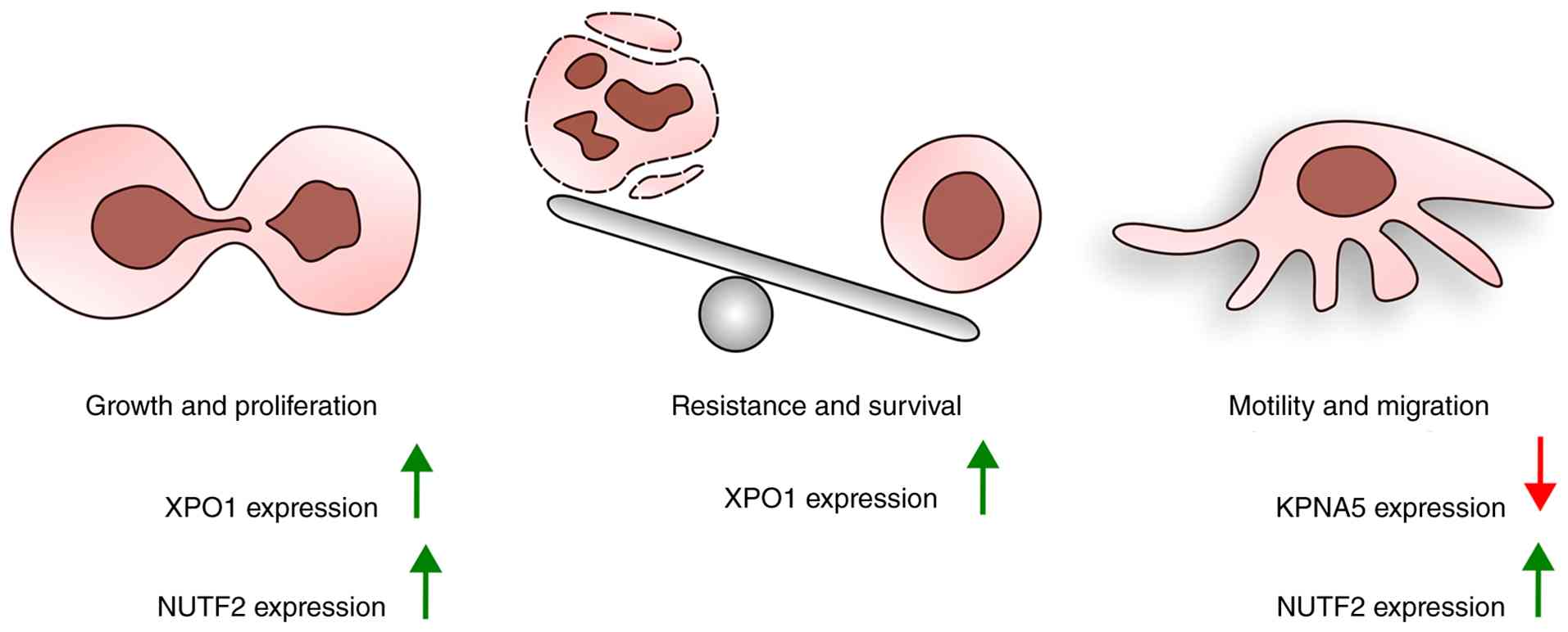
Fig. 2 summarizes
alterations in nuclear transport proteins in gynaecological cancers
and their associations with specific cancer hallmarks.
Targeting nuclear transport to treat
gynaecological cancer
Discoveries of dysregulated nucleocytoplasmic
transport in gynaecological tumours have highlighted its
therapeutic potential. Nucleocytoplasmic transport inhibitors fall
into two broad categories: Nuclear import inhibitors and nuclear
export inhibitors (97,98).
Nuclear export inhibition
XPO1 is a ubiquitously expressed protein in
eukaryotic cells. First identified in yeast, it has since been
shown to mediate the nuclear export of hundreds of proteins
(99). Studies of the natural
compound leptomycin B (LMB), an inhibitor of XPO1, facilitated the
identification of XPO1 cargo proteins. Among all exportins, XPO1
uniquely exports a broad spectrum of biologically important
proteins, including tumour suppressors and growth regulators such
as p53, p21 and FOXO, as well as key signalling molecules involved
in the PI3K/AKT, Wnt/β-catenin, activator protein-1 and NF-κB
pathways (100).
Mechanistically, XPO1 inhibitors promote the nuclear retention of
tumour suppressor proteins, thereby restoring their function. Even
when a specific tumour suppressor, such as p53, is mutated or
inactive, the abnormal accumulation of multiple tumour suppressors
and growth-inhibitory factors within the nucleus can overwhelm
cellular defence mechanisms and trigger programmed cell death
(101). This class of inhibitors
targets the cysteine residue Cys528 within the NES-binding groove
of XPO1, preventing nuclear export of regulatory molecules and
suppressing cell proliferation (100,102).
Research on nuclear export inhibitors began with
LMB, a naturally occurring antifungal compound and the first
identified XPO1 inhibitor (100). Similar to several subsequently
developed compounds, LMB binds covalently to the Cys528 residue;
however, its clinical development was discontinued because of
severe dose-limiting gastrointestinal toxicity (89). Other naturally occurring XPO1
inhibitors include ratjadone C, anguinomycin and goniothalamin
(100). Curcumin has also been
reported to inhibit XPO1 by covalently binding to Cys528, resulting
in nuclear accumulation of cargo proteins such as forkhead box
protein O1 (FOXO1) (103).
Although curcumin suppresses cell proliferation, its efficacy
appears to be lower than that of synthetic XPO1 inhibitors
(104).
The limitations of LMB prompted the development of
synthetic compounds with improved safety profiles, most notably the
selective inhibitors of nuclear export (SINEs). This group includes
Selinexor (KPT-330), Verdinexor (KPT-335), KPT-185, KPT-251 and
KPT-276. Similar to LMB, these compounds target the Cys528 residue
of XPO1, but their interaction is reversible (Fig. 3A and B) (100). This reversibility reduces
toxicity while maintaining anticancer activity. Selinexor is the
most clinically advanced SINE and has been evaluated in >40
clinical trials across over 10 cancer types, including
gynaecological malignancies (105). These studies ultimately led to
approval by the Food and Drug Administration (FDA) for
penta-refractory multiple myeloma (102). Verdinexor has also been
investigated for treating viral infections, particularly influenza
(106). Although compounds such
as KPT-185 have demonstrated promising activity in vitro,
their clinical development has been limited by unfavourable
pharmacokinetic properties (107).
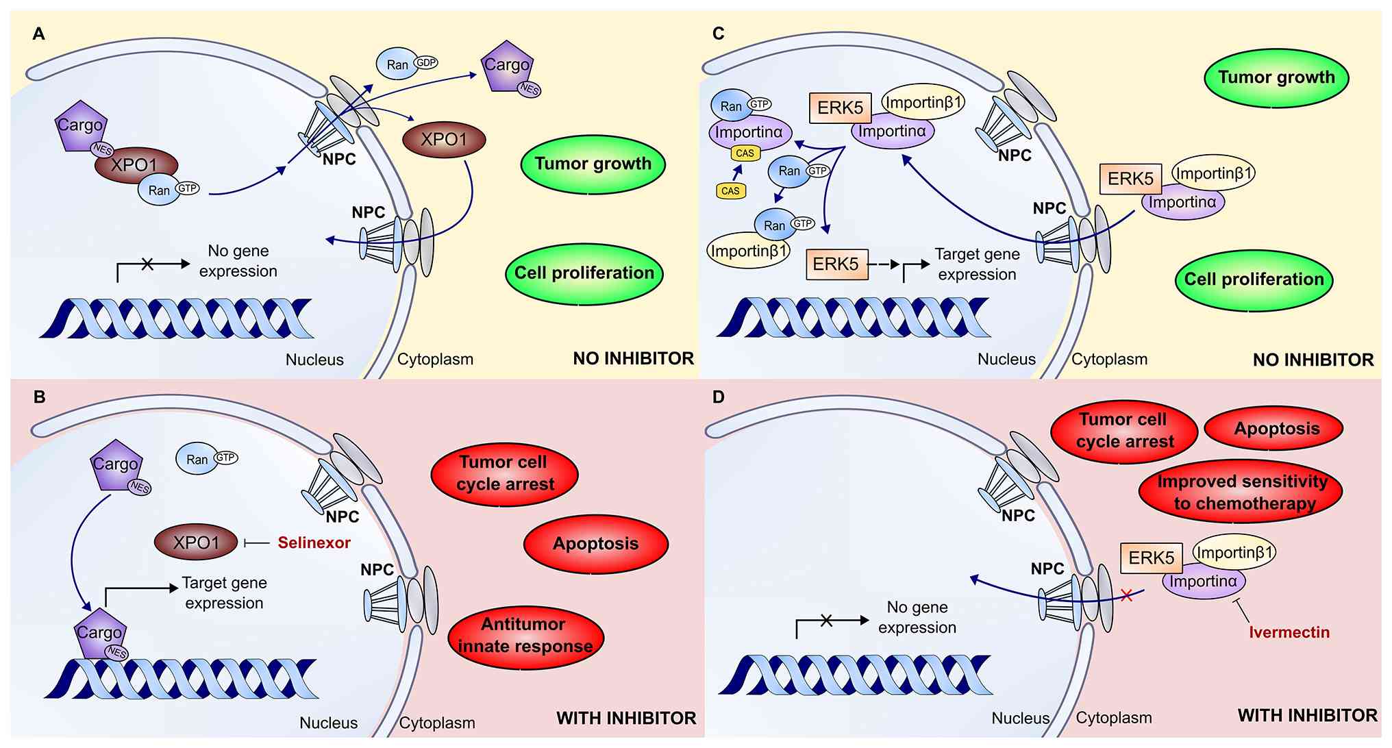 | Figure 3Mechanisms of action of nuclear
transport inhibitors in tumour cells. (A) The mechanism of nuclear
export mediated by XPO1 and the inhibitory effects of Selinexor in
tumour cells. In cancer cells, XPO1 binds nuclear cargo proteins
carrying an NES, along with RanGTP, and transports them through the
NPC into the cytoplasm. After dissociation of the complex, RanGTP
is hydrolysed to RanGDP, and XPO1 is transported back into the
nucleus to sustain the export cycle. The continuous nuclear export
of tumour suppressor proteins disables their nuclear function and
gene expression, thereby supporting tumour cell proliferation and
tumour growth. (B) With XPO1 inhibition by Selinexor, the
interaction between XPO1 and NES-containing cargo proteins is
blocked, leading to their nuclear retention and restoration of
transcriptional activity. This leads to tumour cell cycle arrest,
apoptosis and an antitumor innate response. (C) In cancer cells,
ERK5 binds to the importinα/β1 complex, enabling its nuclear import
through the NPC. Once inside the nucleus, the import complex
dissociates: ERK5 is released and functions as a transcriptional
regulator, promoting target gene expression that drives tumour cell
proliferation and tumour growth, whereas importinα and importinβ1
dissociate, bind RanGTP and return to the import cycle. (D) When
ERK5 nuclear import is inhibited by ivermectin, the interaction
between ERK5 and the importin transport machinery is blocked,
preventing ERK5 from entering the nucleus. Consequently,
ERK5-mediated transcriptional activity is suppressed, resulting in
tumour cell cycle arrest, apoptosis and improved sensitivity to
chemotherapy. NES, nuclear export signal; NPC, nuclear pore
complex; XPO1, exportin 1. |
CBS9106, also known as felezonexor or SL-801,
represents a distinct class of synthetic XPO1 inhibitors with dual
mechanisms of action. In addition to reversibly inhibiting XPO1 and
inducing apoptosis, this orally bioavailable compound markedly
reduces XPO1 protein levels without affecting corresponding mRNA
expression (108). XPO1
degradation occurs through the Cullin-RING ligase system and the
neddylation pathway. Understanding this mechanism matters because
it suggests that CBS9106 may show antagonistic interactions when
combined with neddylation inhibitors or proteasome inhibitors
(109). At present, no studies
have specifically evaluated CBS9106 in gynaecological cancers. The
CBS9106 derivative S109 is currently under development because it
is simpler and more cost-effective to synthesize, facilitating
large-scale production (110).
Nuclear import inhibition
The preclinical and clinical success of
XPO1-targeted therapies has stimulated the development of nuclear
import inhibitors. Although this field remains at an early stage,
targeting importins has shown considerable promise, with several
small-molecule inhibitors already demonstrating anticancer activity
in preclinical studies. In contrast to XPO1 inhibitors, which
uniformly target the well-characterised NES-binding pocket, the
binding sites and mechanisms of action of several nuclear import
inhibitors remain incompletely understood.
Nuclear import inhibitors can be broadly classified
into two groups: Direct importin inhibitors, which target the
transport receptor itself, and cargo-specific inhibitors, which
prevent the import machinery from recognizing individual cargo
proteins (102). Direct importin
inhibitors aim to disrupt the core nuclear transport apparatus. For
example, the FDA-approved antiparasitic drug ivermectin binds the
importin α/β1 heterodimer and blocks the nuclear import of viral
proteins, including HIV-1 integrase, dengue virus NS5 and influenza
virus proteins, thereby exerting broad-spectrum antiviral activity.
However, this mechanism also affects host signalling pathways,
including the HIF-1α pathway (73,111). The pleiotropic effects of
importin inhibition are particularly relevant in cancer, as
numerous oncogenic transcription factors and signalling mediators
depend on importin-mediated nuclear translocation to exert their
tumour-promoting functions.
In cancer cells, ivermectin binds importin β1 and
inhibits nuclear transport (Fig. 3C
and D). This prevents activated ERK5 from entering the nucleus,
retaining it in the cytoplasm and inhibiting its pro-proliferative
activity (112). Given the
recognised role of ERK5 signalling in cell proliferation, survival
and therapeutic resistance, including in gynaecological
malignancies, these findings provide a mechanistic basis for
targeting nuclear import pathways in cancer treatment (113,114). Similarly, the synthetic compound
importazole directly targets importin β1 and impairs cargo
transport by disrupting its interaction with RanGTP (115-117). This mechanism was first
demonstrated for the importin β1 inhibitor karyostatin 1A,
described in 2010, which prevents dissociation of the importin
β1/importin α/cargo complex within the nucleus and thereby blocks
classical nuclear import (118).
Dissociation of this complex is essential for successful transport
because it ensures cargo release within the nucleus rather than its
return to the cytoplasm alongside the transport receptor. More
recently, synthetic compounds such as INI-43 and INI-60, identified
through in silico screening approaches (119), have further demonstrated the
therapeutic potential of this strategy. INI-43 inhibits cancer cell
growth and induces G2/M cell-cycle arrest by preventing nuclear
import of pro-survival proteins, including NF-κB (120,121).
Aberrant activation of NF-κB has been extensively
documented in ovarian cancer, where it contributes to
chemoresistance, stemness, metastatic progression and survival
signalling (122). Dysregulated
NF-κB signalling has also been implicated in endometrial carcinoma
and other endometrial disorders, linking this pathway to multiple
gynaecological malignancies (123). Nevertheless, caution is
warranted when extrapolating these findings to gynaecological
cancers. Although dysregulation of NF-κB and ERK5 signalling is a
recognised feature of gynaecological tumour biology, most
mechanistic and efficacy studies involving ivermectin, importazole,
INI-43 and INI-60 have been performed in non-gynaecological
malignancies, including glioblastoma, oesophageal cancer and
haematological cancers (120,124-127), or in preliminary cell-line
models (120). Direct validation
in disease-specific gynaecological models, particularly primary
ovarian and endometrial tumours, patient-derived xenografts and
clinical studies, remains lacking and represents an important area
for future research.
In contrast to broadly acting importin inhibitors,
cargo-specific inhibitors offer a more selective therapeutic
approach by binding directly to the NLS of a specific cargo
protein. This prevents cargo recognition by the import machinery
and may reduce off-target toxicity (Fig. 4A and B). Examples include the
corticosteroids budesonide and flunisolide, which bind the NLS of
HIV-1 integrase and block its importin α/β1-mediated nuclear
import. Mifepristone exerts a similar effect on HIV-1 integrase,
although its clinical development was discontinued (128,129). Another example is the retinoid
4-HPR (fenretinide), which selectively disrupts the NS5/importin
α/importin β1 interaction in flaviviruses, thereby inhibiting
dengue and Zika virus infection without disturbing essential host
nuclear import pathways (130).
Although most cargo-specific inhibitors have been developed in
antiviral settings, their mechanism of action is attractive for
cancer therapy because it enables selective inhibition of oncogenic
nuclear signalling while potentially minimising systemic
toxicity.
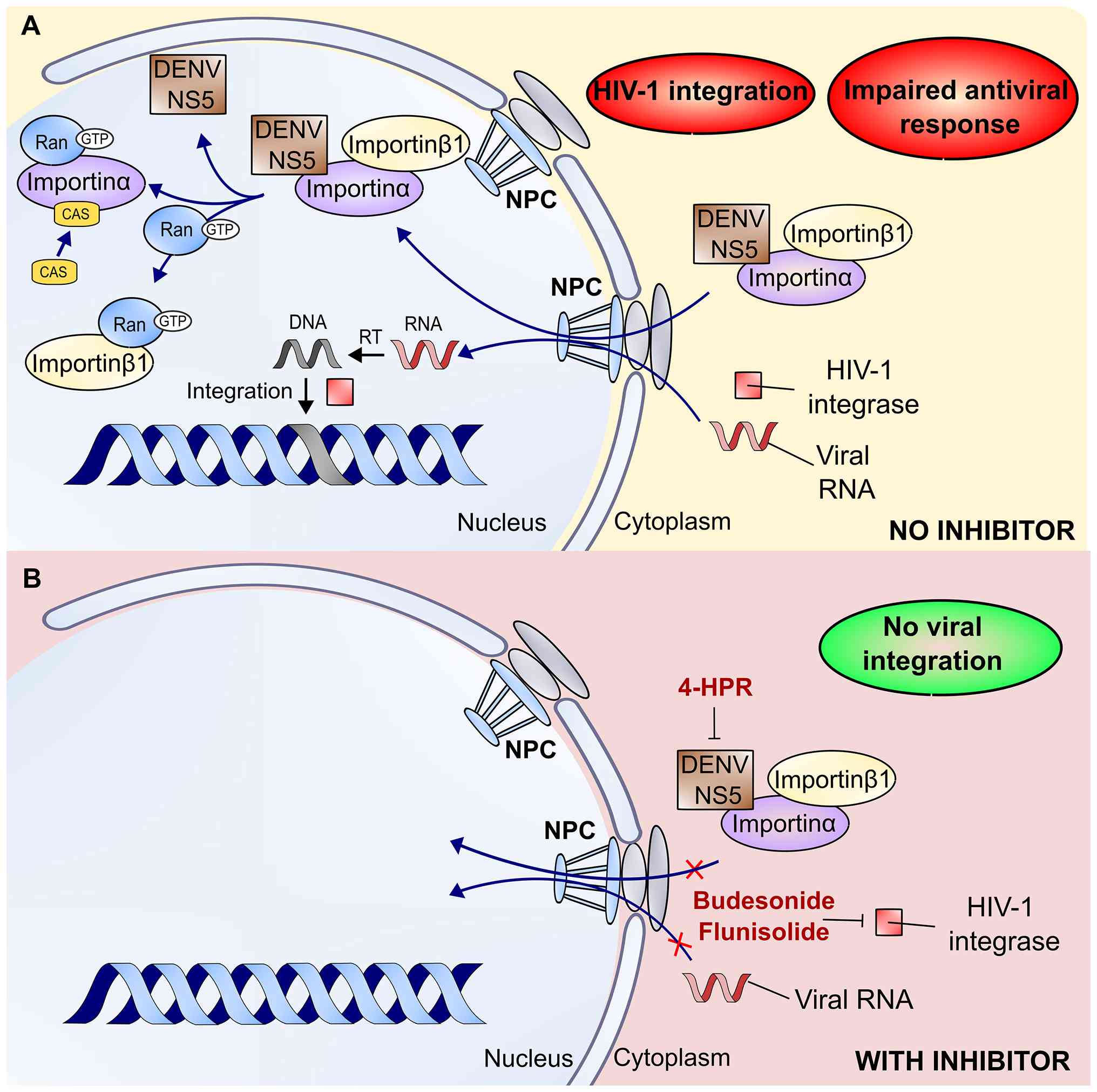 | Figure 4Mechanisms of action of nuclear
transport inhibitors in virus-infected host cells. (A) Infected
host cells use the importinα/β1 complex to recognize and transport
the viral proteins DENV NS5 and HIV-1 integrase through the NPC
into the nucleus. Once inside the nucleus, the importinα/β1 complex
dissociates, releasing viral proteins to interfere with host
cellular functions-DENV NS5 by disrupting antiviral responses and
integrase by integrating viral DNA, which is reverse transcribed
from the viral RNA, into the host genome. (B) Cargo-specific
inhibitors work by binding directly to the viral cargo; 4-HPR binds
to DENV NS5, whereas budesonide and flunisolide bind to the HIV-1
integrase. Direct binding prevents viral proteins from being
recognized by the host's importinα/β1 complex, thereby effectively
blocking their nuclear import and leading to a lack of viral
integration. 4-HPR, N-(4-hydroxyphenyl) retinamide; NPC, nuclear
pore complex; RT, reverse transcriptase; DENV, dengue virus. |
An increasing number of studies have explored
cargo-specific nuclear import inhibitors in cancer treatment
(131-133). Collectively, current evidence
suggests that although direct importin inhibitors provide potent,
broad-spectrum activity, cargo-specific approaches may offer
greater selectivity and reduced toxicity (102). Taken together, these findings
support nuclear import inhibition as a mechanistically rational,
albeit largely preclinical, therapeutic strategy that merits
further investigation alongside the more clinically advanced
nuclear export inhibitors.
However, the literature on cargo-specific nuclear
import inhibitors in gynaecological cancers remains limited.
Although this approach offers potential to avoid the toxicities
associated with global inhibition of nucleocytoplasmic transport,
most validated cargo-specific inhibitors, including budesonide,
flunisolide and fenretinide, currently target viral proteins rather
than oncogenic drivers relevant to ovarian, cervical or endometrial
cancers (124). Future research
should therefore focus on identifying critical nuclear localisation
sequences within key gynaecological oncogenes to facilitate the
development of tumour-specific nuclear import inhibitors and
address this important translational gap.
Preclinical studies of nuclear export and
import inhibitors
In gynaecological cancers, particularly endometrial
and ovarian cancers, several compounds targeting nuclear transport
pathways have shown notable anticancer effects. Among nuclear
export inhibitors, Selinexor has shown clinically meaningful
activity in endometrial cancer. In the phase III SIENDO trial, a
prespecified subgroup analysis demonstrated that Selinexor
significantly prolonged progression-free survival (PFS) in patients
with TP53 wild-type tumours, with a median PFS of 28.4
months compared with 5.2 months for placebo [hazard ratio 0.44;
95%; confidence interval (CI), 0.27-0.73; nominal P=0.0005]. These
results support Selinexor as a biomarker-driven therapeutic
strategy beyond its current FDA approval in hematologic
malignancies. However, the authors note two main limitations of the
SIENDO trial: A low number of patients with TP53-wt (113) and that certain patients (in both
the Selinexor and placebo groups) discontinued before completing
treatment. Given the fact that analysis was prespecified and the
primary endpoint in the intent-to-treat population was not met, the
subgroup analysis is exploratory, and the results are not
conclusive. Additionally, >94% of patients were white and had
endometrioid carcinoma; thus, the results do not reflect diverse
populations and rare subtypes of endometrial cancer. As the study
evaluated Selinexor in pretreated patients who previously received
chemotherapy, the efficacy of Selinexor in untreated patients
remains unknown. The SIENDO trial motivated the subsequent
XPORT-EC-042 trial [NCT05611931, registered (first posted) at
ClinicalTrials.gov 2022-11-10] (134). Although this study also focuses
on pretreated patients with TP53-wt, the group size is
approximately twice as large (276 participants), and the
distribution between treatment arms is more balanced (1:1 Selinexor
vs. placebo compared with 2:1 in the SIENDO trial). Selinexor also
demonstrated activity in heavily pretreated ovarian and endometrial
cancers. In an open-label, single-arm phase II study, median PFS
was 2.6 months in ovarian cancer and 2.8 months in endometrial
cancer patients treated with Selinexor (135). Similar to the SIENDO trial, this
study also included previously pretreated patients; the groups
differed in size and were relatively small (66 patients with
ovarian cancer, 23 patients with endometrial cancer and 25 patients
with cervical cancer). The authors assessed treatment response in
98 of 114 patients, and the absence of a placebo group limits
direct comparison with other clinical trials. Although the authors
performed circulating tumour cell analysis to assess treatment
outcome, the subset of patients was small (31 patients with ovarian
cancer, 8 patients with endometrial cancer and 6 patients with
cervical cancer), so the findings are preliminary. The modest PFS
and their differences in ovarian, endometrial and cervical cancer
could be explained by a small sample size and different numbers of
patients with highly resistant platinum-refractory tumours in the
groups. The authors also note that Selinexor may yield better
outcomes in combination with other treatment modalities (radiation
therapy, targeted therapy or immunotherapy), especially because
combination results in preclinical models (cell lines and mice) are
promising (136-138). Identifying biomarkers to predict
sensitivity to Selinexor in patients with gynaecological cancer
could also improve outcomes. In platinum-resistant ovarian cancer
models, XPO1 inhibition restored platinum sensitivity and induced
apoptosis independently of p53 status (77). By contrast, among nuclear import
inhibitors, ivermectin and the structurally related compounds
INI-60 and INI-43 have shown anticancer activity by inhibiting
importin β1, with selective cytotoxicity observed in cervical
cancers (73,120,121). Ivermectin alone inhibited
ovarian cancer growth by inducing cell cycle arrest and
caspase-dependent apoptosis (139). Additionally, ivermectin showed
importin β1-dependent antitumor activity in epithelial ovarian
cancer, highlighting its potential for drug repurposing in this
setting (73). Other compounds
with anticancer effects in gynaecological cancers include
budesonide, which has demonstrated antitumor activity in ovarian
cancer cells (140), and
fenretinide, which reduces viability and induces apoptosis in
endometrial cancer Ishikawa cells in a dose-dependent manner
(131). Notably, the effects of
fenretinide were independent of retinoic acid receptor signalling
and appeared to be mediated by increased retinol uptake via
stimulated by retinoic acid 6 (STRA6), as silencing STRA6 notably
reduced apoptosis. In vivo, intraperitoneal fenretinide
treatment effectively suppressed tumour growth in mice bearing
Ishikawa-derived endometrial tumours (131).
Combination therapies that include
nuclear transport inhibitors
Conventional chemotherapies disrupt fundamental
cellular processes, such as DNA replication and cell proliferation.
However, they cannot distinguish between malignant and healthy
cells, often resulting in substantial toxicity. By contrast,
targeted therapies selectively inhibit cancer-associated pathways
that normal cells depend on less. Nevertheless, dose-limiting
toxicities and the emergence of drug resistance can compromise
therapeutic efficacy. Consequently, combination therapy is
frequently favoured over monotherapy. By concurrently targeting
multiple oncogenic pathways, combination regimens may overcome
resistance mechanisms that limit the effectiveness of both
conventional and emerging anticancer agents (141). Furthermore, identifying
synergistic or additive drug interactions may allow dose reduction
without compromising therapeutic efficacy, thereby decreasing the
frequency and severity of adverse effects (142).
Among gynaecological malignancies, particularly
ovarian and cervical cancers, this strategy has shown considerable
promise. Regarding nuclear import inhibition, ivermectin has shown
importin β1-dependent antitumour activity in epithelial ovarian
cancer, suppressing proliferation and inducing apoptosis by
increasing expression of p21, p27 and Bax (73). Ivermectin also exhibited
synergistic activity with paclitaxel in chemoresistant epithelial
ovarian cancer models (143),
while nanoparticle-based formulations have been investigated in
combination with pitavastatin to improve therapeutic outcomes in
ovarian cancer (144). In
addition, ivermectin notably enhanced cisplatin efficacy in a
dose-dependent manner. In xenograft mouse models, ivermectin
monotherapy suppressed tumour growth, whereas combination treatment
with cisplatin completely reversed tumour progression without
observable toxicity at pharmacologically relevant concentrations
(139). In cervical cancer, the
nuclear import inhibitor INI-43 showed synergistic cytotoxicity
with cisplatin by activating the p53 and NF-κB signalling pathways,
highlighting its potential to improve outcomes in
cisplatin-resistant disease and reduce treatment-related toxicity
through dose reduction (145).
Conversely, selinexor, a nuclear export inhibitor,
has demonstrated encouraging clinical activity in combination with
weekly paclitaxel in ovarian cancer, with manageable toxicity and
support for further evaluation in randomised trials. In the
intention-to-treat population, the combination achieved a median
PFS of 6.7 months (95% CI, 3.3-10.7 months) (146). Moreover, selinexor showed
synergistic activity when combined with the PARP inhibitor
olaparib, reducing DNA repair protein expression, increasing tumour
suppressor expression and enhancing tumour cell death in ovarian
cancer models (138).
Mechanisms of resistance to nuclear
transport inhibitors
Despite the novel mechanism of action and initial
clinical success in the treatment of refractory haematological and
solid tumours, the efficacy of SINE compounds is ultimately limited
by the development of resistance (Fig. 5). A comprehensive understanding of
these resistance mechanisms is essential for the rational
development of combination therapies, optimisation of current
clinical applications and design of next-generation nuclear
transport inhibitors. In gynaecological cancers, resistance to SINE
compounds may arise from XPO1 mutations that prevent
inhibitor binding. Furthermore, evidence suggests an association
between TP53 mutation status and resistance to Selinexor in
endometrial cancer cells. Other resistance mechanisms, including
global transcriptional reprogramming observed in multiple myeloma
and cytoprotective autophagic responses reported in glioblastoma,
represent broader mechanistic frameworks that are not specific to
gynaecological malignancies. Additionally, SINE compounds have
shown potential to sensitise tumour cells to other anticancer
therapies and overcome existing treatment resistance.
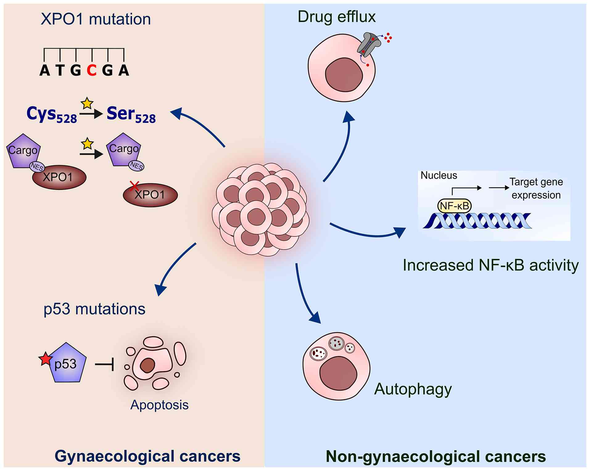 | Figure 5Mechanisms of resistance to nuclear
transport inhibitors. Resistance to selective inhibitors of nuclear
export compounds, such as Selinexor, arises through diverse and
interconnected mechanisms spanning genetic, pharmacokinetic, and
pharmacodynamic challenges. Left side illustrates mechanisms in
gynaecological cancers: a critical genetic escape mechanism is the
dominant XPO1 cysteine 528 to serine mutation, which abolishes the
necessary covalent drug binding, leaving the protein active to
export tumour suppressor proteins; p53 mutations determine the loss
of its ability to induce apoptosis, allowing cancer cells to
survive despite cellular stress or damage. Right side overviews
mechanisms in non-gynaecological cancers: pharmacokinetic
resistance often involves the overexpression of efflux pumps, which
actively expel the drug, reducing its intracellular concentration
below therapeutic levels; pharmacodynamic resistance manifests as
cellular reprogramming, including the activation of compensatory
prosurvival pathways, such as the NF-κB pathway, to bypass the
nuclear retention of tumour suppressor proteins, and the induction
of cytoprotective autophagy. NES, nuclear export signal; XPO1,
exportin 1. |
Mechanisms of SINE resistance in
gynaecological cancers
The principal genetic resistance mechanism
identified across multiple preclinical models involves substitution
of XPO1 cysteine 528 with serine (C528S) (147). This single amino acid
substitution eliminates the reactive thiol group of Cys528,
rendering the binding pocket chemically inert to the covalent
interaction required for SINE-mediated inhibition (148). Cells engineered to express the
XPO1-C528S mutant display complete resistance to Selinexor,
as demonstrated by the continued cytoplasmic localisation of
critical XPO1 cargo proteins, such as RanBP1, following treatment.
These findings indicate that the C528S mutation is sufficient to
preserve the oncogenic nuclear export activity required for
cellular survival despite drug exposure (149).
Notably, this resistance mechanism behaves
dominantly. Experimental modelling using CRISPR/Cas9 genome editing
demonstrated that a heterozygous C528S mutation confers a
resistance phenotype comparable to that observed in homozygous
mutant cells (150). In
heterozygous cells, although the wild-type XPO1 allele
remains susceptible to Selinexor inhibition, the mutant allele
continues to produce fully functional XPO1 protein capable of
exporting tumour suppressor proteins (TSPs) from the nucleus. This
residual export activity is sufficient to maintain essential
nucleocytoplasmic transport processes and to keep nuclear TSP
concentrations, including RanBP1, below the threshold required to
induce effective apoptosis. Consequently, a single mutational event
may be sufficient to negate the therapeutic activity of SINE
compounds, representing an efficient escape mechanism for cancer
cells. Furthermore, because both first-generation (Selinexor) and
second-generation (KPT-8602) SINE compounds depend on covalent
binding to Cys528, this mutation confers cross-resistance to both
classes of inhibitors (151).
The XPO1-C528S mutation has also been
implicated in resistance to the novel XPO1 inhibitor S109 in
ovarian cancer cells. In cells expressing the mutant protein, S109
failed to induce significant nuclear accumulation of the tumour
suppressor proteins Foxo1 and p27, a key component of its mechanism
of action. Moreover, XPO1-C528S-expressing cells resisted
the growth-inhibitory effects of S109 at the tested concentrations,
whereas cells expressing wild-type XPO1 showed significant
growth suppression (152).
However, no published clinical studies have established a role for
the XPO1-C528S mutation as a mechanism of resistance to XPO1
inhibitors in patients with gynaecological cancers. Thus, the
significance of this mutation remains primarily mechanistic rather
than clinical, serving as a model of target-mediated resistance
rather than an established acquired biomarker.
Not all XPO1 mutations confer resistance to
SINE compounds. Clinical genomic profiling has identified
alternative XPO1 alterations in gynaecological malignancies.
Among these, the XPO1-R749Q mutation is a recurrent variant
enriched in endometrial cancer, accounting for ~50% of reported
R749Q cases in solid tumours and frequently co-occurring with
polymerase epsilon hotspot mutations. This variant is particularly
prevalent in tumours with high tumour mutational burden and may
confer resistance to conventional topoisomerase I inhibitors while
retaining sensitivity to Selinexor-based combination therapies
(153).
In addition to XPO1 alterations, resistance
to SINE compounds in gynaecological malignancies may be influenced
by tissue-specific molecular characteristics, particularly the high
prevalence of TP53 mutations. Results from the Phase III
SIENDO trial demonstrated that the efficacy of Selinexor
maintenance therapy in advanced or recurrent endometrial cancer is
strongly dependent on molecular subtype. Patients with TP53
wild-type tumours experienced substantially prolonged median PFS,
whereas those with TP53-mutant tumours derived no notable
benefit from treatment (154,155). Mechanistically, Selinexor
promotes nuclear accumulation of wild-type p53 and restores its
tumour-suppressive functions, leading to cell-cycle arrest and
apoptosis. By contrast, tumours harbouring dysfunctional mutant p53
are less likely to benefit from this therapeutic mechanism
(156).
Several agents that restore p53 function are
currently under clinical investigation and may enhance the efficacy
of Selinexor. APR-246, a small-molecule p53 reactivator, is
undergoing clinical evaluation for high-grade serous ovarian
carcinoma and has shown particularly promising activity in tumours
with low CIP2A expression (157). APR-246 has also been reported to
restore sensitivity to DNA-damaging agents in both established and
primary ovarian cancer cells with acquired drug resistance
(158). Another p53-reactivating
agent, COTI-2, successfully completed a Phase I trial in recurrent
gynaecological cancers (NCT02433626; first registered on ClinicalTrials.gov on 5 May 2015) and has received
FDA orphan drug designation for ovarian cancer treatment (159). A total of ~3% of ovarian tumours
harbour the TP53 Y220C mutation, for which the mutant p53-targeting
agent PC14586 has demonstrated encouraging clinical activity
(160).
Despite their potential, none of these
p53-restoring compounds have yet been evaluated in combination with
nuclear export inhibitors. To date, only combinations of Selinexor
and PARP inhibitors have demonstrated preclinical efficacy,
although not specifically in TP53-mutant gynaecological cancers.
These studies focused primarily on therapeutic synergism rather
than mechanisms of Selinexor resistance. In ovarian cancer,
screening of 688 compounds identified the PARP inhibitor Olaparib
as the most synergistic partner for Selinexor, a finding
subsequently validated in both cell-line and mouse models.
Combination treatment reduced DNA repair protein expression while
increasing tumour suppressor protein levels. However, these
experiments used the A2780 cell line, which carries wild-type
TP53; therefore, the observed synergy cannot be attributed
to restoration of mutant p53 function (138).
The synergistic interaction between Selinexor and
Olaparib was also confirmed in the OVCAR5 mouse model; however, the
TP53 status of OVCAR5 cells remains controversial, with conflicting
reports describing the cell line as either wild-type or
functionally deficient. Notably, the Selinexor-Olaparib combination
was effective in the TP53-mutant breast cancer cell lines
MDA-MB-468 and MDA-MB-436, inducing cell-cycle arrest and cell
death in vitro and suppressing tumour growth in xenograft
models in vivo (161).
Although that study did not focus on TP53 mutation status,
these findings provide preclinical evidence supporting the
potential utility of Selinexor-based combination therapies in
TP53-mutant cancers.
Mechanisms of SINE resistance in
non-gynaecological cancers
Beyond structural alterations in the XPO1 target
protein, resistance mechanisms involving drug efflux and global
cellular adaptation also play important roles in limiting the
efficacy of SINE compounds in several tumour types.
Multidrug resistance (MDR) is frequently associated
with overexpression of ATP-binding cassette (ABC) transporters,
including P-glycoprotein (P-gp/ABCB1), multidrug
resistance-associated proteins (MRPs) and breast cancer resistance
protein (BCRP). These membrane-bound transporters function as
active efflux pumps, utilising ATP hydrolysis to export a broad
range of structurally diverse substrates, including several
hydrophobic small-molecule chemotherapeutic agents, from the cell
(162). In gynaecological
malignancies, including cervical and ovarian cancer, MDR is
commonly mediated by overexpression of drug efflux pumps,
particularly P-gp and MRP1, which reduce intracellular drug
accumulation and consequently diminish therapeutic efficacy
(163-165).
As SINE compounds are small, hydrophobic molecules,
they are recognised and transported by ABC efflux pumps (166). Overexpression of P-gp, often
resulting from gene amplification or transcriptional and epigenetic
alterations, markedly reduces intracellular drug accumulation and
lowers intracellular SINE concentrations (162). This pharmacokinetic mechanism
provides an alternative route to resistance that does not require
genetic alteration of the XPO1 target. Consequently, treatment
failure may occur despite retention of wild-type XPO1, owing
to inadequate intracellular drug concentrations rather than loss of
target sensitivity. In addition, ABC transporters substantially
influence the systemic pharmacokinetics of SINE compounds by
affecting their absorption, distribution and excretion, while also
limiting penetration into pharmacological sanctuary sites such as
the brain and cerebrospinal fluid (167).
Strategies to overcome this pharmacokinetic
resistance include co-administering potent ABC transporter
inhibitors and developing nanoparticle-based delivery systems
designed to bypass efflux pumps (168). Furthermore, second-generation
SINE compounds, such as KPT-8602 (Eltanexor), have been developed
to address some of these limitations. Eltanexor shows reduced
central nervous system penetration and improved tolerability,
allowing more frequent dosing schedules that may sustain higher
intracellular drug concentrations and reduce the impact of
efflux-mediated resistance (169).
Evidence from high-risk multiple myeloma has shown
that XPO1 overexpression itself can contribute to resistance
(170). Increased target-protein
abundance requires higher drug concentrations to fully occupy
Cys528 binding sites, allowing residual oncogenic nuclear export
activity to persist despite treatment. Moreover, resistance to SINE
compounds appears to be a gradual and complex process involving
extensive cellular adaptation. Comparative analyses of parental and
SINE-resistant cells have identified substantial alterations in
signalling pathways, including changes in the expression of genes
associated with inflammation, cell adhesion and apoptosis. These
findings suggest a broad transcriptional reprogramming response
that enables cancer cells to activate compensatory pathways and
circumvent the antitumour effects resulting from SINE-induced
nuclear retention of tumour suppressor proteins. The persistence of
reduced nuclear accumulation of these proteins, together with
sustained alterations in protein expression profiles, further
supports adaptive survival mechanisms in resistant cells (171).
Notably, XPO1 overexpression is a common feature of
gynaecological malignancies, although XPO1 is also markedly
expressed in normal reproductive tissues. A pan-cancer analysis of
normal tissues using data from the Genotype-Tissue Expression
(GTEx) project demonstrated that elevated XPO1 expression is not
restricted to malignant tissues. Expression levels in normal
ovarian and uterine tissues were relatively higher than those
observed in tissues such as the heart and blood (172). Collectively, these findings
indicate that high XPO1 expression is a physiological
characteristic of female reproductive organs. However, persistent
or further elevated XPO1 expression appears to contribute to tumour
progression and chemoresistance in gynaecological malignancies,
including ovarian carcinoma.
Pharmacodynamic resistance refers to the ability of
tumour cells to tolerate target inhibition by activating
compensatory survival mechanisms. Selinexor promotes the nuclear
retention of several negative regulators, including Iκα, PTEN and
FOXO1/3a, thereby suppressing the NF-κB and PI3K-AKT signalling
pathways (173). Nevertheless,
SINE-resistant cells have been reported to exhibit markedly
elevated basal NF-κB activity. Although Selinexor suppresses NF-κB
signalling by retaining Iκα in the nucleus, persistent enhanced
basal NF-κB activity suggests adaptive activation of upstream or
parallel signalling networks that bypass this XPO1-dependent
regulatory checkpoint (174).
In addition, XPO1 inhibition has been shown to
induce dose- and time-dependent autophagy in glioblastoma cells.
Rather than representing a passive consequence of treatment, this
autophagic response functions as a potent cytoprotective mechanism
that enables tumour cells to withstand Selinexor-induced stress.
Mechanistically, this effect has been linked to nuclear retention
of transcription factor EB (TFEB), a recognised XPO1 cargo protein,
which activates genes involved in autophagic initiation and
maintenance. Notably, both genetic and pharmacological inhibition
of autophagy, including chloroquine treatment, markedly enhanced
sensitivity to Selinexor, increased apoptosis, and prolonged
survival in intracranial mouse models. These findings suggest that
targeting treatment-induced autophagy may be a viable strategy to
improve the efficacy of SINE compounds (175). This adaptive mechanism may also
be relevant to ovarian cancer, in which therapeutic stressors,
including cisplatin and SINE treatment, activate ERK signalling and
induce autophagy as a survival response (176). However, although TFEB-mediated
cytoprotective autophagy has been demonstrated in glioblastoma, its
contribution to resistance in gynaecological cancers remains
hypothetical and requires further validation.
SINE therapy for overcoming cancer
resistance
The tumour microenvironment (TME) contributes to
therapeutic resistance by inducing cellular stress responses and
promoting immune evasion. In multiple myeloma, the hypoxic bone
marrow niche plays a central role in disease progression and
resistance to conventional therapies. Notably, Selinexor has
demonstrated the ability to overcome this microenvironment-driven
resistance. In preclinical models, Selinexor reduced tumour burden
and restored sensitivity to bortezomib, with proapoptotic effects
being particularly pronounced under hypoxic conditions. These
observations indicate that XPO1 inhibition disrupts critical
survival pathways supported by the hypoxic TME, supporting the use
of Selinexor to restore drug sensitivity in proteasome
inhibitor-refractory disease (177).
Beyond its role in adaptation to microenvironmental
stress, XPO1 activity has also been implicated in tumour immune
evasion. The TME is characterised by the accumulation of regulatory
T cells, which suppress effector T-cell function by secreting
inhibitory cytokines such as TGF-β and IL-10. At the same time,
increased expression of immune checkpoint molecules, particularly
PD-L1, promotes T-cell exhaustion and facilitates tumour survival
(178). In gynaecological cancer
models, XPO1 inhibition has been shown to convert immunosuppressive
myeloid-derived suppressor cells into neutrophil-like cells with
T-cell-activating properties through nuclear retention of ERK1/2,
thereby enhancing antitumour immune responses (179,180).
If elevated XPO1 activity contributes to the
establishment of an immune-depleted TME, resistance mechanisms that
preserve XPO1 function, including mutation, overexpression or
pharmacokinetic drug efflux, may simultaneously maintain an
immunosuppressive environment. Consequently, effective strategies
to overcome SINE resistance may require integration of nuclear
export inhibitors with immunomodulatory therapies, including immune
checkpoint blockade, to reverse immune suppression and enhance
cytotoxic T-cell activity (181). Supporting this concept,
decitabine priming combined with Selinexor significantly reduced
tumour burden and ascites formation while enhancing effector T-cell
function in ovarian cancer models (182).
High XPO1 expression has also been negatively
associated with interferon-γ (IFN-γ) signalling activity (183). IFN-γ exerts complex and
context-dependent effects in gynaecological malignancies. In
cervical cancer, IFN-γ promotes antitumour immunity by enhancing
tumour cell susceptibility to cytotoxic T lymphocyte-mediated
killing through upregulation of major histocompatibility complex
class I molecules and CD54 expression (184). By contrast, IFN-γ signalling in
ovarian cancer may facilitate immune evasion by activating the
JAK/STAT1 pathway, increasing expression of PD-L1 and the
proto-oncogene BCL3. Furthermore, IFN-γ-induced activation of
JAK/STAT1 signalling and proteasomal pathways promotes expression
of the non-classical major histocompatibility complex molecule
HLA-E, which suppresses natural killer cell-mediated cytotoxicity
and is associated with poor clinical outcomes.
XPO1, the principal nuclear export protein,
reinforces this resistant phenotype in several malignancies,
including ovarian cancer, where its overexpression associates with
poor survival and chemoresistance through aberrant nuclear export
of tumour suppressor proteins such as p53. Although a direct and
universal regulatory relationship between IFN-γ signalling and XPO1
activity has not been established, XPO1 inhibitors such as
Selinexor can restore platinum sensitivity in resistant ovarian
cancer cells. These findings suggest that IFN-γ-mediated immune
escape and XPO1-driven therapeutic resistance represent
complementary but mechanistically distinct pathways that
collectively enhance ovarian tumour fitness (185).
Selective inhibition of XPO1 using SINE compounds
represents an important therapeutic strategy in oncology. However,
complex, multifactorial resistance mechanisms constrain the
long-term efficacy of this approach. Effectively managing SINE
resistance will require a comprehensive strategy that
simultaneously addresses genetic, pharmacokinetic, and
pharmacodynamic contributors to treatment failure. One promising
approach combines SINE compounds with agents that inhibit or
counteract compensatory survival pathways activated in response to
XPO1 inhibition, restoring therapeutic sensitivity and improving
clinical outcomes.
Challenges and future perspectives
An imbalance in bidirectional communication between
the nucleus and cytoplasm is a common feature of several types of
cancer. In gynaecological malignancies, dysregulation of
nucleocytoplasmic transport generally results from altered
expression of proteins involved in nuclear trafficking. Although
these alterations enhance malignant characteristics, including
proliferation, invasiveness and therapeutic resistance, they also
create novel opportunities for targeted intervention. Despite the
identification of numerous natural and synthetic inhibitors of
nuclear import and export, only one nuclear export inhibitor,
Selinexor, has received FDA approval for treating multiple myeloma
and diffuse large B-cell lymphoma. Furthermore, Selinexor has
demonstrated encouraging activity in patients with advanced
gynaecological cancers, both as monotherapy (135,154) and in combination with paclitaxel
and carboplatin (NCT02269293; first registered on ClinicalTrials.
gov on 21 October 2014).
Adverse effects associated with Selinexor treatment
are generally mild to moderate and can usually be managed with
supportive care. Nevertheless, further development of clinically
applicable nuclear transport inhibitors may be constrained by
limited target specificity. Research has indicated that >1,000
XPO1 cargo proteins have been identified in human cells, including
proteins involved in translation, autophagy, signal transduction,
kinase regulation and phosphatase activity (186). Consequently, XPO1 functions as a
central regulator of multiple cellular processes that govern both
survival and cell death. For regulatory proteins, inhibition of
nucleocytoplasmic transport may be rapidly bypassed through
activation of compensatory signalling networks. Given the extensive
number of XPO1 cargoes affected, it remains difficult to identify
the principal determinants of cell survival or to define the
precise mechanisms underlying the antitumour activity of XPO1
inhibitors.
Additional complexity arises from increased drug
efflux and TME-mediated interactions, both of which contribute to
therapeutic resistance. Notably, XPO1 inhibition has been reported
to increase tumour heterogeneity under certain conditions,
potentially creating further challenges for durable treatment
responses (187). Another
important limitation relates to the chemical properties of current
nuclear transport inhibitors. Most known nuclear export inhibitors
are covalent compounds, making them susceptible to off-target
interactions through non-specific bond formation (102). Therefore, future research should
prioritise developing non-covalent SINE compounds, which may offer
improved specificity while overcoming mutation-driven resistance
associated with alterations in XPO1 (188).
Combining SINE compounds with other anticancer
agents may be an effective strategy to overcome treatment
resistance. The PI3K signalling pathway is frequently mutated and
hyperactivated in gynaecological cancers, and several PI3K
inhibitors have been clinically evaluated, with some demonstrating
promising therapeutic activity (189,190). Notably, SINE compounds may
counteract PI3K-mediated resistance mechanisms. Inhibition of
nucleocytoplasmic transport promotes nuclear retention of tumour
suppressor proteins such as FOXO. Nuclear localisation restores
FOXO transcriptional activity and stimulates PTEN expression, a
negative regulator of the PI3K/AKT pathway. Through this mechanism,
SINE compounds may function as potent modulators of therapeutic
resistance and serve as valuable components of combination
treatment strategies (119).
XPO1 inhibition has already been shown to enhance
the efficacy of standard first-line chemotherapeutic agents,
including cisplatin and paclitaxel, in gynaecological cancers. With
regard to targeted therapies, PARP inhibitor treatment, which is
widely used in ovarian cancer, may also benefit from concurrent
XPO1 inhibition; however, direct validation of this strategy in
ovarian cancer remains limited (161). Notably, beyond its direct
cytotoxic activity, Selinexor has also been reported to exert
anti-inflammatory effects. These properties may offer additional
therapeutic opportunities and support the investigation of
combination regimens that incorporate both XPO1 inhibitors and
anti-inflammatory agents as part of future anticancer strategies
(191).
Although cargo-specific nuclear import inhibitors
are likely to cause fewer side effects than importin inhibitors,
the complexity of nuclear import machinery still limits their
clinical development. Most cargoes lack druggable binding sites
because NLSs are short, flexible peptide motifs. There are no flat
surfaces or protein 'pockets' for an inhibitor to bind, and there
is no specificity, as NLSs of different cargoes share sequence
similarity. Additionally, cargo can utilize several NLSs or import
to the nucleus can be NLS-independent. A classic example is tumour
suppressor p53, which has several NLS sequences. The main NLS used
to enter the nucleus is homologous to NLSs on other nuclear
proteins; however, NLS-deprived wild-type p53 can also be imported
into the nucleus in a complex with SV40 (192). Occasionally, NLS cannot be
readily targeted by small-molecule compounds because an inhibitor
protein masks the NLS, which is exposed only after protein
activation; this is the case of NF-κB, which was discussed earlier.
Finally, blocking the import of an individual protein may be less
appealing from a commercial perspective, as the therapeutic benefit
is likely to be confined to cancers that specifically depend on
that protein rather than a variety of tumours. On the other hand,
blocking XPO1 stops the nuclear export of several suppressors;
thus, it can be applied to treat different types of cancer.
To overcome these obstacles, the development of
nuclear import inhibitors should expand beyond conventional
approaches. Stapled peptides that could easily bind flat protein
surfaces (193), single-domain
antibodies that bind cargo with high specificity and mask its NLS
sequence (194) or molecular
glues that prevent cargo import by stabilizing its cytosolic
conformation and NLS exposure (195) are all alternative therapeutic
modalities that could be exploited for nuclear import inhibition.
Clinical development of cargo-specific nuclear import inhibitors
would benefit from studies evaluating cargo expression levels in
gynaecological tumours, as well as identifying gynaecological
cancer subtypes that are particularly dependent on the nuclear
localization of individual cargos.
Transportins showing markedly elevated expression
in gynaecological cancers have been recognized as potential tumour
markers (Table I). Notably,
researchers have detected increased protein or circular RNA
concentrations not only in tumour tissue but also in patient blood
samples, creating opportunities for minimally invasive diagnostics
(70,76,81). Thus, targeting nuclear transport
has potential not only for developing new transformative anticancer
therapies but also for improving gynaecological cancer
screening.
Additionally, clinical practice would benefit from
reliable biomarkers that would help predict patient sensitivity to
nucleocytoplasmic inhibitors. Several candidates for Selinexor
sensitivity have been identified in multiple myeloma, including
transporter ABCC4 (196) and a
three-gene signature WNT10A-DUSP1-ETV7
involved in interferon-mediated apoptosis (197); however, data on gynaecological
tumours remain limited. In addition, some studies promote
repurposing of ivermectin for the treatment of gynaecological
cancer, mostly in combination with other first-line anticancer
drugs (139,198). This should be carefully
considered, as these findings are based on in vitro models
and lack clinical validation (199).
Current evidence suggests that circulating nuclear
transport proteins, including KPNB1, XPO1 and IPO7, are promising
candidate biomarkers for gynaecological cancers because they are
associated with tumour progression and can be detected through
minimally invasive procedures. However, available evidence remains
largely limited to discovery and early validation, highlighting the
need for larger clinical studies to establish their diagnostic and
prognostic utility. Although XPO1 currently has the strongest
clinical relevance, because selective inhibitors such as selinexor
have entered clinical trials (119), several challenges currently
limit their translation into routine clinical screening. Most
studies in gynaecological cancers have been conducted in relatively
small cohorts and lack independent validation in larger
populations. In addition, because these proteins participate in
essential nucleocytoplasmic transport pathways and are dysregulated
across multiple malignancies and non-malignant biological
processes, their cancer specificity remains a major challenge for
clinical translation. Furthermore, it remains unclear whether
circulating levels of proteins originate directly from tumour cells
or other physiological sources (20).
To establish their clinical utility, further
validation is required through large, multicentre studies with
external validation cohorts. Future work should include analyses to
determine whether circulating nuclear transport proteins can detect
early-stage disease or predict recurrence. Analytical validation is
also essential to demonstrate assay accuracy, reproducibility, and
standardized clinical thresholds. Finally, since single biomarkers
rarely achieve sufficient diagnostic accuracy, comparative studies
should evaluate whether combining KPNB1, XPO1, IPO7, and related
proteins with existing biomarkers improves diagnostic sensitivity
and specificity sufficiently for clinical implementation. Given the
complexity and heterogeneity of gynaecological cancers, these
proteins are more likely to achieve clinical utility as components
of biomarker panels rather than standalone screening markers
(119). In summary, the most
notable evidence gap is the lack of large-scale prospective serum
validation studies demonstrating the diagnostic performance of
nuclear transport proteins for screening and early detection of
gynaecological cancers.
Conclusions
In conclusion, dysregulated expression of nuclear
transport proteins contributes to aberrant nucleocytoplasmic
trafficking in endometrial, cervical and ovarian cancers. This
leads to the development of malignant cell phenotypes, including
enhanced cell proliferation, migration, and resistance to therapy.
In this context, SINEs have emerged as a promising class of
targeted therapeutic agents, with growing evidence supporting their
clinical utility.
Availability of data and materials
Not applicable.
Authors' contributions
IN, LMR, EB, AA and EZ wrote the manuscript. IN,
LMR, EB, AA and EZ reviewed and edited the manuscript. EZ prepared
the images. EZ conceived the topic of review. Data authentication
is not applicable. All authors have read and approved the final
manuscript.
Ethics approval and consent to
participate
Not applicable.
Patient consent for publication
Not applicable.
Competing interests
The authors declare that they have no competing
interests.
Abbreviations:
|
ABC
|
ATP-binding cassette
|
|
FDA
|
Food and Drug Administration
|
|
LMB
|
leptomycin B
|
|
MDR
|
multidrug resistance
|
|
NE
|
nuclear envelope
|
|
NES
|
nuclear export signal
|
|
NLS
|
nuclear localization signal
|
|
NPC
|
nuclear pore complex
|
|
Nup
|
nucleoporin
|
|
P-gp
|
P-glycoprotein
|
|
SINE
|
Selective Inhibitors of Nuclear
Export
|
|
TME
|
tumour microenvironment
|
|
TSP
|
tumour suppressor protein
|
Acknowledgments
Not applicable.
Funding
The present review was funded by The Future Biomedicine
Foundation, which supported its open-access publishing [grant no.
(1.78) SU-442].
References
|
1
|
McColloch A, Rabiei M, Rabbani P, Bowling
A and Cho M: Correlation between nuclear morphology and adipogenic
differentiation: Application of a combined experimental and
computational modeling approach. Sci Rep. 9:163812019. View Article : Google Scholar : PubMed/NCBI
|
|
2
|
Heckenbach I, Mkrtchyan GV, Ezra MB,
Bakula D, Madsen JS, Nielsen MH, Oró D, Osborne B, Covarrubias AJ,
Idda ML, et al: Nuclear morphology is a deep learning biomarker of
cellular senescence. Nat Aging. 2:742–755. 2022. View Article : Google Scholar
|
|
3
|
Sung M, Kim JH, Min HS, Jang S, Hong J,
Choi BK, Shin J, Chung KS and Park YR: Three-dimensional label-free
morphology of CD8 + T cells as a sepsis biomarker. Light Sci Appl.
12:2652023. View Article : Google Scholar
|
|
4
|
Koyama AH and Adachi A: Induction of
apoptosis by herpes simplex virus type 1. J Gen Virol.
78:2909–2912. 1997. View Article : Google Scholar : PubMed/NCBI
|
|
5
|
Camozzi D, Capanni C, Cenni V, Mattioli E,
Columbaro M, Squarzoni S and Lattanzi G: Diverse lamin-dependent
mechanisms interact to control chromatin dynamics. Focus on
laminopathies. Nucleus. 5:427–440. 2014. View Article : Google Scholar : PubMed/NCBI
|
|
6
|
Singh I and Lele TP: Nuclear morphological
abnormalities in cancer: A search for unifying mechanisms. Results
Probl Cell Differ. 70:443–467. 2022. View Article : Google Scholar : PubMed/NCBI
|
|
7
|
Davidson PM, Denais C, Bakshi MC and
Lammerding J: Nuclear deformability constitutes a rate-limiting
step during cell migration in 3-D environments. Cell Mol Bioeng.
7:293–306. 2014. View Article : Google Scholar : PubMed/NCBI
|
|
8
|
Lei K, Zhu X, Xu R, Shao C, Xu T, Zhuang Y
and Han M: Inner nuclear envelope proteins SUN1 and SUN2 play a
prominent role in the DNA damage response. Curr Biol. 22:1609–1615.
2012. View Article : Google Scholar : PubMed/NCBI
|
|
9
|
Kim SJ, Park SH, Myung K and Lee KY: Lamin
A/C facilitates DNA damage response by modulating ATM signaling and
homologous recombination pathways. Anim Cells Syst (Seoul).
28:401–416. 2024. View Article : Google Scholar : PubMed/NCBI
|
|
10
|
Kau TR, Way JC and Silver PA: Nuclear
transport and cancer: From mechanism to intervention. Nat Rev
Cancer. 4:106–117. 2004. View Article : Google Scholar : PubMed/NCBI
|
|
11
|
Ledford LRC and Lockwood S: Scope and
epidemiology of gynecologic cancers: An overview. Semin Oncol Nurs.
35:147–150. 2019. View Article : Google Scholar : PubMed/NCBI
|
|
12
|
Bray F, Laversanne M, Sung H, Ferlay J,
Siegel RL, Soerjomataram I and Jemal A: Global cancer statistics
2022: GLOBOCAN estimates of incidence and mortality worldwide for
36 cancers in 185 countries. CA Cancer J Clin. 74:229–263.
2024.PubMed/NCBI
|
|
13
|
Papanicolaou GN and Traut HF: The
diagnostic value of vaginal smears in carcinoma of the uterus.
1941. Arch Pathol Lab Med. 121:211–224. 1997.PubMed/NCBI
|
|
14
|
Palmer JE, Sant Cassia LJ, Irwin CJ,
Morris AG and Rollason TP: The prognostic value of nuclear
morphometric analysis in serous ovarian carcinoma. Int J Gynecol
Cancer. 18:692–701. 2008. View Article : Google Scholar
|
|
15
|
Capo-chichi CD, Cai KQ, Simpkins F,
Ganjei-Azar P, Godwin AK and Xu XX: Nuclear envelope structural
defects cause chromosomal numerical instability and aneuploidy in
ovarian cancer. BMC Med. 9:282011. View Article : Google Scholar : PubMed/NCBI
|
|
16
|
Cicchillitti L, Corrado G, Carosi M,
Dabrowska ME, Loria R, Falcioni R, Cutillo G, Piaggio G and Vizza
E: Prognostic role of NF-YA splicing isoforms and lamin A status in
low grade endometrial cancer. Oncotarget. 8:7935–7945. 2017.
View Article : Google Scholar :
|
|
17
|
Chari A, Vogl DT, Gavriatopoulou M, Nooka
AK, Yee AJ, Huff CA, Moreau P, Dingli D, Cole C, Lonial S, et al:
Oral selinexor-dexamethasone for triple-class refractory multiple
myeloma. N Engl J Med. 381:727–738. 2019. View Article : Google Scholar : PubMed/NCBI
|
|
18
|
Kalakonda N, Maerevoet M, Cavallo F,
Follows G, Goy A, Vermaat JSP, Casasnovas O, Hamad N, Zijlstra JM,
Bakhshi S, et al: Selinexor in patients with relapsed or refractory
diffuse large B-cell lymphoma (SADAL): A single-arm, multinational,
multicentre, open-label, phase 2 trial. Lancet Haematol.
7:e511–e522. 2020. View Article : Google Scholar : PubMed/NCBI
|
|
19
|
Raices M and D'Angelo MA: Structure,
maintenance, and regulation of nuclear pore complexes: The
gatekeepers of the eukaryotic genome. Cold Spring Harb Perspect
Biol. 14:a0406912022. View Article : Google Scholar
|
|
20
|
Yang Y, Guo L, Chen L, Gong B, Jia D and
Sun Q: Nuclear transport proteins: Structure, function, and disease
relevance. Signal Transduct Target Ther. 8:4252023. View Article : Google Scholar : PubMed/NCBI
|
|
21
|
Lai C, Xu L and Dai S: The nuclear export
protein exportin-1 in solid malignant tumours: From biology to
clinical trials. Clin Transl Med. 14:e16842024. View Article : Google Scholar : PubMed/NCBI
|
|
22
|
Li Y, Zhu J, Zhai F, Kong L, Li H and Jin
X: Advances in the understanding of nuclear pore complexes in human
diseases. J Cancer Res Clin Oncol. 150:3742024. View Article : Google Scholar : PubMed/NCBI
|
|
23
|
Beck M and Hurt E: The nuclear pore
complex: Understanding its function through structural insight. Nat
Rev Mol Cell Biol. 18:73–89. 2017. View Article : Google Scholar
|
|
24
|
Lin DH and Hoelz A: The structure of the
nuclear pore complex (an update). Annu Rev Biochem. 88:725–783.
2019. View Article : Google Scholar : PubMed/NCBI
|
|
25
|
Kinoshita Y, Kalir T, Rahaman J, Dottino P
and Kohtz DS: Alterations in nuclear pore architecture allow cancer
cell entry into or exit from drug-resistant dormancy. Am J Pathol.
180:375–389. 2012. View Article : Google Scholar
|
|
26
|
Hazawa M, Lin DC, Kobayashi A, Jiang YY,
Xu L, Dewi FRP, Mohamed MS, Hartono, Nakada M, Meguro-Horike M, et
al: ROCK-dependent phosphorylation of NUP62 regulates p63 nuclear
transport and squamous cell carcinoma proliferation. EMBO Rep.
19:73–88. 2018. View Article : Google Scholar
|
|
27
|
Campbell MJ: Genomic crosstalk between
nuclear receptors in hormone-dependent cancers. Endocrinology.
166:bqaf1492025. View Article : Google Scholar : PubMed/NCBI
|
|
28
|
Jahangiri L: A mechanistic insight into
cancer progression mediated by nucleoporins. Cancer Genet.
286-287:35–42. 2024. View Article : Google Scholar : PubMed/NCBI
|
|
29
|
Dultz E, Wojtynek M, Medalia O and
Onischenko E: The nuclear pore complex: Birth, life, and death of a
cellular behemoth. Cells. 11:14562022. View Article : Google Scholar : PubMed/NCBI
|
|
30
|
Makise M, Mackay DR, Elgort S, Shankaran
SS, Adam SA and Ullman KS: The Nup153-Nup50 protein interface and
its role in nuclear import. J Biol Chem. 287:38515–38522. 2012.
View Article : Google Scholar : PubMed/NCBI
|
|
31
|
Walther TC, Pickersgill HS, Cordes VC,
Goldberg MW, Allen TD, Mattaj IW and Fornerod M: The cytoplasmic
filaments of the nuclear pore complex are dispensable for selective
nuclear protein import. J Cell Biol. 158:63–77. 2002. View Article : Google Scholar : PubMed/NCBI
|
|
32
|
Zaitsava H, Gachowska M, Bartoszewska E,
Kmiecik A and Kulbacka J: The potential of nuclear pore complexes
in cancer therapy. Molecules. 29:48322024. View Article : Google Scholar : PubMed/NCBI
|
|
33
|
Engqvist H, Parris TZ, Kovács A, Rönnerman
EW, Sundfeldt K, Karlsson P and Helou K: Validation of novel
prognostic biomarkers for early-stage clear-cell, endometrioid and
mucinous ovarian carcinomas using immunohistochemistry. Front
Oncol. 10:1622020. View Article : Google Scholar : PubMed/NCBI
|
|
34
|
Borden KLB: The nuclear pore complex and
mRNA export in cancer. Cancers (Basel). 13:422020. View Article : Google Scholar : PubMed/NCBI
|
|
35
|
Güzel C, Govorukhina NI, Wisman GBA,
Stingl C, Dekker LJM, Klip HG, Hollema H, Guryev V, Horvatovich PL,
van der Zee AGJ, et al: Proteomic alterations in early stage
cervical cancer. Oncotarget. 9:18128–18147. 2018. View Article : Google Scholar : PubMed/NCBI
|
|
36
|
Khokhar M, Kartha P, Hassan S and Pandey
RK: Decoding dysregulated genes, molecular pathways and microRNAs
involved in cervical cancer. J Gene Med. 26:e37132024. View Article : Google Scholar : PubMed/NCBI
|
|
37
|
Cautain B, Hill R, de Pedro N and Link W:
Components and regulation of nuclear transport processes. FEBS J.
282:445–462. 2015. View Article : Google Scholar
|
|
38
|
Stewart M: Function of the nuclear
transport machinery in maintaining the distinctive compositions of
the nucleus and cytoplasm. Int J Mol Sci. 23:25782022. View Article : Google Scholar : PubMed/NCBI
|
|
39
|
Kimura M and Imamoto N: Biological
significance of the importin-β family-dependent nucleocytoplasmic
transport pathways. Traffic. 15:727–748. 2014. View Article : Google Scholar : PubMed/NCBI
|
|
40
|
Pumroy RA and Cingolani G: Diversification
of importin-α isoforms in cellular trafficking and disease states.
Biochem J. 466:13–28. 2015. View Article : Google Scholar : PubMed/NCBI
|
|
41
|
Cingolani G, Petosa C, Weis K and Müller
CW: Structure of importin-beta bound to the IBB domain of
importin-alpha. Nature. 399:221–229. 1999. View Article : Google Scholar : PubMed/NCBI
|
|
42
|
Azizian NG and Li Y: XPO1-dependent
nuclear export as a target for cancer therapy. J Hematol Oncol.
13:612020. View Article : Google Scholar : PubMed/NCBI
|
|
43
|
Boudhraa Z, Carmona E, Provencher D and
Mes-Masson AM: Ran GTPase: A key player in tumor progression and
metastasis. Front Cell Dev Biol. 8:3452020. View Article : Google Scholar : PubMed/NCBI
|
|
44
|
Scott KL, Halfmann CT, Hoefakker AD,
Purkayastha P, Wang TC, Lele TP and Roux KJ: Nucleocytoplasmic
transport rates are regulated by cellular processes that modulate
GTP availability. J Cell Biol. 223:e2023081522024. View Article : Google Scholar : PubMed/NCBI
|
|
45
|
El-Tanani M, Nsairat H, Mishra V, Mishra
Y, Aljabali AAA, Serrano-Aroca Á and Tambuwala MM: Ran GTPase and
its importance in cellular signaling and malignant phenotype. Int J
Mol Sci. 24:30652023. View Article : Google Scholar : PubMed/NCBI
|
|
46
|
Pascual-Garcia P and Capelson M: The
nuclear pore complex and the genome: Organizing and regulatory
principles. Curr Opin Genet Dev. 67:142–150. 2021. View Article : Google Scholar : PubMed/NCBI
|
|
47
|
Fatima I, Barman S, Rai R, Thiel KWW and
Chandra V: Targeting Wnt signaling in endometrial cancer. Cancers
(Basel). 13:23512021. View Article : Google Scholar : PubMed/NCBI
|
|
48
|
Nguyen VHL, Hough R, Bernaudo S and Peng
C: Wnt/β-catenin signalling in ovarian cancer: Insights into its
hyperactivation and function in tumorigenesis. J Ovarian Res.
12:1222019. View Article : Google Scholar
|
|
49
|
Feng D, Lin J, Wang W, Yan K, Liang H,
Liang J, Yu H and Ling B: Wnt3a/β-catenin/CBP activation in the
progression of cervical intraepithelial neoplasia. Pathol Oncol
Res. 27:6096202021. View Article : Google Scholar
|
|
50
|
Parrish ML, Broaddus RR and Gladden AB:
Mechanisms of mutant beta-catenin in endometrial cancer
progression. Front Oncol. 12:10093452022. View Article : Google Scholar
|
|
51
|
Mao H, Zhao X and Sun SC: NF-κB in
inflammation and cancer. Cell Mol Immunol. 22:811–839. 2025.
View Article : Google Scholar : PubMed/NCBI
|
|
52
|
Byg LM, Vidlund J, Vasiljevic N, Clausen
D, Forslund O and Norrild B: NF-κB signalling is attenuated by the
E7 protein from cutaneous human papillomaviruses. Virus Res.
169:48–53. 2012. View Article : Google Scholar : PubMed/NCBI
|
|
53
|
Chen H, Cui Q and Yang W: NF-κB activation
is essential for cervical cell proliferation and malignant
transformation. Int J Mol Sci. 26:24932025. View Article : Google Scholar
|
|
54
|
Neumann M and Naumann M: Beyond IkappaBs:
Alternative regulation of NF-kappaB activity. FASEB J.
21:2642–2654. 2007. View Article : Google Scholar : PubMed/NCBI
|
|
55
|
Chen LF, Williams SA, Mu Y, Nakano H,
Duerr JM, Buckbinder L and Greene WC: NF-kappaB RelA
phosphorylation regulates RelA acetylation. Mol Cell Biol.
25:7966–7975. 2005. View Article : Google Scholar : PubMed/NCBI
|
|
56
|
Han X, Dong XX, Shi MY, Feng L, Wang XL,
Zhang JS and Yan QC: SUMOylation and deacetylation affect NF-κB p65
activity induced by high glucose in human lens epithelial cells.
Int J Ophthalmol. 12:1371–1379. 2019. View Article : Google Scholar
|
|
57
|
Wu Z, Li J, Zhang Y, Hu L and Peng X:
Synchronous co-expression of Id-1 and nuclear NF-κB p65 promotes
cervical cancer progression and malignancy, and is associated with
a poor prognosis and chemosensitivity. Oncol Rep. 42:2075–2086.
2019.PubMed/NCBI
|
|
58
|
Yang G, Xiao X, Rosen DG, Cheng X, Wu X,
Chang B, Liu G, Xue F, Mercado-Uribe I, Chiao P, et al: The
biphasic role of NF-kappaB in progression and chemoresistance of
ovarian cancer. Clin Cancer Res. 17:2181–2194. 2011. View Article : Google Scholar : PubMed/NCBI
|
|
59
|
Feng HP, Liu YC, Wang CL, Liao WC, Yu JS
and Yu CJ: Acetylation regulates the nucleocytoplasmic distribution
and oncogenic function of karyopherin alpha 2 in lung
adenocarcinoma. Biochem Biophys Res Commun. 659:96–104. 2023.
View Article : Google Scholar : PubMed/NCBI
|
|
60
|
Oka M, Otani M, Miyamoto Y, Oshima R,
Adachi J, Tomonaga T, Asally M, Nagaoka Y, Tanaka K, Toyoda A, et
al: Phase-separated nuclear bodies of nucleoporin fusions promote
condensation of MLL1/CRM1 and rearrangement of 3D genome structure.
Cell Rep. 42:1128842023. View Article : Google Scholar : PubMed/NCBI
|
|
61
|
Mizoguchi Y, Fujita N, Taki T, Hayashi Y
and Hamamoto K: Juvenile myelomonocytic leukemia with
t(7;11)(p15;p15) and NUP98-HOXA11 fusion. Am J Hematol. 84:295–297.
2009. View Article : Google Scholar : PubMed/NCBI
|
|
62
|
Fujino T, Suzuki A, Ito Y, Ohyashiki K,
Hatano Y, Miura I and Nakamura T: Single-translocation and
double-chimeric transcripts: Detection of NUP98-HOXA9 in myeloid
leukemias with HOXA11 or HOXA13 breaks of the chromosomal
translocation t(7;11)(p15;p15). Blood. 99:1428–1433. 2002.
View Article : Google Scholar : PubMed/NCBI
|
|
63
|
Reader JC, Meekins JS, Gojo I and Ning Y:
A novel NUP98-PHF23 fusion resulting from a cryptic translocation
t(11;17)(p15;p13) in acute myeloid leukemia. Leukemia. 21:842–844.
2007. View Article : Google Scholar : PubMed/NCBI
|
|
64
|
Miloudi H, Bohers É, Guillonneau F, Taly
A, Gibouin VC, Viailly PJ, Jego G, Grumolato L, Jardin F and Sola
B: XPO1E571K mutation modifies exportin 1 localisation
and interactome in B-cell lymphoma. Cancers (Basel). 12:28292020.
View Article : Google Scholar
|
|
65
|
García-Santisteban I, Arregi I,
Alonso-Mariño M, Urbaneja MA, Garcia-Vallejo JJ, Bañuelos S and
Rodríguez JA: A cellular reporter to evaluate CRM1 nuclear export
activity: Functional analysis of the cancer-related mutant E571K.
Cell Mol Life Sci. 73:4685–4699. 2016. View Article : Google Scholar : PubMed/NCBI
|
|
66
|
Caillot M, Miloudi H, Taly A,
Profitós-Pelejà N, Santos JC, Ribeiro ML, Maitre E, Saule S, Roué
G, Jardin F and Sola B: Exportin 1-mediated nuclear/cytoplasmic
trafficking controls drug sensitivity of classical Hodgkin's
lymphoma. Mol Oncol. 17:2546–2564. 2023. View Article : Google Scholar : PubMed/NCBI
|
|
67
|
Melo SA, Moutinho C, Ropero S, Calin GA,
Rossi S, Spizzo R, Fernandez AF, Davalos V, Villanueva A, Montoya
G, et al: A genetic defect in exportin-5 traps precursor microRNAs
in the nucleus of cancer cells. Cancer Cell. 18:303–315. 2010.
View Article : Google Scholar : PubMed/NCBI
|
|
68
|
Sun HL, Cui R, Zhou J, Teng KY, Hsiao YH,
Nakanishi K, Fassan M, Luo Z, Shi G, Tili E, et al: ERK activation
globally downregulates miRNAs through phosphorylating exportin-5.
Cancer Cell. 30:723–736. 2016. View Article : Google Scholar : PubMed/NCBI
|
|
69
|
Stelma T, Chi A, van der Watt PJ, Verrico
A, Lavia P and Leaner VD: Targeting nuclear transporters in cancer:
Diagnostic, prognostic and therapeutic potential. IUBMB Life.
68:268–280. 2016. View Article : Google Scholar : PubMed/NCBI
|
|
70
|
van der Watt PJ, Okpara MO, Wishart A,
Parker MI, Soares NC, Blackburn JM and Leaner VD: Nuclear transport
proteins are secreted by cancer cells and identified as potential
novel cancer biomarkers. Int J Cancer. 150:347–361. 2022.
View Article : Google Scholar
|
|
71
|
Stelma T and Leaner VD: KPNB1-mediated
nuclear import is required for motility and inflammatory
transcription factor activity in cervical cancer cells. Oncotarget.
8:32833–32847. 2017. View Article : Google Scholar : PubMed/NCBI
|
|
72
|
van der Watt PJ, Maske CP, Hendricks DT,
Parker MI, Denny L, Govender D, Birrer MJ and Leaner VD: The
Karyopherin proteins, Crm1 and Karyopherin beta1, are overexpressed
in cervical cancer and are critical for cancer cell survival and
proliferation. Int J Cancer. 124:1829–1840. 2009. View Article : Google Scholar : PubMed/NCBI
|
|
73
|
Kodama M, Kodama T, Newberg JY, Katayama
H, Kobayashi M, Hanash SM, Yoshihara K, Wei Z, Tien JC, Rangel R,
et al: In vivo loss-of-function screens identify KPNB1 as a new
druggable oncogene in epithelial ovarian cancer. Proc Natl Acad Sci
USA. 114:E7301–E7310. 2017. View Article : Google Scholar : PubMed/NCBI
|
|
74
|
Chen J, Hu Y, Teng Y and Yang B: Increased
nuclear transporter importin 7 contributes to the tumor growth and
correlates with CD8 T cell infiltration in cervical cancer. Front
Cell Dev Biol. 9:7327862021. View Article : Google Scholar : PubMed/NCBI
|
|
75
|
Han Y and Wang X: The emerging roles of
KPNA2 in cancer. Life Sci. 241:1171402020. View Article : Google Scholar
|
|
76
|
Huang L, Zhou Y, Cao XP, Lin JX, Zhang L,
Huang ST and Zheng M: KPNA2 is a potential diagnostic serum
biomarker for epithelial ovarian cancer and correlates with poor
prognosis. Tumour Biol. 39:10104283177062892017. View Article : Google Scholar : PubMed/NCBI
|
|
77
|
Chen Y, Camacho S, Silvers T, Razak AR,
Gabrail NY, Gerecitano JF, Kalir E, Pereira E, Evans BR, Ramus SJ,
et al: Inhibition of the nuclear export receptor XPO1 as a
therapeutic target for platinum-resistant ovarian cancer. Clin
Cancer Res. 23:1552–1563. 2017. View Article : Google Scholar
|
|
78
|
Noske A, Weichert W, Niesporek S, Röske A,
Buckendahl AC, Koch I, Sehouli J, Dietel M and Denkert C:
Expression of the nuclear export protein chromosomal region
maintenance/exportin 1/Xpo1 is a prognostic factor in human ovarian
cancer. Cancer. 112:1733–1743. 2008. View Article : Google Scholar : PubMed/NCBI
|
|
79
|
Huang L, Zhou Y, Cao XP, Lin JX, Zhang L,
Huang ST and Zheng M: KPNA2 promotes migration and invasion in
epithelial ovarian cancer cells by inducing epithelial-mesenchymal
transition via Akt/GSK-3β/Snail activation. J Cancer. 9:157–165.
2018. View Article : Google Scholar
|
|
80
|
Zheng M, Tang L, Huang L, Ding H, Liao WT,
Zeng MS and Wang HY: Overexpression of karyopherin-2 in epithelial
ovarian cancer and correlation with poor prognosis. Obstet Gynecol.
116:884–891. 2010. View Article : Google Scholar : PubMed/NCBI
|
|
81
|
Xia B, Zhao Z, Wu Y, Wang Y, Zhao Y and
Wang J: Circular RNA circTNPO3 regulates paclitaxel resistance of
ovarian cancer cells by miR-1299/NEK2 signaling pathway. Mol Ther
Nucleic Acids. 21:780–791. 2020. View Article : Google Scholar : PubMed/NCBI
|
|
82
|
Stawerski P, Wagrowska-Danilewicz M,
Stasikowska O and Danilewicz M: Immunoexpression of CAS protein is
augmented in high grade serous ovarian tumors. Pol J Pathol.
61:219–223. 2010.
|
|
83
|
Hu Y, Zhou J, Ling X, Zhao K, Huang P, Gao
M, Li X, Sun M, Zou Y and Feng G: KPNA5 suppresses malignant
progression of ovarian cancer through importing the PTPN4 into the
nucleus. Cancer Med. 14:e707312025. View Article : Google Scholar : PubMed/NCBI
|
|
84
|
Li Y, Huang Y, Ren S, Xiao X, Cao H and He
J: A pan-cancer analysis of the oncogenic role of nuclear transport
factor 2 in human cancers. Front Oncol. 12:8293892022. View Article : Google Scholar : PubMed/NCBI
|
|
85
|
Bogani G, Monk BJ, Coleman RL, Vergote I,
Oakin A, Ray-Coquard I, Mariani A, Scambia G, Raspagliesi F and
Bolognese B: Selinexor in patients with advanced and recurrent
endometrial cancer. Curr Probl Cancer. 47:1009632023. View Article : Google Scholar : PubMed/NCBI
|
|
86
|
Ikenberg K, Valtcheva N, Brandt S, Zhong
Q, Wong CE, Noske A, Rechsteiner M, Rueschoff JH, Caduff R, Dellas
A, et al: KPNA2 is overexpressed in human and mouse endometrial
cancers and promotes cellular proliferation. J Pathol. 234:239–252.
2014. View Article : Google Scholar : PubMed/NCBI
|
|
87
|
Zeng B, Hu J, Yuan R, Hu L, Zhong L and
Kang K: Increased expression of importin13 in endometriosis and
endometrial carcinoma. Med Sci Monit. 18:CR361–CR367. 2012.
View Article : Google Scholar : PubMed/NCBI
|
|
88
|
Meng N, Li L, Yang X, Tang H, Wang J,
Zhang S, Xia Z, Yao J, Zhang Q, Hu C, et al: NXF1 suppresses
progression of endometrial cancer by interacting with the SRSF3 to
regulate SP4 splicing. iScience. 28:1131132025. View Article : Google Scholar : PubMed/NCBI
|
|
89
|
Ishizawa J, Kojima K, Hail N Jr, Tabe Y
and Andreeff M: Expression, function, and targeting of the nuclear
exporter chromosome region maintenance 1 (CRM1) protein. Pharmacol
Ther. 153:25–35. 2015. View Article : Google Scholar : PubMed/NCBI
|
|
90
|
Walker JS, Hing ZA, Harrington B,
Baumhardt J, Ozer HG, Lehman A, Giacopelli B, Beaver L, Williams K,
Skinner JN, et al: Recurrent XPO1 mutations alter pathogenesis of
chronic lymphocytic leukemia. J Hematol Oncol. 14:172021.
View Article : Google Scholar : PubMed/NCBI
|
|
91
|
Noske A, Schwabe M, Weichert W,
Darb-Esfahani S, Buckendahl AC, Sehouli J, Braicu EI, Budczies J,
Dietel M and Denkert C: An intracellular targeted antibody detects
EGFR as an independent prognostic factor in ovarian carcinomas. BMC
Cancer. 11:2942011. View Article : Google Scholar : PubMed/NCBI
|
|
92
|
Wang Y, Wang Y, Xiang J, Ji F, Deng Y,
Tang C, Yang S, Xi Q, Liu R and Di W: Knockdown of CRM1 inhibits
the nuclear export of p27(Kip1) phosphorylated at serine 10 and
plays a role in the pathogenesis of epithelial ovarian cancer.
Cancer Lett. 343:6–13. 2014. View Article : Google Scholar
|
|
93
|
Iwahashi N, Ikezaki M, Komohara Y,
Fujiwara Y, Noguchi T, Nishioka K, Sakai K, Nishio K, Ueda M, Ihara
Y, et al: Cytoplasmic p53 aggregates accumulated in p53-mutated
cancer correlate with poor prognosis. PNAS Nexus. 1:pgac1282022.
View Article : Google Scholar
|
|
94
|
Fabi F, Adam P, Vincent K, Demontigny F,
Parent S, Joncas FH and Asselin E: Inhibition of CRM1 activity
sensitizes endometrial and ovarian cell lines to TRAIL-induced cell
death. Cell Commun Signal. 16:392018. View Article : Google Scholar : PubMed/NCBI
|
|
95
|
Li J, Chen Y, Qin X, Wen J, Ding H, Xia W,
Li S, Su X, Wang W, Li H, et al: MiR-138 downregulates miRNA
processing in HeLa cells by targeting RMND5A and decreasing
Exportin-5 stability. Nucleic Acids Res. 42:458–474. 2014.
View Article : Google Scholar
|
|
96
|
Zhang T and Xiaohan C: Unveiling the role
of JAK2/STAT3 signaling in chemoresistance of gynecological
cancers: From mechanisms to therapeutic implications. Crit Rev
Oncol Hematol. 211:1047122025. View Article : Google Scholar : PubMed/NCBI
|
|
97
|
Mackmull MT, Klaus B, Heinze I,
Chokkalingam M, Beyer A, Russell RB, Ori A and Beck M: Landscape of
nuclear transport receptor cargo specificity. Mol Syst Biol.
13:9622017. View Article : Google Scholar
|
|
98
|
Hill R, Cautain B, de Pedro N and Link W:
Targeting nucleocytoplasmic transport in cancer therapy.
Oncotarget. 5:11–28. 2014. View Article : Google Scholar : PubMed/NCBI
|
|
99
|
Dong X, Biswas A, Süel KE, Jackson LK,
Martinez R, Gu H and Chook YM: Structural basis for leucine-rich
nuclear export signal recognition by CRM1. Nature. 458:1136–1141.
2009. View Article : Google Scholar : PubMed/NCBI
|
|
100
|
Das A, Wei G, Parikh K and Liu D:
Selective inhibitors of nuclear export (SINE) in hematological
malignancies. Exp Hematol Oncol. 4:72015. View Article : Google Scholar : PubMed/NCBI
|
|
101
|
Landes JR, Moore SA, Bartley BR, Doan HQ,
Rady PL and Tyring SK: The efficacy of selinexor (KPT-330), an XPO1
inhibitor, on non-hematologic cancers: A comprehensive review. J
Cancer Res Clin Oncol. 149:2139–2155. 2023. View Article : Google Scholar
|
|
102
|
Jans DA, Martin AJ and Wagstaff KM:
Inhibitors of nuclear transport. Curr Opin Cell Biol. 58:50–60.
2019. View Article : Google Scholar : PubMed/NCBI
|
|
103
|
Niu M, Wu S, Mao L and Yang Y: CRM1 is a
cellular target of curcumin: new insights for the myriad of
biological effects of an ancient spice. Traffic. 14:1042–1052.
2013. View Article : Google Scholar : PubMed/NCBI
|
|
104
|
Khamphikham P, Tantiworawit A and
Anuchapreeda S: Inhibition of XPO1 by selinexor enhances terminal
erythroid maturation through modulation of HSP70 trafficking in
severe β0-thalassemia/HbE. PLoS One. 20:e03331272025. View Article : Google Scholar
|
|
105
|
Elfar A, Tran AV, Case J, Wayant C, Hughes
GK, McIntire R, Gardner B, Ladd C, Peña AM, Tuia J, et al: An
evaluation of selinexor's clinical trial portfolio: A
cross-sectional study. Ther Adv Hematol. 16:204062072513291742025.
View Article : Google Scholar : PubMed/NCBI
|
|
106
|
Perwitasari O, Johnson S, Yan X, Register
E, Crabtree J, Gabbard J, Howerth E, Shacham S, Carlson R, Tamir S
and Tripp RA: Antiviral efficacy of verdinexor in vivo in two
animal models of influenza a virus infection. PLoS One.
11:e01672212016. View Article : Google Scholar : PubMed/NCBI
|
|
107
|
Gandhi UH, Senapedis W, Baloglu E, Unger
TJ, Chari A, Vogl D and Cornell RF: Clinical implications of
targeting XPO1-mediated nuclear export in multiple myeloma. Clin
Lymphoma Myeloma Leuk. 18:335–345. 2018. View Article : Google Scholar : PubMed/NCBI
|
|
108
|
Sakakibara K, Saito N, Sato T, Suzuki A,
Hasegawa Y, Friedman JM, Kufe DW, Vonhoff DD, Iwami T and Kawabe T:
CBS9106 is a novel reversible oral CRM1 inhibitor with CRM1
degrading activity. Blood. 118:3922–3931. 2011. View Article : Google Scholar : PubMed/NCBI
|
|
109
|
Saito N, Sakakibara K, Sato T, Friedman
JM, Kufe DW, VonHoff DD and Kawabe T: CBS9106-induced CRM1
degradation is mediated by cullin ring ligase activity and the
neddylation pathway. Mol Cancer Ther. 13:3013–3023. 2014.
View Article : Google Scholar : PubMed/NCBI
|
|
110
|
Liu X, Chong Y, Liu H, Han Y and Niu M:
CRM1 inhibitor S109 suppresses cell proliferation and induces cell
cycle arrest in renal cancer cells. Korean J Physiol Pharmacol.
20:161–168. 2016. View Article : Google Scholar : PubMed/NCBI
|
|
111
|
Kaur B, Blavo C and Parmar MS: Ivermectin:
A multifaceted drug with a potential beyond anti-parasitic therapy.
Cureus. 16:e560252024.PubMed/NCBI
|
|
112
|
Lombardi Z, Gardini L, Kashchuk AV,
Menconi A, Lulli M, Tusa I, Tubita A, Maresca L, Stecca B,
Capitanio M and Rovida E: Importin subunit beta-1 mediates ERK5
nuclear translocation, and its inhibition synergizes with ERK5
kinase inhibitors in reducing cancer cell proliferation. Mol Oncol.
19:99–113. 2025. View Article : Google Scholar :
|
|
113
|
Diéguez-Martínez N, Espinosa-Gil S, Yoldi
G, Megías-Roda E, Bolinaga-Ayala I, Viñas-Casas M, Gorgisen G,
Domingo-Ortí I, Pérez-Montoyo H, Bayascas JR, et al: The ERK5/NF-κB
signaling pathway targets endometrial cancer proliferation and
survival. Cell Mol Life Sci. 79:5242022. View Article : Google Scholar
|
|
114
|
Stecca B and Rovida E: Impact of ERK5 on
the hallmarks of cancer. Int J Mol Sci. 20:14262019. View Article : Google Scholar : PubMed/NCBI
|
|
115
|
Soderholm JF, Bird SL, Kalab P,
Sampathkumar Y, Hasegawa K, Uehara-Bingen M, Weis K and Heald R:
Importazole, a small molecule inhibitor of the transport receptor
importin-β. ACS Chem Biol. 6:700–708. 2011. View Article : Google Scholar : PubMed/NCBI
|
|
116
|
Kuusisto HV and Jans DA: Hyper-dependence
of breast cancer cell types on the nuclear transporter importin β1.
Biochim Biophys Acta. 1853:1870–1878. 2015. View Article : Google Scholar : PubMed/NCBI
|
|
117
|
Yang J, Guo Y, Lu C, Zhang R, Wang Y, Luo
L, Zhang Y, Chu CH, Wang KJ, Obbad S, et al: Inhibition of
karyopherin beta 1 suppresses prostate cancer growth. Oncogene.
38:4700–4714. 2019. View Article : Google Scholar : PubMed/NCBI
|
|
118
|
Hintersteiner M, Ambrus G, Bednenko J,
Schmied M, Knox AJ, Meisner NC, Gstach H, Seifert JM, Singer EL,
Gerace L and Auer M: Identification of a small molecule inhibitor
of importin β mediated nuclear import by confocal on-bead screening
of tagged one-bead one-compound libraries. ACS Chem Biol.
5:967–979. 2010. View Article : Google Scholar : PubMed/NCBI
|
|
119
|
Newell S, van der Watt PJ and Leaner VD:
Therapeutic targeting of nuclear export and import receptors in
cancer and their potential in combination chemotherapy. IUBMB Life.
76:4–25. 2024. View Article : Google Scholar :
|
|
120
|
van der Watt PJ, Chi A, Stelma T, Stowell
C, Strydom E, Carden S, Angus L, Hadley K, Lang D, Wei W, et al:
Targeting the nuclear import receptor Kpnβ1 as an anticancer
therapeutic. Mol Cancer Ther. 15:560–573. 2016. View Article : Google Scholar : PubMed/NCBI
|
|
121
|
Ajayi-Smith A, van der Watt P, Mkwanazi N,
Carden S, Trent JO and Leaner VD: Novel small molecule inhibitor of
Kpnβ1 induces cell cycle arrest and apoptosis in cancer cells. Exp
Cell Res. 404:1126372021. View Article : Google Scholar
|
|
122
|
Harrington BS and Annunziata CM: NF-κB
signaling in ovarian cancer. Cancers (Basel). 11:11822019.
View Article : Google Scholar
|
|
123
|
Zdrojkowski Ł, Jasiński T, Ferreira-Dias
G, Pawliński B and Domino M: The role of NF-κB in endometrial
diseases in humans and animals: A review. Int J Mol Sci.
24:29012023. View Article : Google Scholar
|
|
124
|
Kofler M and Kapus A: Nuclear import and
export of YAP and TAZ. Cancers (Basel). 15:49562023. View Article : Google Scholar : PubMed/NCBI
|
|
125
|
Zhu ZC, Liu JW, Li K, Zheng J and Xiong
ZQ: KPNB1 inhibition disrupts proteostasis and triggers unfolded
protein response-mediated apoptosis in glioblastoma cells.
Oncogene. 37:2936–2952. 2018. View Article : Google Scholar : PubMed/NCBI
|
|
126
|
Chen L, Bi S, Wei Q, Zhao Z, Wang C and
Xie S: Ivermectin suppresses tumour growth and metastasis through
degradation of PAK1 in oesophageal squamous cell carcinoma. J Cell
Mol Med. 24:5387–5401. 2020. View Article : Google Scholar : PubMed/NCBI
|
|
127
|
Juarez M, Schcolnik-Cabrera A and
Duenas-Gonzalez A: The multitargeted drug ivermectin: From an
antiparasitic agent to a repositioned cancer drug. Am J Cancer Res.
8:317–331. 2018.PubMed/NCBI
|
|
128
|
Wagstaff KM, Headey S, Telwatte S, Tyssen
D, Hearps AC, Thomas DR, Tachedjian G and Jans DA: Molecular
dissection of an inhibitor targeting the HIV integrase dependent
preintegration complex nuclear import. Cell Microbiol.
21:e129532019. View Article : Google Scholar :
|
|
129
|
Para MF, Schouten J, Rosenkranz SL, Yu S,
Weiner D, Tebas P, White CJ, Reeds D, Lertora J, Patterson KB, et
al: Phase I/II trial of the anti-HIV activity of mifepristone in
HIV-infected subjects ACTG 5200. J Acquir Immune Defic Syndr.
53:491–495. 2010. View Article : Google Scholar : PubMed/NCBI
|
|
130
|
Fraser JE, Watanabe S, Wang C, Chan WK,
Maher B, Lopez-Denman A, Hick C, Wagstaff KM, Mackenzie JM, Sexton
PM, et al: A nuclear transport inhibitor that modulates the
unfolded protein response and provides in vivo protection against
lethal dengue virus infection. J Infect Dis. 210:1780–1791. 2014.
View Article : Google Scholar : PubMed/NCBI
|
|
131
|
Mittal N, Malpani S, Dyson M, Ono M, Coon
JS, Kim JJ, Schink JC, Bulun SE and Pavone ME: Fenretinide: A novel
treatment for endometrial cancer. PLoS One. 9:e1104102014.
View Article : Google Scholar : PubMed/NCBI
|
|
132
|
Orienti I, Salvati V, Sette G, Zucchetti
M, Bongiorno-Borbone L, Peschiaroli A, Zolla L, Francescangeli F,
Ferrari M, Matteo C, et al: A novel oral micellar fenretinide
formulation with enhanced bioavailability and antitumour activity
against multiple tumours from cancer stem cells. J Exp Clin Cancer
Res. 38:3732019. View Article : Google Scholar : PubMed/NCBI
|
|
133
|
De Angelis ML, Francescangeli F, Aricò E,
Verachi P, Zucchetti M, Matteo C, Petricci E, Pilozzi E, Orienti I,
Boe A, et al: A nanoencapsulated oral formulation of fenretinide
promotes local and metastatic breast cancer dormancy in HER2/neu
transgenic mice. J Exp Clin Cancer Res. 43:2962024. View Article : Google Scholar : PubMed/NCBI
|
|
134
|
Vergote I, Perez Fidalgo A, Valabrega G,
Monk BJ, Herzog T, Cibula D, Colombo N, Pothuri B, Sehouli J,
Korach J, et al: ENGOT-EN20/GOG-3083/XPORT-EC-042-A phase III,
randomized, placebo-controlled, double-blind, multicenter trial of
selinexor in maintenance therapy after systemic therapy for
patients with p53 wild-type, advanced, or recurrent endometrial
carcinoma: Rationale, methods, and trial design. Int J Gynecol
Cancer. 34:1283–1289. 2024. View Article : Google Scholar : PubMed/NCBI
|
|
135
|
Vergote IB, Lund B, Peen U, Umajuridze Z,
Mau-Sorensen M, Kranich A, Van Nieuwenhuysen E, Haslund C, Nottrup
T, Han SN, et al: Phase 2 study of the exportin 1 inhibitor
selinexor in patients with recurrent gynecological malignancies.
Gynecol Oncol. 156:308–314. 2020. View Article : Google Scholar
|
|
136
|
Ferreiro-Neira I, Torres NE, Liesenfeld
LF, Chan CH, Penson T, Landesman Y, Senapedis W, Shacham S, Hong TS
and Cusack JC: XPO1 inhibition enhances radiation response in
preclinical models of rectal cancer. Clin Cancer Res. 22:1663–1673.
2016. View Article : Google Scholar
|
|
137
|
Farren MR, Hennessey RC, Shakya R,
Elnaggar O, Young G, Kendra K, Landesman Y, Elloul S, Crochiere M,
Klebanov B, et al: The exportin-1 inhibitor selinexor exerts
superior antitumor activity when combined with T-cell checkpoint
inhibitors. Mol Cancer Ther. 16:417–427. 2017. View Article : Google Scholar : PubMed/NCBI
|
|
138
|
Handley KF, Rodriguez-Aguayo C, Ma S, Stur
E, Joseph R, Bayraktar E, Dasari SK, Nguyen N, Powell RT, Sobieski
M, et al: Rational combination of CRM1 inhibitor selinexor and
olaparib shows synergy in ovarian cancer cell lines and mouse
models. Mol Cancer Ther. 20:2352–2361. 2021. View Article : Google Scholar : PubMed/NCBI
|
|
139
|
Zhang X, Qin T, Zhu Z, Hong F, Xu Y, Zhang
X, Xu X and Ma A: Ivermectin augments the in vitro and in vivo
efficacy of cisplatin in epithelial ovarian cancer by suppressing
Akt/mTOR signaling. Am J Med Sci. 359:123–129. 2020. View Article : Google Scholar : PubMed/NCBI
|
|
140
|
De Girolamo M, Ibello E, Improda T,
Tedesco I, Dell'Aversana C, Buonaiuto S, Arbucci S, D'Aniello C, De
Cesare D, Franco R, et al: Context-dependent effect of
glucocorticoid receptor activity shapes ovarian cancer cell
plasticity and therapy response. Mol Cancer. 25:1372026. View Article : Google Scholar : PubMed/NCBI
|
|
141
|
Parikh K, Cang S, Sekhri A and Liu D:
Selective inhibitors of nuclear export (SINE)-a novel class of
anti-cancer agents. J Hematol Oncol. 7:782014. View Article : Google Scholar
|
|
142
|
Salas Fragomeni RA, Chung HW, Landesman Y,
Senapedis W, Saint-Martin JR, Tsao H, Flaherty KT, Shacham S,
Kauffman M and Cusack JC: CRM1 and BRAF inhibition synergize and
induce tumor regression in BRAF-mutant melanoma. Mol Cancer Ther.
12:1171–1179. 2013. View Article : Google Scholar : PubMed/NCBI
|
|
143
|
Nunes M and Ricardo S: Ivermectin
strengthens paclitaxel effectiveness in high-grade serous carcinoma
in 3D cell cultures. Pharmaceuticals (Basel). 18:142024. View Article : Google Scholar
|
|
144
|
Jawad MJ and Richardson A: Ivermectin
augments the anti-cancer activity of pitavastatin in ovarian cancer
cells. Diseases. 11:492023. View Article : Google Scholar : PubMed/NCBI
|
|
145
|
Chi RA, van der Watt P, Wei W, Birrer MJ
and Leaner VD: Inhibition of Kpnβ1 mediated nuclear import enhances
cisplatin chemosensitivity in cervical cancer. BMC Cancer.
21:1062021. View Article : Google Scholar
|
|
146
|
Westin SN, Fu S, Tsimberidou A, Piha-Paul
S, Akhmedzhanov F, Yilmaz B, McQuinn L, Brink AL, Gong J, Leung CH,
et al: Selinexor in combination with weekly paclitaxel in patients
with metastatic solid tumors: Results of an open label,
single-center, multi-arm phase 1b study with expansion phase in
ovarian cancer. Gynecol Oncol. 168:76–82. 2023. View Article : Google Scholar
|
|
147
|
Aboukameel A, Muqbil I, Baloglu E,
Senapedis W, Landesman Y, Argueta C, Kauffman M, Chang H, Kashyap
T, Shacham S, et al: Down-regulation of AR splice variants through
XPO1 suppression contributes to the inhibition of prostate cancer
progression. Oncotarget. 9:35327–35342. 2018. View Article : Google Scholar : PubMed/NCBI
|
|
148
|
Gargantilla M, López-Fernández J, Camarasa
MJ, Persoons L, Daelemans D, Priego EM and Pérez-Pérez MJ:
Inhibition of XPO-1 mediated nuclear export through the
michael-acceptor character of chalcones. Pharmaceuticals (Basel).
14:11312021. View Article : Google Scholar : PubMed/NCBI
|
|
149
|
Neggers JE, Vercruysse T, Jacquemyn M,
Vanstreels E, Baloglu E, Shacham S, Crochiere M, Landesman Y and
Daelemans D: Identifying drug-target selectivity of smallmolecule
CRM1/XPO1 inhibitors by CRISPR/Cas9 genome editing. Chem Biol.
22:107–116. 2015. View Article : Google Scholar : PubMed/NCBI
|
|
150
|
Neggers JE, Vanstreels E, Baloglu E,
Shacham S, Landesman Y and Daelemans D: Heterozygous mutation of
cysteine528 in XPO1 is sufficient for resistance to selective
inhibitors of nuclear export. Oncotarget. 7:68842–68850. 2016.
View Article : Google Scholar : PubMed/NCBI
|
|
151
|
Vercruysse T, De Bie J, Neggers JE,
Jacquemyn M, Vanstreels E, Schmid-Burgk JL, Hornung V, Baloglu E,
Landesman Y, Senapedis W, et al: The second-generation exportin-1
inhibitor KPT-8602 demonstrates potent activity against acute
lymphoblastic leukemia. Clin Cancer Res. 23:2528–2541. 2017.
View Article : Google Scholar
|
|
152
|
Liu X, Chong Y, Liu H, Han Y and Niu M:
Novel reversible selective inhibitor of CRM1 for targeted therapy
in ovarian cancer. J Ovarian Res. 8:352015. View Article : Google Scholar : PubMed/NCBI
|
|
153
|
Totiger TM, Chiraphapphaiboon W, Baca Y,
Chaudhry S, Notti RQ, Montoya S, Chojnacka M, Gaidosh G, Afaghani
J, Affer M, et al: XPO1 R749Q mutations Co-occur with POLE
mutations in cancer and can be targeted to overcome
chemoresistance. Cancer Res. 85:3219–3233. 2025. View Article : Google Scholar : PubMed/NCBI
|
|
154
|
Vergote I, Pérez-Fidalgo JA, Hamilton EP,
Valabrega G, Van Gorp T, Sehouli J, Cibula D, Levy T, Welch S,
Richardson DL, et al: Oral selinexor as maintenance therapy after
first-line chemotherapy for advanced or recurrent endometrial
cancer. J Clin Oncol. 41:5400–5410. 2023. View Article : Google Scholar : PubMed/NCBI
|
|
155
|
Makker V, Perez-Fidalgo JA, Valabrega G,
Hamilton E, Van Gorp T, Sehouli J, Regináčová K, Richardson DL,
Perri T, Oza AM, et al: Long-term follow-up of efficacy and safety
of selinexor maintenance treatment in patients with TP53wt advanced
or recurrent endometrial cancer: A subgroup analysis of the
ENGOT-EN5/GOG-3055/SIENDO study. Gynecol Oncol. 185:202–211. 2024.
View Article : Google Scholar : PubMed/NCBI
|
|
156
|
Coleman RL, Kalyanapu P, Walker CJ and
Vergote I: An evaluation of selinexor as a maintenance therapy for
patients with p53 wild-type, advanced, or recurrent endometrial
carcinoma. Expert Rev Anticancer Ther. 25:1007–1019. 2025.
View Article : Google Scholar : PubMed/NCBI
|
|
157
|
Cvrljevic AN, Butt U, Huhtinen K, Grönroos
TJ, Böckelman C, Lassus H, Butzow R, Haglund C, Kaipio K, Arsiola
T, et al: Ovarian cancers with low CIP2A tumor expression
constitute an APR-246-sensitive disease subtype. Mol Cancer Ther.
21:1236–1245. 2022. View Article : Google Scholar : PubMed/NCBI
|
|
158
|
Mohell N, Alfredsson J, Fransson Å,
Uustalu M, Byström S, Gullbo J, Hallberg A, Bykov VJ, Björklund U
and Wiman KG: APR-246 overcomes resistance to cisplatin and
doxorubicin in ovarian cancer cells. Cell Death Dis. 6:e17942015.
View Article : Google Scholar : PubMed/NCBI
|
|
159
|
Şimşek I, Shahbazi-Raz F, Krause MJ,
Mohammadzadeh A, Kosar M, Xu P, Khan S, Tykhoniuk O, DaDalt A,
Almasri D, et al: A tale of two mechanisms: The p53 modulator
COTI-2 is a Zn metallochaperone. Drug Dev Res. 87:e703502026.
View Article : Google Scholar : PubMed/NCBI
|
|
160
|
Schram AM, Fellous M, LeDuke K, Schmid A
and Dumbrava EE: PYNNACLE phase II clinical trial protocol:
Rezatapopt (PC14586) monotherapy in advanced or metastatic solid
tumors with a TP53 Y220C mutation. Future Oncol. 21:3159–3166.
2025. View Article : Google Scholar : PubMed/NCBI
|
|
161
|
Marijon H, Gery S, Chang H, Landesman Y,
Shacham S, Lee DH, de Gramont A and Koeffler HP: Selinexor, a
selective inhibitor of nuclear export, enhances the anti-tumor
activity of olaparib in triple negative breast cancer regardless of
BRCA1 mutation status. Oncotarget. 12:1749–1762. 2021. View Article : Google Scholar : PubMed/NCBI
|
|
162
|
Choi YH and Yu AM: ABC Transporters in
multidrug resistance and pharmacokinetics, and strategies for drug
development. Curr Pharm Des. 20:793–807. 2014. View Article : Google Scholar
|
|
163
|
Yang Z, Liu Z, Ablise M, Jia J, Maimaiti
A, Lv ZY, Mutalipu Z, Yan T, Wang Y, Aihaiti A, et al: Design,
synthesis, and in vitro and in vivo anti-drug resistant cervical
cancer activity of novel licochalcone A derivatives based on dual
targeting of VEGFR-2/P-gp. Bioorg Chem. 163:1086392025. View Article : Google Scholar : PubMed/NCBI
|
|
164
|
Liu J, Wang M, Zhang X, Wang Q, Qi M, Hu
J, Zhou Z, Zhang C, Zhang W, Zhao W and Wang X: CIP2A is associated
with multidrug resistance in cervical adenocarcinoma by a
P-glycoprotein pathway. Tumour Biol. 37:2673–2682. 2016. View Article : Google Scholar
|
|
165
|
Penson RT, Oliva E, Skates SJ, Glyptis T,
Fuller AF Jr, Goodman A and Seiden MV: Expression of multidrug
resistance-1 protein inversely correlates with paclitaxel response
and survival in ovarian cancer patients: A study in serial samples.
Gynecol Oncol. 93:98–106. 2004. View Article : Google Scholar : PubMed/NCBI
|
|
166
|
Glavinas H, Krajcsi P, Cserepes J and
Sarkadi B: The role of ABC transporters in drug resistance,
metabolism and toxicity. Curr Drug Deliv. 1:27–42. 2004. View Article : Google Scholar
|
|
167
|
Te Brake LHM, de Knegt GJ, de Steenwinkel
JE, van Dam TJP, Burger DM, Russel FGM, van Crevel R, Koenderink JB
and Aarnoutse RE: The role of efflux pumps in tuberculosis
treatment and their promise as a target in drug development:
Unraveling the black box. Annu Rev Pharmacol Toxicol. 58:271–291.
2018. View Article : Google Scholar
|
|
168
|
Sun W, Wong ILK, Law HKW, Su X, Chan TCF,
Sun G, Yang X, Wang X, Chan TH, Wan S and Chow LMC: In vivo
reversal of P-glycoprotein-mediated drug resistance in a breast
cancer xenograft and in leukemia models using a novel, potent, and
nontoxic epicatechin EC31. Int J Mol Sci. 24:43772023. View Article : Google Scholar : PubMed/NCBI
|
|
169
|
Lee S, Mohan S, Knupp J, Chamoun K, de
Jonge A, Yang F, Baloglu E, Shah J, Kauffman MG, Shacham S and
Bhatnagar B: Oral eltanexor treatment of patients with higher-risk
myelodysplastic syndrome refractory to hypomethylating agents. J
Hematol Oncol. 15:1032022. View Article : Google Scholar : PubMed/NCBI
|
|
170
|
Sellin M, Berg S, Hagen P and Zhang J: The
molecular mechanism and challenge of targeting XPO1 in treatment of
relapsed and refractory myeloma. Transl Oncol. 22:1014482022.
View Article : Google Scholar : PubMed/NCBI
|
|
171
|
Crochiere M, Kashyap T, Kalid O, Shechter
S, Klebanov B, Senapedis W, Saint-Martin JR and Landesman Y:
Deciphering mechanisms of drug sensitivity and resistance to
selective inhibitor of nuclear export (SINE) compounds. BMC Cancer.
15:9102015. View Article : Google Scholar : PubMed/NCBI
|
|
172
|
Zhao L, Luo B, Wang L, Chen W, Jiang M and
Zhang N: Pan-cancer analysis reveals the roles of XPO1 in
predicting prognosis and tumorigenesis. Transl Cancer Res.
10:4664–4679. 2021. View Article : Google Scholar
|
|
173
|
Nagasaka M, Khan HY, Luo F, Al-Hallak MN,
Saif MW, Mohammad RM and Azmi AS: XPO1 inhibition in KRAS-mutant
cancers: Time for clinical trials but how? Transl Lung Cancer Res.
14:2895–2899. 2025. View Article : Google Scholar : PubMed/NCBI
|
|
174
|
Kashyap T, Argueta C, Aboukameel A, Unger
TJ, Klebanov B, Mohammad RM, Muqbil I, Azmi AS, Drolen C, Senapedis
W, et al: Selinexor, a selective inhibitor of nuclear export (SINE)
compound, acts through NF-κB deactivation and combines with
proteasome inhibitors to synergistically induce tumor cell death.
Oncotarget. 7:78883–78895. 2016. View Article : Google Scholar : PubMed/NCBI
|
|
175
|
Tang Y, Sprinzen L, Terada Y, Kiang KM, Li
C, Zeng Y, Liu F, Zhou H, Liang X, Zhang J, et al: Autophagy
modulates glioblastoma cell sensitivity to Selinexor-mediated XPO1
inhibition. Neuro Oncol. 27:1210–1226. 2025. View Article : Google Scholar :
|
|
176
|
Harrath AH, Jalouli M, Al-Zharani M and
Rahman MA: Recent update targeting autophagy-apoptosis crosstalk
using bioactive natural products for ovarian cancer treatment.
Biomedicines. 14:2122026. View Article : Google Scholar : PubMed/NCBI
|
|
177
|
Muz B, Azab F, de la Puente P, Landesman Y
and Azab AK: Selinexor overcomes hypoxia-induced drug resistance in
multiple myeloma. Transl Oncol. 10:632–640. 2017. View Article : Google Scholar : PubMed/NCBI
|
|
178
|
Garg P, Ramisetty SK, Raghu Subbalakshmi
A, Krishna BM, Pareek S, Mohanty A, Kulkarni P, Horne D, Salgia R
and Singhal SS: Gynecological cancer tumor microenvironment:
Unveiling cellular complexity and therapeutic potential. Biochem
Pharmacol. 229:1164982024. View Article : Google Scholar : PubMed/NCBI
|
|
179
|
Gong Y, Yang J, Wang Y, Xue L and Wang J:
Metabolic factors contribute to T-cell inhibition in the ovarian
cancer ascites. Int J Cancer. 147:1768–1777. 2020. View Article : Google Scholar : PubMed/NCBI
|
|
180
|
Zhang S, Li H, Zhao P, Yao J and Cheng Y:
Targeting XPO1: A new perspective for malignant solid tumor
especially small cell lung cancer. Chin Med J (Engl). 139:148–150.
2026. View Article : Google Scholar :
|
|
181
|
Gianchecchi E and Fierabracci A:
Inhibitory receptors and pathways of lymphocytes: The role of PD-1
in treg development and their involvement in autoimmunity onset and
cancer progression. Front Immunol. 9:23742018. View Article : Google Scholar : PubMed/NCBI
|
|
182
|
Stiff PJ, Mehrotra S, Potkul RK, Banerjee
S, Walker C and Drakes ML: Selinexor in combination with decitabine
attenuates ovarian cancer in mice. Cancers (Basel). 15:45412023.
View Article : Google Scholar : PubMed/NCBI
|
|
183
|
Marullo R, Rutherford SC, Revuelta MV,
Zamponi N, Culjkovic-Kraljacic B, Kotlov N, Di Siervi N,
Lara-Garcia J, Allan JN, Ruan J, et al: XPO1 enables adaptive
regulation of mRNA export required for genotoxic stress tolerance
in cancer cells. Cancer Res. 84:101–117. 2024. View Article : Google Scholar :
|
|
184
|
Street D, Kaufmann AM, Vaughan A, Fisher
SG, Hunter M, Schreckenberger C, Potkul RK, Gissmann L and Qiao L:
Interferon-gamma enhances susceptibility of cervical cancer cells
to lysis by tumor-specific cytotoxic T cells. Gynecol Oncol.
65:265–272. 1997. View Article : Google Scholar : PubMed/NCBI
|
|
185
|
Zheng H, Guan X, Meng X, Tong Y, Wang Y,
Xie S, Guo L and Lu R: IFN-γ in ovarian tumor microenvironment
upregulates HLA-E expression and predicts a poor prognosis. J
Ovarian Res. 16:2292023. View Article : Google Scholar
|
|
186
|
Kirli K, Karaca S, Dehne HJ, Samwer M, Pan
KT, Lenz C, Urlaub H and Görlich D: A deep proteomics perspective
on CRM1-mediated nuclear export and nucleocytoplasmic partitioning.
Elife. 4:e114662015. View Article : Google Scholar : PubMed/NCBI
|
|
187
|
Burke RT, Marcus JM and Orth JD:
Inhibition of exportin-1 function results in rapid cell
cycle-associated DNA damage in cancer cells. Oncotarget.
8:39460–39475. 2017. View Article : Google Scholar : PubMed/NCBI
|
|
188
|
Lei Y, An Q, Shen XF, Sui M, Li C, Jia D,
Luo Y and Sun Q: Structure-guided design of the first noncovalent
small-molecule inhibitor of CRM1. J Med Chem. 64:6596–6607. 2021.
View Article : Google Scholar : PubMed/NCBI
|
|
189
|
Passarelli A, Carbone V, Pignata S, Mazzeo
R, Lorusso D, Scambia G, Canova S, Di Palma T, Tasca G, Mantiero M,
et al: Alpelisib for PIK3CA-mutated advanced gynecological cancers:
First clues of clinical activity. Gynecol Oncol. 183:61–67. 2024.
View Article : Google Scholar : PubMed/NCBI
|
|
190
|
Avila M, Grinsfelder MO, Pham M and Westin
SN: Targeting the PI3K pathway in gynecologic malignancies. Curr
Oncol Rep. 24:1669–1676. 2022. View Article : Google Scholar : PubMed/NCBI
|
|
191
|
Li D, Fang H, Zhang R, Xie Q, Yang Y and
Chen L: Beyond oncology: Selinexor's journey into anti-inflammatory
treatment and long-term management. Front Immunol. 15:13989272024.
View Article : Google Scholar : PubMed/NCBI
|
|
192
|
Shaulsky G, Goldfinger N, Ben-Ze'ev A and
Rotter V: Nuclear accumulation of p53 protein is mediated by
several nuclear localization signals and plays a role in
tumorigenesis. Mol Cell Biol. 10:6565–6577. 1990.PubMed/NCBI
|
|
193
|
Ali AM, Atmaj J, Van Oosterwijk N, Groves
MR and Dömling A: Stapled peptides inhibitors: A new window for
target drug discovery. Comput Struct Biotechnol J. 17:263–281.
2019. View Article : Google Scholar : PubMed/NCBI
|
|
194
|
Böldicke T: Single domain antibodies for
the knockdown of cytosolic and nuclear proteins. Protein Sci.
26:925–945. 2017. View Article : Google Scholar : PubMed/NCBI
|
|
195
|
Katz LS, Visser EJ, Plitzko KF, Pennings
MAM, Cossar PJ, Tse IL, Kaiser M, Brunsveld L, Ottmann C and Scott
DK: Molecular glues of the regulatory ChREBP/14-3-3 complex protect
beta cells from glucolipotoxicity. Nat Commun. 16:21102025.
View Article : Google Scholar : PubMed/NCBI
|
|
196
|
Hu F, Chen XQ, Li XP, Lu YX, Chen SL, Wang
DW, Liang Y and Dai YJ: Drug resistance biomarker ABCC4 of
selinexor and immune feature in multiple myeloma. Int
Immunopharmacol. 108:1087222022. View Article : Google Scholar : PubMed/NCBI
|
|
197
|
Restrepo P, Bhalla S, Ghodke-Puranik Y,
Aleman A, Leshchenko V, Melnekoff DT, Agte S, Jiang J, Madduri D,
Richter J, et al: A three-gene signature predicts response to
selinexor in multiple myeloma. JCO Precis Oncol. 6:e22001472022.
View Article : Google Scholar : PubMed/NCBI
|
|
198
|
Hashimoto H, Messerli SM, Sudo T and
Maruta H: Ivermectin inactivates the kinase PAK1 and blocks the
PAK1-dependent growth of human ovarian cancer and NF2 tumor cell
lines. Drug Discov Ther. 3:243–246. 2009.PubMed/NCBI
|
|
199
|
Straughn JM Jr: Ivermectin treatment for
gynecologic cancers-Hope versus hype. Gynecol Oncol Rep.
60:1019202025. View Article : Google Scholar : PubMed/NCBI
|