Introduction
Cancer progression depends on the coordinated
interplay between cancer-cell-autonomous programs and
non-autonomous remodeling of the tumor microenvironment (TME)
(1,2). Identifying master regulators that
bridge these processes is critical for developing next-generation
therapies. Secreted phosphoprotein 1 (SPP1), also known as
Osteopontin (OPN), is a highly phosphorylated glycoprotein
belonging to the extracellular matrix (ECM) protein family, which
was first identified in bone matrix as an essential protein for
mineralization and bone remodeling (3). Cloning and sequence analysis of rat
osteopontin cDNA reveals an Arg-Gly-Asp cell-binding sequence
(3-5), Subsequent research has revealed its
broader biological functions, including roles in inflammation,
wound healing, immune regulation and tissue repair (4). In cancer biology, SPP1 has emerged
as a critical multifunctional regulator. Its expression is elevated
across diverse malignancies-including pancreatic ductal
adenocarcinoma (PDAC), breast cancer (BC), lung cancer (LC),
colorectal cancer (CRC), hepatocellular carcinoma (HCC), gastric
cancer (GC) and ovarian cancer (OC), where elevated levels
correlate with advanced tumor stage, metastatic burden and poor
patient survival (6).
SPP1 promotes core cancer hallmarks, including
sustained proliferation, evasion of apoptosis, angiogenesis,
invasion, metastasis and therapy resistance (7,8).
Beyond its direct effects on cancer cells, SPP1 uniquely serves as
a central coordinator of the TME, modulating immune cell
polarization, cancer-associated fibroblast (CAF) activation, ECM
remodeling, and angiogenesis to establish an immunosuppressive and
pro-metastatic niche (9,10).
The present review comprehensively examines the
research progress on SPP1 in cancer regulation, focusing on its
molecular biological basis, receptor interactions and signaling
pathways, regulatory roles in tumorigenesis and metastasis,
modulation of the TME and immunity, and emerging therapeutic
targeting strategies. Challenges and future perspectives to
accelerate clinical translation of SPP1-targeted strategies were
also discussed.
Molecular biological basis of SPP1
Structural characteristics of SPP1
gene
The SPP1 gene encoding osteopontin (OPN) is
located on the long arm of human chromosome 4 (4q22.1) and spans
~5.4 kilobases (kb), comprising 7 exons and 6 introns (11). The coding sequence is mainly
distributed across exons 2 to 7, producing 5 major alternative
splicing transcripts (12). The
gene is evolutionarily conserved across vertebrates, with mammalian
SPP1 exhibiting >60% nucleotide sequence identity in the coding
region and >70% amino acid identity in cross-species comparisons
(11). The 5'-end promoter region
contains abundant cis-acting elements, allowing dynamic expression
of SPP1 under stimuli such as inflammatory signals, hypoxia and
carcinogenesis.
Structural characteristics of OPN
protein
OPN is a glycosylated protein encoded by the
SPP1 gene, which is widely present in the ECM and is a key
component of bone matrix. The molecular weight of OPN protein
ranges from 44 to 75 kDa. Alternative splicing of the SPP1
gene gives rise to three primary isoforms of OPN, each with
distinct structural and functional characteristics (13). OPN-a, the full-length isoform, is
the canonical secreted form (sSPP1) and the predominant isoform
within the TME (14). OPN-b lacks
exon 5 (OPN-b Δex5); although it is also secreted, it exhibits
enhanced integrin binding and is associated with aggressive tumor
phenotypes (15). OPN-c lacks
exon 4 (OPN-c Δex4) and is often retained intracellularly (iSPP1)
or localizes to the nucleus (nSPP1) (16). Functionally, secreted OPN-a and
OPN-b mediate paracrine signaling in the TME, while
intracellular/nuclear OPN-c modulates cell cycle progression and
transcriptional regulation (14,17).
Structurally, SPP1 contains three functionally
defined domains that support its diverse biological activities. The
integrin-binding domain is acidic and rich in aspartic acid
residues. It serves as the main site of phosphorylation, enhancing
integrin binding affinity and promoting cell adhesion and migration
(18,19). The central domain contains a
conserved Arg-Gly-Asp (RGD) motif, which is responsible for binding
to various integrins (for example, αvβ3, αvβ5, α5β1) (20). This interaction triggers
downstream signaling cascades such as FAK/PI3K-AKT and MAPK/ERK
pathways to regulate tumor cell proliferation, survival and
epithelial-mesenchymal transition (EMT). The C-terminal domain
contains a non-RGD integrin-binding site (SVVYGLR) and a thrombin
cleavage site (21). Under the
action of thrombin, proteolysis produces N-terminal and C-terminal
fragments with distinct functions: The N-terminal fragment retains
RGD-mediated integrin binding, while the C-terminal fragment
preferentially interacts with the CD44 receptor, promoting
stemness, chemoresistance and immune evasion in the TME (22). The functional diversity of OPN is
largely attributed to its extensive and variable post-translational
modifications (PTMs), which finely regulate its protein-protein
interactions, stability and receptor binding specificity (Fig. 1).
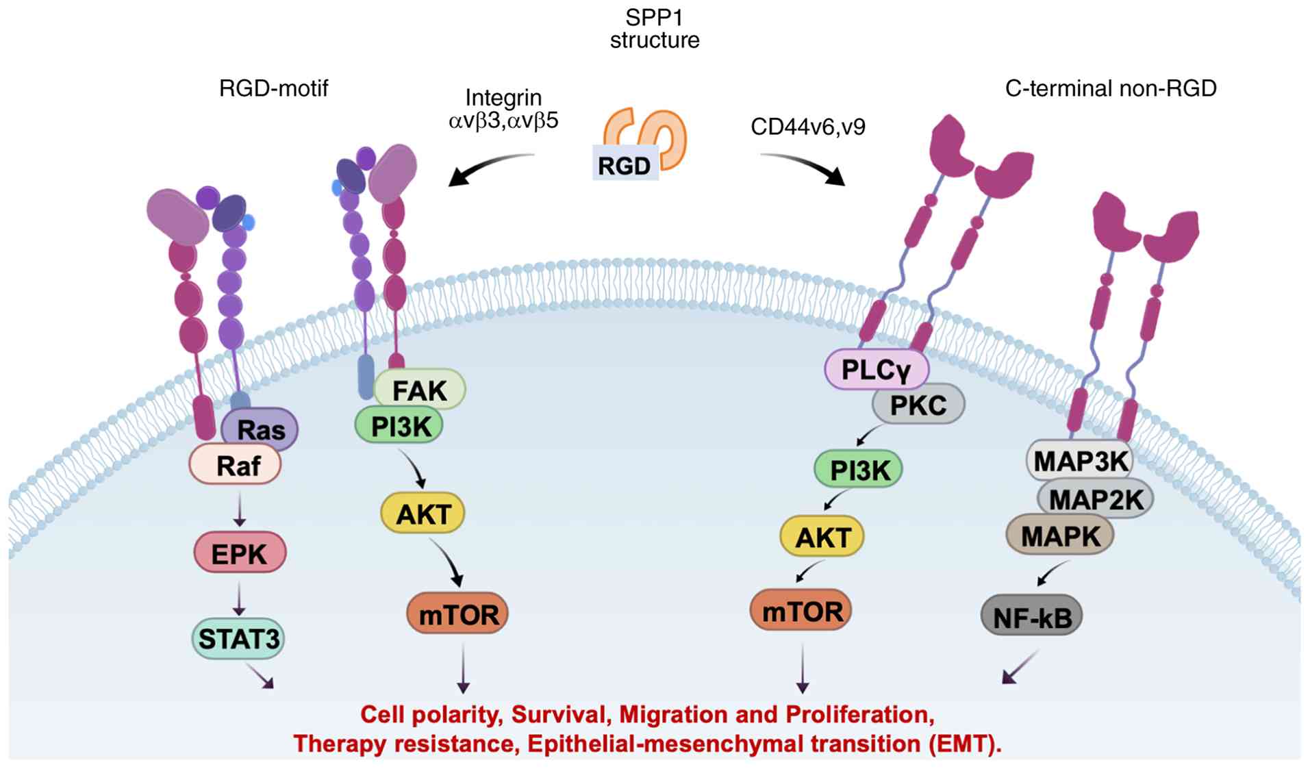 | Figure 1Structural characteristics of SPP1
protein. SPP1, also known as osteopontin, binds to cell surface
receptors integrin αvβ3/αvβ5 via its conserved RGD motif, and to
CD44 (especially variant isoforms CD44v6/v9) via its C-terminal
non-RGD binding domain. Engagement of integrins triggers the
phosphorylation and activation of FAK, which further initiates two
core oncogenic cascades: The PI3K/AKT and MAPK/ERK signaling
pathways. Activated AKT and ERK converge on NF-κB to promote its
nuclear translocation and transcriptional activity. Meanwhile,
SPP1-CD44 binding predominantly activates the JAK/STAT3 signaling
axis, and also cross-activates the PI3K/AKT pathway. These
signaling cascades coordinately drive the core malignant phenotypes
of cancer cells, including uncontrolled proliferation, apoptotic
resistance, EMT, enhanced migration and invasion, cancer stem cell
maintenance, and therapy resistance. SPP1, secreted phosphoprotein
1; RGD, Arg-Gly-Asp; FAK, focal adhesion kinase; EMT,
epithelial-mesenchymal transition. |
PTMs
SPP1 undergoes extensive PTMs that critically
regulate its function:
Phosphorylation: OPN is one of the most highly
phosphorylated proteins in vivo, with numerous serine and
threonine phosphorylation sites, often occurring in acidic
clusters. This modification is essential for high-affinity integrin
binding and downstream signaling activation. Casein kinase II (CK2)
and other kinases phosphorylate specific residues, creating acidic
patches that coordinate calcium ions for integrin recognition
(23,24). This extensive negative charge is
crucial for mediating high-affinity binding to various integrins,
especially αvβ3, through coordination with calcium ions and basic
residues on the integrin surface. Phosphorylation is a prerequisite
for integrin participation in activating downstream pro-survival
and migration signaling pathways such as PI3K/AKT and FAK (25).
Glycosylation: OPN undergoes both N-glycosylation
and O-glycosylation. These carbohydrate modifications significantly
enhance protein stability, increase its resistance to proteolytic
degradation, and affect its secretion (26). Most importantly, glycosylation
directly regulates the interaction between OPN and its other major
receptor, CD44. Specific glycan structures can either promote or
sterically hinder binding to CD44 variants, adding an additional
layer of regulation that determines whether OPN signals primarily
through integrin-mediated or CD44-mediated pathways in a
context-dependent manner (27,28).
Proteolytic cleavage: Thrombin cleavage at
Arg156-Ser157 generates N-terminal (containing RGD) and C-terminal
fragments. The N-terminal fragment retains integrin-binding
activity, while the C-terminal fragment preferentially binds CD44
to promote stemness, chemoresistance and immune evasion (29).
In summary, the multi-domain structure of OPN
combined with its complex PTMs enables interactions with multiple
cell surface receptors, forming the structural basis for its
functional pleiotropy.
Main receptors and downstream signaling
pathways of SPP1
As a multifunctional secreted protein, the
biological functions of SPP1 depend on its specific binding to
various cell surface receptors. Signaling pathways mediated by
different receptors play distinct roles at different stages of
cancer, collectively constituting the complex regulatory network of
SPP1. Currently identified SPP1 receptors mainly include the
integrin receptor family, CD44 and its splice variants, as well as
other receptors such as CD40 and TLR4. Notably, these receptor
interactions are primarily mediated by secreted isoforms (OPN-a and
OPN-b), whereas the intracellular/nuclear OPN-c isoform exerts
distinct, receptor-independent functions in cell cycle regulation
and transcriptional control (13,14,17).
Integrin receptors
Integrins are a class of heterodimeric transmembrane
proteins composed of α and β subunits, which are recognized as the
main cell adhesion transmembrane receptors. They play multiple
roles in transducing biochemical and mechanical signals between
cells and the environment as ECM-cytoskeleton linkers in both
health and disease (30). SPP1
binds to several integrins via its RGD motif, including: αvβ3,
αvβ5, αvβ1, αvβ6 and αIIbβ3 (CD61) (31-33). In cancer, αvβ3 and αvβ5 are the
most extensively studied integrin receptors. They are primarily
responsible for regulating the adhesion, migration and invasion
abilities of cancer cells (34).
After binding to αvβ3, SPP1 can activate the downstream FAK and
PI3K/AKT signaling pathways, promote cytoskeletal rearrangement,
and enhance the motility of cancer cells. In addition, αvβ3 is also
involved in SPP1-induced angiogenesis by upregulating the
expression of pro-angiogenic factors such as VEGF to promote the
formation of tumor neovascularization (35) (Fig.
1).
A landmark study first confirmed that αIIbβ3 (CD61)
is the key receptor through which SPP1 regulates stromal cell fate
in pancreatic cancer (PC) (36).
The study found that SPP1 secreted by epithelial PC cells
specifically binds to the CD61 receptor on the surface of
mesenchymal PC cells (MPCs), activates the downstream BMP2-GREM1
signaling axis, forms an intercellular positive feedback loop, and
maintains the mesenchymal phenotype of MPCs. Knockout of
CD61 completely mimics the phenotype of SPP1
knockout, leading to the transformation of MPCs into
epithelial-like cells and significantly inhibiting the metastatic
ability of PC. This discovery reveals a novel mechanism by which
SPP1 regulates PC cell plasticity.
CD44 receptor and other receptors
CD44 is a transmembrane glycoprotein with multiple
isoforms generated by alternative splicing. CD44v6 and CD44v9 are
particularly associated with cancer stem cells (CSCs). SPP1 binds
CD44 via a region C-terminal to the RGD motif, overlapping with the
SVVYGLR domain (37,38). This interaction activates
JAK2/STAT3, PI3K/AKT and MAPK pathways, promoting stemness,
chemoresistance and immune evasion (39). Unlike integrins, the binding of
SPP1 to CD44 does not depend on the RGD sequence but is achieved
through specific regions at its N-terminal and C-terminal ends.
CD44 exists in multiple alternative splicing variants, among which
CD44v6 plays a specific role in gastrointestinal cancers (40,41), whereas CD44v9 marks CSCs in
cholangiocarcinoma and drives stemness, tumor progression,
metastasis and chemoresistance by regulating Wnt/β-catenin, EMT and
cellular redox status (42)
(Fig. 1).
Existing studies have confirmed that SPP1 secreted
by CAFs binds to the CD44 receptor on the surface of PC cells,
activates the downstream FAK/PI3K/AKT/ERK signaling pathway,
upregulates the expression of stemness transcription factors such
as NANOG, KLF4, BMI-1 and ABCG2, and significantly enhances the
self-renewal ability and tumor sphere formation ability of
pancreatic CSCs (5). CD44v6 is
not only involved in SPP1-induced maintenance of tumor stemness but
also closely related to invasion, metastasis and chemoresistance of
PC (43). In addition, SPP1
binding to CD44 on CAFs activates β-catenin/HIF-1α-related
transcription, leading to increased production of CXCL12 by CAFs
(44). This
SPP1-CD44-β-catenin/HIF-1α-CXCL12 axis establishes a feedback loop
that promotes cancer progression, creates an immunosuppressive
microenvironment, and contributes to both primary and acquired
resistance to immunotherapy (44).
Beyond integrins and CD44, SPP1 interacts with
several additional receptors and co-receptors that modulate its
signaling and functional outcomes. Heparan sulfate proteoglycans
serve as co-receptors that modulate SPP1-mediated signaling, while
calcium-binding proteins interact with SPP1 through its
calcium-binding properties to influence ECM interactions (45).
Notably, CD61 (integrinβ3) has recently been
identified as a critical receptor for SPP1 in PC, where it
maintains mesenchymal cell fate through the SPP1-CD61-BMP2-GREM1
signaling axis (36). These
alternative interactions expand the functional repertoire of SPP1
beyond its canonical integrin and CD44 pathways, highlighting its
context-dependent roles in the TME.
Key downstream signaling pathways of
SPP1
Recent studies have uncovered context-specific roles
of these pathways. In the TME, SPP1+ macrophages secrete
SPP1, which binds to CD44 on CD8+ T cells and activates
PI3K/AKT signaling. This aberrant activation induces a unique
effector-exhaustion intermediate state (T cell stress response),
where T cells retain some effector molecules (for example, IFN-γ)
but upregulate exhaustion markers (PD-1 and TIM-3), leading to
impaired proliferation and cytotoxicity, thereby driving colon
cancer liver metastasis (46). In
prostate cancer, PTEN deletion constitutively activates
PI3K/AKT/mTOR signaling. Activated AKT phosphorylates and degrades
the translation inhibitor PDCD4 (relieving eIF4A inhibition) and
phosphorylates 4E-BP1 (releasing eIF4E). These parallel pathways
increase eIF4F complex availability and cooperate with MNK/eIF4E to
selectively enhance translation of mRNAs with complex
5'-untranslated region structures, promoting SPP1 secretion
(47). Collectively, these
studies reveal a bidirectional immunosuppressive feedback loop:
Tumor cell-intrinsic PI3K/AKT activation drives SPP1 secretion via
translational regulation; secreted SPP1 activates PI3K/AKT in
immune cells (T cells, MDSCs) to induce immunosuppression; and the
resulting immunosuppressive microenvironment sustains tumor cell
survival and pathway activation. This loop, validated in colon and
prostate cancers, may represent a universal immune evasion
mechanism in solid tumors. Targeting this axis, for example,
combining AKT inhibitors with SPP1-neutralizing antibodies, could
overcome immunotherapy resistance. As depicted in Fig. 1, the PI3K/AKT/mTOR pathway is
activated downstream of integrin and CD44 engagement and represents
a central node in SPP1-mediated survival signaling.
Beyond PI3K/AKT, SPP1 activates MAPK/ERK signaling
to promote c-Myc and Cyclin D1 expression, accelerating cell cycle
progression, and upregulates MMP-2/MMP-9 to degrade ECM and enhance
invasion (48). SPP1-integrin
binding also induces FAK phosphorylation, which not only activates
downstream PI3K/AKT and MAPK/ERK but also directly regulates
cytoskeletal rearrangement and cell motility (49). Furthermore, the JAK1/JAK3-STAT1
pathway is an upstream regulator of SPP1 expression in macrophages:
STAT1 activation drives SPP1 transcription, forming pathogenic
SPP1+ macrophage subsets. JAK inhibitors (for example,
tofacitinib) significantly suppress this pathway, downregulate
SPP1, and reduce pathogenic macrophages, showing superior efficacy
to TNF inhibitors, while SPP1 paracrine signaling amplifies JAK
inflammatory signals in a positive feedback loop (50). Additionally, in tumor-associated
macrophages (TAMs), SPP1 promotes angiogenesis through activation
of the ERK/p38 signaling pathway, further highlighting the
context-dependent utilization of p38 MAPK downstream of SPP1
(5). Moreover, In the TME,
SPP1-stimulated macrophages (SPP1+ TAMs) promote
angiogenesis via NF-κB activation (5,51).
This NF-κB activation downstream of SPP1 is a critical node linking
inflammation to tumor progression (Table I).
 | Table IReceptors and downstream signaling
pathways of SPP1 in various cancers. |
Table I
Receptors and downstream signaling
pathways of SPP1 in various cancers.
| First author/s,
year | Cancer type | Receptors | Pathways | Function
mechanism | (Refs.) |
|---|
| Deng et al,
2025 | PDAC | / | PI3K/AKT
pathway | Promotes cell
mobility and invasion metastasis | (59) |
| Nallasamy et
al, 2021 | PDAC | CD44 | / | Promotes stemness
carcinogenesis | (37) |
| Li et al,
2025 | PDAC | CD61 | BMP2-GREM1
pathway | Promotes
metastasis | (36) |
| Huang et al,
2017 | CRC | / | p38 MAPK
pathway | Promotes cell
migration and invasion, and inhibits apoptosis and autophagy | (60) |
| Cheng et al,
2019 | CRC | / |
PI3K-AKT-GSK/3β-β-Catenin pathway | Promotes cell
invasion and stem cell-like properties | (61) |
| Wei et al,
2018 | CRC | / | PI3K/AKT
pathway | Promotes cell
migration and invasion | (62) |
| CRC | CD44 | β-catenin/HIF-1α
pathway | Promotes metastasis
and immunotherapy resistance | (44) |
| Dong et al,
2016 | HCC | / | Vimentin (residues
between 246 and 406) | Promotes EMT
through stablizing vimentin protein | (63) |
| Tong et al,
2024 | HCC | CD44 | PI3K/AKT
pathway | Promotes the
differentiation of HSCs into CAFs | (64) |
| Zhang et al,
2024 | HCC | Integrin | PI3K/AKT/mTOR
signaling pathway | Attracts MDSCs and
Tregs to facilitate immune evasion | (65) |
| Deng et al,
2021 | GC | / | NF-κB pathway | Promoted TIC-like
phenotypes and chemoresistance | (51) |
| Song et al,
2024 | GC | / | PI3K/AKT
pathway | Promotes
chemoresistance | (66) |
| Liu et al,
2020 | LC | / | PI3K/AKT
pathway | Promotes cell
viability and reduces cell apoptosis | (67) |
| Kang et al,
2015 | LC | / | FAK/PI3K/AKT
pathway | Promotes the
lamellipodia formation to allow cell invasion | (68) |
| Fu et al,
2020 | LC | Integrin αVβ3 | Integrin αVβ3/FAK
pathway | Promotes FAK
phosphorylation to enhance cell proliferation | (56) |
| Jia et al,
2016 | LC | / | HIF2α (target) and
AKT1/miR-429/ZEB pathway | Promotes
metastasis | (69) |
| Guo et al,
2024 | BC | / | PI3K/AKT/mTOR
pathway | Promotes metastasis
and invasion, angiogenesis | (70) |
| Zhang et al,
2014 | BC | Integrin αVβ3 | PI3K/AKT/mTOR
pathway | Promotes
metastasis | (71) |
| Pang et al,
2011 | BC | / | p38 MAPK
pathway | Inhibits cell
apoptosis | (72) |
| Mi et al,
2013 | BC | Integrin αVβ3 | Integrin/AP-1 | Promotes
metastasis | (73) |
| Blasberg et
al, 2010; Sun et al, 2013; Sun et al, 2013 | BC | CD44 | MCP-1 and
TGF-β1 | Promotes immune
escape | (57,58) |
| Song et al,
2008 | OC | / | PI3K/AKT
pathway | Promotes
progression and survival | (74) |
In short, SPP1 engagement with integrin or CD44
receptors triggers multiple intracellular signaling cascades
critical for tumor progression. The PI3K/AKT/mTOR pathway,
activated via FAK, supports cell survival, proliferation, metabolic
reprogramming and stemness maintenance (36,52); MAPK/ERK signaling drives
proliferation, differentiation and migration; NF-κB activation
promotes inflammation, survival and invasion through upregulation
of BCL2, MMP9 and CXCL8 (35,53); and CD44-dependent JAK/STAT3
signaling enhances stemness, immune evasion and angiogenesis, while
integrin clustering-induced FAK/Src activation facilitates adhesion
and invasion (54-56) (Table
I). SPP1 splice variants expressed by breast tumors regulate
monocyte activation via MCP-1 and TGF-β1, promoting an
immunosuppressive myeloid phenotype (57,58). This SPP1-MCP-1/TGF-β1 axis
contributes to the establishment of an immunosuppressive TME by
skewing monocyte differentiation toward a pro-tumorigenic state. As
schematically illustrated in Fig.
1, SPP1 functions as a signaling hub that integrates
receptor-specific inputs into coordinated oncogenic outputs.
Collectively, these findings position SPP1 as a
central signaling coordinator rather than a mere ligand: By
engaging distinct receptor complexes, integrins (αvβ3, αvβ5 and
αIIbβ3) and CD44 variants (CD44v6 and CD44v9), SPP1 converges on
and amplifies core oncogenic cascades (PI3K/AKT, MAPK/ERK,
JAK/STAT3 and NF-κB) in a context-dependent manner, thereby
coordinating proliferation, survival, EMT, stemness and immune
evasion as an integrated program (Fig. 1, Table I).
Regulatory roles of SPP1 in tumors
SPP1 in tumor cell proliferation and
survival
SPP1 promotes cancer cell proliferation through
multiple mechanisms. Activation of PI3K/AKT signaling enhances cell
cycle progression by upregulating Cyclin D1 and downregulating p27
(36). SPP1-integrin engagement
activates the Ras-Raf-MEK-ERK cascade, which translocates to the
nucleus and phosphorylates transcription factors including ELK1,
c-FOS and c-JUN. The MAPK/ERK pathway increases MYC expression,
driving metabolic reprogramming and ribosome biogenesis (75). SPP1 also inhibits apoptosis by
upregulating BCL2 and BCL-XL via NF-κB and AKT signaling (35). SPP1 also upregulates IAP family
members that directly bind and inhibit caspases-3, caspases-7 and
caspases-9, further suppressing both intrinsic and extrinsic
apoptotic pathways (8).
SPP1-induced NF-κB increases c-FLIP expression, which inhibits
death receptor-mediated apoptosis by competing with caspase-8
assembly (6) (Table I).
In OC, SPP1-induced STAT3 activation is required for
survival under hypoxic and nutrient-deprived conditions (54). Furthermore, SPP1 promotes
autophagy under stress conditions, providing metabolic
intermediates that support survival. Conversely, excessive SPP1
signaling may suppress autophagic cell death by activating AKT
(76).
Together, these mechanisms establish SPP1 as a
central coordinator of tumor cell proliferation and survival: It
integrates growth factor signaling, stress adaptation (for example,
hypoxia and nutrient deprivation), and autocrine/paracrine cues to
sustain cell cycle progression, apoptotic resistance and metabolic
fitness across diverse cancer types.
SPP1 in EMT and metastasis
SPP1 serves as a potent, multifaceted inducer of
EMT, a cellular reprogramming process essential for metastatic
dissemination, therapy resistance and stemness acquisition. SPP1
regulates EMT at transcriptional, post-transcriptional and
post-translational levels via autocrine and paracrine
mechanisms.
SPP1-induced AKT activation stabilizes Snail by
phosphorylating GSK-3β, preventing Snail degradation. Snail
directly represses CDH1 (E-cadherin) transcription by binding to
E-box elements in its promoter. In CRC, Slug directly binds to the
SPP1 promoter, creating a positive feedback loop that amplifies EMT
(77). SPP1 signaling upregulates
EMT transcription factors including Snail, Slug, Twist and ZEB1/2,
leading to downregulation of E-cadherin and upregulation of
N-cadherin, vimentin and fibronectin (64,78). Additionally, SPP1 activates matrix
metalloproteinases (MMP2 and MMP9) that degrade ECM components,
facilitating invasion (79). SPP1
directly binds to and stabilizes vimentin, preventing its
ubiquitination and degradation, thereby promoting EMT independent
of transcriptional changes (80).
SPP1-induced COL11A1 expression drives EMT and metastasis via
integrin-mediated FAK signaling. SPP1+ TAMs and
FAP+ CAFs synergistically promote EMT and liver
metastasis via β-catenin/HIF-1α activation (81). Several novel molecular mechanisms
underlying SPP1 function in PC have been recently elucidated.
Epithelial PDAC cells secrete SPP1, which activates CD61 on
mesenchymal cells, thereby inducing BMP2 and GREM1 expression and
establishing a bidirectional regulatory loop that maintains
cellular heterogeneity (36).
Additionally, LINC01133 has been identified as a long non-coding
RNA that binds to Arp3 and reduces SPP1 mRNA degradation,
consequently increasing SPP1 expression and promoting EMT in PDAC
(82). Furthermore,
VSIG4+ TAMs epigenetically regulate SPP1 transcription;
VSIG4 deficiency reduces lactate production and histone H3K18
lactylation, leading to decreased STAT3-mediated SPP1 transcription
and disruption of TAM-neutrophil immunosuppressive interactions
(83).
In PC, SPP1 maintains mesenchymal cell fate through
the SPP1-CD61-BMP2-GREM1 axis. Epithelial PDAC cells secrete SPP1,
which binds to CD61 on mesenchymal PDAC cells, activating NF-κB and
inducing BMP2 expression. BMP2 maintains mesenchymal identity,
while GREM1 (BMP antagonist) feeds back to epithelial cells to
sustain SPP1 secretion (36).
In summary, SPP1 functions as a master coordinator
of EMT and metastatic progression rather than a single-pathway
inducer: It orchestrates EMT at transcriptional
(Snail/Slug/Twist/ZEB), post-transcriptional and post-translational
levels, couples EMT to ECM remodeling and stemness acquisition, and
maintains mesenchymal cell fate through reciprocal paracrine axes
(for example, SPP1-CD61-BMP2-GREM1 in PDAC), thereby integrating
invasion, therapy resistance and metastatic competence into a
coordinated phenotypic program.
Epidemiological evidence of SPP1
dysregulation in tumorigenesis
Epidemiological studies of multiple human cancers
consistently show that SPP1 expression is dysregulated in tumor
tissues. This repeated overexpression emphasizes the potential of
SPP1 as a pan-cancer molecule and a promising diagnostic biomarker.
Compared with normal tissues, SPP1 is significantly overexpressed
in multiple solid tumors, including HCC, GC, PDAC, CRC, esophageal
squamous cell carcinoma (ESCC), OC and BC (84-89). Correspondingly, elevated SPP1
protein levels have also been detected in the serum of patients
with these malignancies. Clinically, high SPP1 expression is a
well-recognized predictor of poor prognosis in cancers such as PC,
CRC, clear cell renal cell carcinoma and LC (90-93). In addition, SPP1 overexpression is
closely associated with metastatic progression in BC and melanoma
(94-96). Overall, these epidemiological and
clinical evidence confirm that SPP1 is a molecule that is
consistently upregulated in cancer. Its strong correlation with
aggressive disease and poor patient outcomes makes it a clinically
relevant target for diagnosis, prognostic stratification and
therapeutic development.
Roles of SPP1 in different tumors
SPP1 is widely overexpressed across multiple human
malignancies and exhibits distinct regulatory mechanisms in
different tumor types. This chapter systematically reviews the
research progress of SPP1 in PC, GC, CRC, BC, LC and HCC, with
particular emphasis on its roles in tumor initiation and
progression, metastasis, TME remodeling, immune regulation and
therapeutic targeting.
PC/PDAC
PDAC is one of the most lethal malignancies
worldwide, with a five-year survival rate below 10%. SPP1 plays a
crucial role in maintaining the mesenchymal cell fate in PC. It has
been identified as a key downstream effector of PTHrP, activating
the EMT program (99).
Functionally, SPP1 activates the PI3K/Akt/mTOR signaling pathway to
promote the motility and invasive ability of PC cells (59). Additionally, SPP1 induces aberrant
angiogenesis, which further hinders T cell infiltration and
promotes tumor progression (100).
Long-term co-culture of PC cells with
CAF-conditioned medium enriches CSCs, as evidenced by increased
proportions of CD44+, ALDH+ and
AF+ cells, along with enhanced tumor sphere formation
and self-renewal potential. Knockdown of SPP1 in CAFs or CD44 in
cancer cells completely reverses CAF-induced stemness and drug
resistance, confirming that the SPP1-CD44 axis is a key signaling
pathway connecting the stroma to tumor stemness (37).
SPP1 also regulates PDAC progression by remodeling
the TME through paracrine signaling. A key mechanism for
maintaining epithelial-mesenchymal cell heterogeneity in PDAC
involves GREM1, which is secreted by mesenchymal cells. By
inhibiting BMP signaling in epithelial cells, GREM1 sustains the
continued high expression of SPP1 in epithelial cells, ultimately
forming a transcellular positive feedback regulatory loop:
Epithelial SPP1 → mesenchymal GREM1 → epithelial SPP1. This loop
directly determines the metastatic and invasive potential of tumor
cells (36) (Table II).
| Table IIClinical significance of SPP1
expression in cancers. |
Table II
Clinical significance of SPP1
expression in cancers.
| First author/s,
year | Cancer type | Expression
pattern | Major
functions | Biomarkers | (Refs.) |
|---|
| Li et al,
2025 | PDAC | Significantly
elevated | Maintains
mesenchymal cell fate via SPP1-CD61-BMP2-GREM1 axis; promotes
metastasis | Diagnostic,
prognostic | (36) |
| Song et al,
2024 | GC | Upregulated | Promotes
chemoresistance via POU5F1/SPP1 axis; induces T cell
inactivation | Prognostic | (66) |
| Qi et al,
2022 | CRC | Highly
upregulated | Drives liver
metastasis; promotes CAF-mediated CXCL12 production; induces T cell
stress | Predictive | (81) |
| Feng et al,
2025; Liu et al, 2024 | BC | Overexpressed | Promotes stemness
and chemoresistance via macrophage-tumor SPP1-CD44 crosstalk;
drives lung metastasis | Prognostic | (35,97) |
| Fu et al,
2020; Chen et al, 2023 | LC | Elevated | Drives
CD8+ T exhaustion via SPP1+
malignant-CXCR4+ TAM crosstalk; promotes migration via
COL11A1 | Predictive | (56,75) |
| Xu et al,
2025 | HCC | Elevated | SPP1+
TAMs impair antitumor immunity; SPP1+BCL2A1+
TAMs promote immune suppression | Prognostic | (98) |
CRC
SPP1 is a key driver of CRC progression and
metastasis, with roles spanning malignant transformation,
microenvironment remodeling and immune evasion. In primary tumors,
SPP1 directly enhances the invasive ability of CRC cells through
the PI3K-AKT-GSK3-β-β-catenin signaling pathway and induces stem
cell-like properties, thereby promoting tumor initiation and
recurrence (61). More
importantly, SPP1 plays a central role in constructing the
immunosuppressive microenvironment of CRC. TGF-β signaling induces
the generation of immunosuppressive SPP1+ macrophage
subsets, which form a unique spatial network with CAFs within
primary tumors and liver metastases, jointly establishing a dual
immune barrier (101). On one
hand, these macrophages secrete chemokines that hinder the
recruitment and infiltration of effector T cells (102); on the other hand,
SPP1+ macrophages stimulate CAFs to produce large
amounts of CXCL12, further exacerbating immunosuppression and
driving resistance to immunotherapy (44).
The role of SPP1 is particularly prominent during
CRC liver metastasis. Tumor cell-derived extracellular vesicles
enriched with circ-0034880 induce the formation of pre-metastatic
niches dominated by SPP1 high CD206+ pro-tumor
macrophages in distant organs, creating favorable conditions for
circulating tumor cell colonization. Following the establishment of
liver metastases, the spatial interaction between SPP1+
macrophages and fibroblasts further strengthen local
immunosuppression, leading to poor response to immune checkpoint
blockade (ICB) therapy (103).
Therefore, targeting SPP1 and its mediated intercellular
communication network represents a promising strategy to improve
the therapeutic efficacy of CRC, particularly in metastatic
disease.
OC
SPP1 is a core regulatory molecule in the malignant
progression of OC. Its roles span tumor cell metabolic
reprogramming, microenvironment remodeling, immune exhaustion and
metastasis. SPP1 has been confirmed as a reliable prognostic
biomarker, closely associated with the composition and functional
state of tumor-infiltrating immune cells (104).
Within the TME, the intercellular communication
network mediated by SPP1 is particularly critical. In
ascites-associated macrophages, PLIN2 significantly promotes lipid
accumulation via the HIF1α/SPP1 signaling pathway, thereby
enhancing the pro-tumor phenotype of macrophages (105). Meanwhile, ubiquitous
environmental bisphenol exposure can also accelerate OC progression
and remodel the immunosuppressive microenvironment by activating
the β-catenin/SPP1 axis (106).
More importantly, SPP1+ macrophages serve as core
executors of immunosuppression in OC. They interact with tumor
cells and other stromal cells through the SPP1-CD44 axis to
construct a robust immune barrier (38). Targeting this axis has been shown
to effectively reverse immunosuppression and inhibit tumor
growth.
Notably, SPP1-mediated immune abnormalities are not
limited to macrophages. A recent study identified unique
SPP1+ T cell and NKT cell subsets in OC (107), and their presence is
significantly associated with more severe immune exhaustion and
more aggressive clinical progression. Furthermore, hypoxia-driven
remodeling of SELENOP+ macrophages is also closely
linked to SPP1 signaling, which collectively affects T cell
infiltration and function, ultimately promoting distant metastasis
of OC (108). Therefore,
targeting SPP1 and its upstream and downstream signaling pathways
holds promise as an important breakthrough for breaking immune
tolerance and improving therapeutic outcomes in OC.
HCC and GC
SPP1 is a key inducer of invasion and metastasis in
both HCC and GC. In HCC, the CSC marker SOX9 regulates the
Wnt/β-catenin signaling pathway to influence SPP1 expression
(109), while SPP1 interacts
with and stabilizes vimentin to promote EMT (63). Under hypoxic conditions, specific
enrichment of SPP1+ macrophages in the liver cancer
microenvironment is induced via activation of the HIF-1α pathway.
These macrophages form spatial co-localization with CSCs in hypoxic
regions and directly enhance the stemness, self-renewal and
tumorigenic capacity of CSCs through the SPP1-CD44 axis,
representing a key mechanism underlying the poor prognosis of HCC
(110).
In GC, SPP1 mainly drives malignant progression,
especially tumor metastasis, by mediating intricate crosstalk
between TAMs, stromal cells and immune cells. Multi-omics analyses
have confirmed a specific interaction between GREM1+
fibroblasts and SPP1+ macrophages in GC, with both cell
types jointly constructing a pro-TME (111). This CAF-macrophage crosstalk is
especially critical in GC peritoneal metastasis; it not only
remodels the local stromal architecture but also directly
determines patient response to ICB therapy (112). Patients with high expression of
this interaction signature often show significant ICB
resistance.
In metastatic GC, SPP1+ TAMs can also
engage in direct harmful crosstalk with CD8+ exhausted T
cells, further amplifying immunosuppression (113). These macrophages secrete factors
such as SPP1 to induce CD8+ T cells to express
exhaustion markers including PD-1 and TIM-3, thereby weakening
their antitumor cytotoxic function and ultimately promoting tumor
invasion and metastasis. Therefore, blocking the intercellular
communication network mediated by SPP1 holds promise as an
important strategy to enhance the efficacy of GC immunotherapy and
inhibit peritoneal metastasis (Fig.
2, Table II).
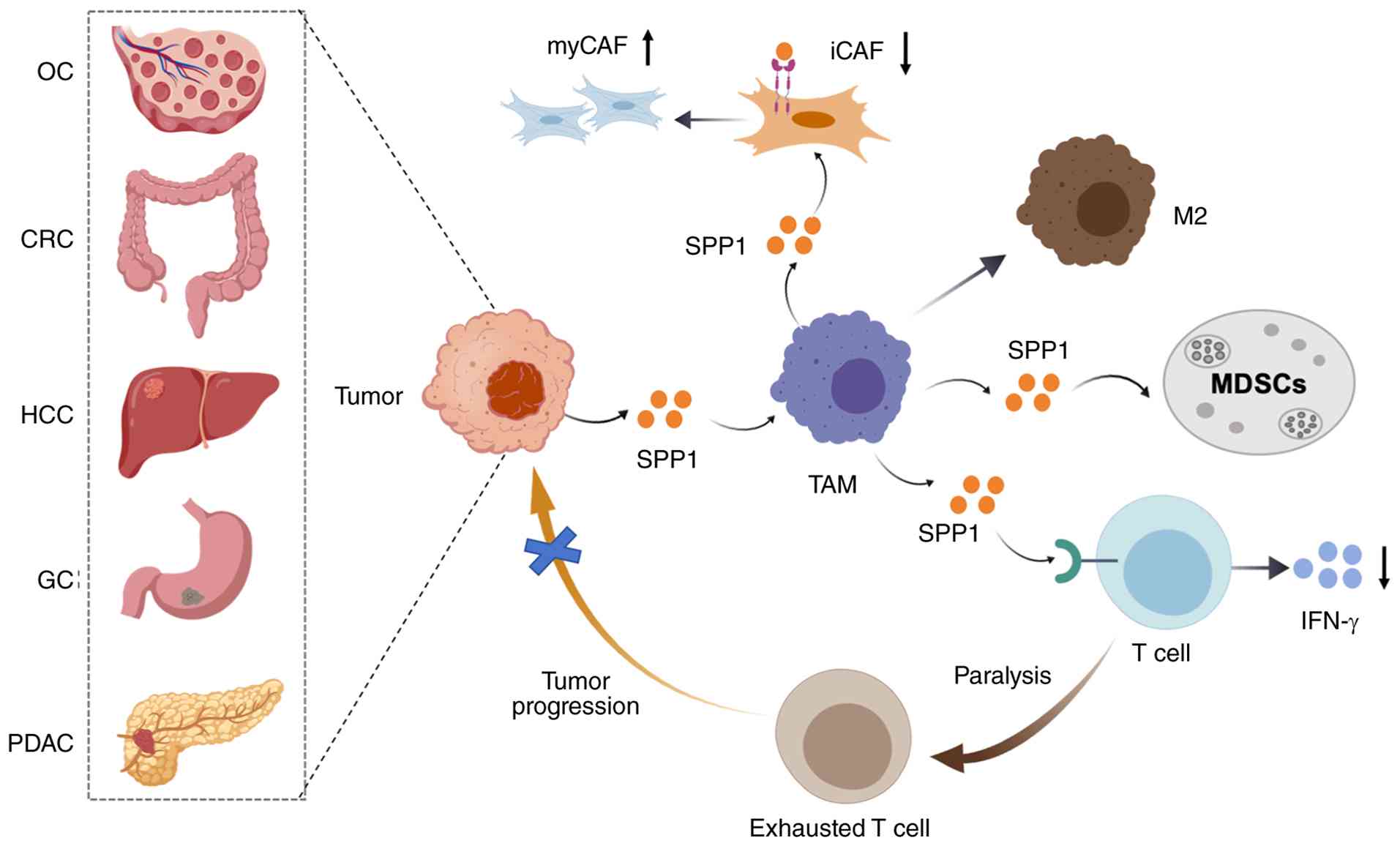 | Figure 2SPP1-mediated crosstalk in the TME.
This schematic depicts that SPP1 secreted by TAMs acts as the key
effector to mediate paracrine and autocrine intercellular crosstalk
via binding to integrins and CD44 splice variants. SPP1 drives
CD8+ T cell exhaustion and MDSC activation to suppress
antitumor immunity; it activates CAFs to promote extracellular
matrix remodeling and T cell exclusion; it sustains tumor cell
stemness, invasion and metastasis via autocrine signaling; and it
amplifies pro-tumor effects by promoting M2-like macrophage
polarization. Ultimately, the SPP1-centered regulatory network
drives immunosuppressive TME formation, tumor invasion and
metastasis, cancer stem cell maintenance, and therapy resistance.
SPP1, secreted phosphoprotein 1; TME, tumor microenvironment;
MDSCs, myeloid-derived suppressor cells; CAFs, cancer-associated
fibroblasts; myCAF, myofibroblastic CAF; iCAF, inflammatory CAF;
TAMs, tumor-associated macrophages; PDAC, pancreatic ductal
adenocarcinoma; CRC, colorectal cancer; HCC, hepatocellular
carcinoma; GC, gastric cancer; OC, ovarian cancer. |
LC
In non-small cell LC (NSCLC), SPP1 drives malignant
progression by promoting cell invasion, metastasis and therapeutic
resistance. Mechanistically, SPP1 induces NSCLC cell invasion via
activating the Rho-associated kinase pathway and inactivating
cofilin (68), and facilitates
metastatic dissemination by regulating epithelial-mesenchymal
plasticity (69). In terms of
drug resistance, SPP1 enhances cisplatin resistance in A549 lung
adenocarcinoma cells through PI3K pathway activation and subsequent
ERCC1 upregulation (67). In
EGFR-mutant NSCLC, aberrant activation of the SPP1/integrin
αVβ3/FAK signaling axis mediates resistance to EGFR tyrosine kinase
inhibitors (TKIs), which is tightly linked to SPP1-induced EMT
(56,75).
Regulation of TME by SPP1
TAMs
SPP1 is a key regulator of TAM recruitment,
polarization and function, contributing to the formation of an
immunosuppressive and pro-TME. It promotes the recruitment and
polarization of macrophages toward a pro-tumor M2-like phenotype
through signaling pathways such as CD44/PI3K/AKT and
CD44/JAK2/STAT3. This leads to increased secretion of factors that
support tumor proliferation, angiogenesis, and ECM remodeling,
including VEGFA and IL-6 (54,55,114,115). Hypoxia further upregulates SPP1
in macrophages and enhances their pro-tumor functions (116). In CRC, SPP1+ TAMs are
enriched at the invasive front via the HIF-1α/STAT3 pathway, and
their density correlates with lymph node metastasis (117). Similarly, single-cell RNA
sequencing (scRNA-seq) of PDAC organoids showed that
SPP1+ macrophages are associated with tumor angiogenesis
and proliferation (118)
(Fig. 2).
SPP1+ TAMs engage in critical crosstalk
with other stromal cells. They interact with CAFs and neutrophils
to promote ECM remodeling, angiogenesis and immune exclusion
(83,119,120). In PDAC, spatially co-localized
SPP1+ APOE+ TAMs and CTHRC1+
GREM1+ myofibroblastic CAFs (myCAFs) synergistically
form an immunosuppressive microenvironment through active ECM
deposition and induction of EMT (121). Analysis of 14 independent CRC
cohorts found a positive correlation between FAP+
fibroblasts and SPP1+ TAMs, which is regulated by
chemerin, TGF-β and interleukin-1. This interaction stimulates the
formation of immune-excluded, desmoplastic structures that limit T
cell infiltration and reduce the efficacy of anti-PD-L1 therapy
(81). In HCC, DAB2+
and SPP1+ TAMs enhance the function of FAP+
CAFs through signals such as TGF-β, PDGF and ADM, participating in
ECM organization and angiogenesis (122) (Fig. 2). Importantly, the CXCL9:SPP1
macrophage polarity index, defined by the expression ratio of CXCL9
and SPP1 (rather than conventional M1/M2 markers), has a strong
prognostic association with survival across multiple cancers and
predicts anti-PD-1 response (123). TAM-derived secreted SPP1
(predominantly OPN-a) mediates these paracrine effects,
highlighting the isoform- and source-specificity of SPP1 function
in the TME.
T cells and immune evasion
SPP1 directly impairs antitumor T cell function.
Macrophage-derived SPP1 interacts with CD44 on T cells, inducing
exhaustion markers (PD-1, TIM-3 and LAG-3) and suppressing
cytotoxic activity via the SPP1-CD44 and CD155-CD226 pathways,
thereby limiting tumor elimination (115,124,125). High SPP1 expression in the TME
is associated with reduced T cell infiltration and proliferation,
while inhibition of SPP1 can restore T cell activation and enhance
immunotherapy efficacy (126).
In metastatic GC, crosstalk between SPP1+ TAMs and
exhausted CD8+ T cells is the key immunosuppressive
mechanism (113). Furthermore,
spatial transcriptomics in primary PDAC tissues identified SPP1 as
a promoter of the immunosuppressive microenvironment at the tumor
front, characterized by accumulation of FOXP3+
regulatory T cells (Tregs) (127). SPP1 can also be directly
expressed by T cells. SPP1+ exhausted T (Tex) cells are
associated with NKT cell activity and poor prognosis in OC and may
be induced by immune checkpoint inhibition (107). Additionally, CAF-derived SPP1
activates CD44 on tumor cells to enhance stemness features in PC
cells, as evidenced by increased CD44+, ALDH+
and AF+ populations and elevated self-renewal and
drug-resistance markers (37).
Thus, SPP1+ TAM-CAF interactions create a desmoplastic
barrier that physically excludes T cells from tumor nests. In CRC,
SPP1+ TAMs and FAP+ fibroblasts form
immune-excluded structures that limit T cell infiltration and
reduce the benefit of anti-PD-L1 therapy (15,81,98). Spatial transcriptomics in PDAC
reveals that SPP1 promotes accumulation of FOXP3+ Tregs
at tumor fronts (64). As
depicted in Fig. 2, this
SPP1-mediated T cell exhaustion represents a key node in the
immunosuppressive network, where SPP1+ TAMs directly
impair cytotoxic T cell function through SPP1-CD44
interactions.
Myeloid-derived suppressor cells
(MDSCs)
SPP1 promotes the formation of an immunosuppressive
microenvironment by recruiting and activating MDSCs. It is a key
factor for recruiting MDSCs into the TME in prostate cancer
(47). In BC models, SPP1
knockdown reduces MDSC infiltration and restores the cytotoxicity
of CD8+ T cells (15).
In monocytic MDSCs, SPP1 expression enhances the activities of
arginase 1 and inducible nitric oxide synthase. This consumes local
L-arginine and produces nitric oxide, which together inhibit T cell
proliferation and function (128). This MDSC recruitment,
illustrated in Fig. 2, further
amplifies the SPP1-centered immunosuppressive circuit.
CAFs
SPP1 reprograms quiescent fibroblasts into
activated, pro-tumor CAFs. SPP1 binds to integrin αvβ5 on
fibroblasts, triggering the FAK-Src-Ras-MAPK cascade, which
phosphorylates the transcription factor AP-1 and upregulates CAF
marker genes including FAP, α-SMA and PDGFRβ (129,130). Activated CAFs then secrete dense
ECM, increase TME stiffness, and activate YAP signaling in tumor
cells, promoting proliferation and invasion (131). Conversely, a positive feedback
loop exists: SPP1-activated CAFs secrete TGF-β and IL-1, which in
turn enhance the pro-inflammatory activity of SPP1+
macrophages. Conversely, in the context of PTEN deletion,
activation of YAP1 in macrophages can directly induce SPP1
secretion through the β1 integrin-PYK2 pathway, thereby stimulating
angiogenesis in glioma (132).
Importantly, CAFs themselves can serve as a source of SPP1. In
prostate cancer, SPP1+ myCAFs promote androgen
deprivation therapy resistance through the SPP1-ERK paracrine
mechanism (133). In NSCLC,
co-culture with SPP1+ CAFs increases the metastatic
potential of tumor cells (134)
(Table II). As shown in Fig. 2, SPP1-induced CAF activation
creates a feedforward loop that reinforces ECM deposition and
immune exclusion, thereby establishing a physical barrier against T
cell infiltration.
ECM
SPP1 fundamentally remodels the ECM to promote an
invasive and immunosuppressive TME. SPP1+ macrophages
and CAFs synergistically remodel the ECM by upregulating
metalloproteinases (for example, MMP9, MMP12 and MMP7) that degrade
structural components, and lysyl oxidase that cross-links collagen,
increasing stiffness (81,116,126).
This remodeling is closely related to hypoxic conditions and the
maintenance of CSCs. Additionally, SPP1 enhances ECM stiffness,
forming a dense desmoplastic barrier that physically excludes
cytotoxic lymphocytes and contributes to immunotherapy resistance.
In PDAC, SPP1+ CAFs are associated with a rigid matrix,
and targeting SPP1 reduces this stiffness (135). In HCC, the interaction between
SPP1+ macrophages and CAFs stimulates the formation of
the tumor immune barrier, limiting immune cell infiltration into
the tumor core (126). This ECM
remodeling, summarized in Fig. 2,
physically excludes T cells and creates a niche conducive to tumor
invasion and therapy resistance.
Collectively, these findings establish SPP1 as a
central orchestrator of the immunosuppressive TME: Secreted
predominantly by SPP1+ TAMs and CAFs, SPP1 coordinates a
multi-component regulatory network, driving M2-like macrophage
polarization, CD8+ T cell exhaustion, MDSC recruitment,
CAF activation and ECM remodeling, through reciprocal paracrine
crosstalk that converges to form an immune-excluded,
therapy-resistant (Fig. 2,
Table III). Its multifaceted
interactions with T cells, MDSCs, CAFs and the ECM make it a
promising therapeutic target for reprogramming the TME to enhance
immune infiltration and improve cancer treatment outcomes.
| Table IIICrosstalk between SPP1 and tumor
microenvironment components. |
Table III
Crosstalk between SPP1 and tumor
microenvironment components.
| First author/s,
year | Cancer type | Cells expressing
SPP1 | Receptors | Interacting
cells | Pathway | Function | (Refs.) |
|---|
| Xu et al,
2024 | Multiple
cancers | SPP1+
TAMs | CD44 | Tumor cells | CD44/PI3K/AKT
pathway | Promotes TAMs
recruitment and polarization | (114) |
| Qu et al,
2025 | CRC | SPP1+
TAMs | Integrin αvβ3 | Tumor cells | Integrin αvβ3 | Promotes metastasis
by activating EMT | (117) |
| Li et al,
2024 | PC | SPP1+
TAMs | / | CTHRC1+
myofibroblastic CAFs | ECM signaling and
EMT | Promotes an
immunosuppressive TME through active ECM deposition and EMT | (121) |
| Qi et al,
2022 | CRC | SPP1+
TAMs |
COL1A1/LAMA1-ITGB1 | FAP+
CAFs | / | Promotes
immune-excluded desmoplastic structure and limits the T cell
infiltration | (81) |
| Long et al,
2024 | HCC | SPP1+
TAMs | / | FAP+
CAFs | TGF-β, PDGF and
ADM | Promotes ECM
organization and angiogenesis | (122) |
| Cheng et al,
2023 | ICC | Tumor cells | CD44 | T cells | CD44 | Inhibits T cells
proliferation | (125) |
| Fan et al,
2024 | HCC | SPP1+
TAMs | CD44 and
integrin | Cancer stem
cells | CCL3, CCL16, and
CCL15 | Limits cell
infiltration into the tumor core | (110) |
| Liu et al,
2023 | HCC | SPP1+
TAMs | / | CAFs | Cytokine receptor
interactions, ECM pathways, TNF pathway, and IL-17 pathway | Promotes TIB
structure formation and immunosuppressive TME | (126) |
| Du et al,
2024 | GC | SPP1+
TAMs | / | CD8+
exhausted T cells | GDF pathway | Promotes
immunosuppressive TME | (113) |
| Liu et al,
2025 | PC | Tumor cells | CD44 | FOXP3-related Treg
cells | / | Promotes EMT and
liver metastasis | (127) |
| Wang et al,
2023 | OC | SPP1+
exhausted T cells | / | NKT cells | Cytokine receptor
interactions, ECM receptor interactions, and local adhesion
pathways | Promotes
immunosuppressive TME | (107) |
| Wang et al,
2023 | PC | SPP1+
CAFs | Integrin | Tumor cells | SPP1-ERK paracrine
pathway | Promotes castration
resistance | (133) |
| Xu et al,
2025 | LC | SPP1+
CAFs | CD44 and
PTGER4 | Tumor cells | SPP1-CD44 and
SPP1-PTGER4 | Promotes
metastasis | (134) |
Targeting SPP1 for tumor therapy
SPP1 as a diagnostic and prognostic
biomarker
Single-cell transcriptome analyses have revealed
that the expression of CXCL9 and SPP1 in TAMs is almost completely
mutually exclusive at the single-cell level, forming two macrophage
subsets with opposing functions: CXCL9+SPP1−
antitumor TAMs and CXCL9−SPP1+ pro-tumor
TAMs. Traditional M1 (CD80 and ITGAX) and M2 (CD163 and MRC1)
markers cannot distinguish these two subsets and are not
significantly associated with patient prognosis, whereas a higher
proportion of SPP1-high TAMs correlates with shorter overall
survival (OS) (123).
SPP1+ TAMs are predominantly distributed
in hypoxic regions of tumors and exhibit strong co-localization
with GLUT1+ cells (a hypoxia marker). In vitro
experiments have confirmed that hypoxia directly induces high SPP1
expression in macrophages via the HIF-1α pathway, which is the key
regulator of their spatial distribution and functional
polarization. Functionally, SPP1+ TAMs are highly
coordinated with the pro-tumor programs of the entire TME: Their
presence is significantly associated with the activation of EMT,
angiogenesis, glycolysis and mTOR signaling pathways. As a secreted
protein, SPP1 promotes tissue remodeling, angiogenesis and tumor
metastasis by acting on integrin and CD44 receptors on the surface
of fibroblasts and endothelial cells. Concurrently,
SPP1+ TAMs inhibit the infiltration of T cells, B cells
and dendritic cells, thereby establishing an immunosuppressive
microenvironment. SPP1 inhibition combined with ICB synergistically
reverses immunosuppression and overcomes immunotherapy resistance
(36,38,81). SPP1 inhibition also sensitizes
tumors to chemotherapy, targeted therapy and stromal-targeting
agents (81). These findings have
been validated in more than 10 human cancer types, including head
and neck cancer, LC, CRC and liver cancer, indicating that TAM
polarity defined by SPP1 is a ubiquitous feature of the TME with
significant clinical prognostic value and therapeutic guiding
implications (136).
In sum, elevated SPP1 expression in tumor tissues
and patient serum correlates with poor prognosis across multiple
cancers. Meta-analyses have confirmed that high SPP1 expression is
associated with reduced OS and disease-free survival (90).
Therapeutic strategies targeting
SPP1
SPP1 is a key driver of tumor progression and
immune suppression, making it an attractive therapeutic target.
Small molecule inhibitors offer a promising approach for targeting
SPP1 in tumor therapy, acting either on SPP1 itself or its
receptors (Table IV).
| Table IVSPP1-targeted therapeutic
strategies. |
Table IV
SPP1-targeted therapeutic
strategies.
| First author/s,
year | Strategy | Therapeutic
drugs | Preclinical
efficacy; Mechanism | Clinical
status | (Refs.) |
|---|
| Li et al,
2025 | Neutralizing
antibodies | MPIIIB10,
ASKP1240 | Inhibits tumor
growth, metastasis; extends survival in PDAC models | Preclinical/Phase
1 | (36) |
| Wang et al,
2024; Wei et al, 2019 | RNA aptamers | SPP1-targeting
aptamers, 4-1BB-SPP1 bispecific | Reduces M2
macrophage infiltration; enhances T cell co-stimulation | Preclinical | (55,137) |
| Hou et al,
2025; Deng et al, 2020 | Small molecule
inhibitors | CANDI460,
nilotinib, NHWD-870, Static | Reduces SPP1
expression; blocks downstream signaling | Preclinical/Early
clinical | (38,138) |
| Krieger et
al, 2021; Xelwa et al, 2024 | Gene silencing
(siRNA/shRNA) | SPP1-specific
siRNAs | Reduces migration,
invasion, chemoresistance | Preclinical | (139,140) |
| Reardon et
al, 2008 | RGD-mimetic
peptides | Cilengitide | Inhibits
integrin-mediated signaling | Phase II
(discontinued) | (141) |
| Liu et al,
2025 | Combination with
chemotherapy | SPP1
knockdown + gemcitabine | Enhanced apoptosis,
reduced invasiveness | Preclinical | (142) |
| Qi et al,
2022; Zhang et al, 2026 | Combination with
immunotherapy | SPP1 inhibition +
anti-PD-1 | Restores T cell
infiltration, enhances immune checkpoint inhibitor efficacy | Preclinical | (81,143) |
Small molecule inhibitors targeting SPP1
or its receptors
Direct inhibition of SPP1
expression
Emerging studies have identified compounds that
directly target SPP1. The lead compound CANDI460, discovered
through phenotypic screening, directly inhibits SPP1 in TAMs,
thereby reducing tumor burden in preclinical models (144). Additionally, molecular dynamics
simulations have identified nilotinib, a TKI, as a high-affinity
binder of SPP1, forming multiple hydrogen bonds and pi-pi
interactions. This binding significantly reduces SPP1 protein
levels in OC models (38). To
avoid disrupting physiological OPN-a, targeting cancer-associated
OPN-c is preferred (13).
Approaches include monoclonal antibodies against unique exon
junctions (for example, OPN-c Δex4 and OPN-b Δex5) and small
molecules selective for isoform-specific conformations (14).
Indirect suppression of SPP1
expression
Small molecules can also downregulate SPP1
indirectly by targeting upstream regulatory pathways. For example,
the bromodomain and extra-terminal inhibitor NHWD-870 downregulates
SPP1 by inhibiting BRD4 and inactivating NF-κB2 transcription,
thereby reducing melanoma growth and metastasis (138). In CRC, the HIF-1α/STAT3 pathway
is critical for activating TAM-derived SPP1 (117); pharmacological inhibition of
STAT3 with Stattic, a small-molecule STAT3 inhibitor, yields robust
antitumor effects across various cancers, and this intervention
effectively represses SPP1 expression (145-147) (Table IV, Fig. 3).
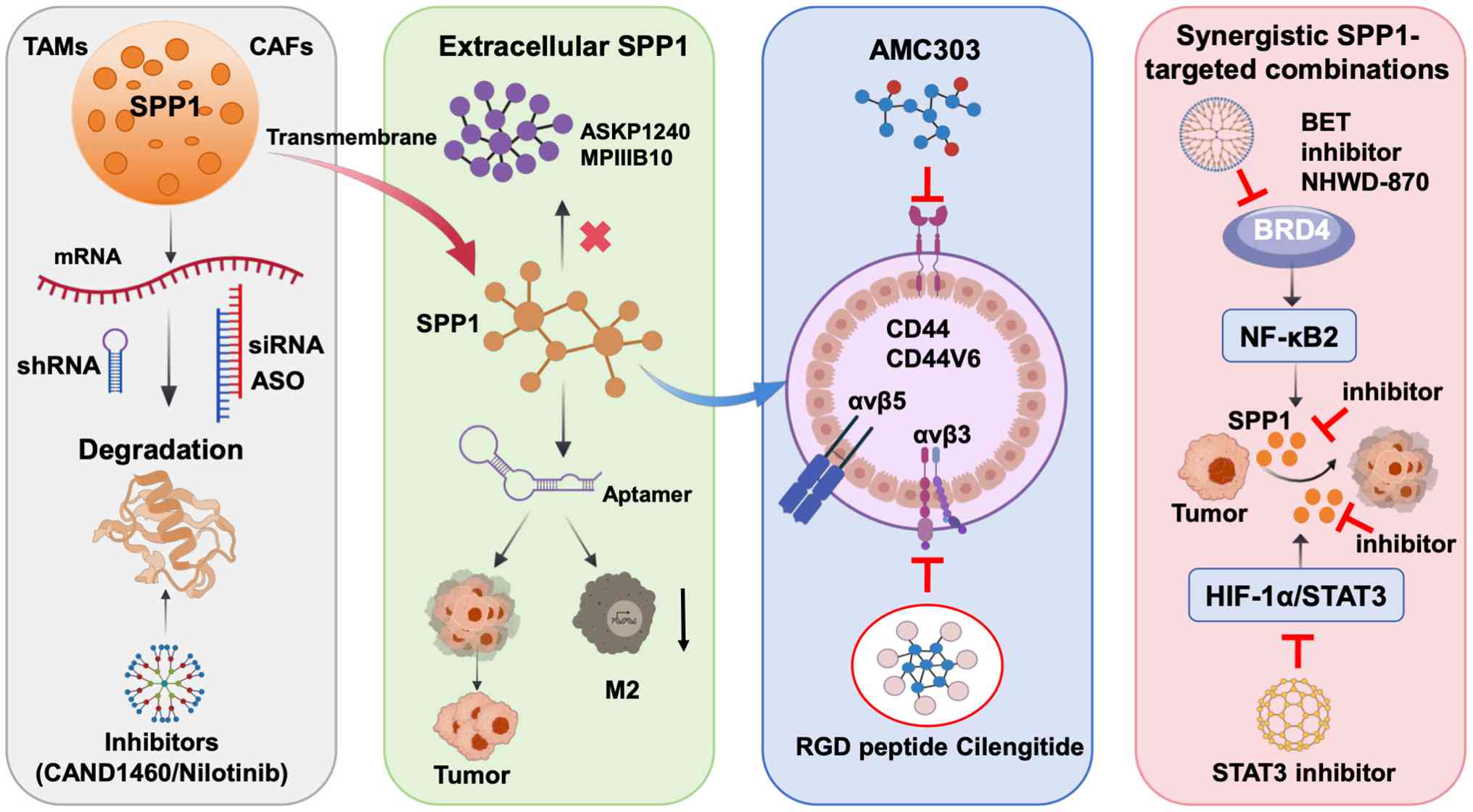 | Figure 3Therapeutic strategies targeting SPP1
in cancer. SPP1/osteopontin is mainly secreted by tumor cells, TAMs
and CAFs in the TME. It drives malignant progression by binding to
integrin receptors and CD44 splice variants, activating downstream
oncogenic signaling cascades, and mediating tumor cell-autonomous
malignant phenotypes and non-autonomous TME immunosuppression. This
diagram systematically summarizes the intervention nodes of
SPP1-targeted therapeutic strategies, including: i) Inhibiting SPP1
transcription and protein synthesis via small molecule inhibitors
and siRNA/shRNA; ii) Neutralizing extracellular secreted SPP1 via
neutralizing antibodies and RNA aptamers; iii) Blocking the
interaction between SPP1 and its membrane receptors via RGD-mimetic
peptides and receptor-targeted inhibitors; iv) Synergistic
combination therapy, including SPP1 targeting combined with
chemotherapy, immune checkpoint inhibitors and pathway inhibitors,
to reverse chemoresistance and immunosuppression, ultimately
exerting comprehensive antitumor effects. SPP1, secreted
phosphoprotein 1; TAMs, tumor-associated macrophages; CAFs,
cancer-associated fibroblasts; TME, tumor microenvironment; siRNA,
small interfering RNA; shRNA, short hairpin RNA. |
Blockade of SPP1-receptor
interactions
Another approach is to disrupt the binding of SPP1
to its receptors, such as integrins and CD44. The cyclic RGD
peptide cilengitide is a characteristic inhibitor of the key SPP1
receptors αvβ3 and αvβ5 integrins. Cilengitide competitively
occupies the ligand binding site, potentially blocking
SPP1-integrin interactions (141,148). In addition, blocking this
interaction can also inhibit downstream FAK signaling, which is
crucial for SPP1-mediated cell migration and invasion. As shown in
Fig. 3, inhibition of the key
SPP1 receptors αvβ3 and αvβ5 integrins will attenuate downstream
FAK signaling, thereby reducing tumor cell migration and invasion,
and potentially synergizing with immunotherapy by reversing the
immunosuppressive TME (Fig.
3).
In summary, although SPP1 expression can be
regulated through various upstream pathways and receptor-blocking
drugs show antitumor effects, the research on highly specific,
direct SPP1-targeting drugs is still in the early stage,
representing a key area for future development.
Nucleic acid aptamers targeting SPP1
Nucleic acid aptamers, especially RNA aptamers, are
short, structured oligonucleotides that can bind to SPP1 with high
affinity and specificity, thereby blocking its pro-cancer
functions. This class of therapeutic agents has the advantages of
low immunogenicity and the ability to be chemically modified to
enhance stability and delivery (149). Their applications are further
expanded, such as aptamer-drug conjugates (ApDCs) and aptamers
constructed entirely from therapeutic nucleoside analogs (150). These modalities enable precise
delivery by discriminating isoforms and cellular sources.
OPN-c-specific ADCs deliver cytotoxic payloads to tumor cells,
improving therapeutic index (144). SPP1 RNA aptamers offer low
immunogenicity and modifiability, with preclinical efficacy in
esophageal cancer (55). The
diverse therapeutic strategies targeting SPP1, ranging from
neutralizing antibodies and RNA aptamers to small molecule
inhibitors and combination immunotherapies, each with distinct
mechanisms of action, are illustrated in Fig. 3.
Preclinical studies have demonstrated the
effectiveness of SPP1-targeting aptamers. In ESCC models, SPP1 RNA
aptamers significantly inhibited tumor growth and reduced M2
macrophage infiltration (55). In
mouse glioblastoma models, systemic administration of 4-1BB
(CD137)-SPP1 bispecific aptamers improved survival by triggering
co-stimulatory immune responses. Notably, SPP1 aptamers alone did
not significantly improve survival, highlighting the benefits of
combinatorial approaches (137).
Beyond oncology, SPP1-specific aptamers have shown protective
effects by regulating immune responses in non-neoplastic diseases
such as chronic liver disease (151).
Overall, these findings highlight the great
potential and clinical application prospects of SPP1-targeting
aptamers in cancer therapy (Table
IV, Fig. 3).
Challenges and future directions of
SPP1-targeted therapy
The clinical translation of SPP1-targeted therapy
faces challenges, mainly due to the complexity of SPP1 interactions
with multiple receptors and its indispensable role in normal
physiology. SPP1 participates in key processes such as cell
adhesion, tissue integrity and immune regulation, affecting the
activation and migration of macrophages and T cells (152). Therefore, systemic inhibition of
SPP1 has the potential to disrupt these functions, possibly leading
to impaired tissue repair and dysregulated immune responses.
Despite these promising therapeutic strategies, the
clinical development of SPP1 pathway inhibitors has encountered
significant setbacks, underscoring the need for a more circumspect
assessment. A cautionary precedent is cilengitide, a cyclic RGD
peptide targeting αvβ3/αvβ5 integrins, key receptors mediating SPP1
signaling. Despite robust preclinical evidence of anti-angiogenic
and anti-invasive activity and encouraging phase I/II results
(141), the phase III CENTRIC
trial (EORTC 26071-22072) in newly diagnosed MGMT-methylated
glioblastoma failed to demonstrate any survival benefit (median OS
26.3 months in both cilengitide and control arms), leading to the
termination of cilengitide development as an anticancer agent
(153). The companion CORE phase
II trial in MGMT-unmethylated glioblastoma yielded paradoxical
results, with standard-dose cilengitide showing modest survival
improvement while dose intensification did not improve outcomes,
further highlighting the unpredictability of integrin-targeted
therapy in clinical settings.
Several interconnected factors likely account for
these failures and pose broader challenges to SPP1-targeted
therapy. First, off-target toxicity remains a fundamental concern:
SPP1/OPN plays indispensable physiological roles in bone
remodeling, wound healing and immune homeostasis. OPN-deficient
mice exhibit impaired wound healing with disorganized collagen
fibrillogenesis and reduced tissue debridement (154), suggesting that systemic SPP1
inhibition could compromise tissue repair and bone integrity in
patients. Second, the failure of integrin-targeted agents such as
cilengitide indicates that broad-spectrum blockade of integrin
signaling is often compromised by functional redundancy among
integrin family members, interference with normal physiological
processes, and paradoxical dose-response relationships. These
limitations suggest that SPP1-targeted therapy should not aim for
global suppression of SPP1 but rather focus on selectively
disrupting the pro-tumorigenic signaling axis between SPP1 and key
integrins, particularly αvβ3. αvβ3 plays a central role in
therapeutic resistance by activating FAK/Src and PI3K/Akt pathways,
thereby promoting cell survival, DNA damage repair and EMT, which
collectively reduce the sensitivity of tumor cells to radiotherapy,
chemotherapy and targeted agents. As one of the major ligands of
αvβ3, elevated SPP1 expression is likely to drive acquired
resistance through sustained activation of these pathways.
Therefore, targeting the SPP1-αvβ3 interaction may offer greater
specificity than direct integrin inhibition, while avoiding broad
interference with the physiological functions of other integrins
(155,156).
To overcome these hurdles, future drug development
must prioritize precision-enhancing strategies, including
tissue-specific targeting of tumor-associated SPP1 isoforms or
TME-activated therapeutics to minimize off-target effects. Advanced
modalities, such as bispecific antibodies co-targeting SPP1 and
immune checkpoints, lipid nanoparticle-delivered siRNA for SPP1
silencing and SPP1-directed ApDCs, represent next-generation
approaches. Rational combination therapy, pairing SPP1 inhibitors
with chemotherapy, TGF-β blockers, or immune checkpoint inhibitors
(ICIs; for example, anti-PD-1), may overcome drug resistance.
Finally, biomarker-driven patient stratification using predictive
biomarkers will ensure that SPP1-targeted therapies are
administered to populations most likely to benefit.
Limitations and perspectives
In the present review, the well-documented
pro-tumorigenic functions of SPP1/OPN were emphasized across a
broad spectrum of malignancies. Nonetheless, it is essential to
acknowledge that the biological effects of OPN are highly
context-dependent, and conflicting outcomes have been reported
under certain conditions. Of particular interest, OPN has been
established as a direct transcriptional target of the tumor
suppressor TP53; activation of p53 in response to DNA damage leads
to upregulated OPN expression. This observation implicates OPN in
TP53-mediated immune surveillance, where it may function to impede
malignant transformation (157).
In cutaneous squamous cell carcinoma models, host-derived OPN
suppressed early tumor progression by eliciting an acute innate
inflammatory response, as wild-type mice exhibited significantly
lower tumor incidence than OPN-null mice (158). A landmark study further revealed
that OPN exerts dual, source-dependent functions: Host-derived OPN
acts as a macrophage chemoattractant to mediate host defense,
whereas tumor-derived OPN suppresses macrophage activity and
enhances the survival of metastatic tumor cells (159). At the isoform level,
overexpression of OPN-c in NSCLC significantly reduced cell
proliferation, colony formation and invasive capacity relative to
controls, a finding that stands in stark contrast to the
pro-tumorigenic activity of the full-length OPN-a isoform (160). These contradictory observations
highlight the complexity of SPP1 biology and caution against a
unidimensional interpretation of its role in cancer.
Context-dependent and contradictory roles
of SPP1 in cancer
Evidence demonstrating that SPP1 can exert opposing
functions depending on the cellular and tissue context was
discussed. For example, the tumor suppressor Chromobox Homolog 7
(CBX7) has been shown to directly inhibit SPP1 gene expression, and
loss of CBX7 correlates with SPP1 overexpression and malignant
progression in various carcinomas, including thyroid, breast and
lung cancers (161,162). Conversely, the CBX7-interacting
protein HMGA1b acts as a positive regulator of SPP1 gene
expression, counteracting CBX7-mediated repression and highlighting
the complex transcriptional regulation of SPP1 (161). This transcriptional duality
reflects a broader theme in SPP1 biology: The same molecule can be
co-opted for tumor promotion or tumor suppression depending on the
regulatory networks active in a given cellular context. Additional
studies have shown that OPN can act as a tumor suppressor in
certain models; for instance, in prostate cancer, OPN has been
reported to induce apoptosis in early-stage lesions, while
promoting metastasis in advanced disease (163). These paradoxical observations
underscore the importance of considering the specific biological
context when interpreting SPP1 functions.
Tumor type and disease stage
SPP1 may play fundamentally different roles in
early vs. late stages of tumorigenesis, and across different tumor
types. While SPP1 is generally pro-tumorigenic in established
cancers, its role in early-stage lesions or specific subtypes may
differ. For instance, in prostate cancer, OPN has been reported to
suppress tumor growth in early stages by inducing apoptosis, yet
promote metastasis in advanced disease (163). Similarly, in BC, OPN may exert
antitumor effects in certain molecular subtypes while promoting
progression in others (5). This
stage- and subtype-specific functionality highlights the need for
careful contextualization when evaluating SPP1's role.
TME composition
Differences in immune infiltration, stromal
activation and ECM composition can markedly alter SPP1's functional
outcomes. In TMEs dominated by M1-like macrophages, SPP1 may
promote antitumor immune responses; conversely, in TMEs enriched
with M2-like macrophages, MDSCs and CAFs, SPP1 reinforces
immunosuppression (164). The
spatial organization of SPP1+ cells relative to immune
cells, whether they co-localize in immune-excluded regions or
infiltrate tumor nests, can also dictate whether SPP1 exerts
immune-stimulatory or immune-suppressive functions (5). In hypoxic, macrophage-dominant
microenvironments, SPP1 primarily mediates immunosuppression; under
other conditions, however, it promotes innate immune activation and
contributes to host defense (158,159). Moreover, the dual roles of
CXCL9/SPP1-polarized TAMs observed in HCC further illustrate that
the microenvironmental context can completely reverse the
functional outcomes of this protein (120).
Cellular source of SPP1
Tumor cell-derived vs. TAM-derived SPP1 may exert
distinct functions, with differential impacts on tumor progression.
Autocrine SPP1 from cancer cells primarily drives cell-intrinsic
malignant phenotypes (proliferation, survival and EMT), while
paracrine SPP1 from TAMs and CAFs shapes the immunosuppressive TME
(163). The distinct PTM
profiles of SPP1 secreted by different cell types further
contribute to functional divergence.
Isoform detection and functional
heterogeneity
Different studies may detect different isoforms or
fail to distinguish between OPN-a, OPN-b and OPN-c, leading to
conflicting conclusions. OPN-a (secreted) and OPN-c
(intracellular/nuclear) have fundamentally distinct functions.
Studies that detect total OPN without isoform discrimination may
inadvertently conflate isoform-specific activities, leading to
contradictory interpretations (13,165). This is particularly relevant for
OPN-c, which is often upregulated in cancer but rarely expressed in
normal tissues, and may mediate tumor-specific functions not
captured by pan-OPN detection. The three major splice variants of
SPP1, OPN-a, OPN-b and OPN-c, display distinct subcellular
localization patterns and exert non-overlapping, and in some cases
even antagonistic, biological functions (13,14,160).
PTMs
Phosphorylation and glycosylation patterns can
markedly alter SPP1's receptor binding and function. For instance,
extensive phosphorylation of serine and threonine residues is
critical for high-affinity integrin binding, while specific
O-glycosylation patterns modulate CD44 interaction (166,167). The degree of phosphorylation and
the specific glycan structures attached to SPP1 can vary across
cancer types, disease stages and cellular sources, thereby
influencing receptor preference (integrin vs. CD44) and downstream
signaling outcomes. Consequently, studies that do not characterize
the PTM status of SPP1 may arrive at different functional
conclusions.
Methodological advances for mechanistic
studies
Single-cell multi-omics and spatial
transcriptomics
Single-cell and spatial multi-omics technologies
have revolutionized cancer research by providing unprecedented
resolution of TME heterogeneity and cell-cell interactions. These
technologies are particularly powerful for studying SPP1-mediated
signaling networks. scRNA-seq enables the identification of
SPP1-producing cell types and the characterization of
SPP1-responsive cell populations, revealing the specific cell
states associated with SPP1 signaling (123). Although SPP1 is known to promote
M2-like TAM polarization, T cell exhaustion, MDSC recruitment and
CAF activation, the precise molecular mechanisms governing these
interactions, particularly the receptor utilization and downstream
signaling in different cell types, remain incompletely defined.
Future studies should integrate single-cell multi-omics, spatial
transcriptomics and advanced imaging approaches to map
SPP1-mediated intercellular crosstalk in the TME with cellular
resolution. Single-cell sequencing is particularly critical for
dissecting the functional heterogeneity of SPP1: It can distinguish
SPP1 originating from tumor cells vs. immune or stromal cell
subsets, resolve isoform expression at single-cell resolution, and
identify spatially defined SPP1+ niches that drive
distinct phenotypic outcomes.
Spatial transcriptomics (for example, 10x Visium
and Xenium) adds a crucial spatial dimension, revealing the spatial
organization of SPP1+ cells relative to SPP1
receptor+ cells and immune populations (97,121). This spatial information is
essential for understanding whether SPP1-mediated signaling occurs
in specific TME niches (for example, tumor-stroma interface,
perivascular regions and immune-excluded zones). The integration of
multiple omics layers, transcriptome, epigenome and spatial
architecture, will be essential for constructing comprehensive
models of SPP1 function in cancer.
Future studies should also incorporate multi-omics
approaches to profile the SPP1 interactome, including both
protein-protein interactions and PTM landscapes. These integrated
approaches will provide a holistic view of SPP1 function, bridging
the gap between molecular mechanisms and clinical outcomes.
Proteomic and PTM analysis
SPP1 undergoes extensive PTMs, including
phosphorylation, glycosylation and proteolytic cleavage, that
critically regulate its function. However, these modifications are
often overlooked in standard studies, leading to incomplete
understanding of SPP1 biology. Advanced proteomic approaches,
including mass spectrometry-based phosphor-proteomics and
glycol-proteomics, are essential for comprehensively characterizing
the PTM landscape of SPP1 in different cancer types, disease stages
and cellular contexts (24,26).
These approaches can reveal modifications that are
specific to cancer vs. normal tissues, modifications that are
altered by drug treatment, and modifications that determine
receptor binding specificity. Furthermore, the development of
modification-specific antibodies would enable the detection of
particular PTM forms of SPP1 in clinical samples, providing
biomarkers of SPP1 activation and receptor preference. The
integration of proteomic data with transcriptomic and epigenomic
data will provide a multi-dimensional understanding of SPP1
function, enabling the development of more precise therapeutic
strategies.
Key challenges and future directions
Isoform-specific functional
heterogeneity
The functional heterogeneity of SPP1 isoforms
(OPN-a, OPN-b and OPN-c) remains a critical knowledge gap. Most
studies treat SPP1 as a single entity despite isoform-specific
localization and functions (13,14,17). Key limitations include the
scarcity of isoform-specific detection reagents and knockout
models, the unknown mechanisms of intracellular OPN-c vs. secreted
isoforms, and the need for isoform-targeted therapies to avoid
off-tumor toxicity. Addressing these gaps through isoform-specific
antibodies, genetic models, and mechanistic studies is essential
for clinical translation and precision oncology: i) Detection
tools: Lack of isoform-specific antibodies (for example, targeting
exon junctions OPN-c Δex4 and OPN-b Δex5) hinders precise
quantification and clinical correlation; ii) Genetic models:
Current knockout strategies ablate all isoforms; isoform-specific
knockout mice are needed to dissect individual contributions to
tumorigenesis and metastasis; iii) Therapeutic targeting: Pan-SPP1
inhibition risks disrupting physiological functions (for example,
OPN-a in bone); isoform-selective inhibitors (antibodies, aptamers
and ADCs) offer greater precision; iv) Intracellular functions:
OPN-c's nuclear/cytoplasmic roles in transcription, cell cycle, and
DNA repair remain poorly understood. v) Clinical translation:
Isoform-specific biomarkers are required for patient stratification
in clinical trials. Future research should prioritize
isoform-specific knockout models and selective detection reagents
to dissect their individual roles and develop isoform-targeted
therapies that minimize off-tumor effects.
Development of isoform-specific
inhibitors
Preclinical studies have validated SPP1 as a
promising therapeutic target, yet clinical translation remains in
its infancy, largely owing to the lack of isoform-specific
inhibitors. Key challenges include the absence of isoform-specific
inhibitors, potential off-target toxicity due to SPP1's
physiological roles (bone remodeling, wound healing and immune
homeostasis), and limited understanding of resistance mechanisms to
SPP1-targeted therapies. Because distinct SPP1 isoforms may exert
opposing functions, broad-spectrum SPP1 inhibition not only fails
to selectively target pathogenic isoforms but may also
inadvertently abrogate isoform-specific protective effects. Future
drug development should therefore prioritize isoform-selective
small molecules, antibodies, or aptamers to achieve specific
targeting of pro-tumorigenic SPP1 isoforms.
Mitigation of off-target toxicity
SPP1 participates in essential physiological
processes, including bone remodeling, wound healing and immune
homeostasis. Systemic inhibition of SPP1 therefore carries inherent
risks of off-tumor toxicity. To address this, future efforts should
focus on developing tissue-specific or TME-activated delivery
systems, such as antibody-drug conjugates targeting SPP1-producing
cells, lipid nanoparticle-encapsulated siRNA with tumor-homing
ligands, or hypoxia-responsive prodrugs that are selectively
activated in the TME. Additionally, designing rational combination
regimens with chemotherapy, targeted therapy and immunotherapy may
allow dose reduction of SPP1-targeted agents while enhancing
therapeutic efficacy.
Biomarker development for patient
stratification
Circulating SPP1 and isoform
profiles
The development of reliable circulating biomarkers
is essential for patient stratification and monitoring of therapy
response. Serum SPP1 levels are already being evaluated as
prognostic biomarkers in numerous cancers; however, current assays
typically measure total SPP1 without isoform discrimination. The
development of isoform-specific ELISA assays for circulating OPN-a,
OPN-b and OPN-c would enable more precise assessment of SPP1
isoform profiles and their correlation with clinical outcomes.
Studies have shown that serum SPP1 levels are elevated in patients
with PDAC, HCC, NSCLC and other cancers, and correlate with tumor
stage and poor prognosis (65,75,121,137). However, the prognostic value of
individual isoforms remains unclear. Isoform-specific circulating
SPP1 detection could identify patients with OPN-c-driven tumors who
may be candidates for OPN-c-targeted therapies. Additionally,
changes in serum SPP1 isoform profiles during treatment could serve
as early markers of response or resistance.
Spatial signatures and TME
architecture
The spatial organization of SPP1-expressing cells
within the TME provides powerful prognostic and predictive
information. Spatial imaging techniques, including multiplex
immunohistochemistry, imaging mass cytometry and spatial
transcriptomics, can reveal the density and distribution of
SPP1+ cells relative to immune cells, CAFs and cancer
cells. Key spatial signatures with clinical relevance include: i)
SPP1+ TAM-CAF co-localization, where high
co-localization of SPP1+ TAMs and FAP+ CAFs
predicts poor response to checkpoint blockade (81); ii) the location of
SPP1+ cells in immune-excluded vs. immune-infiltrated
regions, with SPP1+ cells in immune-excluded regions
contributing to T cell exclusion, while those in immune-infiltrated
regions directly suppress T cell function; and iii) SPP1 density at
invasive fronts, where high SPP1 density at tumor-stroma interfaces
predicts metastatic potential and poor prognosis (117). Together, these spatial
signatures could serve as companion diagnostics for SPP1-targeted
therapies, identifying patients whose tumors display the
immunologically 'cold' and desmoplastic phenotype likely to respond
to SPP1 modulation.
Functional immune ratios
Functional immune ratios derived from
transcriptomic data provide a measure of the balance between immune
activation and immune suppression. The CXCL9:SPP1 macrophage
polarity index is a prime example; a low ratio (more SPP1 than
CXCL9) indicates strong immunosuppression and predicts poorer
response to immunotherapy (123,125). Other functional ratios may also
hold predictive value, including the IFNG: SPP1 ratio (reflecting T
cell activation vs. macrophage-mediated immunosuppression), the
CD8A:SPP1 ratio (indicating cytotoxic T cell infiltration vs.
macrophage-derived immune suppression), and the TGFB1:SPP1 ratio
(reflecting CAF-mediated fibrosis vs. immune suppression). These
ratios could be used to identify patients with 'SPP1-dominant'
immunosuppression who are most likely to benefit from SPP1-targeted
therapies, as well as to monitor therapeutic responses and the
emergence of resistance.
Overall, while SPP1 has emerged as a central
coordinator of cancer progression and a promising therapeutic
target, its context-dependent functional duality, isoform
complexity and multifaceted TME interactions present significant
challenges; overcoming these limitations through mechanistic
dissection, advanced therapeutic modalities and biomarker-guided
clinical trials will be essential to translate SPP1-targeted
strategies into clinical practice and ultimately improve outcomes
for cancer patients.
Conclusions
The present review has systematically synthesized
the multifaceted roles of SPP1 in cancer, from its structural and
molecular basis to its profound impact on tumorigenesis,
metastasis, TME remodeling, immune regulation and therapeutic
targeting (Fig. 4). Accumulating
evidence firmly establishes SPP1 not merely as a bystander but as a
central coordinator of cancer progression.
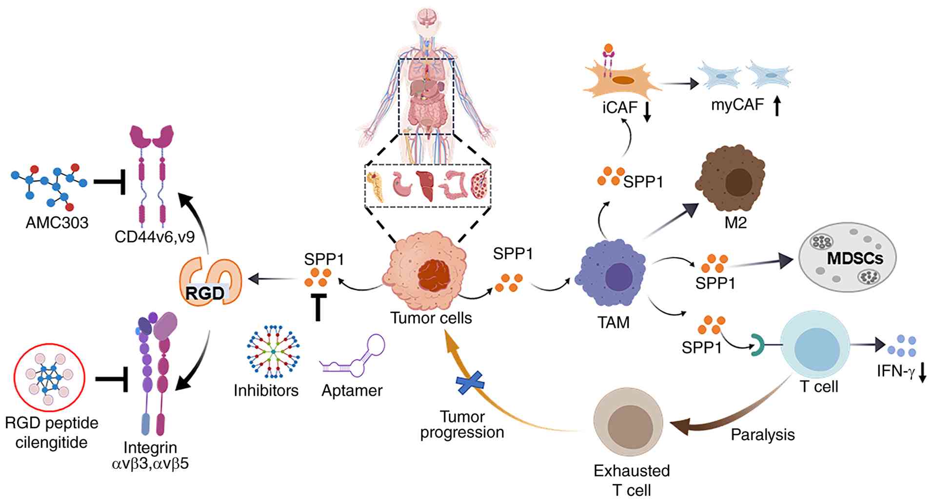 | Figure 4SPP1 as a central oncogenic hub in
multiple malignancies: From receptor signaling to therapeutic
targeting. The present review systematically delineates the core
oncogenic mechanisms of SPP1 (SPP1/osteopontin) in multiple
malignancies, including ovarian cancer, colorectal cancer,
hepatocellular carcinoma, gastric cancer and pancreatic ductal
adenocarcinoma. SPP1 is predominantly produced by tumor cells and
M2-like TAMs within the TME. Through its RGD motif, SPP1 binds
integrin αvβ3/αvβ5, and via its C-terminal non-RGD domain, it
interacts with CD44 and its splice variants v6/v9, thereby
activating PI3K/AKT, MAPK/ERK and JAK/STAT3 signaling cascades.
These pathways drive cell-autonomous malignant phenotypes and
orchestrate an immunosuppressive tumor microenvironment. Based on
these mechanistic insights, the present review further summarizes
multi-node targeted intervention strategies against the SPP1
signaling axis, providing a critical theoretical foundation for
developing novel antitumor therapies. SPP1, secreted phosphoprotein
1; MDSCs, myeloid-derived suppressor cells; CAFs, cancer-associated
fibroblasts; myCAF, myofibroblastic CAF; iCAF, inflammatory CAF;
TAMs, tumor-associated macrophages. |
In summary, SPP1 emerges as a pivotal regulator of
tumor progression by orchestrating oncogene activation, tumor
suppressor inhibition and extensive TME remodeling, including
immune cell polarization, CAF activation, angiogenesis and ECM
stiffening. Its functional effects are mediated through
interactions with key receptors, integrins (αvβ3 and αvβ5) and
CD44, forming a pro-tumor signaling network (Fig. 4). Although SPP1-targeted therapies
(small molecules, aptamers and bispecific agents) exhibit
therapeutic potential, off-target effects remain a major challenge.
Future research should leverage single-cell sequencing to dissect
TME heterogeneity, advance tissue-specific drug delivery systems,
and validate rational combination strategies (chemotherapy, ICIs)
integrated with SPP1-based companion biomarkers. These advances
will eventually translate mechanistic findings into improved
clinical outcomes for patients with SPP1-driven malignancies.
Availability of data and materials
Not applicable.
Authors' contributions
HL conceived and designed the scope of the review.
YX, DH, ST, HY, RZ, BC, PZ and AS performed the literature search
and drafted the initial manuscript. YX, DH, and HL revised
manuscript. HL provided funding support. All authors read and
approved the final version of the manuscript and agreed to be
accountable for all aspects of the work. Data authentication is not
applicable.
Ethics approval and consent to
participate
Not applicable.
Patient consent for publication
Not applicable.
Competing interests
The authors declare that they have no competing
interests.
Abbreviations:
|
SPP1
|
secreted phosphoprotein 1
|
|
OPN
|
osteopontin
|
|
TME
|
tumor microenvironment
|
|
TAMs
|
tumor-associated macrophages
|
|
CAFs
|
cancer-associated fibroblasts
|
|
MDSCs
|
myeloid-derived suppressor cells
|
|
Tregs
|
regulatory T cells
|
|
RGD
|
Arg-Gly-Asp
|
|
PDAC
|
pancreatic ductal adenocarcinoma
|
|
CRC
|
colorectal cancer
|
|
HCC
|
hepatocellular carcinoma
|
|
GC
|
gastric cancer
|
|
BC
|
breast cancer
|
|
LC
|
lung cancer
|
|
OC
|
ovarian cancer
|
|
ECM
|
extracellular matrix
|
|
PTMs
|
post-translational modifications
|
|
CDH1
|
E-cadherin
|
|
CK2
|
Casein kinase II
|
|
MPCs
|
mesenchymal pancreatic cancer
cells
|
|
CSCs
|
cancer stem cells
|
|
ApDCs
|
aptamer-drug conjugates
|
|
NSCLC
|
non-small cell lung cancer
|
|
ICB
|
immune checkpoint blockade
|
|
scRNA-seq
|
single-cell RNA sequencing
|
Acknowledgements
Not applicable.
Funding
The present study was supported by The National Natural Science
Foundation of China (grant nos. 82270193 and 82470156), the
Zhejiang Provincial Natural Science Foundation of China (grant no.
YXD24H0801), the Strategic Priority Research Program of the Chinese
Academy of Grant (grant no. XDB1020000) and the Joint Fund Project
of Tongde Hospital of Zhejiang Province and Hangzhou Institute of
Medicine, Chinese Academy of Sciences (grant no. 2025LHJJ08).
References
|
1
|
Hanahan D: Hallmarks of cancer: New
dimensions. Cancer Discov. 12:31–46. 2022. View Article : Google Scholar : PubMed/NCBI
|
|
2
|
Sung H, Ferlay J, Siegel RL, Laversanne M,
Soerjomataram I, Jemal A and Bray F: Global cancer statistics 2020:
GLOBOCAN estimates of incidence and mortality worldwide for 36
cancers in 185 countries. CA Cancer J Clin. 71:209–249.
2021.PubMed/NCBI
|
|
3
|
Oldberg A, Franzén A and Heinegård D:
Cloning and sequence analysis of rat bone sialoprotein
(osteopontin) cDNA reveals an Arg-Gly-Asp cell-binding sequence.
Proc Natl Acad Sci USA. 83:8819–8823. 1986. View Article : Google Scholar : PubMed/NCBI
|
|
4
|
Wang KX and Denhardt DT: Osteopontin: Role
in immune regulation and stress responses. Cytokine Growth Factor
Rev. 19:333–345. 2008. View Article : Google Scholar : PubMed/NCBI
|
|
5
|
Tan Y, Zhao L, Yang YG and Liu W: The role
of osteopontin in tumor progression through tumor-associated
macrophages. Front Oncol. 12:9532832022. View Article : Google Scholar : PubMed/NCBI
|
|
6
|
Xu W, Bi Z, Lu L, Feng F, Chen L and Zhang
C: Role of osteopontin in cancer: From pathogenesis to therapeutics
(Review). Oncol Rep. 54:1362025. View Article : Google Scholar : PubMed/NCBI
|
|
7
|
Denhardt DT and Guo X: Osteopontin: A
protein with diverse functions. FASEB J. 7:1475–1482. 1993.
View Article : Google Scholar : PubMed/NCBI
|
|
8
|
Rangaswami H, Bulbule A and Kundu GC:
Osteopontin: Role in cell signaling and cancer progression. Trends
Cell Biol. 16:79–87. 2006. View Article : Google Scholar : PubMed/NCBI
|
|
9
|
Wai PY and Kuo PC: Osteopontin: Regulation
in tumor metastasis. Cancer Metastasis Rev. 27:103–118. 2008.
View Article : Google Scholar
|
|
10
|
Weber GF: The metastasis gene osteopontin:
A candidate target for cancer therapy. Biochim Biophys Acta.
1552:61–85. 2001.
|
|
11
|
Kazanecki CC, Uzwiak DJ and Denhardt DT:
Control of osteopontin signaling and function by post-translational
phosphorylation and protein folding. J Cell Biochem. 102:912–924.
2007. View Article : Google Scholar : PubMed/NCBI
|
|
12
|
Kazanecki CC, Kowalski AJ, Ding T,
Rittling SR and Denhardt DT: Characterization of anti-osteopontin
monoclonal antibodies: Binding sensitivity to post-translational
modifications. J Cell Biochem. 102:925–935. 2007. View Article : Google Scholar : PubMed/NCBI
|
|
13
|
Gimba ER and Tilli TM: Human osteopontin
splicing isoforms: Known roles, potential clinical applications and
activated signaling pathways. Cancer Lett. 331:11–17. 2013.
View Article : Google Scholar
|
|
14
|
He B, Mirza M and Weber GF: An osteopontin
splice variant induces anchorage independence in human breast
cancer cells. Oncogene. 25:2192–2202. 2006. View Article : Google Scholar
|
|
15
|
Liu C, Xia S, Wang B, Li J, Wang X, Ren Y
and Zhou X: Osteopontin promotes tumor microenvironment remodeling
and therapy resistance. Cancer Lett. 617:2176182025. View Article : Google Scholar : PubMed/NCBI
|
|
16
|
Yan Z, Hu X, Tang B and Deng F: Role of
osteopontin in cancer development and treatment. Heliyon.
9:e210552023. View Article : Google Scholar : PubMed/NCBI
|
|
17
|
Briones-Orta MA, Avendaño-Vázquez SE,
Aparicio-Bautista DI, Coombes JD, Weber GF and Syn WK: Osteopontin
splice variants and polymorphisms in cancer progression and
prognosis. Biochim Biophys Acta Rev Cancer. 1868:93–108.A. 2017.
View Article : Google Scholar : PubMed/NCBI
|
|
18
|
Lin EYH, Xi W, Aggarwal N and Shinohara
ML: Osteopontin (OPN)/SPP1: From its biochemistry to biological
functions in the innate immune system and the central nervous
system (CNS). Int Immunol. 35:171–180. 2023. View Article : Google Scholar :
|
|
19
|
Schytte GN, Christensen B, Bregenov I,
Kjøge K, Scavenius C, Petersen SV, Enghild JJ and Sørensen ES:
FAM20C phosphorylation of the RGDSVVYGLR motif in osteopontin
inhibits interaction with the αvβ3 integrin. J Cell Biochem.
121:4809–4818. 2020. View Article : Google Scholar : PubMed/NCBI
|
|
20
|
Yokosaki Y, Tanaka K, Higashikawa F,
Yamashita K and Eboshida A: Distinct structural requirements for
binding of the integrins alphavbeta6, alphavbeta3, alphavbeta5,
alpha5beta1 and alpha9beta1 to osteopontin. Matrix Biol.
24:418–427. 2005. View Article : Google Scholar : PubMed/NCBI
|
|
21
|
Green PM, Ludbrook SB, Miller DD, Horgan
CM and Barry ST: Structural elements of the osteopontin SVVYGLR
motif important for the interaction with alpha(4) integrins. FEBS
Lett. 503:75–79. 2001. View Article : Google Scholar : PubMed/NCBI
|
|
22
|
Shao Z, Morser J and Leung LLK: Thrombin
cleavage of osteopontin disrupts a pro-chemotactic sequence for
dendritic cells, which is compensated by the release of its
pro-chemotactic C-terminal fragment. J Biol Chem. 289:27146–27158.
2014. View Article : Google Scholar : PubMed/NCBI
|
|
23
|
Anborgh PH, Mutrie JC, Tuck AB and
Chambers AF: Pre- and post-translational regulation of osteopontin
in cancer. J Cell Commun Signal. 5:111–122. 2011. View Article : Google Scholar : PubMed/NCBI
|
|
24
|
Christensen B, Kläning E, Nielsen MS,
Andersen MH and Sørensen ES: C-terminal modification of osteopontin
inhibits interaction with the αVβ3-integrin. J Biol Chem.
287:3788–3797. 2012. View Article : Google Scholar
|
|
25
|
Al-Shami R, Sorensen ES, Ek-Rylander B,
Andersson G, Carson DD and Farach-Carson MC: Phosphorylated
osteopontin promotes migration of human choriocarcinoma cells via a
p70 S6 kinase-dependent pathway. J Cell Biochem. 94:1218–1233.
2005. View Article : Google Scholar : PubMed/NCBI
|
|
26
|
Shirasaki T, Honda M, Yamashita T, Nio K,
Shimakami T, Shimizu R, Nakasyo S, Murai K, Shirasaki N, Okada H,
et al: The osteopontin-CD44 axis in hepatic cancer stem cells
regulates IFN signaling and HCV replication. Sci Rep. 8:131432018.
View Article : Google Scholar : PubMed/NCBI
|
|
27
|
Fnu G, Agrawal P, Kundu GC and Weber GF:
Structural constraint of osteopontin facilitates efficient binding
to CD44. Biomolecules. 11:8132021. View Article : Google Scholar : PubMed/NCBI
|
|
28
|
Kariya Y, Kanno M, Matsumoto-Morita K,
Konno M, Yamaguchi Y and Hashimoto Y: Osteopontin O-glycosylation
contributes to its phosphorylation and cell-adhesion properties.
Biochem J. 463:93–102. 2014. View Article : Google Scholar : PubMed/NCBI
|
|
29
|
Leung LL, Myles T and Morser J: Thrombin
cleavage of osteopontin and the host anti-tumor immune response.
Cancers (Basel). 15:34802023. View Article : Google Scholar : PubMed/NCBI
|
|
30
|
Pang X, He X, Qiu Z, Zhang H, Xie R, Liu
Z, Gu Y, Zhao N, Xiang Q and Cui Y: Targeting integrin pathways:
Mechanisms and advances in therapy. Signal Transduct Target Ther.
8:12023. View Article : Google Scholar : PubMed/NCBI
|
|
31
|
Wang JB, Zhang Z, Li JN, Yang T, Du S, Cao
RJ and Cui SS: SPP1 promotes Schwann cell proliferation and
survival through PKCα by binding with CD44 and αvβ3 after
peripheral nerve injury. Cell Biosci. 10:982020. View Article : Google Scholar
|
|
32
|
Messex JK, Byrd CJ, Thomas MU and Liou GY:
Macrophages cytokine Spp1 increases growth of prostate
intraepithelial neoplasia to promote prostate tumor progression.
Int J Mol Sci. 23:42472022. View Article : Google Scholar : PubMed/NCBI
|
|
33
|
Pang X, Gong K, Zhang X, Wu S, Cui Y and
Qian BZ: Osteopontin as a multifaceted driver of bone metastasis
and drug resistance. Pharmacol Res. 144:235–244. 2019. View Article : Google Scholar : PubMed/NCBI
|
|
34
|
Chen Y, Yang S, Tavormina J, Tampe D,
Zeisberg M, Wang H, Mahadevan KK, Wu CJ, Sugimoto H, Chang CC, et
al: Oncogenic collagen I homotrimers from cancer cells bind to α3β1
integrin and impact tumor microbiome and immunity to promote
pancreatic cancer. Cancer Cell. 40:818–834.e9. 2022. View Article : Google Scholar
|
|
35
|
Feng Y, Shao X, Xu ZF, Liu P, Wu H, Cheng
Y, Qi M, Meng H, Zhang B, Jian FZ, et al: SPP1 regulates tumor
progression through modulation of signaling pathways and the tumor
microenvironment. Discov Oncol. 17:1152025. View Article : Google Scholar : PubMed/NCBI
|
|
36
|
Li H, Lan L, Chen H, Zaw Thin M, Ps H,
Nelson JK, Evans IM, Ruiz EJ, Cheng R, Tran L, et al: SPP1 is
required for maintaining mesenchymal cell fate in pancreatic
cancer. Nature. 648:203–209. 2025. View Article : Google Scholar : PubMed/NCBI
|
|
37
|
Nallasamy P, Nimmakayala RK, Karmakar S,
Leon F, Seshacharyulu P, Lakshmanan I, Rachagani S, Mallya K, Zhang
C, Ly QP, et al: Pancreatic tumor microenvironment factor promotes
cancer stemness via SPP1-CD44 axis. Gastroenterology.
161:1998–2013.e7. 2021. View Article : Google Scholar : PubMed/NCBI
|
|
38
|
Hou L, Jiang M, Li Y, Cheng J, Liu F, Han
X, Guo J, Feng L, Li Z, Yi J, et al: Targeting SPP1+
macrophages via the SPP1-CD44 axis reveals a key mechanism of
immune suppression and tumor progression in ovarian cancer. Int
Immunopharmacol. 160:1149062025. View Article : Google Scholar
|
|
39
|
Poggio P, Branchetti E, Grau JB, Lai EK,
Gorman RC, Gorman JH III, Sacks MS, Bavaria JE and Ferrari G:
Osteopontin-CD44v6 interaction mediates calcium deposition via
phospho-Akt in valve interstitial cells from patients with
noncalcified aortic valve sclerosis. Arterioscler Thromb Vasc Biol.
34:2086–2094. 2014. View Article : Google Scholar : PubMed/NCBI
|
|
40
|
Nian H, Bai Y, Wang HY, Yu H, Zhang ZL,
Shi RH, Zhang S, Wu YB, Zhou DH and Du QC: Targeting the
osteopontin-regulated PI3K/AKT signaling pathway: A molecular
approach to overcome drug resistance and metastasis in
gastrointestinal tumors. World J Gastrointest Oncol. 17:1099232025.
View Article : Google Scholar : PubMed/NCBI
|
|
41
|
Lin YH and Yang-Yen HF: The
osteopontin-CD44 survival signal involves activation of the
phosphatidylinositol 3-kinase/Akt signaling pathway. J Biol Chem.
276:46024–46030. 2001. View Article : Google Scholar : PubMed/NCBI
|
|
42
|
Suwannakul N, Ma N, Midorikawa K, Oikawa
S, Kobayashi H, He F, Kawanishi S and Murata M: CD44v9 induces stem
cell-like phenotypes in human cholangiocarcinoma. Front Cell Dev
Biol. 8:4172020. View Article : Google Scholar : PubMed/NCBI
|
|
43
|
Li Z, Chen K, Jiang P, Zhang X, Li X and
Li Z: CD44v/CD44s expression patterns are associated with the
survival of pancreatic carcinoma patients. Diagn Pathol. 9:792014.
View Article : Google Scholar : PubMed/NCBI
|
|
44
|
Liu S, Zhang Z, Wang Z, Liu C, Liang G, Xu
T, Li Z, Duan X, Xu G, Feng X, et al: SPP1 drives colorectal cancer
liver metastasis and immunotherapy resistance by stimulating CXCL12
production in cancer-associated fibroblasts. Cancer Res. 86:58–79.
2026. View Article : Google Scholar :
|
|
45
|
Chaikiawkeaw D, Khorattanakulchai N,
Nammultriputtar K, Rattanapisit K, Everts V, Kubera A, Phoolcharoen
W and Pavasant P: Osteopontin induces osteogenic differentiation by
human periodontal ligament cells via calcium binding domain-ALK-1
interaction. J Periodontol. 93:e13–e23. 2022. View Article : Google Scholar
|
|
46
|
Ding D, Li W, Ren J, Li Y, Jiang G, Liang
R, Huang H, You J, Wang Y and Wei B: SPP1high
macrophage-induced T-cell stress promotes colon cancer liver
metastasis through SPP1/CD44/PI3K/AKT signaling. J Immunother
Cancer. 13:e0123302025. View Article : Google Scholar
|
|
47
|
Brina D, Ponzoni A, Troiani M, Calì B,
Pasquini E, Attanasio G, Mosole S, Mirenda M, D'Ambrosio M, Colucci
M, et al: The Akt/mTOR and MNK/eIF4E pathways rewire the prostate
cancer translatome to secrete HGF, SPP1 and BGN and recruit
suppressive myeloid cells. Nat Cancer. 4:1102–1121. 2023.
View Article : Google Scholar : PubMed/NCBI
|
|
48
|
Nagel S and Meyer C: Aberrant expression
and oncogenic activity of SPP1 in hodgkin lymphoma. Biomedicines.
13:7352025. View Article : Google Scholar : PubMed/NCBI
|
|
49
|
Zhuang F, Liu ZT, Zhou G, Liang F, Wang
YH, Chen L, Zhang WF, Shen LH, Lu YQ, Huo HH, et al: Integrin
β3-mediated platelet extracellular vesicle adhesion facilitates
vascular smooth muscle cell dysfunction in postinjury intimal
hyperplasia. Int J Biol Sci. 21:2380–2395. 2025. View Article : Google Scholar
|
|
50
|
Xia X, He C, Xue Z, Wang Y, Qin Y, Ren Z,
Huang Y, Luo H, Chen HN, Zhang WH, et al: Single cell immunoprofile
of synovial fluid in rheumatoid arthritis with TNF/JAK inhibitor
treatment. Nat Commun. 16:21522025. View Article : Google Scholar : PubMed/NCBI
|
|
51
|
Deng S, Li L, Xu S, Wang X and Han T:
Promotion of gastric tumor initiating cells in a 3D collagen gel
culture model via YBX1/SPP1/NF-κB signaling. Cancer Cell Int.
21:5992021. View Article : Google Scholar
|
|
52
|
Ji X, Liu Y, Mei F, Li X, Zhang M, Yao B,
Wu R, You J and Pei F: SPP1 overexpression is associated with poor
outcomes in ALK fusion lung cancer patients without receiving
targeted therapy. Sci Rep. 11:140312021. View Article : Google Scholar : PubMed/NCBI
|
|
53
|
Cui J, Wang J, Lin C, Liu J and Zuo W:
Osteopontin mediates cetuximab resistance via the MAPK pathway in
NSCLC cells. Onco Targets Ther. 12:10177–10185. 2019. View Article : Google Scholar
|
|
54
|
Huang Z, Li Y, Liu Q, Chen X, Lin W, Wu W,
Chen Z, Chen X, Pan Y and Qiu S: SPP1-mediated M2 macrophage
polarization shapes the tumor microenvironment and enhances
prognosis and immunotherapy guidance in nasopharyngeal carcinoma.
Int Immunopharmacol. 147:1139442025. View Article : Google Scholar : PubMed/NCBI
|
|
55
|
Wang C, Li Y, Wang L, Han Y, Gao X, Li T,
Liu M, Dai L and Du R: SPP1 represents a therapeutic target that
promotes the progression of oesophageal squamous cell carcinoma by
driving M2 macrophage infiltration. Br J Cancer. 130:1770–1782.
2024. View Article : Google Scholar : PubMed/NCBI
|
|
56
|
Fu Y, Zhang Y, Lei Z, Liu T, Cai T, Wang
A, Du W, Zeng Y, Zhu J, Liu Z and Huang JA: Abnormally activated
OPN/integrin αVβ3/FAK signalling is responsible for EGFR-TKI
resistance in EGFR mutant non-small-cell lung cancer. J Hematol
Oncol. 13:1692020. View Article : Google Scholar
|
|
57
|
Blasberg JD, Goparaju CM, Pass HI and
Donington JS: Lung cancer osteopontin isoforms exhibit angiogenic
functional heterogeneity. J Thorac Cardiovasc Surg. 139:1587–1593.
2010. View Article : Google Scholar
|
|
58
|
Sun J, Feng A, Chen S, Zhang Y, Xie Q,
Yang M, Shao Q, Liu J, Yang Q, Kong B and Qu X: Osteopontin splice
variants expressed by breast tumors regulate monocyte activation
via MCP-1 and TGF-β1. Cell Mol Immunol. 10:176–182. 2013.
View Article : Google Scholar : PubMed/NCBI
|
|
59
|
Deng L, Ren J, Liu D, Li H, Yang G, Wang
K, Song Y and Su H: Ran drives pancreatic cancer metastasis by
activating the osteopontin-PI3K/AKT-androgen receptor signaling
cascade. Toxicol Appl Pharmacol. 499:1173282025. View Article : Google Scholar : PubMed/NCBI
|
|
60
|
Huang RH, Quan YJ, Chen JH, Wang TF, Xu M,
Ye M, Yuan H, Zhang CJ, Liu XJ and Min ZJ: Osteopontin promotes
cell migration and invasion, and inhibits apoptosis and autophagy
in colorectal cancer by activating the p38 MAPK signaling pathway.
Cell Physiol Biochem. 41:1851–1864. 2017. View Article : Google Scholar : PubMed/NCBI
|
|
61
|
Cheng Y, Wen G, Sun Y, Shen Y, Zeng Y, Du
M, Zhu G, Wang G and Meng X: Osteopontin promotes colorectal cancer
cell invasion and the stem cell-like properties through the
PI3K-AKT-GSK/3β-β/catenin pathway. Med Sci Monit. 25:3014–3025.
2019. View Article : Google Scholar : PubMed/NCBI
|
|
62
|
Wei R, Wong JPC, Lyu P, Xi X, Tong O,
Zhang SD, Yuen HF, Shirasawa S and Kwok HF: In vitro and clinical
data analysis of osteopontin as a prognostic indicator in
colorectal cancer. J Cell Mol Med. 22:4097–4105. 2018. View Article : Google Scholar : PubMed/NCBI
|
|
63
|
Dong Q, Zhu X, Dai C, Zhang X, Gao X, Wei
J, Sheng Y, Zheng Y, Yu J, Xie L, et al: Osteopontin promotes
epithelial-mesenchymal transition of hepatocellular carcinoma
through regulating vimentin. Oncotarget. 7:12997–13012. 2016.
View Article : Google Scholar : PubMed/NCBI
|
|
64
|
Tong W, Wang T, Bai Y, Yang X, Han P, Zhu
L, Zhang Y and Shen Z: Spatial transcriptomics reveals
tumor-derived SPP1 induces fibroblast chemotaxis and activation in
the hepatocellular carcinoma microenvironment. J Transl Med.
22:8402024. View Article : Google Scholar : PubMed/NCBI
|
|
65
|
Zhang Z, Chen X, Li Y, Zhang F, Quan Z,
Wang Z, Yang Y, Si W, Xiong Y, Ju J, et al: The resistance to
anoikis, mediated by Spp1, and the evasion of immune surveillance
facilitate the invasion and metastasis of hepatocellular carcinoma.
Apoptosis. 29:1564–1583. 2024. View Article : Google Scholar : PubMed/NCBI
|
|
66
|
Song H, Yao X, Zheng Y and Zhou L:
Helicobacter pylori infection induces POU5F1 upregulation and SPP1
activation to promote chemoresistance and T cell inactivation in
gastric cancer cells. Biochem Pharmacol. 225:1162532024. View Article : Google Scholar : PubMed/NCBI
|
|
67
|
Liu D, Luo M, Hu J, Chen C and Mei H:
Osteopontin enhances cisplatin resistance of human A549 lung cancer
cells via stimulating the PI3K signaling pathway and upregulating
ERCC1 expression. Transl Cancer Res. 9:3258–3265. 2020. View Article : Google Scholar : PubMed/NCBI
|
|
68
|
Kang CG, Han HJ, Lee HJ, Kim SH and Lee
EO: Rho-associated kinase signaling is required for
osteopontin-induced cell invasion through inactivating cofilin in
human non-small cell lung cancer cell lines. Bioorg Med Chem Lett.
25:1956–1960. 2015. View Article : Google Scholar : PubMed/NCBI
|
|
69
|
Jia R, Liang Y, Chen R, Liu G, Wang H,
Tang M, Zhou X, Wang H, Yang Y, Wei H, et al: Osteopontin
facilitates tumor metastasis by regulating epithelial-mesenchymal
plasticity. Cell Death Dis. 7:e25642016. View Article : Google Scholar : PubMed/NCBI
|
|
70
|
Guo M, Liu M, Li W, Wang C, Zhang L and
Zhang H: Osteopontin promotes tumor growth and metastasis and
GPX4-mediated anti-lipid peroxidation in triple-negative breast
cancer by activating the PI3k/Akt/mTOR pathway. J Cancer Res Clin
Oncol. 150:1552024. View Article : Google Scholar : PubMed/NCBI
|
|
71
|
Zhang H, Guo M, Chen JH, Wang Z, Du XF,
Liu PX and Li WH: Osteopontin knockdown inhibits αv,β3
integrin-induced cell migration and invasion and promotes apoptosis
of breast cancer cells by inducing autophagy and inactivating the
PI3K/Akt/mTOR pathway. Cell Physiol Biochem. 33:991–1002. 2014.
View Article : Google Scholar
|
|
72
|
Pang H, Cai L, Yang Y, Chen X, Sui G and
Zhao C: Knockdown of osteopontin chemosensitizes MDA-MB-231 cells
to cyclophosphamide by enhancing apoptosis through activating p38
MAPK pathway. Cancer Biother Radiopharm. 26:165–173.
2011.PubMed/NCBI
|
|
73
|
Mi Z, Bhattacharya SD, Kim VM, Guo H,
Talbot LJ and Kuo PC: Osteopontin promotes CCL5-mesenchymal stromal
cell-mediated breast cancer metastasis. Carcinogenesis. 32:477–487.
2011. View Article : Google Scholar : PubMed/NCBI
|
|
74
|
Song G, Cai QF, Mao YB, Ming YL, Bao SD
and Ouyang GL: Osteopontin promotes ovarian cancer progression and
cell survival and increases HIF-1alpha expression through the
PI3-K/Akt pathway. Cancer Sci. 99:1901–1907. 2008. View Article : Google Scholar : PubMed/NCBI
|
|
75
|
Chen J, Shi L, Qian Y, Jin Y, Dong N, Chen
C and Wang B: Epithelial-mesenchymal transition is associated with
osteopontin-induced EGFR-TKI resistance in EGFR mutant non-small
cell lung cancer. J Thorac Dis. 15:3359–3371. 2023. View Article : Google Scholar : PubMed/NCBI
|
|
76
|
Gu Y, Taifour T, Bui T, Zuo D, Pacis A,
Poirier A, Attalla S, Fortier AM, Sanguin-Gendreau V, Pan TC, et
al: Osteopontin is a therapeutic target that drives breast cancer
recurrence. Nat Commun. 15:91742024. View Article : Google Scholar : PubMed/NCBI
|
|
77
|
Amilca-Seba K, Tan TZ, Thiery JP, Louadj
L, Thouroude S, Bouygues A, Sabbah M, Larsen AK and Denis JA:
Osteopontin (OPN/SPP1), a mediator of tumor progression, is
regulated by the mesenchymal transcription factor Slug/SNAI2 in
colorectal cancer (CRC). Cells. 11:18082022. View Article : Google Scholar : PubMed/NCBI
|
|
78
|
Xie S, Li J, Heng S, Xiao J, Chen J and Qi
X: Integrated single-cell and spatial transcriptomics uncover
SPP1+ and HLA-DRB5+ macrophages as key
modulators of the immune microenvironment in colorectal cancer
liver metastasis. J Transl Med. 24:3702026. View Article : Google Scholar
|
|
79
|
Castellano G, Malaponte G, Mazzarino MC,
Figini M, Marchese F, Gangemi P, Travali S, Stivala F, Canevari S
and Libra M: Activation of the osteopontin/matrix
metalloproteinase-9 pathway correlates with prostate cancer
progression. Clin Cancer Res. 14:7470–7480. 2008. View Article : Google Scholar : PubMed/NCBI
|
|
80
|
Liu Y, Yan X and Fan R: Relationship
between SPP1 (Osteopontin) and extracellular matrix dynamics: A
comprehensive review. Front Med (Lausanne). 12:17006522025.
View Article : Google Scholar : PubMed/NCBI
|
|
81
|
Qi J, Sun H, Zhang Y, Wang Z, Xun Z, Li Z,
Ding X, Bao R, Hong L, Jia W, et al: Single-cell and spatial
analysis reveal interaction of FAP+ fibroblasts and
SPP1+ macrophages in colorectal cancer. Nat Commun.
13:17422022. View Article : Google Scholar
|
|
82
|
Yang Y, Gong Y, Ding Y, Sun S, Bai R, Zhuo
S and Zhang Z: LINC01133 promotes pancreatic ductal adenocarcinoma
epithelial-mesenchymal transition mediated by SPP1 through binding
to Arp3. Cell Death Dis. 15:4922024. View Article : Google Scholar : PubMed/NCBI
|
|
83
|
Pan Z, Chen J, Xu T, Cai A, Han B, Li Y,
Fang Z, Yu D, Wang S, Zhou J, et al: VSIG4+
tumor-associated macrophages mediate neutrophil infiltration and
impair antigen-specific immunity in aggressive cancers through
epigenetic regulation of SPP1. J Exp Clin Cancer Res. 44:452025.
View Article : Google Scholar
|
|
84
|
Duarte-Salles T, Misra S, Stepien M,
Plymoth A, Muller D, Overvad K, Olsen A, Tjønneland A, Baglietto L,
Severi G, et al: Circulating osteopontin and prediction of
hepatocellular carcinoma development in a large European
population. Cancer Prev Res (Phila). 9:758–765. 2016. View Article : Google Scholar : PubMed/NCBI
|
|
85
|
Junnila S, Kokkola A, Mizuguchi T, Hirata
K, Karjalainen-Lindsberg ML, Puolakkainen P and Monni O: Gene
expression analysis identifies over-expression of CXCL1, SPARC,
SPP1, and SULF1 in gastric cancer. Genes Chromosomes Cancer.
49:28–39. 2010. View Article : Google Scholar
|
|
86
|
Fiorino S, Visani M, Masetti M, Acquaviva
G, Tallini G, De Leo A, Fornelli A, Ragazzi M, Vasuri F, Grifoni D,
et al: Periostin, tenascin, osteopontin isoforms in long- and
non-long survival patients with pancreatic cancer: A pilot study.
Mol Biol Rep. 47:8235–8241. 2020. View Article : Google Scholar : PubMed/NCBI
|
|
87
|
Nastase A, Pâslaru L, Niculescu AM,
Ionescu M, Dumitraşcu T, Herlea V, Dima S, Gheorghe C, Lazar V and
Popescu I: Prognostic and predictive potential molecular biomarkers
in colon cancer. Chirurgia (Bucur). 106:177–185. 2011.PubMed/NCBI
|
|
88
|
Wang C, Sun G, Wang H, Dai L, Zhang J and
Du R: Serum anti-SPP1 autoantibody as a potential novel biomarker
in detection of esophageal squamous cell carcinoma. BMC Cancer.
22:9322022. View Article : Google Scholar : PubMed/NCBI
|
|
89
|
Bellahcène A and Castronovo V: Increased
expression of osteonectin and osteopontin, two bone matrix
proteins, in human breast cancer. Am J Pathol. 146:95–100.
1995.PubMed/NCBI
|
|
90
|
Poruk KE, Firpo MA, Scaife CL, Adler DG,
Emerson LL, Boucher KM and Mulvihill SJ: Serum osteopontin and
tissue inhibitor of metalloproteinase 1 as diagnostic and
prognostic biomarkers for pancreatic adenocarcinoma. Pancreas.
42:193–197. 2013. View Article : Google Scholar : PubMed/NCBI
|
|
91
|
Giannos P, Triantafyllidis KK, Giannos G
and Kechagias KS: SPP1 in infliximab resistant ulcerative colitis
and associated colorectal cancer: An analysis of differentially
expressed genes. Eur J Gastroenterol Hepatol. 34:598–606. 2022.
View Article : Google Scholar : PubMed/NCBI
|
|
92
|
Rabjerg M, Bjerregaard H, Halekoh U,
Jensen BL, Walter S and Marcussen N: Molecular characterization of
clear cell renal cell carcinoma identifies CSNK2A1, SPP1 and DEFB1
as promising novel prognostic markers. APMIS. 124:372–383. 2016.
View Article : Google Scholar : PubMed/NCBI
|
|
93
|
Donati V, Boldrini L, Dell'Omodarme M,
Prati MC, Faviana P, Camacci T, Lucchi M, Mussi A, Santoro M,
Basolo F and Fontanini G: Osteopontin expression and prognostic
significance in non-small cell lung cancer. Clin Cancer Res.
11:6459–6465. 2005. View Article : Google Scholar : PubMed/NCBI
|
|
94
|
Singhal H, Bautista DS, Tonkin KS,
O'Malley FP, Tuck AB, Chambers AF and Harris JF: Elevated plasma
osteopontin in metastatic breast cancer associated with increased
tumor burden and decreased survival. Clin Cancer Res. 3:605–611.
1997.PubMed/NCBI
|
|
95
|
Rangel J, Nosrati M, Torabian S, Shaikh L,
Leong SP, Haqq C, Miller JR III, Sagebiel RW and Kashani-Sabet M:
Osteopontin as a molecular prognostic marker for melanoma. Cancer.
112:144–150. 2008. View Article : Google Scholar
|
|
96
|
Maier T, Laubender RP, Sturm RA,
Klingenstein A, Korting HC, Ruzicka T and Berking C: Osteopontin
expression in plasma of melanoma patients and in melanocytic
tumours. J Eur Acad Dermatol Venereol. 26:1084–1091. 2012.
View Article : Google Scholar
|
|
97
|
Liu F, Zhang J, Gu X, Guo Q and Guo W:
Single-cell transcriptome sequencing reveals SPP1-CD44-mediated
macrophage-tumor cell interactions drive chemoresistance in TNBC. J
Cell Mol Med. 28:e185252024. View Article : Google Scholar : PubMed/NCBI
|
|
98
|
Xu WX, Ye YM, Chen JL, Guo XY, Li C, Luo
J, Lu LB and Chen X: SPP1+ tumor-associated macrophages define a
high-risk subgroup and inform personalized therapy in
hepatocellular carcinoma. Front Oncol. 15:16061952025. View Article : Google Scholar : PubMed/NCBI
|
|
99
|
Pitarresi JR, Norgard RJ, Chiarella AM,
Suzuki K, Bakir B, Sahu V, Li J, Zhao J, Marchand B, Wengyn MD, et
al: PTHrP drives pancreatic cancer growth and metastasis and
reveals a new therapeutic vulnerability. Cancer Discov.
11:1774–1791. 2021. View Article : Google Scholar : PubMed/NCBI
|
|
100
|
Du Y, Zhao Y, Li J, Wang J, You S, Zhang
Y, Zhang L, Yang J, Alinejad-Rokny H, Cheng S, et al:
PLXDC1+ tumor-associated pancreatic stellate cells
promote desmoplastic and immunosuppressive niche in pancreatic
ductal adenocarcinoma. Adv Sci (Weinh). 12:e24157562025. View Article : Google Scholar
|
|
101
|
Henriques A, Salvany-Celades M, Nieto P,
Palomo-Ponce S, Sevillano M, Hernando-Momblona X, Middendorp-Guerra
E, Llanses Martinez M, Haak EM, Nieto J, et al: TGF-β builds a dual
immune barrier in colorectal cancer by impairing T cell recruitment
and instructing immunosuppressive SPP1+ macrophages. Nat
Genet. 57:3050–3065. 2025. View Article : Google Scholar : PubMed/NCBI
|
|
102
|
Sathe A, Mason K, Grimes SM, Zhou Z, Lau
BT, Bai X, Su A, Tan X, Lee H, Suarez CJ, et al: Colorectal cancer
metastases in the liver establish immunosuppressive spatial
networking between tumor-associated SPP1+ macrophages and
fibroblasts. Clin Cancer Res. 29:244–260. 2023. View Article : Google Scholar :
|
|
103
|
Zhou J, Song Q, Li H, Han Y, Pu Y, Li L,
Rong W, Liu X, Wang Z, Sun J, et al: Targeting
circ-0034880-enriched tumor extracellular vesicles to impede
SPP1highCD206+ pro-tumor macrophages mediated
pre-metastatic niche formation in colorectal cancer liver
metastasis. Mol Cancer. 23:1682024. View Article : Google Scholar
|
|
104
|
Gao W, Liu D, Sun H, Shao Z, Shi P, Li T,
Yin S and Zhu T: SPP1 is a prognostic related biomarker and
correlated with tumor-infiltrating immune cells in ovarian cancer.
BMC Cancer. 22:13672022. View Article : Google Scholar
|
|
105
|
Luo H, She X, Zhang Y, Xie B, Zhang S, Li
Q, Zhou Y, Guo S, Zhang S, Jiang Y, et al: PLIN2 promotes lipid
accumulation in ascites-associated macrophages and ovarian cancer
progression by HIF1α/SPP1 signaling. Adv Sci (Weinh).
12:e24113142025. View Article : Google Scholar
|
|
106
|
Xu S, Yuan X, Wang Y, Fu Z, Chen K, Cui Z,
Xu L, Zhang H, Xia D and Wu Y: Bisphenols exposure at
environmentally relevant dose promoted ovarian cancer progression
and modulated tumor microenvironment through β-catenin/SPP1 axis. J
Hazard Mater. 490:1378242025. View Article : Google Scholar
|
|
107
|
Wang K, Hou H, Zhang Y, Ao M, Luo H and Li
B: Ovarian cancer-associated immune exhaustion involves SPP1+ T
cell and NKT cell, symbolizing more malignant progression. Front
Endocrinol (Lausanne). 14:11682452023. View Article : Google Scholar : PubMed/NCBI
|
|
108
|
Liu Q, Feng C, Wu T, Zhang S, Wang X, Zhao
Q, Song X, Liu S, Quan L, Zhang Y, et al: Hypoxia-driven remodeling
of SELENOP+ macrophages shapes T cell dynamics and
promotes ovarian cancer metastasis. Nat Commun. 17:10972026.
View Article : Google Scholar
|
|
109
|
Kawai T, Yasuchika K, Ishii T, Miyauchi Y,
Kojima H, Yamaoka R, Katayama H, Yoshitoshi EY, Ogiso S, Kita S, et
al: SOX9 is a novel cancer stem cell marker surrogated by
osteopontin in human hepatocellular carcinoma. Sci Rep.
6:304892016. View Article : Google Scholar : PubMed/NCBI
|
|
110
|
Fan G, Xie T, Li L, Tang L, Han X and Shi
Y: Single-cell and spatial analyses revealed the co-location of
cancer stem cells and SPP1+ macrophage in hypoxic region that
determines the poor prognosis in hepatocellular carcinoma. NPJ
Precis Oncol. 8:752024. View Article : Google Scholar : PubMed/NCBI
|
|
111
|
Qiu L, Zhao X, Yao S, Fei Y, Gong Y, Zhou
Z, Jiao S and Xu J: Multi-omics analyses reveal interactions
between GREM1+ fibroblasts and SPP1+ macrophages in gastric cancer.
NPJ Precis Oncol. 9:1642025. View Article : Google Scholar : PubMed/NCBI
|
|
112
|
Li Y, Zheng Y, Huang J, Nie RC, Wu QN, Zuo
Z, Yuan S, Yu K, Liang CC, Pan YQ, et al: CAF-macrophage crosstalk
in tumour microenvironments governs the response to immune
checkpoint blockade in gastric cancer peritoneal metastases. Gut.
74:350–363. 2025. View Article : Google Scholar :
|
|
113
|
Du Y, Lin Y, Gan L, Wang S, Chen S, Li C,
Hou S, Hu B, Wang B, Ye Y and Shen Z: Potential crosstalk between
SPP1 + TAMs and CD8 + exhausted T cells promotes an
immunosuppressive environment in gastric metastatic cancer. J
Transl Med. 22:1582024. View Article : Google Scholar
|
|
114
|
Xu L, Chen Y, Liu L, Hu X, He C, Zhou Y,
Ding X, Luo M, Yan J, Liu Q, et al: Tumor-associated macrophage
subtypes on cancer immunity along with prognostic analysis and
SPP1-mediated interactions between tumor cells and macrophages.
PLoS Genet. 20:e10112352024. View Article : Google Scholar : PubMed/NCBI
|
|
115
|
Zhang Y, Du W, Chen Z and Xiang C:
Upregulation of PD-L1 by SPP1 mediates macrophage polarization and
facilitates immune escape in lung adenocarcinoma. Exp Cell Res.
359:449–457. 2017. View Article : Google Scholar : PubMed/NCBI
|
|
116
|
Wei J, Chen Z, Hu M, He Z, Jiang D, Long J
and Du H: Characterizing intercellular communication of pan-cancer
reveals SPP1+ tumor-associated macrophage expanded in hypoxia and
promoting cancer malignancy through single-cell RNA-Seq data. Front
Cell Dev Biol. 9:7492102021. View Article : Google Scholar : PubMed/NCBI
|
|
117
|
Qu Y, Wang X, Li J, Luo H, Liu H, Wang T
and Han X: TAMs-derived SPP1, regulated by HIF-1α/STAT3 signaling
pathway, influences colorectal cancer malignant progression by
activation of EMT via integrin αvβ3. Int Immunopharmacol.
159:1149472025. View Article : Google Scholar
|
|
118
|
Tabe S, Takeuchi K, Aoshima K, Okumura A,
Yamamoto Y, Yanagisawa K, Eto R, Matsuo M, Ueno Y, Konishi T, et
al: A pancreatic cancer organoid incorporating macrophages reveals
the correlation between the diversity of tumor-associated
macrophages and cancer cell survival. Biomaterials. 314:1228382025.
View Article : Google Scholar
|
|
119
|
Matsubara E, Yano H, Pan C, Komohara Y,
Fujiwara Y, Zhao S, Shinchi Y, Kurotaki D and Suzuki M: The
significance of SPP1 in lung cancers and its impact as a marker for
protumor tumor-associated macrophages. Cancers (Basel).
15:22502023. View Article : Google Scholar : PubMed/NCBI
|
|
120
|
Gu Y, Zhang Z, Huang H, Zhu W, Liu H,
Zhang R, Weng N and Sun X: The dual role of CXCL9/SPP1 polarized
tumor-associated macrophages in modulating anti-tumor immunity in
hepatocellular carcinoma. Front Immunol. 16:15281032025. View Article : Google Scholar : PubMed/NCBI
|
|
121
|
Li E, Cheung HCZ and Ma S:
CTHRC1+ fibroblasts and SPP1+ macrophages
synergistically contribute to pro-tumorigenic tumor
microenvironment in pancreatic ductal adenocarcinoma. Sci Rep.
14:174122024. View Article : Google Scholar
|
|
122
|
Long F, Zhong W, Zhao F, Xu Y, Hu X, Jia
G, Huang L, Yi K, Wang N, Si H, et al: DAB2 +
macrophages support FAP + fibroblasts in shaping tumor
barrier and inducing poor clinical outcomes in liver cancer.
Theranostics. 14:4822–4843. 2024. View Article : Google Scholar
|
|
123
|
Bill R, Wirapati P, Messemaker M, Roh W,
Zitti B, Duval F, Kiss M, Park JC, Saal TM, Hoelzl J, et al:
CXCL9:SPP1 macrophage polarity identifies a network of cellular
programs that control human cancers. Science. 381:515–524. 2023.
View Article : Google Scholar : PubMed/NCBI
|
|
124
|
Dong L, Hu S, Li X, Pei S, Jin L, Zhang L,
Chen X, Min A and Yin M: SPP1+ TAM regulates the
metastatic colonization of CXCR4+ metastasis-associated
tumor cells by remodeling the lymph node microenvironment. Adv Sci
(Weinh). 11:e24005242024. View Article : Google Scholar
|
|
125
|
Cheng M, Liang G, Yin Z, Lin X, Sun Q and
Liu Y: Immunosuppressive role of SPP1-CD44 in the tumor
microenvironment of intrahepatic cholangiocarcinoma assessed by
single-cell RNA sequencing. J Cancer Res Clin Oncol. 149:5497–5512.
2023. View Article : Google Scholar
|
|
126
|
Liu Y, Xun Z, Ma K, Liang S, Li X, Zhou S,
Sun L, Liu Y, Du Y, Guo X, et al: Identification of a tumour immune
barrier in the HCC microenvironment that determines the efficacy of
immunotherapy. J Hepatol. 78:770–782. 2023. View Article : Google Scholar : PubMed/NCBI
|
|
127
|
Liu H, Chen M, Hong B, Xiao Y, Chen Q and
Qian Y: Single-nucleus RNA sequencing and spatial transcriptomics
reveal an immunosuppressive tumor microenvironment related to
metastatic dissemination during pancreatic cancer liver metastasis.
Theranostics. 15:5337–5357. 2025. View Article : Google Scholar : PubMed/NCBI
|
|
128
|
Sangaletti S, Tripodo C, Sandri S,
Torselli I, Vitali C, Ratti C, Botti L, Burocchi A, Porcasi R,
Tomirotti A, et al: Osteopontin shapes immunosuppression in the
metastatic niche. Cancer Res. 74:4706–4719. 2014. View Article : Google Scholar : PubMed/NCBI
|
|
129
|
Heim JB, Squirewell EJ, Neu A, Zocher G,
Sominidi-Damodaran S, Wyles SP, Nikolova E, Behrendt N, Saunte DM,
Lock-Andersen J, et al: Myosin-1E interacts with FAK proline-rich
region 1 to induce fibronectin-type matrix. Proc Natl Acad Sci USA.
114:3933–3938. 2017. View Article : Google Scholar : PubMed/NCBI
|
|
130
|
Wang JX, Xu XY, Dong HK, Wang YM, Dai M,
Zhou B, Li YH, Zhu GQ and Xiong XQ: Exomeres from adventitial
fibroblasts of spontaneously hypertensive rats promote vascular
remodelling via transferring osteopontin. J Extracell Vesicles.
14:e701462025. View Article : Google Scholar : PubMed/NCBI
|
|
131
|
Wang Y, Wang Q, Tao S, Li H, Zhang X, Xia
Y, Wang Y, Yang C and Sui C: Identification of SPP1+
macrophages in promoting cancer stemness via vitronectin and CCL15
signals crosstalk in liver cancer. Cancer Lett. 604:2171992024.
View Article : Google Scholar
|
|
132
|
Chen P, Zhao D, Li J, Liang X, Li J, Chang
A, Henry VK, Lan Z, Spring DJ, Rao G, et al: Symbiotic
macrophage-glioma cell interactions reveal synthetic lethality in
PTEN-null glioma. Cancer Cell. 35:868–884.e6. 2019. View Article : Google Scholar : PubMed/NCBI
|
|
133
|
Wang H, Li N, Liu Q, Guo J, Pan Q, Cheng
B, Xu J, Dong B, Yang G, Yang B, et al: Antiandrogen treatment
induces stromal cell reprogramming to promote castration resistance
in prostate cancer. Cancer Cell. 41:1345–1362.e9. 2023. View Article : Google Scholar : PubMed/NCBI
|
|
134
|
Xu K, Wang H, Zou YX, Zhang HH, Wang YN,
Ren XR, Wang HQ, Xu YH, Li JJ, Tang H, et al: Distinct fibroblast
subpopulations associated with bone, brain or intrapulmonary
metastasis in advanced non-small-cell lung cancer. Clin Transl Med.
14:e16052024. View Article : Google Scholar : PubMed/NCBI
|
|
135
|
Yao N, Fu Y, Wang T, Qin J, Cao Y, Shi M,
Ma F, Shen B and Jiang L: CEMIP2 sensitizes PDAC to chemotherapy
through extracellular matrix remodeling by hyaluronan degradation.
Cancer Lett. 630:2178982025. View Article : Google Scholar : PubMed/NCBI
|
|
136
|
Su X, Liang C, Chen R and Duan S:
Deciphering tumor microenvironment: CXCL9 and SPP1 as crucial
determinants of tumor-associated macrophage polarity and prognostic
indicators. Mol Cancer. 23:132024. View Article : Google Scholar : PubMed/NCBI
|
|
137
|
Wei J, Marisetty A, Schrand B,
Gabrusiewicz K, Hashimoto Y, Ott M, Grami Z, Kong LY, Ling X,
Caruso H, et al: Osteopontin mediates glioblastoma-associated
macrophage infiltration and is a potential therapeutic target. J
Clin Invest. 129:137–149. 2019. View Article : Google Scholar :
|
|
138
|
Deng G, Zeng F, Su J, Zhao S, Hu R, Zhu W,
Hu S, Chen X and Yin M: BET inhibitor suppresses melanoma
progression via the noncanonical NF-κB/SPP1 pathway. Theranostics.
10:11428–11443. 2020. View Article : Google Scholar
|
|
139
|
Krieger TG, Le Blanc S, Jabs J, Ten FW,
Ishaque N, Jechow K, Debnath O, Leonhardt CS, Giri A, Eils R, et
al: Single-cell analysis of patient-derived PDAC organoids reveals
cell state heterogeneity and a conserved developmental hierarchy.
Nat Commun. 12:58262021. View Article : Google Scholar : PubMed/NCBI
|
|
140
|
Xelwa N, Naicker P, Omoshoro-Jones JAO,
Devar J, Smith M, Candy G, Augustine T and Nweke EE: The
combination of SPP1 knockdown and Gemcitabine treatment enhances
apoptosis and reduces invasiveness of pancreatic cancer cells.
medRxiv. 2024.
|
|
141
|
Reardon DA, Nabors LB, Stupp R and
Mikkelsen T: Cilengitide: An integrin-targeting
arginine-glycine-aspartic acid peptide with promising activity for
glioblastoma multiforme. Expert Opin Investig Drugs. 17:1225–1235.
2008. View Article : Google Scholar : PubMed/NCBI
|
|
142
|
Liu J, Li Z, Liu G, Pei Z, Liu W, Yang W,
Li F and Huo L: Single-cell and spatial transcriptomics reveal the
interaction between PRRX2-driven epithelial cells and SPP1+
macrophages in mediating gemcitabine resistance in pancreatic
ductal adenocarcinoma. Chin Med J (Engl). Dec 26–2025.Epub ahead of
print. View Article : Google Scholar
|
|
143
|
Zhang G, Wu Y, Qi D, Zhou J, Wu X, Du J,
Xu R and Du X: M2 macrophages modulate the differentiation of CD8 +
CD101-TIM3 + T cells via the SPP1-CD44 pathway, influencing the
immunotherapeutic response in NSCLC. J Transl Med. 24:1342026.
View Article : Google Scholar
|
|
144
|
Kartal B, Garris CS, Kim HS, Kohler RH,
Carrothers J, Halabi EA, Iwamoto Y, Goubet AG, Xie Y, Wirapati P,
et al: Targeted SPP1 inhibition of tumor-associated myeloid cells
effectively decreases tumor sizes. Adv Sci (Weinh).
12:e24103602025. View Article : Google Scholar :
|
|
145
|
Mikyskova R, Sapega O, Psotka M, Novotny
O, Hodny Z, Balintova S, Malinak D, Svobodova J, Andrys R, Rysanek
D, et al: STAT3 inhibitor Stattic and its analogues inhibit STAT3
phosphorylation and modulate cytokine secretion in senescent tumour
cells. Mol Med Rep. 27:812023. View Article : Google Scholar : PubMed/NCBI
|
|
146
|
Zhong H, Wang L, Zhu X, Li S, Li X, Ding
C, Wang K and Wang X: STAT3 inhibitor stattic exhibits the
synergistic effect with FGFRs inhibitor erdafitinib in
FGFR1-positive lung squamous cell carcinoma. J Cancer.
15:5415–5424. 2024. View Article : Google Scholar : PubMed/NCBI
|
|
147
|
Zhang Q, Zhang C, He J, Guo Q, Hu D, Yang
X, Wang J, Kang Y, She R, Wang Z, et al: STAT3 inhibitor stattic
enhances radiosensitivity in esophageal squamous cell carcinoma.
Tumour Biol. 36:2135–2142. 2015. View Article : Google Scholar
|
|
148
|
Yamamiya M, Tanabe S and Muramatsu R:
Microglia promote the proliferation of neural precursor cells by
secreting osteopontin. Biochem Biophys Res Commun. 513:841–845.
2019. View Article : Google Scholar : PubMed/NCBI
|
|
149
|
Mahmoudian F, Ahmari A, Shabani S, Sadeghi
B, Fahimirad S and Fattahi F: Aptamers as an approach to targeted
cancer therapy. Cancer Cell Int. 24:1082024. View Article : Google Scholar : PubMed/NCBI
|
|
150
|
Zhu L, Yang J, Ma Y, Zhu X and Zhang C:
Aptamers entirely built from therapeutic nucleoside analogues for
targeted cancer therapy. J Am Chem Soc. 144:1493–1497. 2022.
View Article : Google Scholar : PubMed/NCBI
|
|
151
|
Coombes JD, Manka PP, Swiderska-Syn M,
Vannan DT, Riva A, Claridge LC, Moylan C, Suzuki A, Briones-Orta
MA, Younis R, et al: Osteopontin promotes cholangiocyte secretion
of chemokines to support macrophage recruitment and fibrosis in
MASH. Liver Int. 45:e161312025. View Article : Google Scholar
|
|
152
|
Zhao Y, Huang Z, Gao L, Ma H and Chang R:
Osteopontin/SPP1: A potential mediator between immune cells and
vascular calcification. Front Immunol. 15:13955962024. View Article : Google Scholar : PubMed/NCBI
|
|
153
|
Mason WP: End of the road: Confounding
results of the CORE trial terminate the arduous journey of
cilengitide for glioblastoma. Neuro Oncol. 17:634–635. 2015.
View Article : Google Scholar : PubMed/NCBI
|
|
154
|
Liaw L, Birk DE, Ballas CB, Whitsitt JS,
Davidson JM and Hogan BL: Altered wound healing in mice lacking a
functional osteopontin gene (spp1). J Clin Invest. 101:1468–1478.
1998. View Article : Google Scholar : PubMed/NCBI
|
|
155
|
Pachane BC and Selistre-de-Araujo HS: The
role of αvβ3 integrin in cancer therapy resistance. Biomedicines.
12:11632024. View Article : Google Scholar
|
|
156
|
Bergonzini C, Kroese K, Zweemer AJM and
Danen EHJ: Targeting integrins for cancer therapy-disappointments
and opportunities. Front Cell Dev Biol. 10:8638502022. View Article : Google Scholar
|
|
157
|
Morimoto I, Sasaki Y, Ishida S, Imai K and
Tokino T: Identification of the osteopontin gene as a direct target
of TP53. Genes Chromosomes Cancer. 33:270–278. 2002. View Article : Google Scholar : PubMed/NCBI
|
|
158
|
Hsieh YH, Margaret Juliana M, Ho KJ, Kuo
HC, van der Heyde H, Elmets C and Chang PL: Host-derived
osteopontin maintains an acute inflammatory response to suppress
early progression of extrinsic cancer cells. Int J Cancer.
131:322–333. 2012. View Article : Google Scholar
|
|
159
|
Crawford HC, Matrisian LM and Liaw L:
Distinct roles of osteopontin in host defense activity and tumor
survival during squamous cell carcinoma progression in vivo. Cancer
Res. 58:5206–5215. 1998.PubMed/NCBI
|
|
160
|
Goparaju CM, Pass HI, Blasberg JD, Hirsch
N and Donington JS: Functional heterogeneity of osteopontin
isoforms in non-small cell lung cancer. J Thorac Oncol.
5:1516–1523. 2010. View Article : Google Scholar : PubMed/NCBI
|
|
161
|
Sepe R, Formisano U, Federico A, Forzati
F, Bastos AU, D'Angelo D, Cacciola NA, Fusco A and Pallante P: CBX7
and HMGA1b proteins act in opposite way on the regulation of the
SPP1 gene expression. Oncotarget. 6:2680–2692. 2015. View Article : Google Scholar : PubMed/NCBI
|
|
162
|
Pallante P, Sepe R, Federico A, Forzati F,
Bianco M and Fusco A: CBX7 modulates the expression of genes
critical for cancer progression. PLoS One. 9:e982952014. View Article : Google Scholar : PubMed/NCBI
|
|
163
|
Kundu G and Elangovan S: Investigating the
role of osteopontin (OPN) in the progression of breast, prostate,
renal and skin cancers. Biomedicines. 13:1732025. View Article : Google Scholar : PubMed/NCBI
|
|
164
|
Panda VK, Mishra B, Nath AN, Butti R,
Yadav AS, Malhotra D, Khanra S, Mahapatra S, Mishra P, Swain B, et
al: Osteopontin: A key multifaceted regulator in tumor progression
and immunomodulation. Biomedicines. 12:15272024. View Article : Google Scholar : PubMed/NCBI
|
|
165
|
Gu Y and Muller WJ: The multifaceted role
of osteopontin in modulating the tumor microenvironment. Cancer
Res. 85:4049–4061. 2025. View Article : Google Scholar : PubMed/NCBI
|
|
166
|
Oh HJ, Kim ST, Kim HJ, Lee KT and Yim H:
Multifaceted roles of CD44 in cancer progression and targeted
therapeutic strategies. Exp Mol Med. 58:2451–2469. 2026. View Article : Google Scholar : PubMed/NCBI
|
|
167
|
O'Young J, Chirico S, Al Tarhuni N, Grohe
B, Karttunen M, Goldberg HA and Hunter GK: Phosphorylation of
osteopontin peptides mediates adsorption to and incorporation into
calcium oxalate crystals. Cells Tissues Organs. 189:51–55. 2009.
View Article : Google Scholar
|

















