Usefulness of magnifying endoscopy with narrow-band imaging for diagnosis of depressed gastric lesions

  • Authors:
    • Hiroaki Sumie
    • Shuji Sumie
    • Keita Nakahara
    • Yasutomo Watanabe
    • Ken Matsuo
    • Michita Mukasa
    • Takeshi Sakai
    • Hikaru Yoshida
    • Osamu Tsuruta
    • Michio Sata
  • View Affiliations

  • Published online on: November 13, 2013     https://doi.org/10.3892/mco.2013.213
  • Pages: 129-133
Metrics: Total Views: 0 (Spandidos Publications: | PMC Statistics: )
Total PDF Downloads: 0 (Spandidos Publications: | PMC Statistics: )


Abstract

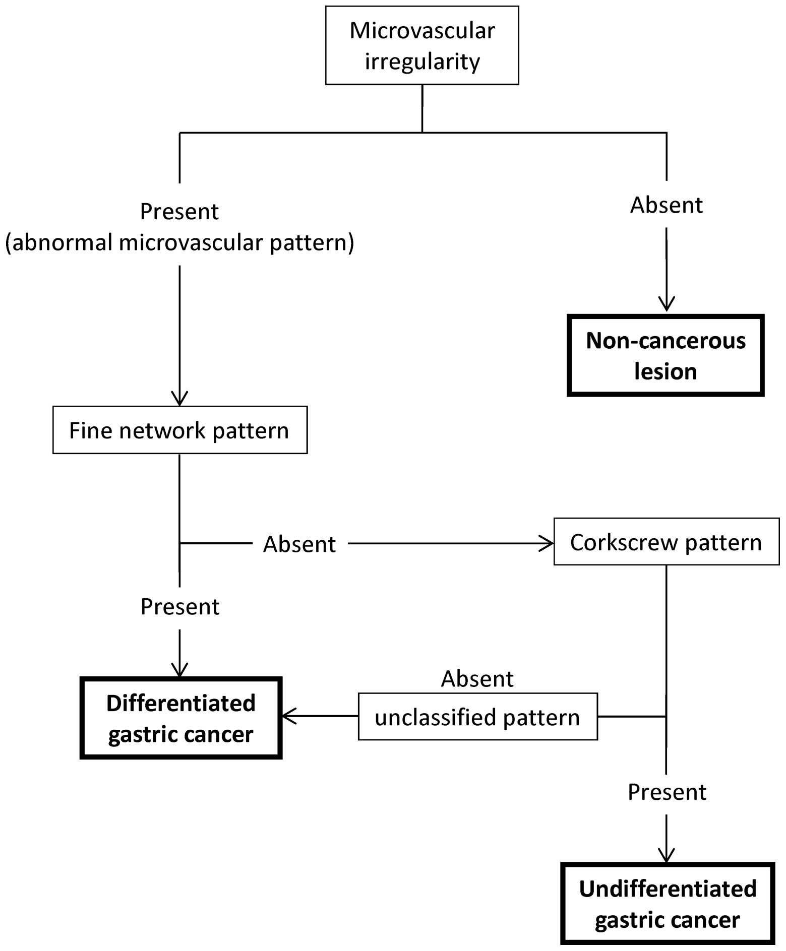
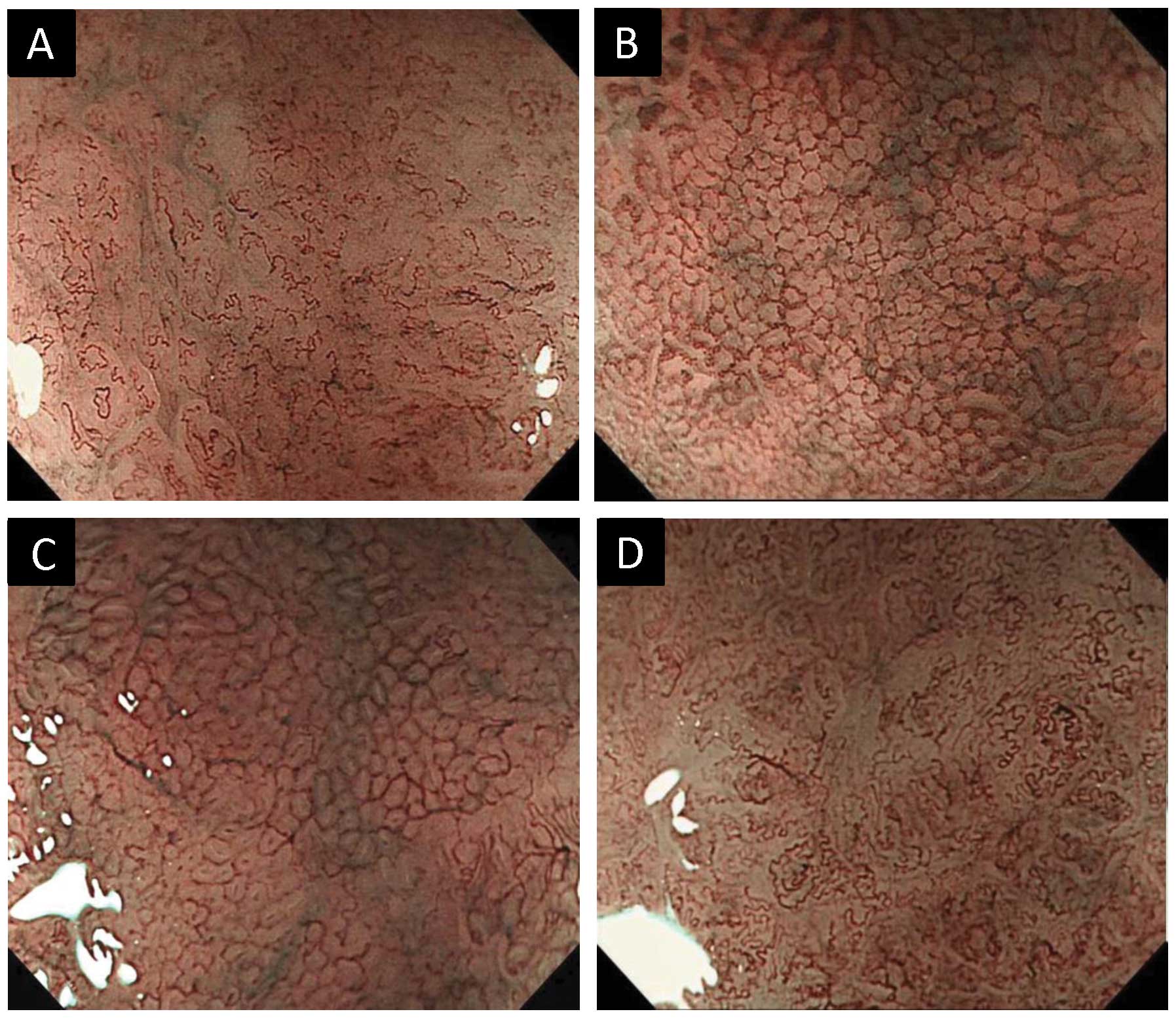
The usefulness of magnifying endoscopy with narrow‑band imaging (ME-NBI) for the diagnosis of early gastric cancer is well known, however, there are no evaluation criteria. The aim of this study was to devise and evaluate a novel diagnostic algorithm for ME-NBI in depressed early gastric cancer. Between August, 2007 and May, 2011, 90 patients with a total of 110 depressed gastric lesions were enrolled in the study. A diagnostic algorithm was devised based on ME-NBI microvascular findings: microvascular irregularity and abnormal microvascular patterns (fine network, corkscrew and unclassified patterns). The diagnostic efficiency of the algorithm for gastric cancer and histological grade was assessed by measuring its mean sensitivity, specificity, positive predictive value (PPV), negative predictive value (NPV), and accuracy. Furthermore, inter- and intra-observer variation were measured. In the differential diagnosis of gastric cancer from non‑cancerous lesions, the mean sensitivity, specificity, PPV, NPV, and accuracy of the diagnostic algorithm were 86.7, 48.0, 94.4, 26.7, and 83.2%, respectively. Furthermore, in the differential diagnosis of undifferentiated adenocarcinoma from differentiated adenocarcinoma, the mean sensitivity, specificity, PPV, NPV, and accuracy of the diagnostic algorithm were 61.6, 86.3, 69.0, 84.8, and 79.1%, respectively. For the ME-NBI final diagnosis using this algorithm, the mean κ values for inter- and intra-observer agreement were 0.50 and 0.77, respectively. In conclusion, the diagnostic algorithm based on ME-NBI microvascular findings was convenient and had high diagnostic accuracy, reliability and reproducibility in the differential diagnosis of depressed gastric lesions.
View Figures
View References

Related Articles

Journal Cover

January-February 2014
Volume 2 Issue 1

Print ISSN: 2049-9450
Online ISSN:2049-9469

Sign up for eToc alerts

Recommend to Library

Copy and paste a formatted citation
x
Spandidos Publications style
Sumie H, Sumie S, Nakahara K, Watanabe Y, Matsuo K, Mukasa M, Sakai T, Yoshida H, Tsuruta O, Sata M, Sata M, et al: Usefulness of magnifying endoscopy with narrow-band imaging for diagnosis of depressed gastric lesions. Mol Clin Oncol 2: 129-133, 2014
APA
Sumie, H., Sumie, S., Nakahara, K., Watanabe, Y., Matsuo, K., Mukasa, M. ... Sata, M. (2014). Usefulness of magnifying endoscopy with narrow-band imaging for diagnosis of depressed gastric lesions. Molecular and Clinical Oncology, 2, 129-133. https://doi.org/10.3892/mco.2013.213
MLA
Sumie, H., Sumie, S., Nakahara, K., Watanabe, Y., Matsuo, K., Mukasa, M., Sakai, T., Yoshida, H., Tsuruta, O., Sata, M."Usefulness of magnifying endoscopy with narrow-band imaging for diagnosis of depressed gastric lesions". Molecular and Clinical Oncology 2.1 (2014): 129-133.
Chicago
Sumie, H., Sumie, S., Nakahara, K., Watanabe, Y., Matsuo, K., Mukasa, M., Sakai, T., Yoshida, H., Tsuruta, O., Sata, M."Usefulness of magnifying endoscopy with narrow-band imaging for diagnosis of depressed gastric lesions". Molecular and Clinical Oncology 2, no. 1 (2014): 129-133. https://doi.org/10.3892/mco.2013.213