Introduction
Although polypectomy has long been recommended for
the early detection of and rapid cure from colorectal carcinoma, it
has not led to a decrease in colorectal carcinoma (1). Colorectal carcinoma is one of the
most common cancers worldwide and has the highest incidence of
cancer patients by site of the disease in Japan. Additionally,
colorectal carcinoma is the second most common cancer after gastric
cancer among males as well as the second most common cancer after
breast cancer in females, with a significant increase in recent
years (2,3). Furthermore, a review of surgical
resection cases in elderly patients in the last 10 years has shown
a tendency for advanced cancer in the right side colon (4–6). A
molecular agent (cetuximab) targeting epidermal growth factor
receptor (EGFR) in unresectable advanced colorectal carcinomas
(refractory colorectal carcinomas defined by unresectable advanced
or metastatic/recurrent colorectal carcinomas) has been developed
and is considered to be effective in patients with the wild-type
K-ras gene (7).
K-ras gene protein (KRAS), a small G protein that functions
as a signal transducer and downstream integrator of EGFR, is a key
component in the EGFR signal cascade. Moreover, b-raf gene
protein (BRAF) is an immediate downstream effector of K-ras
in this signaling pathway. However, considering the EGFR signal
transduction mechanism and carcinogenic mechanism of colorectal
carcinoma, the wild-type K-ras/mutant b-raf genes are
also present (8,9). Carcinomas with these genetic
characteristics are described as a serrated neoplasia pathway,
which is considered to be characteristic of right-sided colon in
elderly patients. Therefore, clarification of clinicopathological
features of unresectable advanced colorectal carcinomas may be a
clinically important issue when considering the therapeutic
modality.
In the present study, 49 cases of unresectable
advanced colorectal carcinomas were analyzed to clarify the
association between their clinicopathological characteristics and
the molecular alterations in K-ras gene.
Materials and methods
Patients and tissue samples
We obtained 49 samples of advanced colorectal
carcinomas by surgical resection from 49 patients at the Dokkyo
Medical University School of Medicine Hospital (Tochigi, Japan)
between January, 2005 and December, 2009. This study was performed
following approval by the ethics committee of the Dokkyo Medical
University Surgical Pathology. Informed consent was obtained from
the patients.
For the ethics procedure, a linkable anonymizing
method was used to ensure the study was conducted in a blinded
manner. Samples used in this study were materials for biopsy or
surgery obtained for diagnosis or treatment and not for research
purposes. Participation in the present study did not increase
medical disadvantage or risk for patients and data were used
strictly for analysis of information as part of therapeutic
intervention.
Age and gender of patients, as well as subsites of
cancer, were confirmed from the pathological report. The cases were
staged using Turnbull’s modification of Dukes’ classification.
Classification of macroscopic type and depth of tumor invasion were
assessed according to the Japanese classification of colorectal
carcinoma edited by the Japanese Society for Cancer of the Colon
and Rectum (JSCCR) (10) and
International Union Against Cancer (TNM classification of malignant
tumours) (11). Gross
classification was as follows: type 1, protuberant type; type 2,
ulcerated type with clear margin; type 3, ulcerated type with
infiltration; type 4, diffusely infiltrating type and type 5,
unclassified type.
In classifying the carcinoma, the predominant
pattern was adopted as its representative histological type. For
example, for a tumor comprising mainly of well-differentiated
adenocarcinoma with partially moderately differentiated adenocarci
noma or mucinous adenocarcinoma, a diagnosis of well-differentiated
type was made. The clinicopathological characteristics of patients
are shown in Tables I and II.
 | Table IPatient characteristics (age, gender,
gross and microscopic findings) in the unresectable advanced
colorectal carcinomas. |
Table I
Patient characteristics (age, gender,
gross and microscopic findings) in the unresectable advanced
colorectal carcinomas.
| Patient
characteristics | No. of patients
(n=49) |
|---|
| Age (years) | <65 (n=34) | ≥65 (n=15) |
| Gender (M:F) | 23:11 | 9:6 |
| Gross (type) | | |
| 1 | 1:0 | 0 |
| 2 | 13:6 | 6:4 |
| 3 | 7:5 | 3:2 |
| 4 | 1:0 | 0 |
| 5 | 1:0 | 0 |
| Microscopic
findings | | |
| Tub 1 | 5:2 | 2:1 |
| Tub 2 | 12:8 | 5:5 |
| Por+sig | 5:1 | 1:0 |
| Others (Muc) | 1:0 | 1:0 |
| Table IIPatients characteristics (primary
subsites, VI, DM and Dukes’ classification) in the unresectable
advanced colorectal carcinomas. |
Table II
Patients characteristics (primary
subsites, VI, DM and Dukes’ classification) in the unresectable
advanced colorectal carcinomas.
| VI
| DM
| Dukes’
|
|---|
| Primary tumor
site | (+) | (−) | Liver | Others | A | B | C | D |
|---|
| Right side
colon | 9 | 0 | 4 | 0 | 0 | 2 | 3 | 4 |
| Left side
colon | 23 | 0 | 6 | 3 | 0 | 6 | 9 | 8 |
| Rectum | 16 | 0 | 6 | 0 | 1 | 0 | 10 | 6 |
Screening of K-ras gene mutations
DNA extraction
Formalin-fixed, paraffin-embedded samples were cut
serially at a thickness of 10 μm. Based on
histolopathological findings, the tumor and corresponding normal
tissues were microdissected from each of five serial sections and
the tissues were deparaffinized in xylene and rehydrated in graded
ethanol series.
DNA was then extracted from the whole dissected
tissue using a DNA isolator PS kit (Wako Pure Chemical, Osaka,
Japan) according to the supplied protocol.
Analysis of K-ras codon 12
mutations
Extracted DNA was amplified using the polymerase
chain reaction (PCR), which was carried out using the following
amplification profile: denaturation for 20 min at 4°C once;
annealing for 30 sec at 94°C, 30 sec at 55°C, extension for 30 sec
at 72°C for 40 cycles; and a final extension for 4 min at 72°C. The
reaction mixture (100 μl) contained 1 μg genomic DNA,
10 μl Ex Taq buffer, 8 μl dNTP mixture, 2.5 units Ex
Taq polymerase (Takara Bio, Kyoto, Japan), 25 pmol of forward
primer (5′-GACT-GAA TAT AAA CTT GTG G-3′), and 25 pmol of reverse
primer (3′-CCA GGT CCT GGT AAG AAACT-5′). The DNA extracted from
the gallbladder carcinoma cell line NOZ (kindly provided by Dr S.
Nagamori, Jikei University School of Medicine, Tokyo, Japan), in
which K-ras codon 12 is mutated (12), was used the positive control,
whereas DNA from the HeLa cell line, the K-ras codon 12 of
which is not mutated (13), was
used as the negative control.
Mutations of K-ras codon 12 in amplified DNA
were screened using PCR-restriction fragment length polymorphism
(PCR-RFRP) (14) and analysed
further by fluorescence direct sequencing. Briefly, the PCR
products were digested with MvaI (Takara, Kyoto, Japan) to
distinguish the mutant allele from the wild-type allele and
electrophoresed on 3% agarose gels, followed by staining with
ethidium bromide. PCR products encoding the wild-type and mutant
alleles were detected as 114 and 143 bp fragments, respectively.
Subsequently, the PCR products encoding the mutant allele were
extracted from agarose gels using a Sephagras™ BandPrep kit
(Amersham Pharmacia Biotech, Tokyo, Japan) and purified using
MicroSpin™ columns (Amersham Pharmacia Biotech). Extracted DNA was
sequenced using a Thermo Sequence™ fluorescence-labeled primer
cycle sequencing kit (Amersham International PLC, Buckinghamshire,
UK). The sequence reaction was carried out using the
above-mentioned forward primer labeled by the Fluoro-Prime method
at its 5′ end with a fluorescent derivative. The reaction products
were analysed using ALF express™ automatic DNA sequencer (Amersham
Pharmacia Biotech).
Immunohistochemical analysis of EGFR
protein
Immunohistochemical staining for EGFR was performed
with an anti-mouse monoclonal antibody (clone 31G7, Nichirei
Biosciences, Inc., Tokyo, Japan) on paraffin-embedded tissue
sections using EnVision™ + Mouse/HRP kit (Dako Japan, Kyoto,
Japan). For EGFR, positive and negative controls were used for each
set of experiments. Sections of colorectal adenocarcinomas that
were confirmed to overexpress this protein were used as positive
controls. However, the surrounding normal epithelial cells also
showed faint positive cytoplasm staining. Immunoreactivity was
considered positive if strong staining of EGFR was detected in at
least a focal cytoplasmic membrane (Fig. 1).
Statistical analysis
Data were presented as the means ± SD. To determine
the association of clinicopathological variables and tumor location
with gender and age, Fisher’s exact test was used. Statistical
analysis was performed using R (version 2.15.0). P<0.05 was
considered to indicate a statistically significant difference.
Results
Clinical and histopathological features
of unresectable advanced colorectal carcinomas
Thirty-two males and 17 females with a mean age of
59.1 years (range, 34–75) were included in this study. All patients
had unresectable advanced colorectal carcinomas. There were 34
patients aged <65 years with an M/F ratio of 23/11 (female
ratio, 32.4%) and 15 patients were aged ≥65 years with an M/F ratio
of 9/6 (40%). Gross and histological findings were not
characterized by the specificity of unresectable colorectal
carcinomas. Concerning the subsite of cancer, unresectable advanced
colorectal carcinomas developed in right-sided colon in 13
patients, left-sided colon in 19 patients and rectum in 17
patients. Right-sided colon carcinomas were frequently identified
in elderly patients aged ≥65 years and this tendency was more
marked in the female patients (Tables
I–III).
| Table IIIDifferences between subsites of
unresectable advanced colorectal carcinomas and age/gender.a |
Table III
Differences between subsites of
unresectable advanced colorectal carcinomas and age/gender.a
| No. of patients
| |
|---|
| Variables | Age ≥65 years | Age <65
years | P-value |
|---|
| Female | | | |
| Right side
colon | 4 | 1 | 0.024 |
| Left side
colon | 2 | 4 | |
| Rectum | 0 | 6 | |
| Male | | | |
| Right side
colon | 3 | 5 | 0.88 |
| Left side
colon | 3 | 10 | |
| Rectum | 3 | 8 | |
| Total | | | |
| Right side
colon | 7 | 6 | 0.10 |
| Left side
colon | 5 | 14 | |
| Rectum | 3 | 14 | |
Screening of K-ras mutation
When the pH of buffered form-aldehyde is kept
neutral, the purified DNA does not degrade. However, our study
showed that high molecular weight DNA degrades in unbuffered
formaldehyde, as noted in a previous study (12,15).
The results of various formaldehyde fixations suggest that DNAse,
which plays an important role in degrading genomic DNA during
fixation, and other enzymes may degrade genomic DNA. Subsequently,
40 of 49 (81.6%) samples were examined.
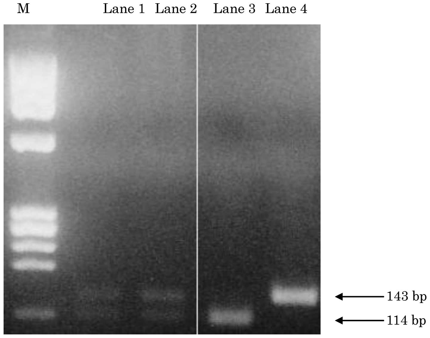
As shown in Fig. 2,
the mutated samples of the K-ras codon 12 were screened by
PCR-RFLP. The wild-type fragment was digested with a restriction
enzyme (MvaI) and detected as a band of 114 bp, whereas the
mutant-type fragment was not digested and was detected as a band of
143 bp. Eight of the 40 (20%) samples had a K-ras codon 12
point mutation (Fig. 2) (Table IV).
| Table IVPrimary subsites and status of
K-ras gene in the unresectable advanced colorectal
carcinomas. |
Table IV
Primary subsites and status of
K-ras gene in the unresectable advanced colorectal
carcinomas.
| K-ras
mutant/<65
| K-ras
mutant/≥65
|
|---|
| Primary tumor
site | Male | Female | Total | Male | Female | Total |
|---|
| Right side
colon | 2/5 | 1/1 | 3/6 | 0/3 | 1/4 | 1/7 |
| Left side
colon | 1/10 | 0/4 | 1/14 | 0/3 | 0/2 | 0/5 |
| Rectum | 0/8 | 3/6 | 3/14 | 0/3 | 0 | 0/3 |
No significant correlation was detected between the
anatomical subsites, age and K-ras mutations, while the
incidence of K-ras gene mutations was relatively low
compared with that noted in previous studies (?).
Result of immunohistochemical staining
for EGFR protein
Result of immunohistochemical staining for EGFR
protein, carried out with an anti-mouse monoclonal antibody (31G7)
on paraffin-embedded tissue sections, was positive in the 40 cases
for which screening of K-ras mutations was readily
detectable.
Discussion
Colorectal carcinoma has high morbidity and
mortality rates worldwide, and according to the estimated number of
cancer patients identified in 2008 in the GLOBOCAN, published by
the International Agency for Research on Cancer (IARC), 1.2 million
people had colorectal carcinoma, accounting for approximately 10%
of cancer patients (1). Colorectal
carcinoma is the third most common cancer after lung and prostate
cancer in males and the second most common cancer after breast
cancer in females. Although a high incidence of colorectal
carcinoma was not identified in Japan until approximately 1970, the
morbidity rate has rapidly increased since 1975 and it was
estimated that ∼104,000 people had colorectal carcinoma in 2005 and
∼43,000 succumbed to the disease in 2008. Over the last 30 years,
the incidence of colorectal carcinoma has increased >5-fold and
the incidence of unresectable advanced/recurrent metastatic
colorectal carcinoma (unresectable advanced colorectal carcinomas)
has been on the increase with an increase in the incidence of
colorectal carcinoma (2,3).
Anti-EGFR antibody (cetuximab), a molecularly
targeted agent, was developed as a treatment option for
unresectable advanced colorectal carcinomas. In Japan, cetuximab is
indicated only for cases of positive EGFR immunostaining and the
wild-type ras gene has been investigated for
immunohistochemical study. The patients in this study tested
positive for EGFR. Standardization of the criteria for EGFR
antibody expression and a positive result are necessary, however, a
correlation between colorectal carcinoma and EGFR expression was
detected in this study.
By contrast, incidence of the mutant K-ras
gene was lower compared with the controlled statistics in the
previously published literature (15,16),
likely due to the fact that the wild-type K-ras gene
colorectal carcinoma is frequently observed in unresectable
advanced colorectal carcinomas. The wild-type K-ras gene
colorectal carcinoma is a candidate lesion of de novo cancer
and morphologic type IIc (flat and depressed type). It is known
that this type is smaller than the elevated type and that it
accelerates the progression of cancer more readily (17). Similar conditions include laterally
spreading tumor and non-granular type (LST-NG) (16).
There were 32 males and 17 female patients (mean
age, 59.2 years; range, 34–75 years) with unresectable advanced
colorectal carcinomas in our hospital. Twenty-four patients were
aged <60 years with an M/F ratio of 16/8 and 25 patients aged
≥60 years with an M/F ratio of 16/9. Additionally, of the patients
aged ≥65 years, 15 patients were enrolled as controls and the M/F
ratio was 9/6. Concerning the cancer subsite, unresectable advanced
colorectal carcinomas developed in the right-sided colon in 13
patients, left-sided colon in 19 patients and rectum in 17
patients. Right-sided colon carcinomas were frequently identified
in elderly patients aged ≥65 years, with this tendency being more
marked in the female patients.
Furthermore, mucinous carcinoma was observed in two
groups including patients in their 30s and elderly patients.
Mucinous carcinoma is similar to cancer type associated with
abnormalities of germline and somatic line mismatch-repair
(MMR) genes in the young and elderly patients, respectively,
and the results of the present study together with future studies
are likely to further elucidate this type of carcinoma. To confirm
that the high incidence of this right-sided colon carcinoma, it is
necessary to predict the precursor lesion that develops in the
right-sided colon based on the origin of unresectable advanced
colorectal carcinomas.
In general, colorectal carcinoma has a multi-step
process characterized by a sequence of genetic alterations in cell
growth regulatory genes. Mutational activation of the ras
gene, in particular the K-ras oncogene, is an early event
and is considered to play a role in the progression of size and
grade of atypia. Ras is part of the
ras/raf/MEK/MAP kinase cascade, which is an essential
component of intracellular signaling from activated cell surface
receptors to transcription factors in the cell nucleus. Recently,
the activating mutation of b-raf, a member of the raf
gene family, has been found in malignant melanoma and in a wide
range of human carcinomas.
Although the incidence of b-raf mutation is
reported to be ∼10% in colorectal carcinoma (12,15),
more recent data (16) have
indicated that b-raf mutation is associated with a high
frequency of microsatellite instability (MSI) and inactivation of
the MMR gene in colorectal carcinoma, in particular the
so-called serrated neoplasia pathway which may be associated with
sessile serrated adenoma/polyp.
Approximately 50% of the colorectal carcinomas occur
in rectosigmoid area, although their relative incidence appears to
be on the decrease, in that a shift in location towards the proxi
mal colon during the past few decades has been noted. Noteworthy
correlations between certain clinical features and the anatomic
subsite of the tumor have been made. Right-sided tumors are said to
be more common in the Japanese elderly and in patients with
surveillance for clean colon (so-called interval cancer) (18).
As a direct sequence reaction of this study,
mutations of K-ras codon 13 were detected concurrently with
those of codon 12 (data not shown). The mutations of the
K-ras codon 12 were different transversions: GGT (glycine)
to GTT (valine) and GGT (glycine) to TGT (cystein), and the
transition: GGT (glycine) to GAT (aspartic acid). In addition,
activating mutations involving codon 13 of the K-ras gene
were detected in three cases. Although the reason for the
occurrence of varying mutations remains to be elucidated and data
are conflicting, these different mutations seem to be prospectively
correlated with the biological behavior or prognosis of the
unresectable advanced colorectal carcinomas. To clarify these
issues, more studies based on key components (KRAS and BRAF) in the
EGFR signal cascade should be carried out.
In conclusion, the increase in the incidence of
unresectable advanced colorectal carcinomas in elderly females,
located on the right side colon is clinically important and should
be elucidated further. Although no investigation on b-raf
mutations was performed in this study, the abnormality of the
b-raf gene and the wild-type K-ras should be examined
for a more accurate analysis of the effect of anti-EGFR antibody
(cetuximab).
In addition, the multidisciplinary and multicenter
assessments on the anatomic subsites, gender and age in
unresectable advanced colorectal carcinomas, suggesting a shift in
location towards the right-sided colon in elderly patients is to be
demonstrated based on cumulative results, such as the effect of
these treatments.
References
|
1
|
Ferlay F, Shin HR and Bray F: Estimates of
worldwide burden of cancer in 2008: GLOBOCAN 2008. Int J Cancer.
127:2893–2917. 2010. View Article : Google Scholar : PubMed/NCBI
|
|
2
|
Japanese Society for Cancer of the Colon
and Rectum: Multi institutional Registry of Large Bowel Cancer in
Japan. Case treated in 1999. 28:(Prospective registry date).
|
|
3
|
Watanabe T, Itabashi M, Shimoda Y, et al:
Japanese society for cancer of the colon and rectum (JSCCR)
guidelines 2010 for the treatment of colorectal cancer. Int J Clin
Oncol. 17:1–29. 2012. View Article : Google Scholar : PubMed/NCBI
|
|
4
|
Ghahremani GG and Dowlatshahi K:
Colorectal carcinomas: diagnostic implications of their changing
frequency and anatomic distribution. World J Surg. 13:321–325.
1989. View Article : Google Scholar
|
|
5
|
Cady B, Stone MD and Wayne J: Continuing
trends in the prevalence of right-sided lesions among colorectal
carcinomas. Arch Surg. 128:505–509. 1993. View Article : Google Scholar : PubMed/NCBI
|
|
6
|
Dubrow R, Bernstein J and Holford TR:
Age-period-cohort modeling of large-bowel-cancer by anatomic
sub-site and sex in Conneticut. Int J Cancer. 53:907–913. 1993.
View Article : Google Scholar : PubMed/NCBI
|
|
7
|
Karapetis CS, Khambate-Ford S, Jonker DJ,
et al: K-ras mutations and benefit from cetuximab in
advanced colorectal cancer. N Engl J Med. 359:1757–1765. 2008.
View Article : Google Scholar
|
|
8
|
Vogelstein B, Fearon ER, Hamilton SR, et
al: Genetic alterations during colorectal-tumor development. N Engl
J Med. 319:525–532. 1988. View Article : Google Scholar : PubMed/NCBI
|
|
9
|
Snover DC: Update on the serrated pathway
to colorectal carcinoma. Hum Pathol. 42:1–10. 2011. View Article : Google Scholar : PubMed/NCBI
|
|
10
|
Japanese Society for Cancer of the Colon
and Rectum (JSCCR): Japanese Classification of Colorectal
Carcinoma. Kanehara; Tokyo: pp. 4–32. 1997
|
|
11
|
Hamilton SR, Bosman FT, Boffetta P, et al:
Carcinoma of the colon and rectum. WHO Classification of Tumours of
the Digestive System. Bosman FT, Carneiro F, Hruban RH, et al: 4th
edition. IARC; Lyon: pp. 134–146. 2010
|
|
12
|
Ajiki T, Onoyama H, Yamamoto M, et al:
Detection of point mutations in K-ras gene at codon 12 in
bile from percutaneous transhepatic choledochal drainage tubes for
diagnosis of biliary strictures. Int J Pancreatol. 18:215–220.
1995.
|
|
13
|
Lehman TA, Scott F, Seddon M, et al:
Detection of K-ras oncogene mutations by polymerase chain
reaction-based ligase chain reaction. Anal Biochem. 239:153–159.
1996.PubMed/NCBI
|
|
14
|
Jiang W, Kahn SM, Guillem JG, et al: Rapid
detection of ras oncogenes in human tumors: applications to
colon, esophageal and gastric cancer. Oncogene. 4:923–928.
1989.
|
|
15
|
Fujimori T, Satonaka K, Yamamura-Idei Y,
et al: Non-involvement of ras mutations in flat colorectal
adenomas and carcinomas. Int J Cancer. 57:51–55. 1994.
|
|
16
|
Kusaka T, Fukui H, Sano Y, et al: Analysis
of K-ras codon 12 mutations and p53 overexpression in
colorectal nodule-aggregating tumors. J Gastroenterol Hepatol.
15:1151–1157. 2000.
|
|
17
|
Kudo S, Kashida H, Nakajima T, et al:
Endoscopic diagnosis and treatment of early colorectal cancer.
World J Surg. 21:694–701. 1997. View Article : Google Scholar
|
|
18
|
Matsuda T, Fujii T, Sano Y, et al:
Five-year incidence of advanced neoplasia after initial colonoscopy
in Japan: a multicenter retrospective cohort study. Jpn J Clin
Oncol. 39:435–442. 2009.PubMed/NCBI
|