Spandidos Publications Logo
  • About
    • About Spandidos
    • Aims and Scopes
    • Abstracting and Indexing
    • Editorial Policies
    • Reprints and Permissions
    • Job Opportunities
    • Terms and Conditions
    • Contact
  • Journals
    • All Journals
    • Oncology Letters
      • Oncology Letters
      • Information for Authors
      • Editorial Policies
      • Editorial Board
      • Aims and Scope
      • Abstracting and Indexing
      • Bibliographic Information
      • Archive
    • International Journal of Oncology
      • International Journal of Oncology
      • Information for Authors
      • Editorial Policies
      • Editorial Board
      • Aims and Scope
      • Abstracting and Indexing
      • Bibliographic Information
      • Archive
    • Molecular and Clinical Oncology
      • Molecular and Clinical Oncology
      • Information for Authors
      • Editorial Policies
      • Editorial Board
      • Aims and Scope
      • Abstracting and Indexing
      • Bibliographic Information
      • Archive
    • Experimental and Therapeutic Medicine
      • Experimental and Therapeutic Medicine
      • Information for Authors
      • Editorial Policies
      • Editorial Board
      • Aims and Scope
      • Abstracting and Indexing
      • Bibliographic Information
      • Archive
    • International Journal of Molecular Medicine
      • International Journal of Molecular Medicine
      • Information for Authors
      • Editorial Policies
      • Editorial Board
      • Aims and Scope
      • Abstracting and Indexing
      • Bibliographic Information
      • Archive
    • Biomedical Reports
      • Biomedical Reports
      • Information for Authors
      • Editorial Policies
      • Editorial Board
      • Aims and Scope
      • Abstracting and Indexing
      • Bibliographic Information
      • Archive
    • Oncology Reports
      • Oncology Reports
      • Information for Authors
      • Editorial Policies
      • Editorial Board
      • Aims and Scope
      • Abstracting and Indexing
      • Bibliographic Information
      • Archive
    • Molecular Medicine Reports
      • Molecular Medicine Reports
      • Information for Authors
      • Editorial Policies
      • Editorial Board
      • Aims and Scope
      • Abstracting and Indexing
      • Bibliographic Information
      • Archive
    • World Academy of Sciences Journal
      • World Academy of Sciences Journal
      • Information for Authors
      • Editorial Policies
      • Editorial Board
      • Aims and Scope
      • Abstracting and Indexing
      • Bibliographic Information
      • Archive
    • International Journal of Functional Nutrition
      • International Journal of Functional Nutrition
      • Information for Authors
      • Editorial Policies
      • Editorial Board
      • Aims and Scope
      • Abstracting and Indexing
      • Bibliographic Information
      • Archive
    • International Journal of Epigenetics
      • International Journal of Epigenetics
      • Information for Authors
      • Editorial Policies
      • Editorial Board
      • Aims and Scope
      • Abstracting and Indexing
      • Bibliographic Information
      • Archive
    • Medicine International
      • Medicine International
      • Information for Authors
      • Editorial Policies
      • Editorial Board
      • Aims and Scope
      • Abstracting and Indexing
      • Bibliographic Information
      • Archive
  • Articles
  • Information
    • Information for Authors
    • Information for Reviewers
    • Information for Librarians
    • Information for Advertisers
    • Conferences
  • Language Editing
Spandidos Publications Logo
  • About
    • About Spandidos
    • Aims and Scopes
    • Abstracting and Indexing
    • Editorial Policies
    • Reprints and Permissions
    • Job Opportunities
    • Terms and Conditions
    • Contact
  • Journals
    • All Journals
    • Biomedical Reports
      • Information for Authors
      • Editorial Policies
      • Editorial Board
      • Aims and Scope
      • Abstracting and Indexing
      • Bibliographic Information
      • Archive
    • Experimental and Therapeutic Medicine
      • Information for Authors
      • Editorial Policies
      • Editorial Board
      • Aims and Scope
      • Abstracting and Indexing
      • Bibliographic Information
      • Archive
    • International Journal of Epigenetics
      • Information for Authors
      • Editorial Policies
      • Editorial Board
      • Aims and Scope
      • Abstracting and Indexing
      • Bibliographic Information
      • Archive
    • International Journal of Functional Nutrition
      • Information for Authors
      • Editorial Policies
      • Editorial Board
      • Aims and Scope
      • Abstracting and Indexing
      • Bibliographic Information
      • Archive
    • International Journal of Molecular Medicine
      • Information for Authors
      • Editorial Policies
      • Editorial Board
      • Aims and Scope
      • Abstracting and Indexing
      • Bibliographic Information
      • Archive
    • International Journal of Oncology
      • Information for Authors
      • Editorial Policies
      • Editorial Board
      • Aims and Scope
      • Abstracting and Indexing
      • Bibliographic Information
      • Archive
    • Medicine International
      • Information for Authors
      • Editorial Policies
      • Editorial Board
      • Aims and Scope
      • Abstracting and Indexing
      • Bibliographic Information
      • Archive
    • Molecular and Clinical Oncology
      • Information for Authors
      • Editorial Policies
      • Editorial Board
      • Aims and Scope
      • Abstracting and Indexing
      • Bibliographic Information
      • Archive
    • Molecular Medicine Reports
      • Information for Authors
      • Editorial Policies
      • Editorial Board
      • Aims and Scope
      • Abstracting and Indexing
      • Bibliographic Information
      • Archive
    • Oncology Letters
      • Information for Authors
      • Editorial Policies
      • Editorial Board
      • Aims and Scope
      • Abstracting and Indexing
      • Bibliographic Information
      • Archive
    • Oncology Reports
      • Information for Authors
      • Editorial Policies
      • Editorial Board
      • Aims and Scope
      • Abstracting and Indexing
      • Bibliographic Information
      • Archive
    • World Academy of Sciences Journal
      • Information for Authors
      • Editorial Policies
      • Editorial Board
      • Aims and Scope
      • Abstracting and Indexing
      • Bibliographic Information
      • Archive
  • Articles
  • Information
    • For Authors
    • For Reviewers
    • For Librarians
    • For Advertisers
    • Conferences
  • Language Editing
Login Register Submit
  • This site uses cookies
  • You can change your cookie settings at any time by following the instructions in our Cookie Policy. To find out more, you may read our Privacy Policy.

    I agree
Search articles by DOI, keyword, author or affiliation
Search
Advanced Search
presentation
Molecular and Clinical Oncology
Join Editorial Board Propose a Special Issue
Print ISSN: 2049-9450 Online ISSN: 2049-9469
Journal Cover
March-April 2013 Volume 1 Issue 2

Full Size Image

Sign up for eToc alerts
Recommend to Library

Journals

International Journal of Molecular Medicine

International Journal of Molecular Medicine

International Journal of Molecular Medicine is an international journal devoted to molecular mechanisms of human disease.

International Journal of Oncology

International Journal of Oncology

International Journal of Oncology is an international journal devoted to oncology research and cancer treatment.

Molecular Medicine Reports

Molecular Medicine Reports

Covers molecular medicine topics such as pharmacology, pathology, genetics, neuroscience, infectious diseases, molecular cardiology, and molecular surgery.

Oncology Reports

Oncology Reports

Oncology Reports is an international journal devoted to fundamental and applied research in Oncology.

Experimental and Therapeutic Medicine

Experimental and Therapeutic Medicine

Experimental and Therapeutic Medicine is an international journal devoted to laboratory and clinical medicine.

Oncology Letters

Oncology Letters

Oncology Letters is an international journal devoted to Experimental and Clinical Oncology.

Biomedical Reports

Biomedical Reports

Explores a wide range of biological and medical fields, including pharmacology, genetics, microbiology, neuroscience, and molecular cardiology.

Molecular and Clinical Oncology

Molecular and Clinical Oncology

International journal addressing all aspects of oncology research, from tumorigenesis and oncogenes to chemotherapy and metastasis.

World Academy of Sciences Journal

World Academy of Sciences Journal

Multidisciplinary open-access journal spanning biochemistry, genetics, neuroscience, environmental health, and synthetic biology.

International Journal of Functional Nutrition

International Journal of Functional Nutrition

Open-access journal combining biochemistry, pharmacology, immunology, and genetics to advance health through functional nutrition.

International Journal of Epigenetics

International Journal of Epigenetics

Publishes open-access research on using epigenetics to advance understanding and treatment of human disease.

Medicine International

Medicine International

An International Open Access Journal Devoted to General Medicine.

Journal Cover
March-April 2013 Volume 1 Issue 2

Full Size Image

Sign up for eToc alerts
Recommend to Library

  • Article
  • Citations
    • Cite This Article
    • Download Citation
    • Create Citation Alert
    • Remove Citation Alert
    • Cited By
  • Similar Articles
    • Related Articles (in Spandidos Publications)
    • Similar Articles (Google Scholar)
    • Similar Articles (PubMed)
  • Download PDF
  • Download XML
  • View XML
Case Report

A type 2 papillary renal cell carcinoma presenting as an intracystic necrotic lesion: A case report

  • Authors:
    • Zhenyu Fu
    • Liguo Sun
    • Yuhua Huang
    • Jie Zhang
    • Zichao Zhang
    • Lijun Wang
    • Shengyu Wang
    • Ge Zhang
  • View Affiliations / Copyright

    Affiliations: Department of Urology, The No. 2 People's Hospital of Changshu, Suzhou, Jiangsu 215500, P.R. China, Department of Urology, The First Affiliated Hospital of Soochow University, Suzhou, Jiangsu 215006, P.R. China, Institute of Biosciences and Technology, Texas A__AMB__M Health Science Center, Houston, TX 77030, USA, Department of Orthopaedics, The No. 2 People's Hospital of Changshu, Suzhou, Jiangsu 215500, P.R. China, Department of Radiography, The No. 2 People's Hospital of Changshu, Suzhou, Jiangsu 215500, P.R. China
  • Pages: 318-320
    |
    Published online on: January 11, 2013
       https://doi.org/10.3892/mco.2013.64
  • Expand metrics +
Metrics: Total Views: 0 (Spandidos Publications: | PMC Statistics: )
Metrics: Total PDF Downloads: 0 (Spandidos Publications: | PMC Statistics: )
Cited By (CrossRef): 0 citations Loading Articles...

This article is mentioned in:


Abstract

Papillary renal carcinoma (papillary RCC) is a histological subtype of the renal carcinoma, which in turn, has two morphological subtypes that correlate with prognosis. The present study reported an unexpected finding of type 2 papillary renal cell carcinoma (papillary RCC) presenting intracystic necrosis cavity. A cystic renal lesion was identified incidentally in a 66‑year‑old man during an abdominal computed tomography (CT) scan performed for the evaluation of a gastrointestinal stromal tumor. Subsequent contrast material‑enhanced CT scan and magnetic resonance imaging (MRI) examination labeled the mass as category Ⅲ degree on the basis of the Bosniak classification scheme. Surgical exploration by laparoscopic radical nephrectomy was performed to determine the diagnosis. Definitive pathological study confirmed a type 2 papillary RCC with an intracystic necrosis cavity. To the best of our knowledge, this case demonstrated for the first time a cavity within a papillary RCC, supporting the hypothesis that type 2 papillary RCC could develop cavity avascular necrosis during its cystic degeneration.

Introduction

Papillary renal carcinoma (papillary RCC) is a histological subtype of the renal carcinoma. There are two morphological subtypes of papillary RCC that are correlated with their prognosis. Type 1 tumor consists of papillae enclosed with a single layer of small cells with scanty cytoplasm and low-grade nuclei. In type 2 tumor, the cells covering the papillae are pseudostratified, generally have eosinophilic cytoplasm and are usually of higher nuclear grade compared to the cells of type 1 tumors. Papillary RCC is usually associated with a more favorable prognosis compared to the carcinoma of renal clear cells, while type 2 tumors carry a worse prognosis compared to type 1 tumors. At the cytogenetic level, the most common karyotypic changes in papillary RCC are trisomy of chromosomes 7 and 17, as well as loss of chromosome Y in males (1–3). Microscopically, papillary RCCs were predominantly papillary or tubulopapillary, often with foam cells, necrosis, hemorrhage and multifocality (4). This case report described an incidental finding of a necrotic cavity within a papillary RCC. The imaging data documented the dynamic progress of the cavitation within the cystic mass. This study was approved by the Institutional Review Board of the No. 2 People’s Hospital of Changshu. Informed consent was obtained from the patient.

Case report

A 66-year-old male patient underwent a second abdominal computed tomography (CT) scan while being monitored for a renal cyst at the upper pole of the left kidney that was inadvertently detected 22 months earlier in another hospital. The patient’s medical history showed surgery had been performed for gastric adenocarcinoma in January, 2010 at the same institution. 
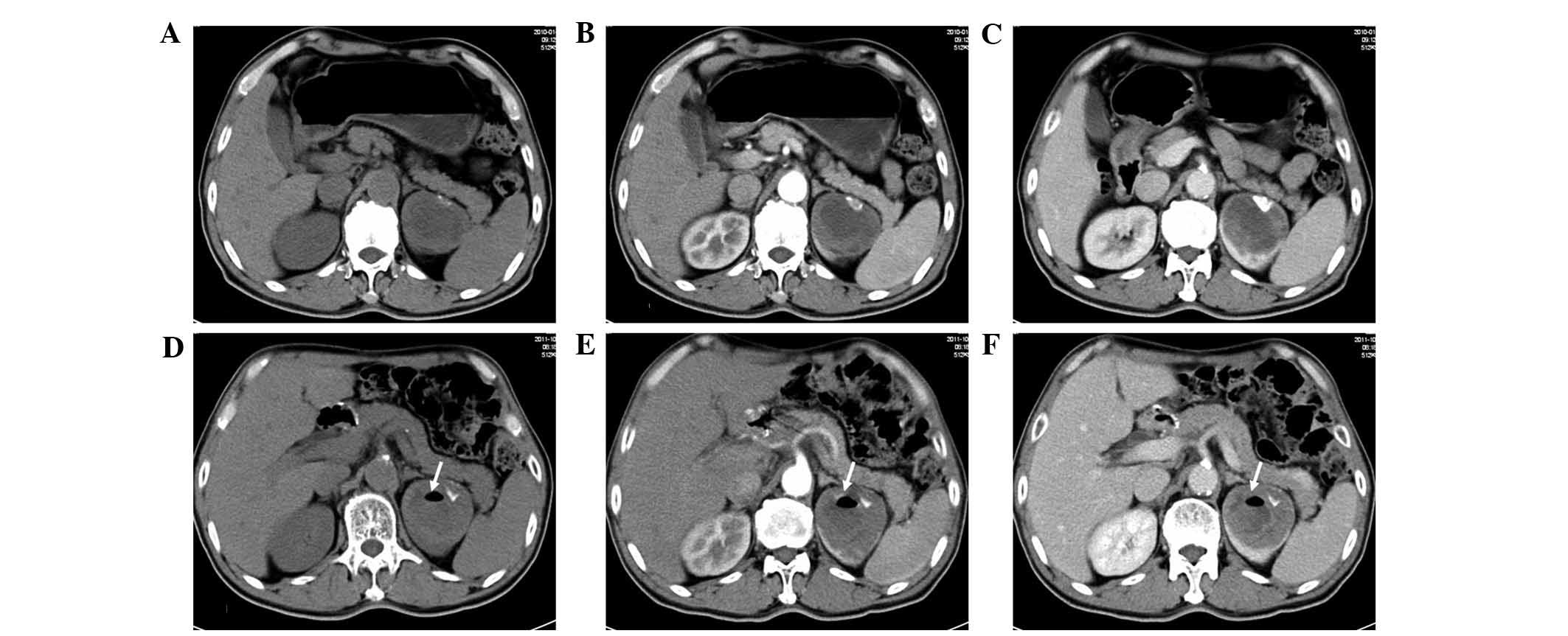
Pre-operative abdominal CT in 2010 detected an unexpected complex renal cyst at the upper pole of the left kidney (Fig. 1A–C), but no relevant treatment was 
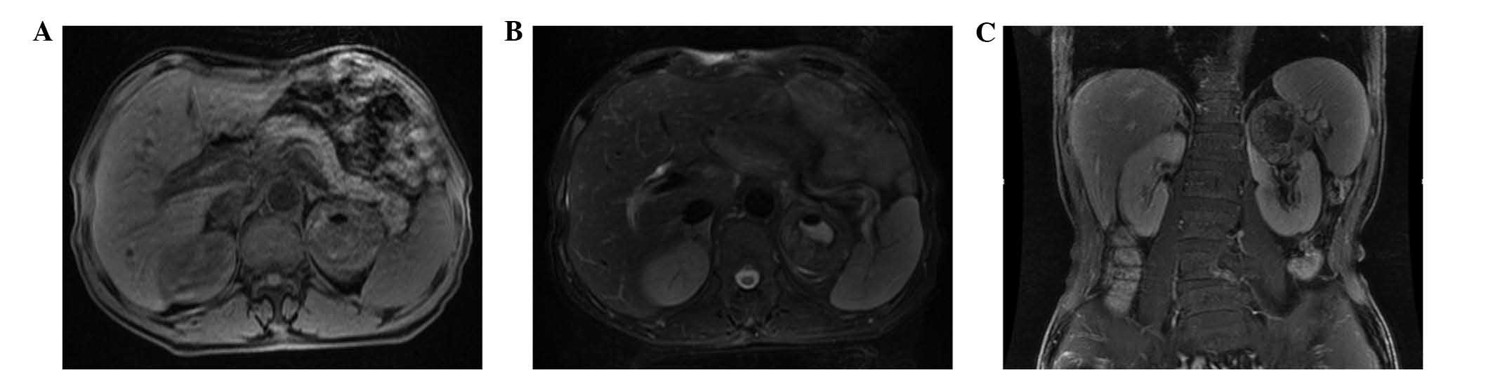
administered. Following admission, the patient underwent a contrast material-enhanced CT and magnetic resonance imaging (MRI) examination to evaluate the origin of the mass, its correlation with surrounding structures and possible evidence of metastatic involvement. CT scans demonstrated a 5.7×5.4 cm well-circumscribed, exophytic mass arising from the upper pole of the left kidney. A distinct lesion of 1.8 cm at its greatest diameter within the exophytic mass was also detected, with a CT value between −600 to −970 HU (Fig. 1D–F). MRI results showed that the mass exhibited a pseudocapsule and had low signal intensity on T1- and T2-weighted images (Fig. 2). This cystic renal lesion was confirmed as category III on the basis of the Bosniak classification scheme. The patient analysis did not show any abnormalities. Concerning current diagnostic criteria, probable diagnosis for such a heterogeneous cystic mass was limited to a hemorrhagic renal cyst or cell carcinoma.

Figure 1

(A) Unenhanced, (B) corticomedullary phase and (C) nephrographic phase CT scans showing a 5.2×4.4-cm low-attenuation mass in the left renal cortex, with extensive necrotic areas, calcification and peripheral enhancement. (D–F) CT scans 22 months later showed a distinct lesion with a maximum diameter of 1.8 cm within the exophytic renal mass (arrow), with a suspected air CT value between −600 and −970 HU.

Figure 2

Magnetic resonance imaging (MRI) shows the renal cystic mass exhibiting a pseudocapsule with low signal intensity on T1- and T2-weighted images.

Since renal carcinoma involves a potential risk of metastasis, radical nephrectomy was scheduled for removal of the left kidney. After general anesthesia was induced, the patient was placed in the lateral recumbent position and laparoscopic left radical nephrectomy was successfully performed in October, 2011. 
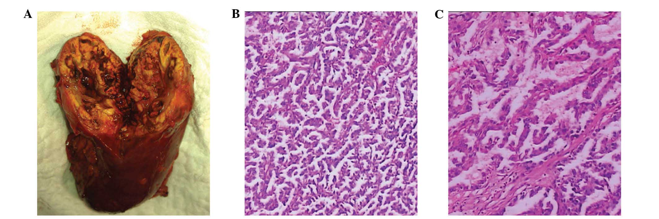
Gross examination was characterized by an irregular-shaped lesion of mahogany-brown color in the upper pole of the left kidney, with a maximum diameter of 5.7 cm. The central area in the exophytic lesion was identified as a 1.8-cm circumscribed cavity, with no liquid inside but with viscous, chocolate-brown, necrotic and hemorrhagic material on the cavity wall. The harvested specimen was processed according to standard surgical pathology protocols. Microscopic examination results (Fig. 3) showed that the specimen exhibited an outer cystic mass in the papillary architecture with pleomorphic cells showing prominent nucleoli and abundant eosinophilic cytoplasm. Papillary interstitial structure comprises cells arranged on a delicate fibrovascular core. Infiltration of foamy macrophages into the papillary structures and extensive tumor necrosis with abundant hemosiderin granules were detected. Immunohistochemical analyses showed the tumor cells to be strongly positive for vimentin, epithelial membrane antigen (EMA) and cytokeratins 7, focally positive for CD10 and negative for Malan A. Overall, the cyst characteristics are those of a type 2 papillary RCC of Fuhrman nuclear grade 3. There was no tumor involvement of the renal capsule or perinephric fat, no vascular space invasion was identified and the margins of resection were free of tumor. The patient was discharged after an uneventful post-operative course. Follow-up control 6 months later showed normal values in the urine analysis and renal function.

Figure 3

(A) Image of the cut surface of the specimen from radical nephrectomy shows the heterogeneous tumor with a large cavity component secondary to necrosis. (B–C) Histological analysis: representative photomicrographs of tumor sections (original magnification, ×100 and ×400, respectively; hematoxylin and eosin-stained), showing fibrovascular papillae lined by pseudostratified, large pleomorphic cells with eosinophilic cytoplasm and large, prominent nucleoli.

Discussion

Papillary RCC is the second most frequent carcinoma of the proximal renal tubules (10–15% of the cases) (5,6). The clinical and histological details were first described by Mancilla-Jiménez et al (7) in 1976. Commonly, it is diagnosed histologically and based on its characteristic enhanced pattern on CT and MRI. Certain characteristics of a cystic renal mass including the character of the lesion wall, septation, calcification, nodularity, CT attenuation values and enhancement are suggestive of a particular diagnosis. However, there is a continuum of radiologic findings often rendering the confident labeling of a cystic renal lesion benign or malignant difficult.

The papillary RCC in the present case was initially characterized by a single cyst with an internal cavity and a calcification filling as part of the cyst. The 1.8-cm diameter distinct cavity was measured using a CT value between −600 and −970 HU, potentially appreciated as air density. To the best of our knowledge, the presence of a necrotic cavity within a papillary RCC has rarely been described. This finding may be considered of interest with respect to the aetiology of the cavity within the tumor mass. We first hypothesized that the intracystic vacuum phenomenon is simply a result of the migration of a gastrointestinal gaseous collection through disrupted renal parenchyma or a renal cyst with inflammatory changes. However, the patient did not experience any fever or abdominal discomfort during the past 22 months. Additionally, the intact renal capsule of the renal specimen and evidence of non-infection of perinephric capsule thickening allows us to exclude the aetiology of gas deposits or inflammatory changes. At histological analysis, infiltration of foamy macrophages into the papillary structures and extensive necrotic tissue with abundant hemosiderin granules suggest a predisposition for degenerative cyst formation.

Extensive necrosis and bleeding within a tumor is often thought to reflect poor tumor vascularization, a characteristic well recognized in angiographic studies of papillary RCC. The comparison between the CT scan in January, 2010 and 22 months later documented the marked progress of the necrotic cavitation of the papillary RCC mass.

Acknowledgements

The authors thank Dr Kai Ye for his valuable technical assistance during the immunohistochemical analysis.

References

1 

Reuter VE: The pathology of renal epithelial neoplasms. Semin Oncol. 33:534–543. 2006. View Article : Google Scholar : PubMed/NCBI

2 

Presti JC Jr, Rao PH, Chen Q, Reuter VE, Li FP, Fair WR and Jhanwar SC: Histopathological, cytogenetic, and molecular characterization of renal cortical tumors. Cancer Res. 51:1544–1552. 1991.PubMed/NCBI

3 

Kovacs G, Wilkens L, Papp T and de Riese W: Differentiation between papillary and nonpapillary renal cell carcinomas by DNA analysis. J Natl Cancer Inst. 81:527–530. 1989. View Article : Google Scholar : PubMed/NCBI

4 

Yamada T, Endo M, Tsuboi M, Matsuhashi T, Takase K, Higano S and Takahashi S: Differentiation of pathologic subtypes of papillary renal cell carcinoma on CT. AJR Am J Roentgenol. 191:1559–1563. 2008. View Article : Google Scholar : PubMed/NCBI

5 

Wein AJ, Kavoussi LR, Novick AC, Partin AW and Peters CA: Campbell-Walsh Urology. 10th edition. WB Saunders Company; Philadelphia PA: pp. 1432–1434. 2012

6 

Störkel S, Eble JN, Adlakha K, Amin M, Blute ML, Bostwick DG, Darson M, Delahunt B and Iczkowski K: Classification of renal cell carcinoma: Workgroup No.1. Union Internationale Contre le Cancer (UICC) and the American Joint Committee on Cancer (AJCC). Cancer. 80:987–989. 1997.

7 

Mancilla-Jiménez R, Stanley RJ and Blath RA: Papillary renal cell carcinoma: a clinical, radiologic, and pathologic study of 34 cases. Cancer. 38:2469–2480. 1976.PubMed/NCBI

Related Articles

  • Abstract
  • View
  • Download
  • Twitter
Copy and paste a formatted citation
Spandidos Publications style
Fu Z, Sun L, Huang Y, Zhang J, Zhang Z, Wang L, Wang S and Zhang G: A type 2 papillary renal cell carcinoma presenting as an intracystic necrotic lesion: A case report. Mol Clin Oncol 1: 318-320, 2013.
APA
Fu, Z., Sun, L., Huang, Y., Zhang, J., Zhang, Z., Wang, L. ... Zhang, G. (2013). A type 2 papillary renal cell carcinoma presenting as an intracystic necrotic lesion: A case report. Molecular and Clinical Oncology, 1, 318-320. https://doi.org/10.3892/mco.2013.64
MLA
Fu, Z., Sun, L., Huang, Y., Zhang, J., Zhang, Z., Wang, L., Wang, S., Zhang, G."A type 2 papillary renal cell carcinoma presenting as an intracystic necrotic lesion: A case report". Molecular and Clinical Oncology 1.2 (2013): 318-320.
Chicago
Fu, Z., Sun, L., Huang, Y., Zhang, J., Zhang, Z., Wang, L., Wang, S., Zhang, G."A type 2 papillary renal cell carcinoma presenting as an intracystic necrotic lesion: A case report". Molecular and Clinical Oncology 1, no. 2 (2013): 318-320. https://doi.org/10.3892/mco.2013.64
Copy and paste a formatted citation
x
Spandidos Publications style
Fu Z, Sun L, Huang Y, Zhang J, Zhang Z, Wang L, Wang S and Zhang G: A type 2 papillary renal cell carcinoma presenting as an intracystic necrotic lesion: A case report. Mol Clin Oncol 1: 318-320, 2013.
APA
Fu, Z., Sun, L., Huang, Y., Zhang, J., Zhang, Z., Wang, L. ... Zhang, G. (2013). A type 2 papillary renal cell carcinoma presenting as an intracystic necrotic lesion: A case report. Molecular and Clinical Oncology, 1, 318-320. https://doi.org/10.3892/mco.2013.64
MLA
Fu, Z., Sun, L., Huang, Y., Zhang, J., Zhang, Z., Wang, L., Wang, S., Zhang, G."A type 2 papillary renal cell carcinoma presenting as an intracystic necrotic lesion: A case report". Molecular and Clinical Oncology 1.2 (2013): 318-320.
Chicago
Fu, Z., Sun, L., Huang, Y., Zhang, J., Zhang, Z., Wang, L., Wang, S., Zhang, G."A type 2 papillary renal cell carcinoma presenting as an intracystic necrotic lesion: A case report". Molecular and Clinical Oncology 1, no. 2 (2013): 318-320. https://doi.org/10.3892/mco.2013.64
Follow us
  • Twitter
  • LinkedIn
  • Facebook
About
  • Spandidos Publications
  • Careers
  • Cookie Policy
  • Privacy Policy
How can we help?
  • Help
  • Live Chat
  • Contact
  • Email to our Support Team