Spandidos Publications Logo
  • About
    • About Spandidos
    • Aims and Scopes
    • Abstracting and Indexing
    • Editorial Policies
    • Reprints and Permissions
    • Job Opportunities
    • Terms and Conditions
    • Contact
  • Journals
    • All Journals
    • Oncology Letters
      • Oncology Letters
      • Information for Authors
      • Editorial Policies
      • Editorial Board
      • Aims and Scope
      • Abstracting and Indexing
      • Bibliographic Information
      • Archive
    • International Journal of Oncology
      • International Journal of Oncology
      • Information for Authors
      • Editorial Policies
      • Editorial Board
      • Aims and Scope
      • Abstracting and Indexing
      • Bibliographic Information
      • Archive
    • Molecular and Clinical Oncology
      • Molecular and Clinical Oncology
      • Information for Authors
      • Editorial Policies
      • Editorial Board
      • Aims and Scope
      • Abstracting and Indexing
      • Bibliographic Information
      • Archive
    • Experimental and Therapeutic Medicine
      • Experimental and Therapeutic Medicine
      • Information for Authors
      • Editorial Policies
      • Editorial Board
      • Aims and Scope
      • Abstracting and Indexing
      • Bibliographic Information
      • Archive
    • International Journal of Molecular Medicine
      • International Journal of Molecular Medicine
      • Information for Authors
      • Editorial Policies
      • Editorial Board
      • Aims and Scope
      • Abstracting and Indexing
      • Bibliographic Information
      • Archive
    • Biomedical Reports
      • Biomedical Reports
      • Information for Authors
      • Editorial Policies
      • Editorial Board
      • Aims and Scope
      • Abstracting and Indexing
      • Bibliographic Information
      • Archive
    • Oncology Reports
      • Oncology Reports
      • Information for Authors
      • Editorial Policies
      • Editorial Board
      • Aims and Scope
      • Abstracting and Indexing
      • Bibliographic Information
      • Archive
    • Molecular Medicine Reports
      • Molecular Medicine Reports
      • Information for Authors
      • Editorial Policies
      • Editorial Board
      • Aims and Scope
      • Abstracting and Indexing
      • Bibliographic Information
      • Archive
    • World Academy of Sciences Journal
      • World Academy of Sciences Journal
      • Information for Authors
      • Editorial Policies
      • Editorial Board
      • Aims and Scope
      • Abstracting and Indexing
      • Bibliographic Information
      • Archive
    • International Journal of Functional Nutrition
      • International Journal of Functional Nutrition
      • Information for Authors
      • Editorial Policies
      • Editorial Board
      • Aims and Scope
      • Abstracting and Indexing
      • Bibliographic Information
      • Archive
    • International Journal of Epigenetics
      • International Journal of Epigenetics
      • Information for Authors
      • Editorial Policies
      • Editorial Board
      • Aims and Scope
      • Abstracting and Indexing
      • Bibliographic Information
      • Archive
    • Medicine International
      • Medicine International
      • Information for Authors
      • Editorial Policies
      • Editorial Board
      • Aims and Scope
      • Abstracting and Indexing
      • Bibliographic Information
      • Archive
  • Articles
  • Information
    • Information for Authors
    • Information for Reviewers
    • Information for Librarians
    • Information for Advertisers
    • Conferences
  • Language Editing
Spandidos Publications Logo
  • About
    • About Spandidos
    • Aims and Scopes
    • Abstracting and Indexing
    • Editorial Policies
    • Reprints and Permissions
    • Job Opportunities
    • Terms and Conditions
    • Contact
  • Journals
    • All Journals
    • Biomedical Reports
      • Information for Authors
      • Editorial Policies
      • Editorial Board
      • Aims and Scope
      • Abstracting and Indexing
      • Bibliographic Information
      • Archive
    • Experimental and Therapeutic Medicine
      • Information for Authors
      • Editorial Policies
      • Editorial Board
      • Aims and Scope
      • Abstracting and Indexing
      • Bibliographic Information
      • Archive
    • International Journal of Epigenetics
      • Information for Authors
      • Editorial Policies
      • Editorial Board
      • Aims and Scope
      • Abstracting and Indexing
      • Bibliographic Information
      • Archive
    • International Journal of Functional Nutrition
      • Information for Authors
      • Editorial Policies
      • Editorial Board
      • Aims and Scope
      • Abstracting and Indexing
      • Bibliographic Information
      • Archive
    • International Journal of Molecular Medicine
      • Information for Authors
      • Editorial Policies
      • Editorial Board
      • Aims and Scope
      • Abstracting and Indexing
      • Bibliographic Information
      • Archive
    • International Journal of Oncology
      • Information for Authors
      • Editorial Policies
      • Editorial Board
      • Aims and Scope
      • Abstracting and Indexing
      • Bibliographic Information
      • Archive
    • Medicine International
      • Information for Authors
      • Editorial Policies
      • Editorial Board
      • Aims and Scope
      • Abstracting and Indexing
      • Bibliographic Information
      • Archive
    • Molecular and Clinical Oncology
      • Information for Authors
      • Editorial Policies
      • Editorial Board
      • Aims and Scope
      • Abstracting and Indexing
      • Bibliographic Information
      • Archive
    • Molecular Medicine Reports
      • Information for Authors
      • Editorial Policies
      • Editorial Board
      • Aims and Scope
      • Abstracting and Indexing
      • Bibliographic Information
      • Archive
    • Oncology Letters
      • Information for Authors
      • Editorial Policies
      • Editorial Board
      • Aims and Scope
      • Abstracting and Indexing
      • Bibliographic Information
      • Archive
    • Oncology Reports
      • Information for Authors
      • Editorial Policies
      • Editorial Board
      • Aims and Scope
      • Abstracting and Indexing
      • Bibliographic Information
      • Archive
    • World Academy of Sciences Journal
      • Information for Authors
      • Editorial Policies
      • Editorial Board
      • Aims and Scope
      • Abstracting and Indexing
      • Bibliographic Information
      • Archive
  • Articles
  • Information
    • For Authors
    • For Reviewers
    • For Librarians
    • For Advertisers
    • Conferences
  • Language Editing
Login Register Submit
  • This site uses cookies
  • You can change your cookie settings at any time by following the instructions in our Cookie Policy. To find out more, you may read our Privacy Policy.

    I agree
Search articles by DOI, keyword, author or affiliation
Search
Advanced Search
presentation
Molecular and Clinical Oncology
Join Editorial Board Propose a Special Issue
Print ISSN: 2049-9450 Online ISSN: 2049-9469
Journal Cover
May-June 2013 Volume 1 Issue 3

Full Size Image

Sign up for eToc alerts
Recommend to Library

Journals

International Journal of Molecular Medicine

International Journal of Molecular Medicine

International Journal of Molecular Medicine is an international journal devoted to molecular mechanisms of human disease.

International Journal of Oncology

International Journal of Oncology

International Journal of Oncology is an international journal devoted to oncology research and cancer treatment.

Molecular Medicine Reports

Molecular Medicine Reports

Covers molecular medicine topics such as pharmacology, pathology, genetics, neuroscience, infectious diseases, molecular cardiology, and molecular surgery.

Oncology Reports

Oncology Reports

Oncology Reports is an international journal devoted to fundamental and applied research in Oncology.

Experimental and Therapeutic Medicine

Experimental and Therapeutic Medicine

Experimental and Therapeutic Medicine is an international journal devoted to laboratory and clinical medicine.

Oncology Letters

Oncology Letters

Oncology Letters is an international journal devoted to Experimental and Clinical Oncology.

Biomedical Reports

Biomedical Reports

Explores a wide range of biological and medical fields, including pharmacology, genetics, microbiology, neuroscience, and molecular cardiology.

Molecular and Clinical Oncology

Molecular and Clinical Oncology

International journal addressing all aspects of oncology research, from tumorigenesis and oncogenes to chemotherapy and metastasis.

World Academy of Sciences Journal

World Academy of Sciences Journal

Multidisciplinary open-access journal spanning biochemistry, genetics, neuroscience, environmental health, and synthetic biology.

International Journal of Functional Nutrition

International Journal of Functional Nutrition

Open-access journal combining biochemistry, pharmacology, immunology, and genetics to advance health through functional nutrition.

International Journal of Epigenetics

International Journal of Epigenetics

Publishes open-access research on using epigenetics to advance understanding and treatment of human disease.

Medicine International

Medicine International

An International Open Access Journal Devoted to General Medicine.

Journal Cover
May-June 2013 Volume 1 Issue 3

Full Size Image

Sign up for eToc alerts
Recommend to Library

  • Article
  • Citations
    • Cite This Article
    • Download Citation
    • Create Citation Alert
    • Remove Citation Alert
    • Cited By
  • Similar Articles
    • Related Articles (in Spandidos Publications)
    • Similar Articles (Google Scholar)
    • Similar Articles (PubMed)
  • Download PDF
  • Download XML
  • View XML
Case Report

Chronic cholecystitis with hilar bile duct stricture mimicking gallbladder carcinoma on positron emission tomography: A case report

  • Authors:
    • Qing‑Hong Ke
    • Zeng‑Lei He
    • Xin Duan
    • Shu‑Sen Zheng
  • View Affiliations / Copyright

    Affiliations: Department of Hepatobiliary and Pancreatic Surgery, The First Affiliated Hospital, College of Medicine, Key Laboratory of Combined Multi‑Organ Transplantation, Ministry of Health, Zhejiang University, Hangzhou, Zhejiang 310003, P.R.China
  • Pages: 517-520
    |
    Published online on: March 14, 2013
       https://doi.org/10.3892/mco.2013.93
  • Expand metrics +
Metrics: Total Views: 0 (Spandidos Publications: | PMC Statistics: )
Metrics: Total PDF Downloads: 0 (Spandidos Publications: | PMC Statistics: )
Cited By (CrossRef): 0 citations Loading Articles...

This article is mentioned in:


Abstract

Thickening of the gallbladder wall is observed in patients with gallbladder carcinoma, as well as in those with chronic cholecystitis. It is difficult to distinguish between benign and malignant gallbladder wall thickening with conventional diagnostic imaging techniques, such as abdominal ultrasonography (US), computed tomography (CT) and magnetic resonance imaging (MRI), particularly in patients with bile duct strictures. Currently, the fluorine‑18 2‑fluorodeoxyglucose positron emission tomography̸CT (F‑18 FDG PET/CT) scan is widely used in the differentiation of cholecystitis from gallbladder carcinoma. However, the F‑18 FDG PET̸CT scan may also be responsible for false‑positive diagnosis. This case report focuses on a 74‑year‑old male who presented with thickening of the gallbladder wall and hilar bile duct stricture, originally misdiagnosed as gallbladder carcinoma by US and MRI. F‑18 FDG PET/CT also demonstrated increased activity. This case was ultimately proven to be chronic cholecystitis by postoperative pathological examination and it is presented in order to emphasize the significance of considering the possibility of false‑positive diagnosis by PET̸CT, as a result of inflammatory lesions. Therefore, PET̸CT should not be considered the gold standard for the discrimination between benign and malignant gallbladder wall thickening.

Introduction

Gallbladder carcinoma is the most common malignancy of the biliary system (1). The optimal treatment for gallbladder carcinoma is radical resection. Thus, early detection is critical. However, thickening of the gallbladder wall is observed, not only in patients with gallbladder carcinoma, but also in those with chronic cholecystitis (2). It is difficult to distinguish between benign and malignant gallbladder wall thickening with conventional diagnostic imaging techniques, such as abdominal ultrasonography (US), computed tomography (CT) and magnetic resonance imaging (MRI), particularly in patients with bile duct strictures.

Currently, the fluorine-18 2-fluorodeoxyglucose positron emission tomography/CT (F-18 FDG PET/CT) scan is widely used in the differentiation between cholecystitis and gallbladder carcinoma and is also valuable for detection of regional lymph node involvement and unsuspected distant metastases (3). However, F-18 FDG PET/CT may lead to a false-positive diagnosis, particularly in cases of acute cholecystitis (4). We hereby present a case of thickening of the gallbladder wall with concurrent bile duct stricture, originally misdiagnosed as gallbladder carcinoma by US and MRI. F-18 FDG PET/CT also demonstrated increased activity. However, this case was ultimately proven to be chronic cholecystitis by postoperative pathological examination.

This study was approved by the Ethics Committee of Zhejiang University (Hangzhou, China). Written informed consent was obtained from the patient.

Case report

Patient

A 74-year-old man was referred to our hospital with signs of jaundice. On admission, he was free from abdominal pain and fever. The liver and spleen were not palpable and there was no sign of an abdominal mass. Murphy’s sign was negative. In addition, the patient had been suffering from hypothyroidism for 15 years, following treatment with radioiodine (I-131) for hyperthyroidism, but had never received any thyroid hormone replacement medication.

Following admission, his alanine aminotransferase level was 36 U/l (normal range, 5–40 U/l), aspartate aminotransferase level 21 U/l (normal range, 8–40 U/l), γ-glutamyltransferase level 24 U/l (normal range, 15–80 U/l), alkaline phosphatase level 239 U/l (normal range, <50 U/l), total bilirubin level 56 μmol/l (normal range, 1.71–17.1 μmol/l) and direct bilirubin level 31 μmol/l (normal range, 1.71–7 μmol/l). Hepatitis B serology for HBsAg was negative and there was no hepatitis C infection or leucocytosis. The α-fetoprotein and carbohydrate 19-9 levels were normal. C-reactive protein (CRP) level was 35 mg/l (normal, <10 mg/l). The patient had high thyroid-stimulating hormone (TSH) levels, with decreased levels of free T3 (FT3) and free T4 (FT4) (FT3=2.6 pmol/l, normal range 3.1–6.8 pmol/l; FT4=7.6 pmol/l, normal range 10.3–24.45 pmol/l; and TSH=75.0 mIU/l, normal range 0.4–4.0 mIU/l).

Findings

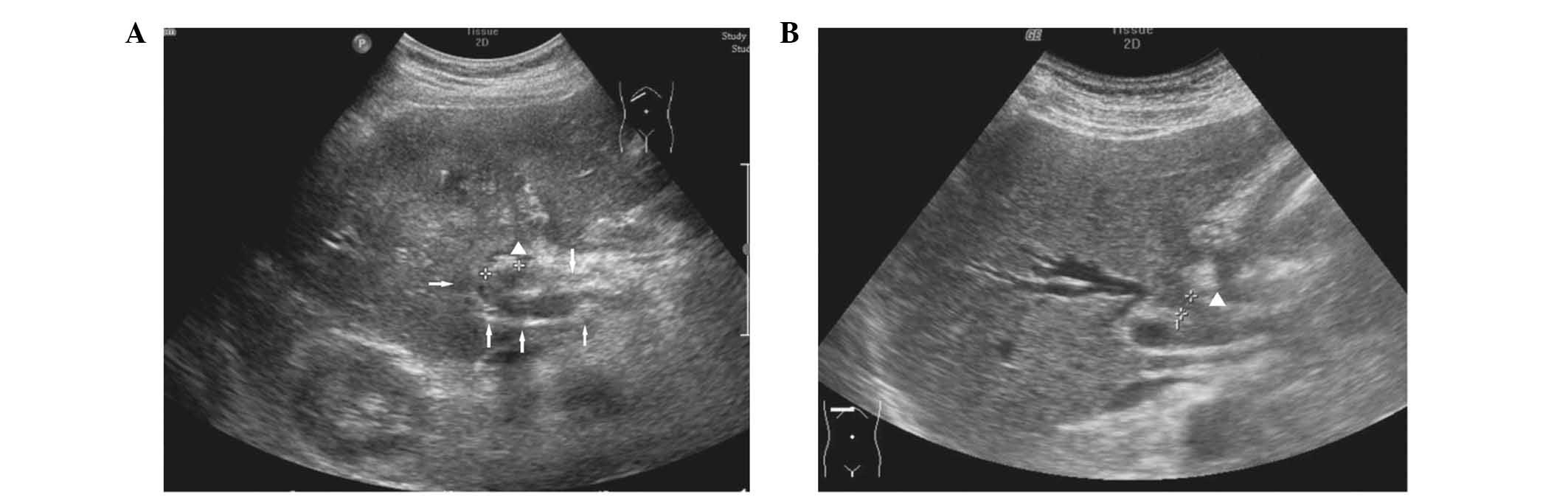
US demonstrated a thickened gallbladder wall and an intraluminal stone, as well as a perihilar bile duct stricture 
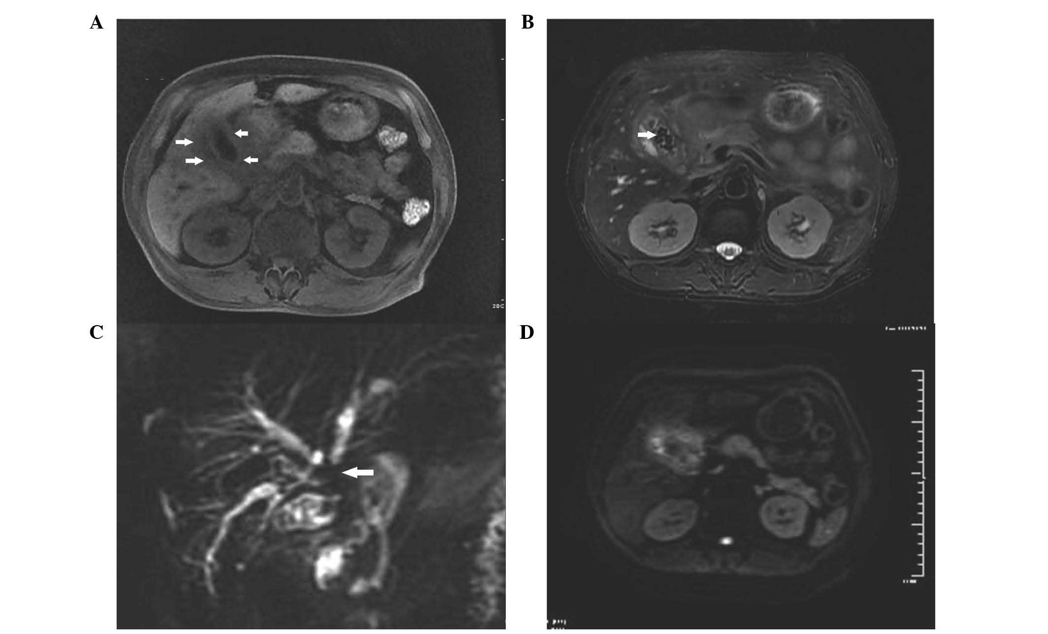
(Fig. 1). MRI demonstrated a thickened gallbladder wall and a positive signal in diffusion-weighted imaging (DWI). Magnetic resonance (MR) cholangiography demonstrated a hilar bile duct stricture and dilation of the intrahepatic bile ducts situated above the 
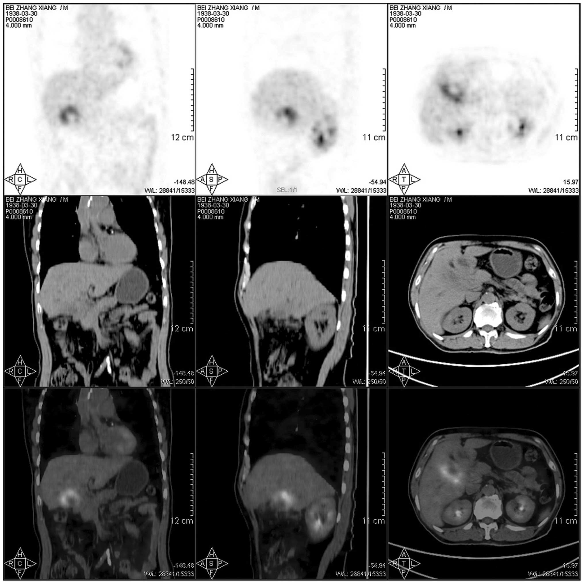
stricture (Fig. 2). F-18 FDG PET/CT also demonstrated increased activity in the gallbladder (Fig. 3), which also indicated the presence of gallbladder carcinoma. During the operation, a hard and 
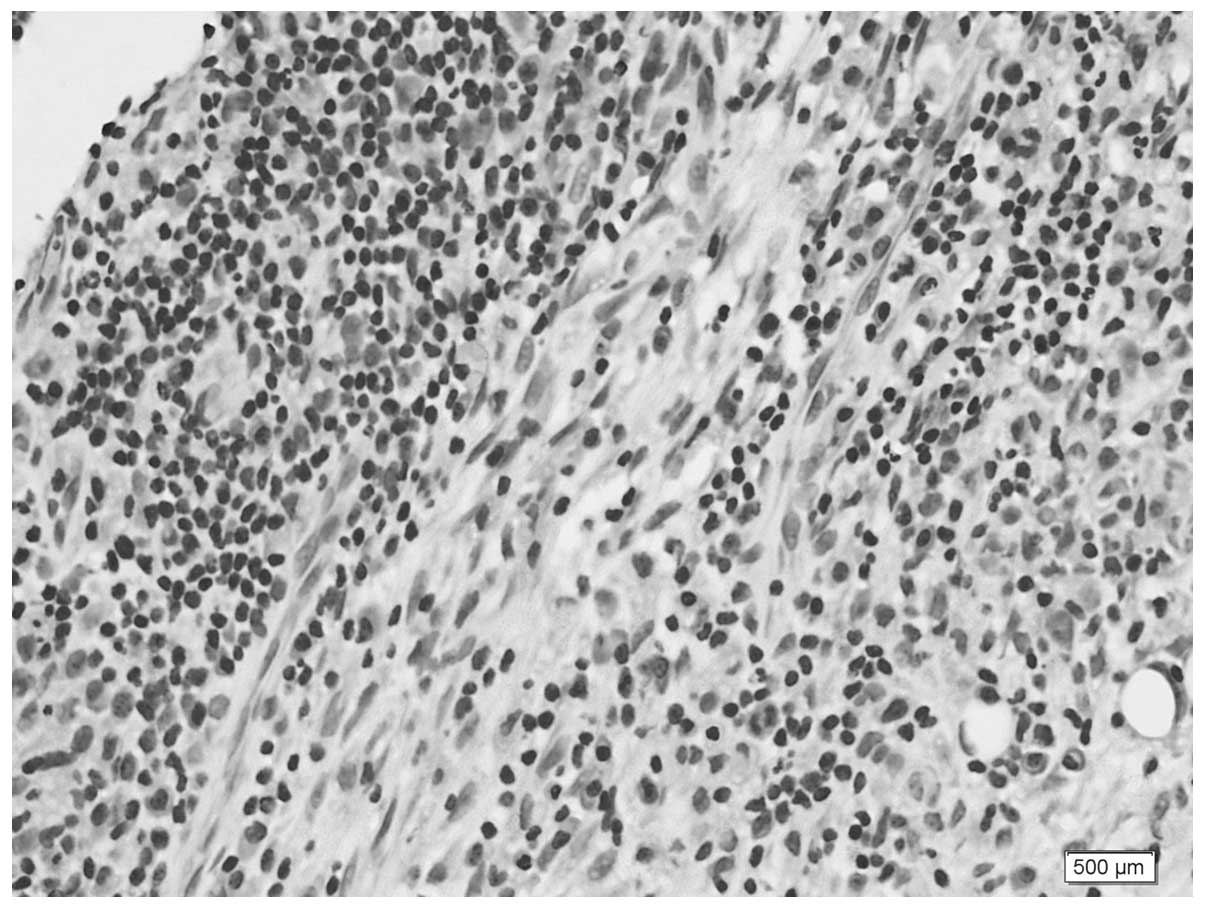
thickened gallbladder wall was identified. Frozen section of the gallbladder was negative for carcinoma and revealed the presence of chronic cholecystitis (Fig. 4). The hilar bile duct stricture was also proven to be an inflammatory stricture. A T-tube was inserted to drain the bile, following bile duct exploration. Intra-operative blood loss was 100 ml. The patient recovered well after the operation and was discharged on the tenth post-operative day. The T-tube was removed two months after the operation and the patient was free from the jaundice.

Figure 1

Ultrasonography demonstrated (A) thickened gallbladder wall (arrows) and intraluminal stone (arrowhead), with (B) perihilar bile duct stricture (arrowhead).

Figure 2

Magnetic resonance imaging (MRI) of the gallbladder. T1-weighted image demonstrated (A) thickened gallbladder wall (arrows) and (B) numerous gallstones (arrow). (C) Magnetic resonance (MR) cholangiography demonstrated hilar bile duct stricture (arrow) and dilation of the intrahepatic bile ducts. (D) Diffusion-weighted imaging demonstrated a positive signal in the gallbladder.

Figure 3

Fluorine-18 2-fluorodeoxyglucose positron emission tomography/computed tomography (F-18 FDG PET/CT) scan demonstrated intense F-18 FDG activity in the gallbladder fossa, which indicated gallbladder carcinoma.

Figure 4

Frozen section of the gallbladder was negative for carcinoma. Final pathology (hematoxylin and eosin staining, magnification, ×400) was consistent with chronic cholecystitis.

Discussion

Thickening of the gallbladder wall has been considered an indication of gallbladder carcinoma or cholecysitis. However, this finding is non-specific and may represent a diagnostic challenge in the sense that its misinterpretation may result in delayed treatment.

CT and MRI scans play a critical role in the differentiation of cholecystitis from gallbladder carcinoma. However, the majority of imaging modalities, such as CT and MRI, are anatomical, which affects the accuracy of the diagnosis. By contrast, PET is a functional diagnostic imaging technique, that uses compounds labelled with F-18 FDG to measure cell metabolism. Therefore, F-18 FDG PET/CT exhibits significant advantages regarding the differentiation between benign and malignant lesions. However, F-18 FDG is not tumor-specific. This tracer may accumulate in inflammatory lesions as well, which results in low specificity in the differentiation of malignant tumors from benign lesions. Kitazono et al(4) reported their findings of ring-like radiotracer uptake in a patient diagnosed with acute cholecystitis. Xanthogranulomatous cholecystitis may also mimic gallbladder carcinoma, with a false-positive result on F-18 FDG PET/CT (5). Therefore, distinguishing benign from malignant gallbladder wall thickening is not always possible using F-18 FDG PET/CT.

In this case, the patient underwent DWI prior to the F-18 FDG PET/CT scan. Ogawa et al(6) reported that DWI may contribute to the improvement of the diagnostic ability for gallbladder wall thickening. This case demonstrated DWI positivity, which indicated gallbladder carcinoma. In addition to DWI positivity, we identified a hilar bile duct stricture. The majority of benign biliary strictures are attributed to iatrogenic injury or stones, although a few cases may be due to primary sclerosing cholangitis (7) or xanthogranulomatous choledochitis (8). The patient had no history of operation and MR cholangiopancreatography showed no multifocal strictures or dilated ducts, apart from the hilar stricture. The biliary stricture was first considered to be due to malignancy. All of the imaging findings favored the diagnosis of gallbladder cancer, which may affect the future diagnostic credibility of the F-18 FDG PET/CT scan.

During the diagnostic process, we also overlooked an important factor, which was the preoperative elevated CRP levels. The elevated CRP levels may also affect the accuracy of F-18 FDG PET/CT. Nishiyama et al(9) reported that the specificity of PET was higher in the group with normal CRP levels, compared to that in the group with elevated CRP levels. In this case, the patient had no fever, no abdominal pain and normal white blood cell count, which led us to mistakenly associate the elevated CRP level with malignancy. However, the postoperative pathological examination confirmed the diagnosis of chronic cholecystitis. We hypothesized that the relapsing cholecystitis induced the formation of fibrotic tissue around the gallbladder, which caused the bile duct stricture. Due to the hypothyroidism, the patient had become insensitive to painful stimuli and only seeked medical advice after noticing the appearance of jaundice. Therefore, the recurrent inflammatory reaction in the gallbladder had been concealed.

PET/CT may be widely applied for the diagnosis of gall-bladder disease. However, the possibility of false-positive results due to inflammatory lesions should be considered to avoid a misdiagnosis which may complicate further treatment.

References

1. 

Zhu AX, Hong TS, Hezel AF and Kooby DA: Current management of gallbladder carcinoma. Oncologist. 15:168–181. 2010. View Article : Google Scholar : PubMed/NCBI

2. 

Oe A, Kawabe J, Torii K, et al: Distinguishing benign from malignant gallbladder wall thickening using FDG-PET. Ann Nucl Med. 20:699–703. 2006. View Article : Google Scholar : PubMed/NCBI

3. 

Hansen N, Brown RK, Khan A, Frey KA and Orringer M: False positive diagnosis of metastatic esophageal carcinoma on positron emission tomography: a case report of cholecystitis simulating a hepatic lesion. Clin Nucl Med. 35:409–412. 2010. View Article : Google Scholar

4. 

Kitazono MT and Colletti PM: FDG PET imaging of acute cholecystitis. Clin Nucl Med. 31:23–24. 2006. View Article : Google Scholar : PubMed/NCBI

5. 

Makino I, Yamaguchi T, Sato N, Yasui T and Kita I: Xanthogranulomatous cholecystitis mimicking gallbladder carcinoma with a false-positive result on fluorodeoxyglucose PET. World J Gastroenterol. 15:3691–3693. 2009. View Article : Google Scholar

6. 

Ogawa T, Horaguchi J, Fujita N, et al: High b-value diffusion-weighted magnetic resonance imaging for gallbladder lesions: differentiation between benignity and malignancy. J Gastroenterol. 47:1352–1360. 2012. View Article : Google Scholar

7. 

Helmberger H, Hellerhoff K, Rüll T and Rösch T: Chronic infections of the biliary system. Radiologe. 40:530–536. 2000.PubMed/NCBI

8. 

Krishna RP, Kumar A, Singh RK, Sikora S, Saxena R and Kapoor VK: Xanthogranulomatous inflammatory strictures of extrahepatic biliary tract: presentation and surgical management. J Gastrointest Surg. 12:836–841. 2008. View Article : Google Scholar : PubMed/NCBI

9. 

Nishiyama Y, Yamamoto Y, Fukunaga K, et al: Dual-time-point 18F-FDG PET for the evaluation of gallbladder carcinoma. J Nucl Med. 47:633–638. 2006.PubMed/NCBI

Related Articles

  • Abstract
  • View
  • Download
  • Twitter
Copy and paste a formatted citation
Spandidos Publications style
Ke QH, He ZL, Duan X and Zheng SS: Chronic cholecystitis with hilar bile duct stricture mimicking gallbladder carcinoma on positron emission tomography: A case report. Mol Clin Oncol 1: 517-520, 2013.
APA
Ke, Q., He, Z., Duan, X., & Zheng, S. (2013). Chronic cholecystitis with hilar bile duct stricture mimicking gallbladder carcinoma on positron emission tomography: A case report. Molecular and Clinical Oncology, 1, 517-520. https://doi.org/10.3892/mco.2013.93
MLA
Ke, Q., He, Z., Duan, X., Zheng, S."Chronic cholecystitis with hilar bile duct stricture mimicking gallbladder carcinoma on positron emission tomography: A case report". Molecular and Clinical Oncology 1.3 (2013): 517-520.
Chicago
Ke, Q., He, Z., Duan, X., Zheng, S."Chronic cholecystitis with hilar bile duct stricture mimicking gallbladder carcinoma on positron emission tomography: A case report". Molecular and Clinical Oncology 1, no. 3 (2013): 517-520. https://doi.org/10.3892/mco.2013.93
Copy and paste a formatted citation
x
Spandidos Publications style
Ke QH, He ZL, Duan X and Zheng SS: Chronic cholecystitis with hilar bile duct stricture mimicking gallbladder carcinoma on positron emission tomography: A case report. Mol Clin Oncol 1: 517-520, 2013.
APA
Ke, Q., He, Z., Duan, X., & Zheng, S. (2013). Chronic cholecystitis with hilar bile duct stricture mimicking gallbladder carcinoma on positron emission tomography: A case report. Molecular and Clinical Oncology, 1, 517-520. https://doi.org/10.3892/mco.2013.93
MLA
Ke, Q., He, Z., Duan, X., Zheng, S."Chronic cholecystitis with hilar bile duct stricture mimicking gallbladder carcinoma on positron emission tomography: A case report". Molecular and Clinical Oncology 1.3 (2013): 517-520.
Chicago
Ke, Q., He, Z., Duan, X., Zheng, S."Chronic cholecystitis with hilar bile duct stricture mimicking gallbladder carcinoma on positron emission tomography: A case report". Molecular and Clinical Oncology 1, no. 3 (2013): 517-520. https://doi.org/10.3892/mco.2013.93
Follow us
  • Twitter
  • LinkedIn
  • Facebook
About
  • Spandidos Publications
  • Careers
  • Cookie Policy
  • Privacy Policy
How can we help?
  • Help
  • Live Chat
  • Contact
  • Email to our Support Team