Introduction
Gastric cancer is one of the most commonly diagnosed
cancers and the second most common cause of cancer-related
mortality worldwide (1,2). Radical gastrectomy and lymph node
dissection with adjuvant chemotherapy are performed for patients
with advanced gastric cancer (3).
However, metastatic gastric cancer has a 5-year survival rate of
5–20% and a median overall survival of <1 year (1,4,5). In
2010, trastuzumab combined with chemotherapy was established as a
new standard treatment option for human epidermal growth factor
receptor 2 (HER2)-positive advanced gastric or gastroesophageal
junction cancer by the ToGA study (6). Although trastuzumab combination
therapy is adopted for inoperable advanced or metastatic disease,
the HER2 status is commonly evaluated in the primary lesion, since
metastatic sites are rarely resected or biopsied prior to
treatment. With breast cancer, however, the concordance ratio for
HER2 status between the primary lesion and metastatic lymph nodes
was reported to be 90–98% (7,8),
whereas the concordance ratio for HER2 status between primary and
metastatic sites other than lymph nodes was reported to be lower
(9,10). However, although a high concordance
ratio for HER2 status between primary and lymph node lesions has
been reported in gastric cancer (11,12),
a concordance ratio for HER2 status between primary and metastatic
lesions other than lymph nodes has not been reported. In addition,
HER2 status is typically evaluated by immunohistochemistry (IHC)
and/or fluorescence in situ hybridization (FISH) and a high
concordance ratio between IHC and FISH has been reported (13).
In this study, HER2 expression was assessed using
IHC (IHC score 2+) and FISH in the primary lesion and in paired
metastatic lesions other than lymph nodes. The aim of this study
was to investigate the concordance of HER2 expression between
primary and metastatic lesions and the feasibility of using HER2
expression in the primary lesion for determining therapy against
metastatic lesions.
Patients and methods
Patients and tissue samples
The samples used in this study were surgically
resected or biopsied at Fukuoka University Hospital between
January, 1998 and September, 2012. A total of 37 patients with
gastric adenocarcinoma (9 biopsies and 28 resection specimens) and
49 paired synchronous or metachronous metastatic tissues (2
biopsies and 47 resection specimens) were analyzed. The invasive
front of the resected tumor tissues was examined
immunohistochemically. None of the patients received neoadjuvant
therapy and 23 metastatic tissue samples were obtained from
patients who had been treated with chemotherapy. No patients
received trastuzumab combination therapy. Metachronous metastasis
was defined as metastasis arising >6 months following curative
resection.
HER2 expression and amplification
HER2 status was examined using 10% formalin-fixed
paraffin-embedded tissues. Immunohistochemical staining was
performed automatically with the Ventana iView PATHWAY HER2 (4B5)
(Ventana Medical Systems, Roche, Tucson, AZ, USA). Antigen
activation was performed in citrate buffer under high pressure.
HER2 immunoreactivity was scored as negative (0 or 1+), equivocal
(2+) and positive (3+) by an experienced pathologist according to
the scoring system described by Hofmann et al (14).
The PathVysion HER2 DNA Probe kit (Abbott Molecular,
Abbott Park, IL, USA) and a BioView Duet-3 scanning system
(BioView, Ltd., Rehovot, Israel) with fluorescence microscopy (BX51
TRF; Olympus, Nagano, Japan) were used for FISH. Gene amplification
was scored when a minimum of 20 cancer cell nuclei exhibited a
HER2/chromosome enumeration probe (CEP)17 ratio of >2.
HER2-positive status was defined as IHC 3+, or HER2
2+ and FISH-positive (HER2/CEP17 ratio >2). HER2-negative status
was defined as IHC 0, 1+, or 2+ and FISH-negative; the positive
groups were considered suitable for trastuzumab combination therapy
(6). All IHC 2+ tumors were
further analyzed with FISH to determine the HER2 gene copy level.
All the tissue samples from metastatic sites enabled the
pathologist to confirm the lesions as being metastatic from gastric
cancer.
Statistical methods
For the evaluation of the correlations of HER2
status between primary and paired metastatic lesion, the Fisher’s
exact probability test was employed. P<0.05 was considered to
indicate a statistically significant difference.
Results
Clinicopathological characteristics
The clinicopathological characteristics of the
patients in this study are summarized in Table I. The metastatic sites included the
peritoneum (n=20), liver (n=10), lung (n=7), skin and subcutaneous
tissue (n=5), colon (n=3) and others (n=4) (data not shown).
 | Table IClinicopathological characteristics of
the study population. |
Table I
Clinicopathological characteristics of
the study population.
| Characteristics | Total (%) | No. of primary sites
(%) (n=37) | No. of metastatic
sites (%) (n=49) |
|---|
| Age (years) | | | |
| Median | 64 | | |
| Range | 45–80 | | |
| Gender | | | |
| Male | 30 (81.0) | | |
| Female | 7 (19.0) | | |
| Type of
intervention | | | |
| Surgical
resection | 75 (87.0) | 28 (76.0) | 47 (96.0) |
| Biopsy | 11 (13.0) | 9 (24.0) | 2 (4.0) |
| Histological
type | | | |
| Differentiated | 13 (35.0) | | |
|
Undifferentiated | 17 (46.0) | | |
| Mixed | 7 (19.0) | | |
| Metastasis | | | |
| Synchronous | 19 (39.0) | | |
| Metachronous | 30 (61.0) | | |
| Pre-intervention
chemotherapy of metastatic sites | | | |
| UFT | 9 (18.0) | | |
| S-1 | 9 (18.0) | | |
| Others | 5 (10.0) | | |
| None | 24 (54.0) | | |
HER2 IHC
The results for HER2 IHC are shown in Table II. The HER2 status of the primary
sites was scored as 0 in 30 specimens (81%), 1+ in 1 (3%), 2+ in 1
(3%) and 3+ in 5 (13%), while metastatic sites were scored as 0 in
29 specimens (78%), 1+ in 1 (3%), 2+ in 0 (0%) and 3+ in 7 (19%).
The HER2 positivity (IHC 3+ or 2+ and FISH-positive) ratio of
primary sites was ~16%. The association of the HER2 status of the
primary site with histological type and type of intervention are
shown in Table III. The HER2
positivity ratio of the differentiated type was significantly
higher compared to that of the undifferentiated type. However,
there was no significant association between the HER2 status of the
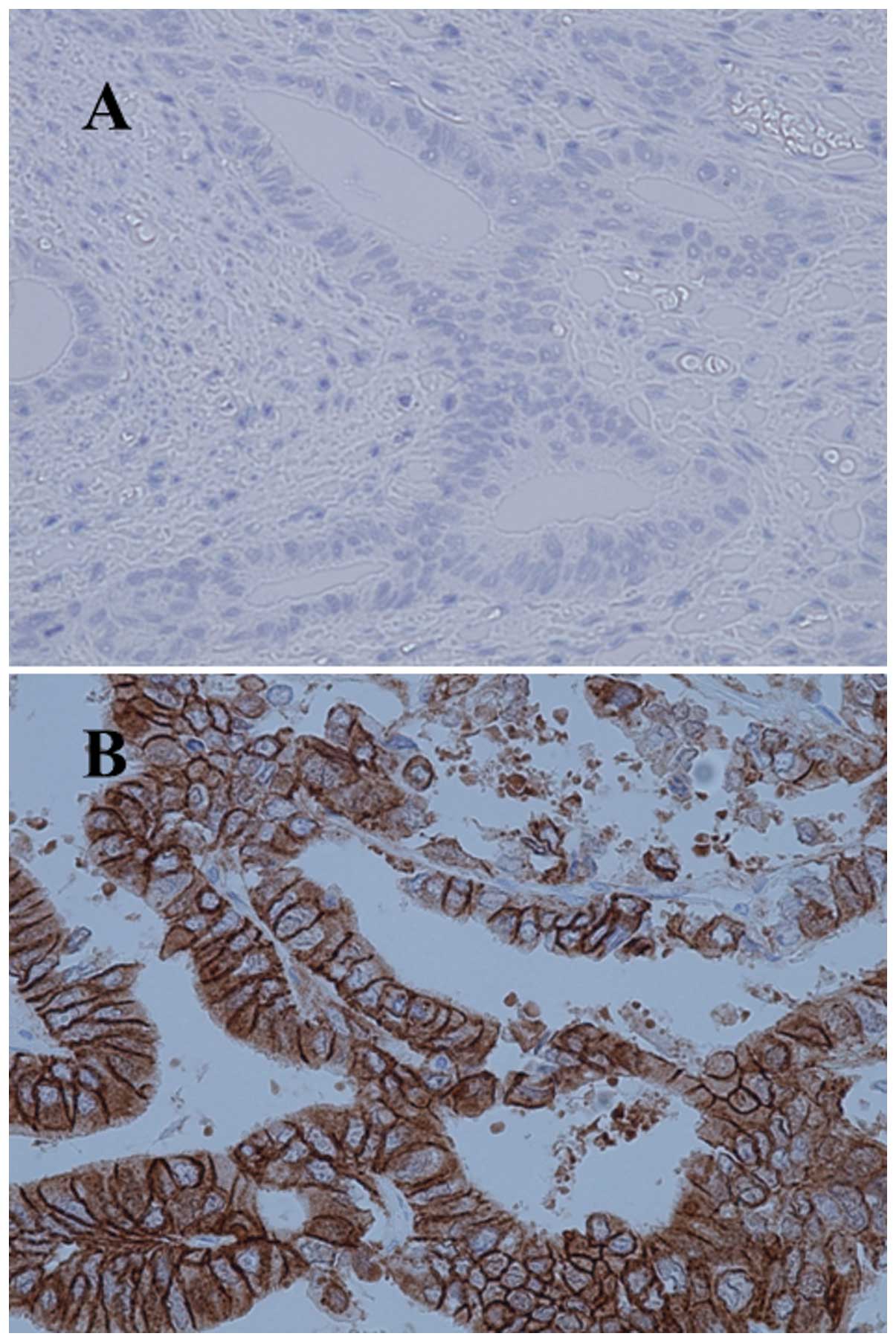
primary site and the type of intervention. The total concordance
ratio between primary sites and metastatic sites was ~97% (Table IV), reflecting a significant
correlation (P<0.0001). Only one case exhibited positive
conversion (Fig. 1A and B). This
case evaluated HER2 status between primary and paired metastatic
sites by resected specimen. Although in this case all the portions
of the primary site were examined, no positive reaction was
observed on the membrane in any of the specimens. The equivocal
(2+) case exhibited amplification by FISH and was therefore
classified as positive. Although one case of discordance between
primary and metastatic sites was identified, there were no
discordances among metachronous metastatic sites (Table V).
| Table IIHuman epidermal growth factor receptor
2 (HER2) status of primary and metastatic sites. |
Table II
Human epidermal growth factor receptor
2 (HER2) status of primary and metastatic sites.
| Metastatic sites |
|---|
|
|
|---|
| Negative | Positive |
|---|
|
|
|
|---|
| HER2 status | 0 | 1+ | 2+ (FISH−) | 2+ (FISH+) | 3+ |
|---|
| Primary sites |
| Negative |
| 0 | 28 | 1 | 0 | 0 | 1 |
| 1+ | 1 | 0 | 0 | 0 | 0 |
| 2+ (FISH−) | 0 | 0 | 0 | 0 | 0 |
| Positive |
| 2+ (FISH+) | 0 | 0 | 0 | 0 | 1 |
| 3+ | 0 | 0 | 0 | 0 | 5 |
| Table IIIAssociation of human epidermal growth
factor receptor 2 (HER2) status of the primary site with
histological type and type of intervention. |
Table III
Association of human epidermal growth
factor receptor 2 (HER2) status of the primary site with
histological type and type of intervention.
| HER2 status | |
|---|
|
| |
|---|
| Variables | Negative | Positive | P-value |
|---|
| Histological
type | | | 0.0244 |
|
Differentiated | 8 | 5 | |
|
Undifferentiated | 16 | 1 | |
| Mixed | 7 | 0 | |
| Type of
intervention | | | >0.9999 |
| Surgical
resection | 23 | 5 | |
| Biopsy | 8 | 1 | |
| Table IVConcordance of human epidermal growth
factor receptor 2 (HER2) status between primary and paired
metastatic lesions. |
Table IV
Concordance of human epidermal growth
factor receptor 2 (HER2) status between primary and paired
metastatic lesions.
| Metastatic
site | |
|---|
|
| |
|---|
| HER2 status | Negative | Positive | P-value |
|---|
| Primary site | | | <0.0001 |
| Negative | 30 | 1 | |
| Positive | 0 | 6 | |
| Table VHuman epidermal growth factor
receptor 2 (HER2) status with metachronous interventions. |
Table V
Human epidermal growth factor
receptor 2 (HER2) status with metachronous interventions.
| Case | Histology | HER2 status of the
primary site | HER2 status in
metachronous interventions |
|---|
|
|---|
| 1st | 2nd | 3rd | 4th | 5th |
|---|
| 1 | Differentiated | 0 | 0: Colon | 0: Skin | 0: Skin | 0: Kidney | 0: Skin |
| 2 |
Undifferentiated | 0 | 0: Colon | 0: Peritoneum | | | |
| 3 |
Undifferentiated | 0 | 0: Colon | 0: Liver | | | |
| 4 | Differentiated | 0 | 3+: Liver | 3+: Peritoneum | | | |
| 5 |
Undifferentiated | 0 | 0: Liver | 0: Peritoneum | | | |
| 6 | Differentiated | 3+ | 3+: Lung | 3+: Lung | | | |
| 7 | Differentiated | 3+ | 3+: Cerebellum | 3+: Lung | | | |
| 8 |
Undifferentiated | 0 | 0: Ovary | 0: Skin | | | |
| 9 |
Undifferentiated | 0 | 0: Peritoneum | 0: Peritoneum | | | |
Discussion
Previous studies have estimated the HER2 positivity
ratio to be 8.1–17.1% in gastric cancer (6,15,16).
Recent studies reported that the HER2 positivity ratio is lower in
patients with curatively resectable gastric cancer compared to that
in unresectable patients (15,16).
The present study estimated an HER2-positive ratio of ~16%,
presumably because the study population comprised patients with
curatively resected and unresected or recurrent gastric cancer. In
this study, the differentiated type of tumor exhibited a
significantly high HER2 positivity, as the consensus reported that
the majority of positive cases were histologically of the
intestinal type (17). However,
Lee et al (18) reported a
discordance of the HER2 IHC score between biopsies and
gastrectomies; in this study, there was no difference between
bioptic and resected specimens, although only a small series was
analyzed.
The HER2 status in gastric cancer is commonly
evaluated immunohistochemically, with a IHC 2+ status further
analyzed by FISH, since previous studies demonstrated a high
concordance ratio between IHC and FISH in gastric cancer (11,13,14,17,19,20)
and the American Society of Clinical Oncology/College of American
Pathologists guidelines recommend that FISH analysis be conducted
for cases with an IHC 2+ lesion in breast cancer (21). In gastric cancer, combination
chemotherapy with trastuzumab was adopted from 2010 onwards and
sufficient scientific evidence regarding HER2 has not yet been
accumulated. Therefore, references have been made to previous
studies regarding HER2 in breast cancer. Since metastatic sites
from gastric cancer patients are rarely resected or biopsied,
evidence on the concordance of HER2 status by IHC between primary
tumor and paired metastatic lesions other than lymph nodes has not
generally been reported (17,22).
In breast cancer, a high concordance ratio between primary and
matched metastatic lymph nodes has been reported (7,8).
However, in parenchymal metastases, the concordance ratio was found
to be lower (9,10). Nakamura et al (23) reported that biopsy of the
metastatic lesions may be useful for determining treatment
strategies. We examined the tumor invasive front at the primary
sites in resected specimens, as it was previously reported that
HER2 staining exhibits no preferential distribution within the
tumor, with negligible variation between the tumor mucosal surface
and the invasive front (24) and
the tumor invasive front is closely involved in the metastatic
process. However, Fusco et al (25) reported that there is discordance in
the HER2 status between the tumor invasive front and other lesions
and gastric cancer is known to exhibit heterogeneous HER2
expression (14,18). Therefore we examined all the sites
of the primary tumor in the discordant case to determine whether
there was heterogeneity of HER2 expression, in order to assess the
discordance between primary and paired metastatic lesions. However,
all the lesions were IHC-scored as 0. In addition, we investigated
whether there exists discordance among metachronous multiple
interventions. A limited number of studies (17,22,26)
have addressed such issues in gastric cancer. Kim et al
(17) reported significant
discordance (13.1%) between primary and metastatic lesions by IHC,
but no discordance by FISH. Bozzetti et al (22) reported a concordance ratio of 94.9%
between primary and matched metastatic lesions by IHC. In addition,
Kochi et al (26) reported
a discordance ratio of 9.8% in the HER2 status between primary
sites and metastatic lymph nodes by FISH and IHC. Our results using
IHC revealed a high concordance ratio (~97%) between primary and
paired metastatic lesions. These results suggest the efficacy of
HER2 status examination in the primary lesion for assessing the
status of parenchymal metastatic lesions. Only one case of positive
conversion was found in our study; likewise, Bozzetti et al
(22) reported only a single case
of discordance between the primary lesion and metastasis. The
discordant case in this study was IHC 0 at the primary site and
underwent liver resection for metachronous liver recurrence
following hepatic intra-arterial chemotherapy (fluorouracil +
cisplatin + irinotecan). The HER2 status of the liver specimen at
that time was IHC 3+ and the HER2 status of the metachronous
peritoneal recurrent specimen was also IHC 3+. These findings
suggest that the transition of the metastatic process strongly
involves the discordance of HER2 status between primary and
metastatic sites rather than heterogeneity of HER2 expression
within the primary lesion. Further investigation of this issue in a
larger series is required.
Nine patients underwent metachronous multiple
interventions for metastatic lesions; no cases of discordance
during the therapeutic period were encountered. To the best of our
knowledge, no previous studies have reported such findings.
Unfortunately, no cases in this study population
received trastuzumab combination therapy and further investigation,
including those cases, is required.
In conclusion, the concordance ratio for HER2 status
between primary and parenchymal metastatic or recurrent lesions was
high. Therefore, determining the HER2 status in the primary lesion
may be acceptable when considering the suitability of anti-HER2
agents for patients with inoperable advanced or recurrent gastric
cancer.
Acknowledgements
The authors wish to thank Yuki Mutsumi for assisting
with IHC and FISH.
References
|
1
|
Kamangar F, Dores GM and Anderson WF:
Patterns of cancer incidence, mortality, and prevalence across five
continents: defining priorities to reduce cancer disparities in
different geographic regions of the world. J Clin Oncol.
24:2137–2150. 2006. View Article : Google Scholar
|
|
2
|
Parkin DM, Bray F, Ferlay J and Pisani P:
Global cancer statistics, 2002. CA Cancer J Clin. 55:74–108. 2005.
View Article : Google Scholar
|
|
3
|
Otsuji E, Yamaguchi T, Sawai K, et al:
Recent advances in surgical treatment have improved the survival of
patients with gastric carcinoma. Cancer. 82:1233–1237. 1998.
View Article : Google Scholar : PubMed/NCBI
|
|
4
|
Horner MJ, Ries LAG, Krapcho M, et al:
SEER Cancer Statistics Review, 1975–2006. http://seer.cancer.gov/archive/csr/1975_2006/results_merged/sect_24_stomach.pdf.
Accessed April 12, 2009
|
|
5
|
Cunningham SC, Kamangar F, Kim MP, et al:
Survival after gastric adenocarcinoma serection: eighteen-year
experience at a single institution. J Gastrointest Surg. 9:718–725.
2005.PubMed/NCBI
|
|
6
|
Bang YJ, Van Cutsem E, Feyereislova A, et
al: Trastuzumab in combination with chemotherapy versus
chemotherapy alone for treatment of HER2-positive advanced gastric
or gastro-oesophageal junction cancer (ToGA): a phase 3,
open-label, randomized controlled trial. Lancet. 376:687–697. 2010.
View Article : Google Scholar
|
|
7
|
Cardoso F, Di Leo A, Larsimont D, et al:
Evaluation of HER2, p53, bcl-2, topoisomerase II-alpha, heat shock
proteins 27 and 70 in primary breast cancer and metastatic
ipsilateral axillary lymph nodes. Ann Oncol. 12:615–620. 2001.
View Article : Google Scholar : PubMed/NCBI
|
|
8
|
Simon R, Nocito A, Huebscher T, et al:
Patterns of her-2/neu amplification and overexpression in primary
and metastatic breast cancer. J Natl Cancer Inst. 93:1141–1146.
2001. View Article : Google Scholar : PubMed/NCBI
|
|
9
|
Chang HJ, Han SW, Oh DY, et al: Discordant
human epidermal growth factor receptor 2 and hormone receptor
status in primary and metastatic breast cancer and response to
trastuzumab. Jpn J Clin Oncol. 41:593–599. 2011. View Article : Google Scholar
|
|
10
|
Chan A, Morey A, Brown B, et al: A
retrospective study investigating the rate of HER2 discordance
between primary breast carcinoma and locoregional or metastatic
disease. BMC Cancer. 12:5552012. View Article : Google Scholar : PubMed/NCBI
|
|
11
|
Marx AH, Tharun L, Muth J, et al: HER-2
amplification is highly homogenous in gastric cancer. Hum Pathol.
40:769–777. 2009. View Article : Google Scholar : PubMed/NCBI
|
|
12
|
Fassan M, Ludwig K, Pizzi M, et al: Human
epithelial growth factor receptor 2 (HER2) status in primary and
metastatic esophagogastric junction adenocarcinomas. Hum Pathol.
43:1206–1212. 2012. View Article : Google Scholar : PubMed/NCBI
|
|
13
|
Tafe LJ, Janjigian YY, Zaidinski M, et al:
Human epidermal growth factor receptor 2 testing in
gastroesophageal cancer: correlation between immunohistochemistry
and fluorescence in situ hybridization. Arch Pathol Lab Med.
135:1460–1465. 2011. View Article : Google Scholar
|
|
14
|
Hofmann M, Stoss O, Shi D, et al:
Assessment of a HER2 scoring system for gastric cancer: results
from a validation study. Histopathology. 52:797–805. 2008.
View Article : Google Scholar : PubMed/NCBI
|
|
15
|
Kunz PL, Mojtahed A, Fisher GA, et al:
HER2 expression in gastric and gastroesophageal junction
adenocarcinoma in a US population: clinicopathologic analysis with
proposed approach to HER2 assessment. Appl Immunohistochem Mol
Morphol. 20:13–24. 2012. View Article : Google Scholar
|
|
16
|
Kataoka Y, Okabe H, Yoshizawa A, et al:
HER2 expression and its clinicopathological features in resectable
gastric cancer. Gastric Cancer. 16:84–93. 2013. View Article : Google Scholar : PubMed/NCBI
|
|
17
|
Kim MA, Lee HJ, Yang HK, et al:
Heterogeneous amplification of ERBB2 in primary lesions is
responsible for the discordant ERBB2 status of primary and
metastatic lesions in gastric carcinoma. Histopathology.
59:822–831. 2011. View Article : Google Scholar
|
|
18
|
Lee S, de Boer WB, Fermoyle S, et al:
Human epidermal growth factor 2 testing in gastric carcinoma:
issues related to heterogeneity in biopsies and resections.
Histopathology. 59:832–840. 2011. View Article : Google Scholar : PubMed/NCBI
|
|
19
|
Takehara T, Kunitomo K, Kono K, et al:
Status of c-erbB-2 in gastric adenocarcinoma: a comparative study
of immunohistochemistry, fluorescence in situ hybridization and
enzyme-linked immuno-sorbent assay. Int J Cancer. 98:833–837. 2002.
View Article : Google Scholar
|
|
20
|
Yano T, Doi T, Ohtsu A, et al: Comparison
of HER2 gene amplification assessed by fluorescence in
situ hybridization and HER2 protein expression assessed by
immunohistochemistry in gastric cancer. Oncol Rep. 15:65–71.
2006.
|
|
21
|
Wolff AC, Hammond ME, Schwartz JN, et al;
American Society of Clinical Oncology; College of American
Pathologists. American Society of Clinical Oncology/College of
American Pathologists guideline recommendations for human epidermal
growth factor receptor 2 testing in breast cancer. J Clin Oncol.
25:118–145. 2007. View Article : Google Scholar
|
|
22
|
Bozzetti C, Negri FV, Lagrasta CA, et al:
Comparison of HER2 status in primary and paired metastatic sites of
gastric carcinoma. Br J Cancer. 104:1372–1376. 2011. View Article : Google Scholar : PubMed/NCBI
|
|
23
|
Nakamura R, Yamamoto N, Onai Y, et al:
Importance of confirming HER2 overexpression of recurrence lesion
in breast cancer patients. Breast Cancer. 20:336–341. 2013.
View Article : Google Scholar : PubMed/NCBI
|
|
24
|
Kim KC, Koh YW, Chang HM, et al:
Evaluation of HER2 protein expression in gastric carcinomas:
comparative analysis of 1,414 cases of whole-tissue sections and
595 cases of tissue microarrays. Ann Surg Oncol. 18:2833–2840.
2011. View Article : Google Scholar
|
|
25
|
Fusco N, Rocco EG, Del Conte C, et al:
HER2 in gastric cancer: a digital image analysis in pre-neoplastic,
primary and metastatic lesions. Mod Pathol. 26:816–824. 2013.
View Article : Google Scholar : PubMed/NCBI
|
|
26
|
Kochi M, Fujii M, Masuda S, et al:
Differing deregulation of HER2 in primary gastric cancer and
synchronous related metastatic lymph nodes. Diagn Pathol.
8:1912013. View Article : Google Scholar : PubMed/NCBI
|