Magnetic resonance imaging shrinkage patterns following neoadjuvant chemotherapy for breast carcinomas with an emphasis on the radiopathological correlations

  • Authors:
    • Kaori Tomida
    • Mitsuaki Ishida
    • Tomoko Umeda
    • Sachiko Sakai
    • Yuki Kawai
    • Tsuyoshi Mori
    • Yoshihiro Kubota
    • Eiji Mekata
    • Shigeyuki Naka
    • Hajime Abe
    • Hidetoshi Okabe
    • Tohru Tani
  • View Affiliations

  • Published online on: June 30, 2014     https://doi.org/10.3892/mco.2014.333
  • Pages: 783-788
Metrics: Total Views: 0 (Spandidos Publications: | PMC Statistics: )
Total PDF Downloads: 0 (Spandidos Publications: | PMC Statistics: )


Abstract

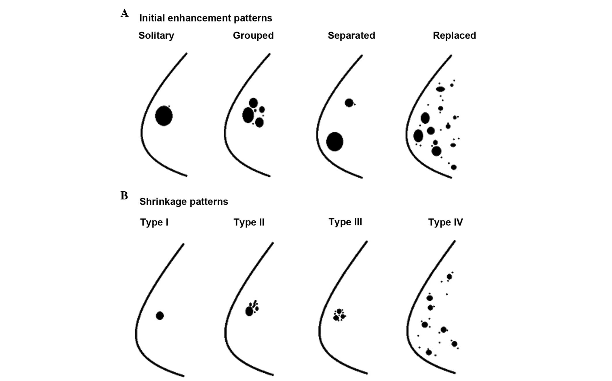
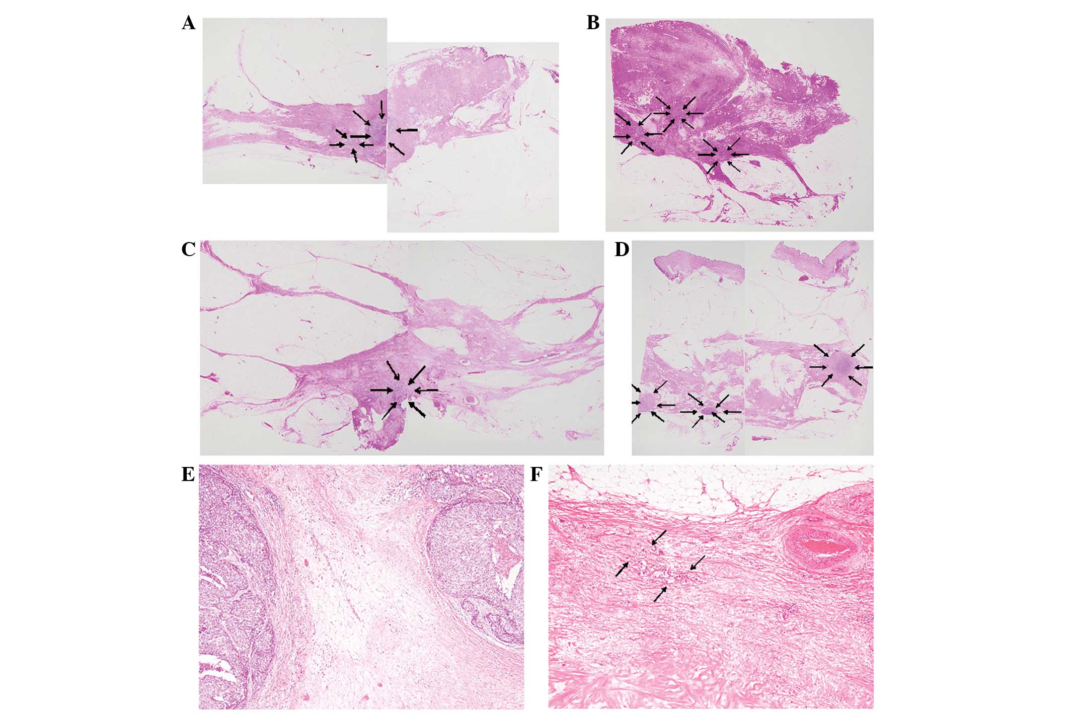
Preoperative neoadjuvant chemotherapy (NAC) is considered to be the standard treatment for locally‑advanced breast carcinomas. Obtaining precise information regarding the tumor extent and distribution by imaging modalities to assess the success of breast‑conserving surgery following NAC is extremely important. Analysis of the detailed radiopathological correlation of magnetic resonance imaging (MRI) following NAC has not been reported previously. The MRI and histopathological shrinkage patterns of residual breast carcinomas in 27 consecutive cases were analyzed following NAC and classified into five categories: Types I and II (concentric shrinkage with and without surrounding lesions, respectively); type III (shrinkage with residual multinodular lesions); type IV (diffuse contrast enhancement in whole quadrant); and non‑visualization. The present study clearly demonstrated that the most common MRI shrinkage pattern was type I (11 cases), followed by type II and non‑visuali­zation, and the most common histopathological shrinkage pattern was type II (11 cases), followed by type III (8 cases). The concordance rate between MRI and pathological patterns was 48% and the worst concordance MRI pattern was type I. MRI is considered to be a useful method for evaluation of the residual carcinoma following NAC. However, the concordance rate was low in the MRI pattern I cases and tiny foci of residual carcinoma were present in half of the non‑visualization cases, as shown by MRI. Therefore, the tumor extent must be completely resected for patients who undergo NAC, and postoperative radiation may be important for preventing local recurrence of breast carcinoma.
View Figures
View References

Related Articles

Journal Cover

September-October 2014
Volume 2 Issue 5

Print ISSN: 2049-9450
Online ISSN:2049-9469

Sign up for eToc alerts

Recommend to Library

Copy and paste a formatted citation
x
Spandidos Publications style
Tomida K, Ishida M, Umeda T, Sakai S, Kawai Y, Mori T, Kubota Y, Mekata E, Naka S, Abe H, Abe H, et al: Magnetic resonance imaging shrinkage patterns following neoadjuvant chemotherapy for breast carcinomas with an emphasis on the radiopathological correlations. Mol Clin Oncol 2: 783-788, 2014
APA
Tomida, K., Ishida, M., Umeda, T., Sakai, S., Kawai, Y., Mori, T. ... Tani, T. (2014). Magnetic resonance imaging shrinkage patterns following neoadjuvant chemotherapy for breast carcinomas with an emphasis on the radiopathological correlations. Molecular and Clinical Oncology, 2, 783-788. https://doi.org/10.3892/mco.2014.333
MLA
Tomida, K., Ishida, M., Umeda, T., Sakai, S., Kawai, Y., Mori, T., Kubota, Y., Mekata, E., Naka, S., Abe, H., Okabe, H., Tani, T."Magnetic resonance imaging shrinkage patterns following neoadjuvant chemotherapy for breast carcinomas with an emphasis on the radiopathological correlations". Molecular and Clinical Oncology 2.5 (2014): 783-788.
Chicago
Tomida, K., Ishida, M., Umeda, T., Sakai, S., Kawai, Y., Mori, T., Kubota, Y., Mekata, E., Naka, S., Abe, H., Okabe, H., Tani, T."Magnetic resonance imaging shrinkage patterns following neoadjuvant chemotherapy for breast carcinomas with an emphasis on the radiopathological correlations". Molecular and Clinical Oncology 2, no. 5 (2014): 783-788. https://doi.org/10.3892/mco.2014.333