Introduction
Periosteal chondroma, also known as juxtacortical
chondroma, is a benign cartilaginous neoplasm of the bone surface
that accounts for <2% of all chondromas (1). Periosteal chondroma exhibits a notable
tendency to involve the proximal humerus and distal femur. Patients
present with a swelling or palpable mass that may be painful.
Surgical excision is the treatment of choice. Local recurrence is
extremely uncommon and is associated with incomplete excision. This
is a presentation of the clinicopathological and radiological
characteristics of a case of periosteal chondroma involving the
distal tibia in a young adult female patient. Written informed
consent was obtained from the patient for publication of this case
report and accompanying images.
Case report
A 25-year-old woman was referred to our hospital for
further evaluation of abnormal findings on an ankle radiograph. The
patient had a history of antecedent trauma to the left distal lower
limb. The physical examination revealed swelling and tenderness in
the anterolateral aspect of the left distal lower limb. The
laboratory data were within normal limits. The patient's medical
history was non-contributory.
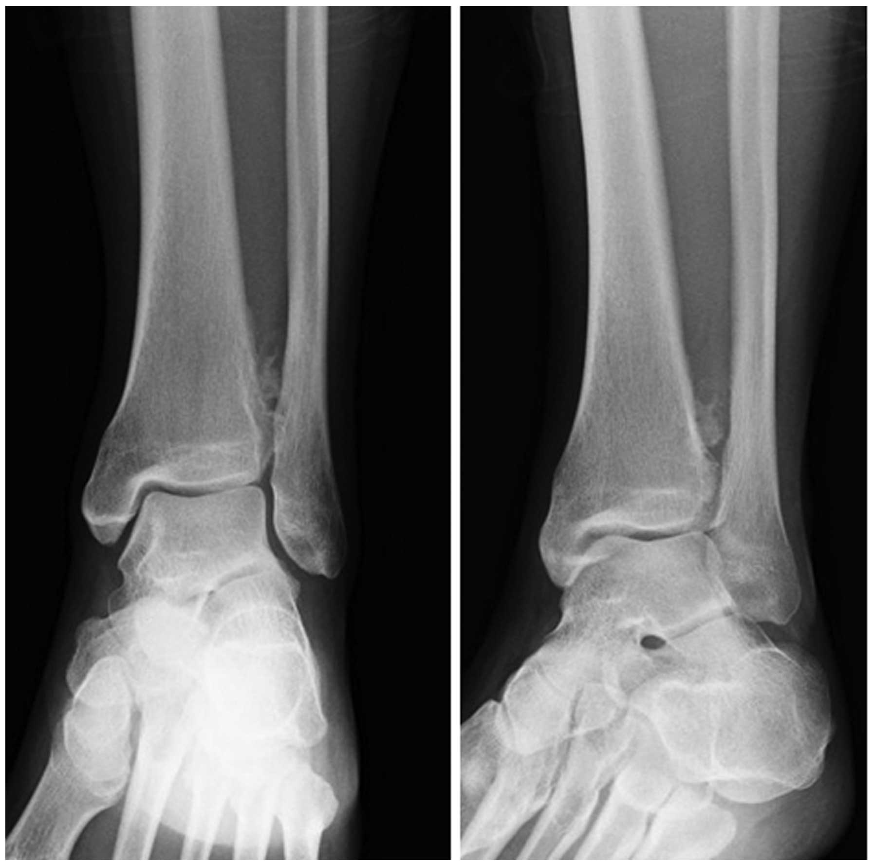
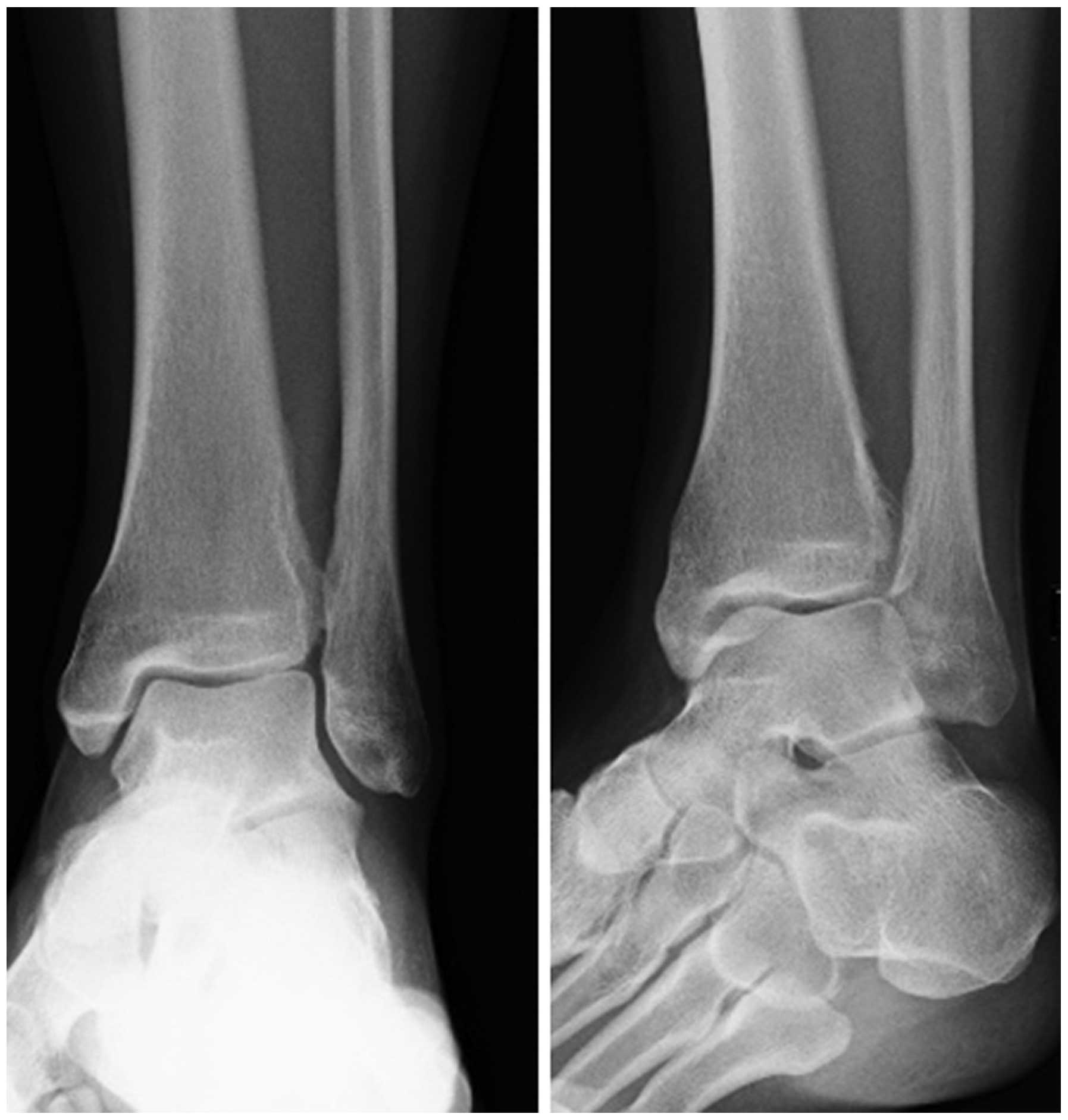
Plain radiographs revealed a discernible soft tissue
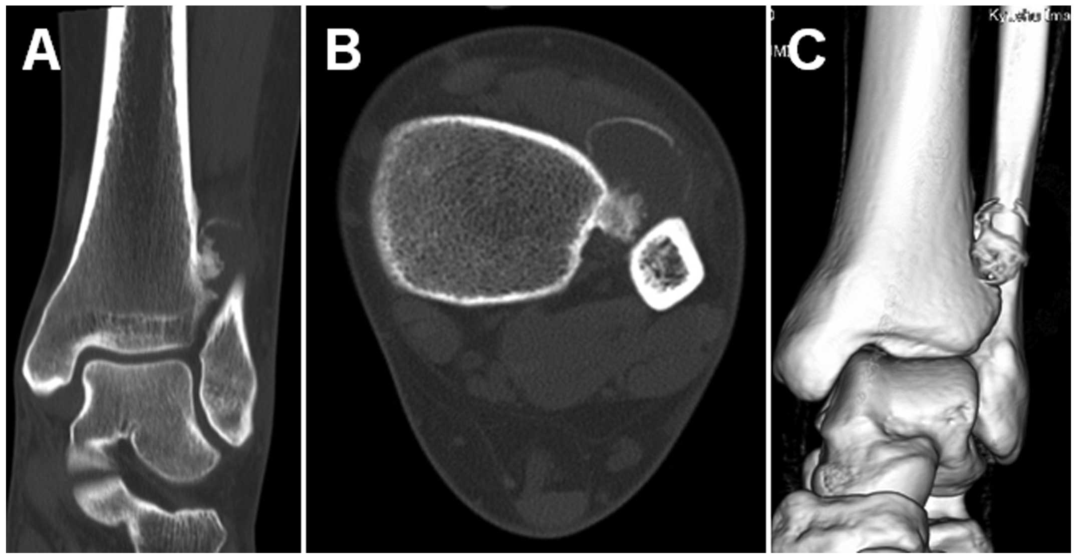
lesion with peripheral foci of mineralization (Fig. 1). Computed tomography (CT) scans
confirmed the presence of a surface-based mass with peripheral
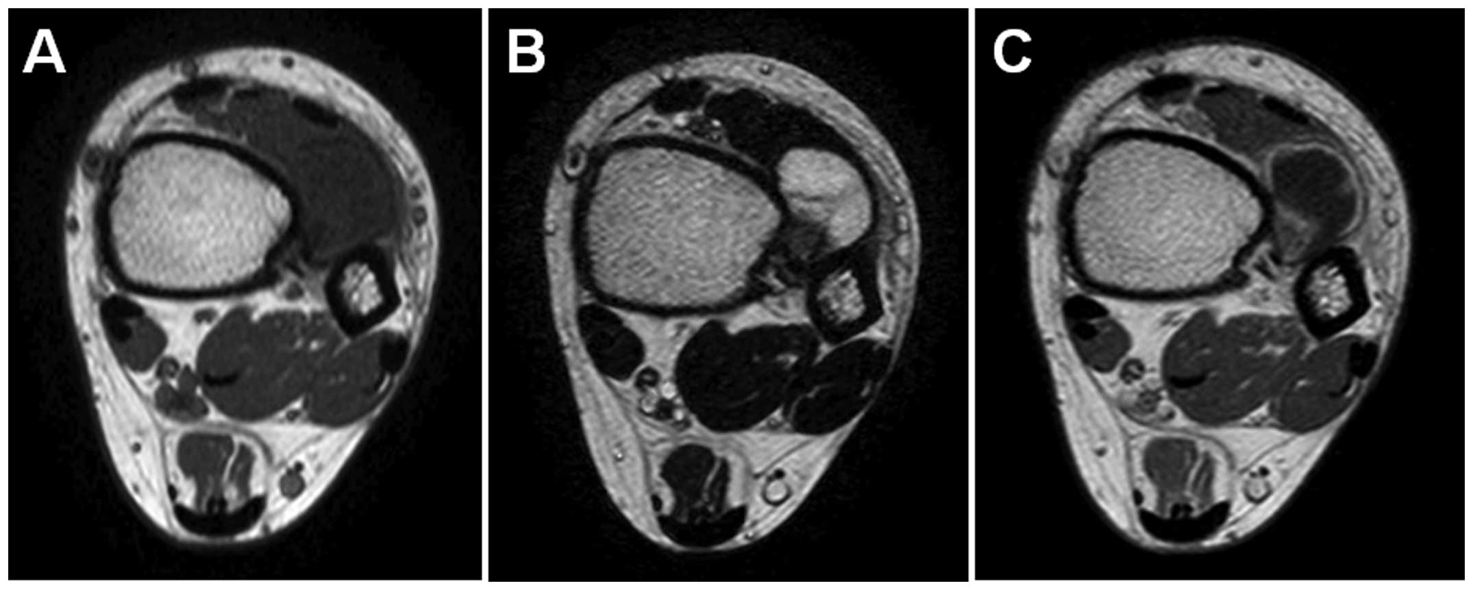
ossification and a thin rim of calcification (Fig. 2). Magnetic resonance imaging (MRI)
revealed a well-circumscribed juxtacortical mass measuring
2.5×1.8×1.5 cm. The mass exhibited intermediate signal intensity on
T1-weighted sequences (Fig. 3A) and
high signal intensity with foci of decreased signal intensity on
T2-weighted sequences (Fig. 3B).
Contrast-enhanced T1-weighted sequences demonstrated predominantly
peripheral enhancement without intramedullary involvement (Fig. 3C). Soft tissue edema adjacent to the
lesion was also observed.
Following an open biopsy, marginal excision with
curettage of the underlying bone cortex was performed.
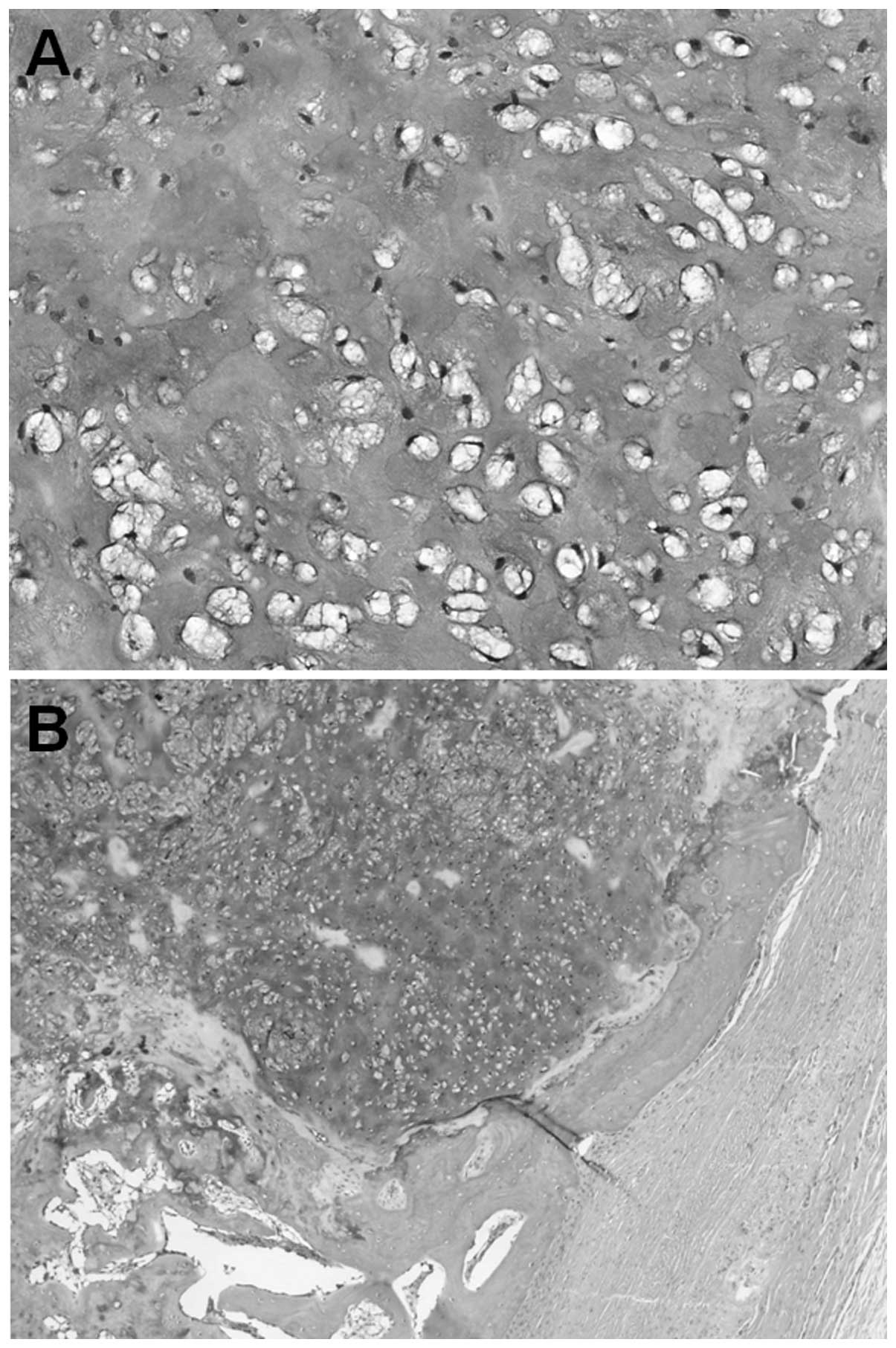
Histologically, the tumor was well defined and surrounded by a
periosteum-like fibrous capsule. The tumor was composed of a
proliferation of chondrocytes in an abundant myxoid or
chondromyxoid matrix (Fig. 4A). Foci
of ossification with mature bone trabeculae forming a thin
shell-like structure were found in the periphery of the tumor
(Fig. 4B). The cytoplasm of the tumor
cells was strongly positive for periodic acid-Schiff reaction.
Alcian blue staining demonstrated an abundanCE OF acid mucin in the
stroma. The mindbomb E3 ubiquitin protein ligase 1 labeling index
was <1%. There was no nuclear atypia and mitotic figures were
not detected. Based on these characteristics, the tumor was
diagnosed as periosteal chondroma.
The postoperative course was uneventful and THERE
WAS NO evidence of local recurrence 4 months after surgery
(Fig. 5).
Discussion
Periosteal chondroma is significantly less common
compared TO enchondroma and predominantly occurs in children and
young adults, with a marginal male predominance (1). Periosteal chondroma tends to arise in
the metaphysis of the proximal humerus or distal femur. The small
tubular bones of the hand are also common sites (2). Periosteal chondroma rarely exceeds 3 cm
in maximum diameter and may erode the underlying cortex without
penetrating into the medullar cavity. Histologically, the lesion
occasionally exhibits hypercellularity, nuclear pleomorphism and
binucleation, which may lead to a misdiagnosis of chondrosarcoma.
It is therefore crucial to be familiar with the imaging
characteristics of periosteal chondroma, in order to avoid a
misdiagnosis.
The pathogenesis of periosteal chondroma is not well
understood. It is of interest that our patient had a history of at
least one traumatic event. In the present case, it was suspected
that trauma may be a related or predisposing factor for the
development of periosteal chondroma. However, congenital periosteal
chondroma has been reported in the literature (3). Recently, heterozygous mutations of the
isocitrate dehydrogenase 1 gene were detected in a number of
periosteal chondromas (4).
Plain radiographs commonly reveal a discernible
soft-tissue lesion with cortical scalloping, underlying cortical
sclerosis and overhanging margins (2,5). The
lesion may exhibit a sclerotic rim or thin cortical shell. CT is
useful in identifying the presence of scattered mineralizationS. On
MRI, periosteal chondroma typically appears as a well-circumscribed
mass with intermediate signal intensity on T1-weighted sequences
and high signal intensity with variable low signal intensity foci
on T2-weighted sequences (2,5,6).
Intramedullary involvement is quite uncommon, although surrounding
soft-tissue edema may occasionally be observed (5). Periosteal chondroma demonstrates
predominantly peripheral enhancement following contrast agent
administration (6). In our case, the
tumor was <3 cm and arose on the surface of the distal tibia.
The imaging characteristics were consistent with those described in
the literature.
The differential diagnosis of the present case
included myositis ossificans, bizarre parosteal osteochondromatous
proliferation (BPOP) and periosteal chondrosarcoma. Myositis
ossificans is the most common benign bone-forming lesion that
mainly affects active adolescents and young adults, with a marginal
male predominance (7). The lesion is
associated with a single traumatic event or repeated minor trauma
in the majority of the cases. The zoning phenomenon of peripheral
maturation is the most significant diagnostic feature (8). BPOP, also referred to as Nora's lesion,
is a surface-based osteocartilaginous lesion that typically affects
the hands and feet in young adults (9) and may also occur in long bones in ~25%
of the cases (10). Antecedent trauma
may be considered as an etiologic factor. Radiographically, BPOP
typically appears as a well-marginated mass of heterotopic
mineralization arising from the cortical surface without affecting
the underlying bone architecture (10). The MRI characteristics are
non-specific, demonstrating intermediate signal intensity on
T1-weighted sequences and intermediate to high signal intensity on
T2-weighted sequences, with marked contrast enhancement (10,11).
Histologically, BPOP consists of three components in different
amounts, namely cartilage, bone and fibrous tissue. The matrix in
cartilage and bone has a characteristic blue tinctorial quality at
the osteocartilaginous interfaces (12). Periosteal chondrosarcoma predominantly
occurs in the metaphyses of long bones and has a peak incidence
between the second and fourth decades of life, with a marginal male
predominance (13). The lesion is
generally >5 cm in diameter. Radiographically, periosteal
chondrosarcoma is often round and displays granular or ‘popcorn’
cartilaginous opacities (14). On
MRI, the mass is well-delineated, with low to intermediate signal
intensity on T1-weighted sequences and high signal intensity on
T2-weighted sequences. There is peripheral and septal enhancement
following contrast agent administration (14). According to Robinson et al
(15), lesion size is the most
reliable predictor for distinguishing periosteal chondrosarcoma
from periosteal chondroma.
In summary, we described the clinicopathological and
radiological characteristics of a periosteal chondroma involving
the distal tibia in a young female patient. Although rare,
periosteal chondroma should be considered in the differential
diagnosis of a surface-based lesion with matrix mineralization in
the metaphysis of long bones.
References
|
1
|
Lucas DR and Bridge JA: Chondromas:
enchondroma, periosteal chondromaWHO Classification of Tumours of
Soft Tissue and Bone, Fletcher CDM. Bridge JA, Hogendoorn PCW and
Mertens F: 5. 4th. IARC Press Lyon; pp. 252–254. 2013
|
|
2
|
Kosaka H, Nishio J, Matsunaga T, Aoki M,
Iwasaki H and Naito M: Imaging features of periosteal chondroma
manifesting as a subcutaneous mass in the index finger. Case Rep
Orthop. 2014:7634802014.PubMed/NCBI
|
|
3
|
Domson GF, Bush CH, Reith JR, Rajaram A,
Scarborough MT and Gibbs CP: Periosteal chondroma at birth.
Skeletal Radiol. 37:559–562. 2008. View Article : Google Scholar : PubMed/NCBI
|
|
4
|
Amary MF, Bacsi K, Maggiani F, et al: IDH1
and IDH2 mutations are frequent events in central chondrosarcoma
and central and periosteal chondromas but not in other mesenchymal
tumours. J Pathol. 224:334–343. 2011. View Article : Google Scholar : PubMed/NCBI
|
|
5
|
Miller FS: Imaging features of
juxtacortical chondroma in children. Pediatr Radiol. 44:56–63.
2014. View Article : Google Scholar : PubMed/NCBI
|
|
6
|
Woertler K, Blasius S, Brinkschmidt C,
Hillmann A, Link TM and Heindel W: Periosteal chondroma: MR
characteristics. J Comput Assist Tomogr. 25:425–430. 2001.
View Article : Google Scholar : PubMed/NCBI
|
|
7
|
Rosenberg AE and Oliveira AM: Myositis
ossificans and fibro-osseous pseudotumour of digitsWHO
Classification of Tumours of Soft Tissue and Bone, Fletcher CDM.
Bridge JAHogendoornPCWMertensF: 5. 4th. IARC Press; Lyon: pp.
50–51. 2013
|
|
8
|
Tyler P and Saifuddin A: The imaging of
myositis ossificans. Semin Musculoskelet Radiol. 14:201–216. 2010.
View Article : Google Scholar : PubMed/NCBI
|
|
9
|
Berber O, Dawson-Bowling S, Jalgaonkar A,
Miles J, Pollock RC, Skinner JA, Aston WJ and Briggs TW: Bizarre
parosteal osteochondromatous proliferation of bone: clinical
management of a series of 22 cases. J Bone Joint Surg Br.
93:1118–1121. 2011. View Article : Google Scholar : PubMed/NCBI
|
|
10
|
Dhondt E, Oudenhoven L, Khan S, Kroon HM,
Hogendoorn PC, Nieborg A, Bloem JL and De Schepper A: Nora's
lesion, a distinct radiological entity? Skeletal Radiol.
35:497–502. 2006. View Article : Google Scholar : PubMed/NCBI
|
|
11
|
Torreggiani WC, Munk PL, Al-Ismail K,
O'Connell JX, Nicolaou S, Lee MJ and Masri BA: MR imaging features
of bizarre parosteal osteochondromatous proliferation of bone
(Nora's lesion). Eur J Radiol. 40:224–231. 2001. View Article : Google Scholar : PubMed/NCBI
|
|
12
|
Abramovici L and Steiner GC: Bizarre
parosteal osteochondromatous proliferation (Nora's lesion): a
retrospective study of 12 cases, 2 arising in long bones. Hum
Pathol. 33:1205–1210. 2002. View Article : Google Scholar : PubMed/NCBI
|
|
13
|
Goedhart LM, Ploegmakers JJ, Kroon HM,
Zwartkruis EC and Jutte PC: The presentation, treatment and outcome
of periosteal chondrosarcoma in the Netherlands. Bone Joint J.
96-B:823–828. 2014. View Article : Google Scholar : PubMed/NCBI
|
|
14
|
Chaabane S, Bouaziz MC, Drissi C, Abid L
and Ladeb MF: Periosteal chondrosarcoma. AJR Am J Roentgenol.
192:W1–W6. 2009. View Article : Google Scholar : PubMed/NCBI
|
|
15
|
Robinson P, White LM, Sundaram M, Kandel
R, Wunder J, McDonald DJ, Janney C and Bell RS: Periosteal
chondroid tumors: radiologic evaluation with pathologic
correlation. AJR Am J Roentgenol. 177:1183–1188. 2001. View Article : Google Scholar : PubMed/NCBI
|