Postoperative brachytherapy and electron beam irradiation for keloids: A single institution retrospective analysis

  • Authors:
    • Qiwen Duan
    • Junhua Liu
    • Zhiguo Luo
    • Chenhao Hu
  • View Affiliations

  • Published online on: January 27, 2015     https://doi.org/10.3892/mco.2015.498
  • Pages: 550-554
Metrics: Total Views: 0 (Spandidos Publications: | PMC Statistics: )
Total PDF Downloads: 0 (Spandidos Publications: | PMC Statistics: )


Abstract

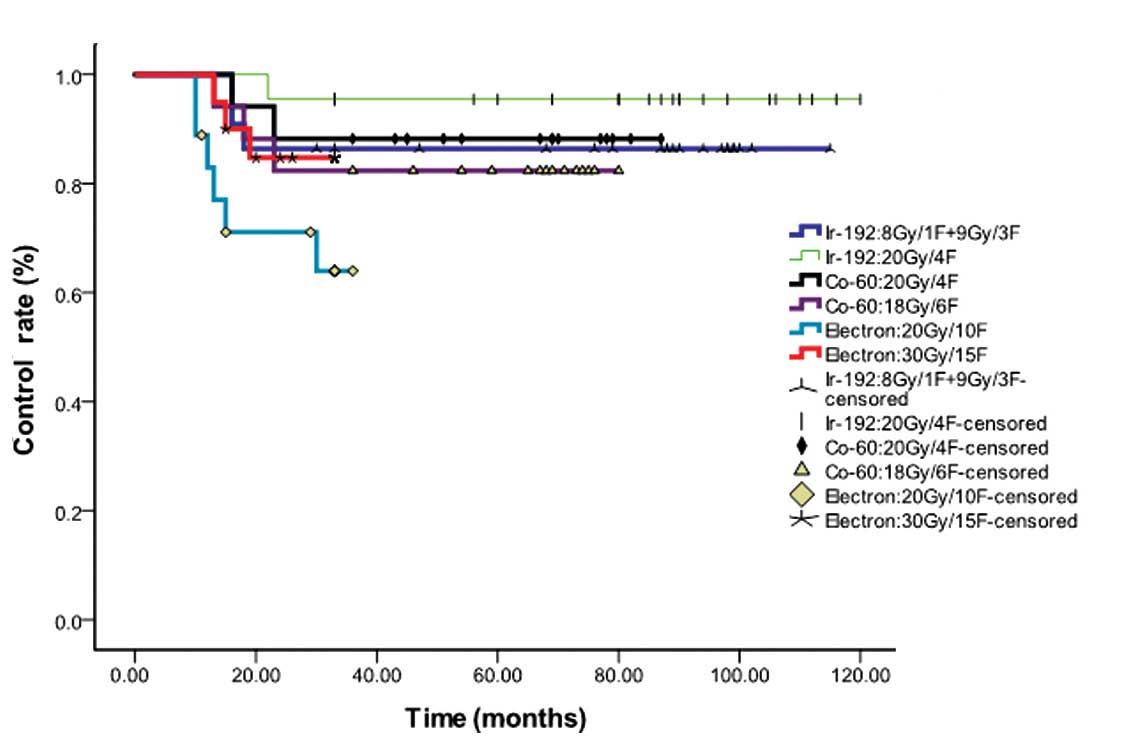
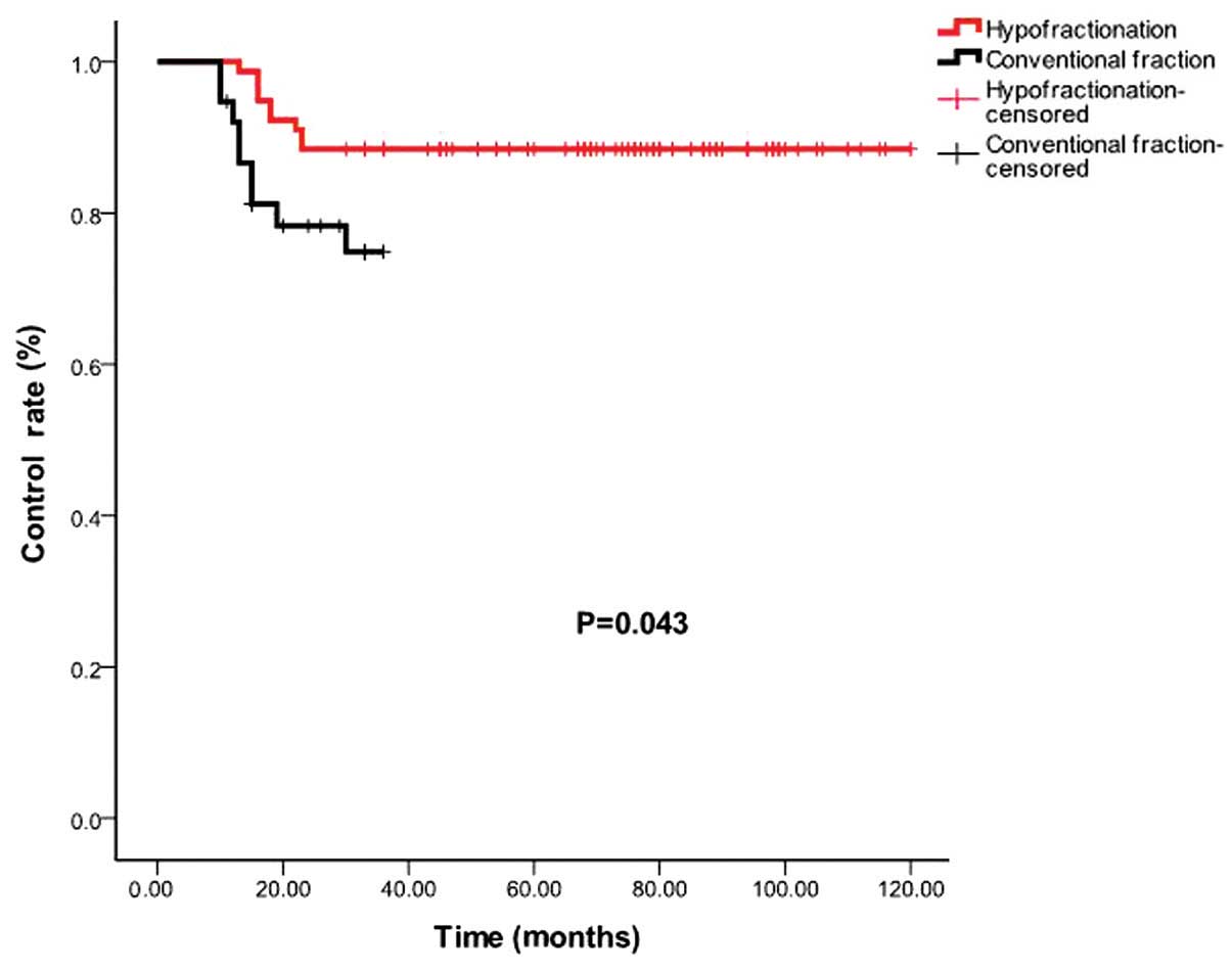
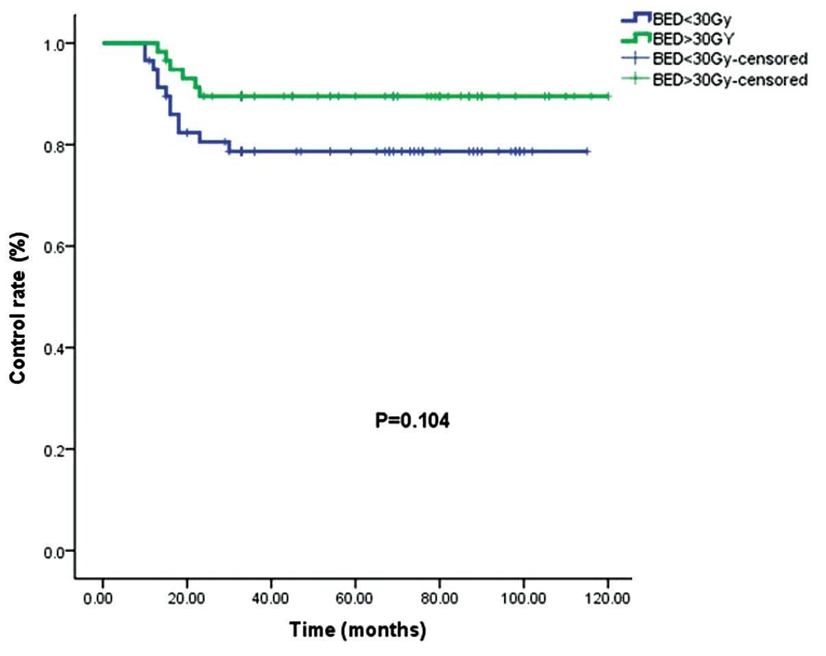
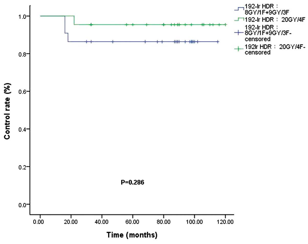
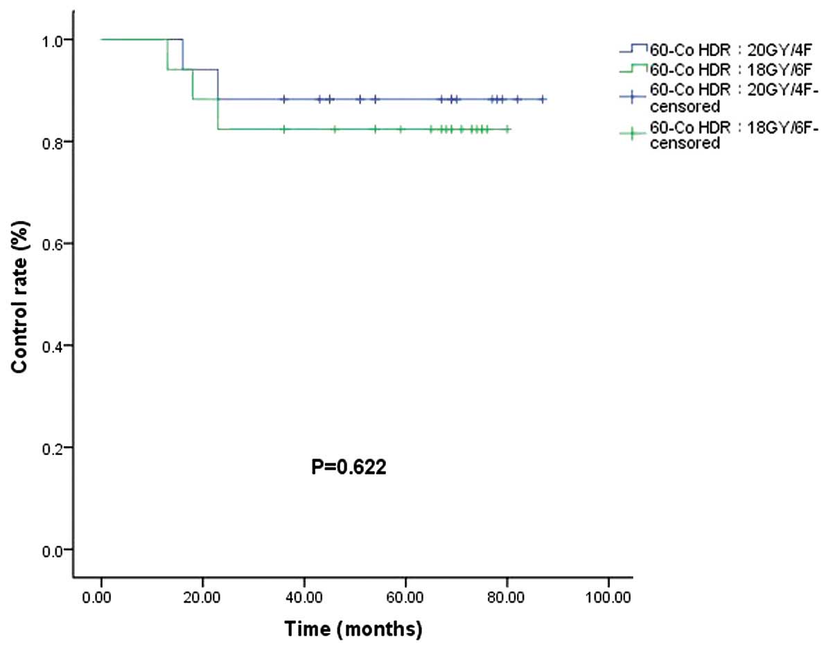
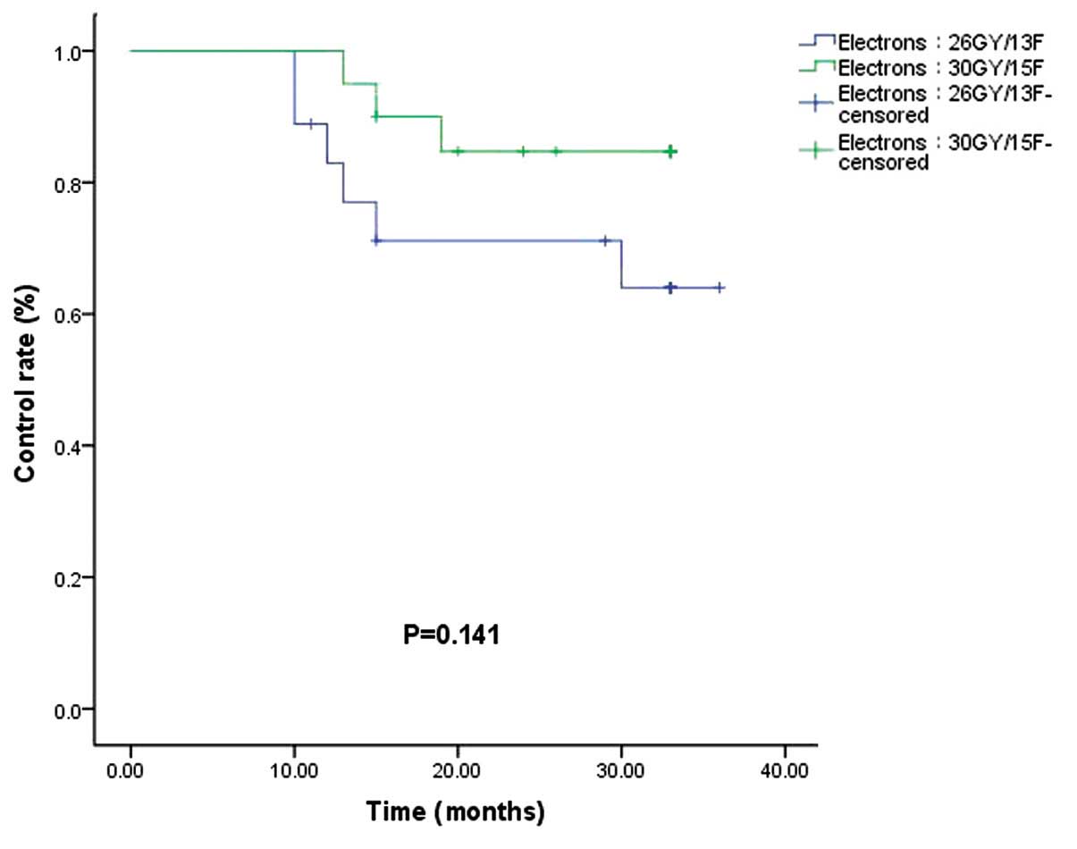
The aim of the present study was to perform a retrospective analysis of the control rate and toxicity of postoperative brachytherapy and electron beam irradiation for keloids. A retrospective review was performed of 116 keloid patients who underwent postoperative brachytherapy and electron beam irradiation between January 1, 2002 and June 30, 2012. Several different radiotherapy techniques and fractionation schedules were performed in the analysis, including high‑dose rate (HDR) irradiation with 192Ir at 8 Gy/1 fraction (F)+9 Gy/3F or 20 Gy/4F; HDR brachytherapy with 60Co at 20 Gy/4F or 18 Gy/6F; or external beam electron therapy at 26 Gy/13F or 30 Gy/15F. The endpoints of the study were analysis of the control rate and toxicity. The median observation period was 46.5 months (range, 10.0‑120.0 months) for all patients. In total, 18 of the 116 patients relapsed, and 16.7 months (range, 10.0‑30.0 months) was the median time to recurrence for these patients. The control rates for the patients who received hypofractionation (>2 Gy per fraction) and conventional fraction (2 Gy per fraction) were 88.5 and 76.3%, respectively (P=0.043). The control rates for the patients whose calculated biological effective doses (BED) were >30 Gy and <30 Gy were 89.7 and 79.3%, respectively (P=0.104). There were no grade 2 or higher adverse effects based on the Common Terminology Criteria for Adverse Events v3.0 in the late phase. No evidence was identified for a link between radiotherapy and the subsequent occurrence of cancer. The results of the present study indicate that hypofractionated radiotherapy played an important role as an adjuvant therapy following surgical excision of keloids. A BED of >30 Gy appears to be sufficient. No definitive evidence was found for an association between radiotherapy and the occurrence of cancer during the follow‑up, however, more cases and longer follow‑up periods are required.
View Figures
View References

Related Articles

Journal Cover

May-2015
Volume 3 Issue 3

Print ISSN: 2049-9450
Online ISSN:2049-9469

Sign up for eToc alerts

Recommend to Library

Copy and paste a formatted citation
x
Spandidos Publications style
Duan Q, Liu J, Luo Z and Hu C: Postoperative brachytherapy and electron beam irradiation for keloids: A single institution retrospective analysis. Mol Clin Oncol 3: 550-554, 2015
APA
Duan, Q., Liu, J., Luo, Z., & Hu, C. (2015). Postoperative brachytherapy and electron beam irradiation for keloids: A single institution retrospective analysis. Molecular and Clinical Oncology, 3, 550-554. https://doi.org/10.3892/mco.2015.498
MLA
Duan, Q., Liu, J., Luo, Z., Hu, C."Postoperative brachytherapy and electron beam irradiation for keloids: A single institution retrospective analysis". Molecular and Clinical Oncology 3.3 (2015): 550-554.
Chicago
Duan, Q., Liu, J., Luo, Z., Hu, C."Postoperative brachytherapy and electron beam irradiation for keloids: A single institution retrospective analysis". Molecular and Clinical Oncology 3, no. 3 (2015): 550-554. https://doi.org/10.3892/mco.2015.498