Spandidos Publications Logo
  • About
    • About Spandidos
    • Aims and Scopes
    • Abstracting and Indexing
    • Editorial Policies
    • Reprints and Permissions
    • Job Opportunities
    • Terms and Conditions
    • Contact
  • Journals
    • All Journals
    • Oncology Letters
      • Oncology Letters
      • Information for Authors
      • Editorial Policies
      • Editorial Board
      • Aims and Scope
      • Abstracting and Indexing
      • Bibliographic Information
      • Archive
    • International Journal of Oncology
      • International Journal of Oncology
      • Information for Authors
      • Editorial Policies
      • Editorial Board
      • Aims and Scope
      • Abstracting and Indexing
      • Bibliographic Information
      • Archive
    • Molecular and Clinical Oncology
      • Molecular and Clinical Oncology
      • Information for Authors
      • Editorial Policies
      • Editorial Board
      • Aims and Scope
      • Abstracting and Indexing
      • Bibliographic Information
      • Archive
    • Experimental and Therapeutic Medicine
      • Experimental and Therapeutic Medicine
      • Information for Authors
      • Editorial Policies
      • Editorial Board
      • Aims and Scope
      • Abstracting and Indexing
      • Bibliographic Information
      • Archive
    • International Journal of Molecular Medicine
      • International Journal of Molecular Medicine
      • Information for Authors
      • Editorial Policies
      • Editorial Board
      • Aims and Scope
      • Abstracting and Indexing
      • Bibliographic Information
      • Archive
    • Biomedical Reports
      • Biomedical Reports
      • Information for Authors
      • Editorial Policies
      • Editorial Board
      • Aims and Scope
      • Abstracting and Indexing
      • Bibliographic Information
      • Archive
    • Oncology Reports
      • Oncology Reports
      • Information for Authors
      • Editorial Policies
      • Editorial Board
      • Aims and Scope
      • Abstracting and Indexing
      • Bibliographic Information
      • Archive
    • Molecular Medicine Reports
      • Molecular Medicine Reports
      • Information for Authors
      • Editorial Policies
      • Editorial Board
      • Aims and Scope
      • Abstracting and Indexing
      • Bibliographic Information
      • Archive
    • World Academy of Sciences Journal
      • World Academy of Sciences Journal
      • Information for Authors
      • Editorial Policies
      • Editorial Board
      • Aims and Scope
      • Abstracting and Indexing
      • Bibliographic Information
      • Archive
    • International Journal of Functional Nutrition
      • International Journal of Functional Nutrition
      • Information for Authors
      • Editorial Policies
      • Editorial Board
      • Aims and Scope
      • Abstracting and Indexing
      • Bibliographic Information
      • Archive
    • International Journal of Epigenetics
      • International Journal of Epigenetics
      • Information for Authors
      • Editorial Policies
      • Editorial Board
      • Aims and Scope
      • Abstracting and Indexing
      • Bibliographic Information
      • Archive
    • Medicine International
      • Medicine International
      • Information for Authors
      • Editorial Policies
      • Editorial Board
      • Aims and Scope
      • Abstracting and Indexing
      • Bibliographic Information
      • Archive
  • Articles
  • Information
    • Information for Authors
    • Information for Reviewers
    • Information for Librarians
    • Information for Advertisers
    • Conferences
  • Language Editing
Spandidos Publications Logo
  • About
    • About Spandidos
    • Aims and Scopes
    • Abstracting and Indexing
    • Editorial Policies
    • Reprints and Permissions
    • Job Opportunities
    • Terms and Conditions
    • Contact
  • Journals
    • All Journals
    • Biomedical Reports
      • Information for Authors
      • Editorial Policies
      • Editorial Board
      • Aims and Scope
      • Abstracting and Indexing
      • Bibliographic Information
      • Archive
    • Experimental and Therapeutic Medicine
      • Information for Authors
      • Editorial Policies
      • Editorial Board
      • Aims and Scope
      • Abstracting and Indexing
      • Bibliographic Information
      • Archive
    • International Journal of Epigenetics
      • Information for Authors
      • Editorial Policies
      • Editorial Board
      • Aims and Scope
      • Abstracting and Indexing
      • Bibliographic Information
      • Archive
    • International Journal of Functional Nutrition
      • Information for Authors
      • Editorial Policies
      • Editorial Board
      • Aims and Scope
      • Abstracting and Indexing
      • Bibliographic Information
      • Archive
    • International Journal of Molecular Medicine
      • Information for Authors
      • Editorial Policies
      • Editorial Board
      • Aims and Scope
      • Abstracting and Indexing
      • Bibliographic Information
      • Archive
    • International Journal of Oncology
      • Information for Authors
      • Editorial Policies
      • Editorial Board
      • Aims and Scope
      • Abstracting and Indexing
      • Bibliographic Information
      • Archive
    • Medicine International
      • Information for Authors
      • Editorial Policies
      • Editorial Board
      • Aims and Scope
      • Abstracting and Indexing
      • Bibliographic Information
      • Archive
    • Molecular and Clinical Oncology
      • Information for Authors
      • Editorial Policies
      • Editorial Board
      • Aims and Scope
      • Abstracting and Indexing
      • Bibliographic Information
      • Archive
    • Molecular Medicine Reports
      • Information for Authors
      • Editorial Policies
      • Editorial Board
      • Aims and Scope
      • Abstracting and Indexing
      • Bibliographic Information
      • Archive
    • Oncology Letters
      • Information for Authors
      • Editorial Policies
      • Editorial Board
      • Aims and Scope
      • Abstracting and Indexing
      • Bibliographic Information
      • Archive
    • Oncology Reports
      • Information for Authors
      • Editorial Policies
      • Editorial Board
      • Aims and Scope
      • Abstracting and Indexing
      • Bibliographic Information
      • Archive
    • World Academy of Sciences Journal
      • Information for Authors
      • Editorial Policies
      • Editorial Board
      • Aims and Scope
      • Abstracting and Indexing
      • Bibliographic Information
      • Archive
  • Articles
  • Information
    • For Authors
    • For Reviewers
    • For Librarians
    • For Advertisers
    • Conferences
  • Language Editing
Login Register Submit
  • This site uses cookies
  • You can change your cookie settings at any time by following the instructions in our Cookie Policy. To find out more, you may read our Privacy Policy.

    I agree
Search articles by DOI, keyword, author or affiliation
Search
Advanced Search
presentation
Molecular and Clinical Oncology
Join Editorial Board Propose a Special Issue
Print ISSN: 2049-9450 Online ISSN: 2049-9469
Journal Cover
February-2016 Volume 4 Issue 2

Full Size Image

Sign up for eToc alerts
Recommend to Library

Journals

International Journal of Molecular Medicine

International Journal of Molecular Medicine

International Journal of Molecular Medicine is an international journal devoted to molecular mechanisms of human disease.

International Journal of Oncology

International Journal of Oncology

International Journal of Oncology is an international journal devoted to oncology research and cancer treatment.

Molecular Medicine Reports

Molecular Medicine Reports

Covers molecular medicine topics such as pharmacology, pathology, genetics, neuroscience, infectious diseases, molecular cardiology, and molecular surgery.

Oncology Reports

Oncology Reports

Oncology Reports is an international journal devoted to fundamental and applied research in Oncology.

Experimental and Therapeutic Medicine

Experimental and Therapeutic Medicine

Experimental and Therapeutic Medicine is an international journal devoted to laboratory and clinical medicine.

Oncology Letters

Oncology Letters

Oncology Letters is an international journal devoted to Experimental and Clinical Oncology.

Biomedical Reports

Biomedical Reports

Explores a wide range of biological and medical fields, including pharmacology, genetics, microbiology, neuroscience, and molecular cardiology.

Molecular and Clinical Oncology

Molecular and Clinical Oncology

International journal addressing all aspects of oncology research, from tumorigenesis and oncogenes to chemotherapy and metastasis.

World Academy of Sciences Journal

World Academy of Sciences Journal

Multidisciplinary open-access journal spanning biochemistry, genetics, neuroscience, environmental health, and synthetic biology.

International Journal of Functional Nutrition

International Journal of Functional Nutrition

Open-access journal combining biochemistry, pharmacology, immunology, and genetics to advance health through functional nutrition.

International Journal of Epigenetics

International Journal of Epigenetics

Publishes open-access research on using epigenetics to advance understanding and treatment of human disease.

Medicine International

Medicine International

An International Open Access Journal Devoted to General Medicine.

Journal Cover
February-2016 Volume 4 Issue 2

Full Size Image

Sign up for eToc alerts
Recommend to Library

  • Article
  • Citations
    • Cite This Article
    • Download Citation
    • Create Citation Alert
    • Remove Citation Alert
    • Cited By
  • Similar Articles
    • Related Articles (in Spandidos Publications)
    • Similar Articles (Google Scholar)
    • Similar Articles (PubMed)
  • Download PDF
  • Download XML
  • View XML
Case Report

A rare case of oligoastrocytoma with atypical symptoms initially diagnosed as multiple sclerosis: A case report

  • Authors:
    • Jiguo Gao
    • Yanli Ti
    • Hongmei Meng
    • Teng Zhao
    • Chunkui Zhou
    • Lijun Zhu
    • Shaokuan Fang
  • View Affiliations / Copyright

    Affiliations: Department of Neurology, The First Teaching Hospital of Jilin University, Changchun, Jilin 130021, P.R. China, Department of Neurology, The Third Teaching Hospital of Jilin University, Changchun, Jilin 130012, P.R. China
  • Pages: 206-208
    |
    Published online on: November 25, 2015
       https://doi.org/10.3892/mco.2015.686
  • Expand metrics +
Metrics: Total Views: 0 (Spandidos Publications: | PMC Statistics: )
Metrics: Total PDF Downloads: 0 (Spandidos Publications: | PMC Statistics: )
Cited By (CrossRef): 0 citations Loading Articles...

This article is mentioned in:


Abstract

Oligoastrocytoma (OA) is an extremely rare tumor that may be difficult to diagnose, as it mimics multiple sclerosis (MS) clinically and radiologically. OA and MS are both space‑occupying lesions. The symptoms of OA are complex and depend on tumor location and size. The clinical symptoms of OA are frequently not typical of glioma; therefore, OA is associated with a high misdiagnosis rate. we herein share our experience with diagnosing a rare OA case with atypical symptoms, which was initially diagnosed as MS, while stereotactic biopsy provided the final diagnosis. Due to the rarity and high misdiagnosis rate of OAs, it is suggested that clinical physicians update their knowledge regarding brain tumor classification and increase their awareness of rare tumor occurrence.

Introduction

Oligoastrocytoma (OA) is a type of brain tumor derived from neuroepithelial tissue that contains a variety of different neoplastic glial cells. OAs may be referred to as mixed gliomas, whereas OA cells originate from oligodendrocytes, ependymal cells and astrocytes. The most common types of mixed OA encountered in clinical practice include OA/OA2 [World Health Organisation (WHO) grade II] and anaplastic OA (AOA)/OA3 (WHO grade III) (1,2) OAs may originate anywhere within the cerebral hemispheres, although the frontal and temporal lobes are the most common locations. OAs comprise 5–10% of gliomas and 1% of all brain tumors and they typically develop in young and middle-aged adults (aged 30–50 years), whereas very few children are diagnosed with OA (3). The typical symptoms include seizures, headaches and personality changes, but the clinical manifestations and imaging characteristics of OAs are highly variable, which may further increase the difficulty of clinical diagnosis. In this report, we present the case of a patient with OA that was initially misdiagnosed as multiple sclerosis (MS).

Case report

A 46-year-old man was admitted to the Neurology Department of the China-Japan Union Hospital (Changchun, China). After recovering from a cold, the patient experienced dizziness and unsteadiness when walking. These symptoms continued for 5 days. The patient's medical history revealed that he had previously suffered from optic neuritis, but his vision improved following corticosteroid injections. A magnetic resonance imaging (MRI) scan of the head revealed abnormal, flaky, hypointense T1 and hyperintense T2 signals near the right basal ganglia and in the posterior horn of the right lateral ventricle. The visually evoked potentials (VEPs) were abnormal. The cerebrospinal fluid (CSF) oligoclonal bands (OCBs) were positive and, therefore, the patient was diagnosed with MS. Following this diagnosis, methylprednisolone was administered and the patient was discharged from the hospital after his symptoms improved. However, 1 week later the patient returned to the hospital with more severe dizziness and unsteadiness when walking and was admitted to the Neurology Department of The First Affiliated Hospital of Jilin University (Changchun, China) on January 17, 2014. Magnetic resonance spectroscopy (MRS) and an enhancement scan of the head revealed multiple abnormal signals, including a significant decrease in N-acetyl aspartate (NAA), a mild decrease in creatine (Cr), a significant increase in choline-containing compounds (Cho) and a mild increase in lactate (Lac). The MRS analysis indicated that a tumorigenic lesion could not be excluded; however, the results of the CSF analysis on January 20, 2014 were normal. The patient was diagnosed with multiple intracranial lesions and a biopsy was suggested, but was rejected by the patients family. On February 6, 2014, the dizziness and unsteadiness became more pronounced, whereas additional symptoms appeared, including dysarthria, dysphagia, blurred vision and numbness of the right-sided limbs. Due to the worsening of the symptoms, the patient returned to our hospital for treatment. A head MRI on February 18, 2014 revealed flaky, hypointense T1-weighted and hyperintense T2-weighted signals and a fluid-attenuated inversion recovery (FLAIR) signal with 
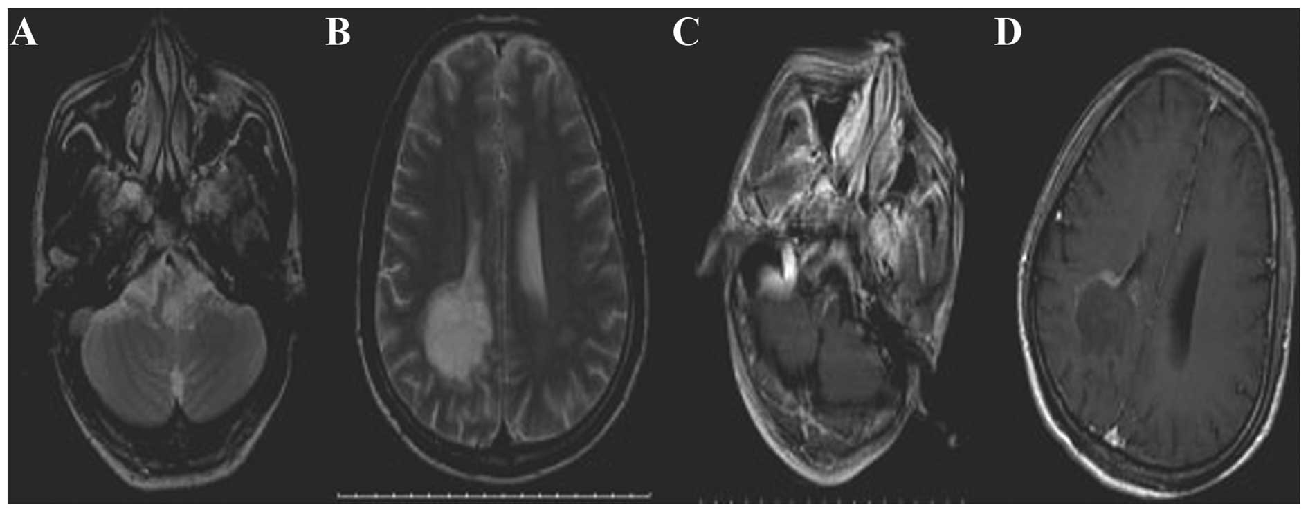
unclear edges in the medulla oblongata. These signals were strengthened after an enhancement scan (Fig. 1A and B). The head MRI also revealed flaky, hypointense T1, hyperintense T2 and FLAIR signals in the right basal ganglia region and in the white matter around the anterior and posterior horn of the right lateral ventricle. The posterior horn lesion appeared flaky, with low-signal intensity that was marginally strengthened after the enhancement scan, particularly close to the edges (Fig. 1C and D). A biopsy below the trigone of the right parieto-occipital cortex was performed, and differential immunohistochemistry-based neuropathological examination using a set of diagnostic glioma markers led to the diagnosis of OA (WHO 
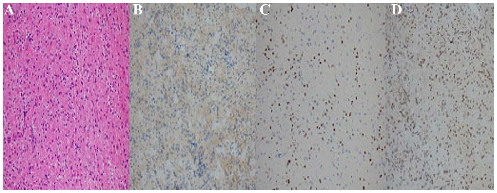
grade II). Immunohistochemistry yielded the following results: Ki-67 (~1%); p53 (−); glial fibrillary acidic protein (GFAP) (+); IDH1-R132H (−); oligodendrocyte lineage transcription factor 2 (Olig2) (+); O6-methylguanine DNA methyltransferase (MGMT) (+ in >75% of the tumor cells); epidermal growth factor receptor (EGFR) (±); CD68 (−); myelin basic protein (partially +); and neurofilament (+) (Fig. 2). The biopsied tissue displayed weak staining for the astrocytoma marker EGFR, but was positive for the oligodendrocyte marker Olig2 and the OA marker MGMT in ~75% of tumor cells. These results combined were indicative of WHO grade II OA. The patient underwent craniotomy, while the symptoms persisted and were gradually exacerbated. The patient succumbed 1 month later in a local hospital due to a pulmonary infection.

Figure 1.

(A) Head magnetic resonance imaging (MRI) showing a flaky hyperintense T2 signal with unclear edges in the medulla oblongata. (B) The signal was strengthened after an enhancement scan. (C) Head MRI showing a flaky hyperintense T2 signal in the white matter around the posterior horn of the right lateral ventricle. (D) The lesion in the posterior horn of the right lateral ventricle appears as a low-intensity flaky signal that was only marginally strengthened after the enhancement scan.

Figure 2.

Immunohistochemistry-based neuropathological examination of the patient's biopsy material leading to the diagnosis of World Health Organisation grade II oligoastrocytoma. Magnification, ×400. (A) Control hematoxylin and eosin staining; (B) weak epithelial growth factor receptor staining; (C) positive oligodendrocyte lineage transcription factor 2 staining; and (D) O6-methylguanine DNA methyltransferase staining in ~75% of the tumor cells.

Discussion

The etiology of OA has not yet been fully elucidated. Long-term ionizing radiation exposure is the only currently known risk factor, although it has been suggested that OA is the result of multiple factors, such as genetic diversity, viral infections and immune factors. The symptoms of OA are complex and depend on tumor location and size. OA commonly occurs in the supratentorial region, particularly in the frontal lobe, followed by the temporal lobe, parietal lobe, thalamus, corpus callosum, ventricle, cerebellopontine angle region and brainstem. The tumor often crosses lobes and, therefore, may involve multiple lobes of the brain and the superficial cortex. The involvement of multiple lobes of the brain may explain the appearance of epilepsy-like symptoms in OA. Epilepsy is the most common clinical symptom in OA and ~80% of OA patients suffer from seizures (3). Other symptoms include ataxia, headaches, memory loss, personality changes and other mental changes, including intelligence decline and dementia (4). Therefore, the clinical symptoms of OA are frequently not characteristic of glioma.

The initial symptoms of the patient in the present case were atypical and contributed to the original misdiagnosis. The patient had previously suffered from optic neuritis, a condition associated with MS, and his vision had improved following corticosteroid injections. Finally, the premonitory cold symptoms, abnormal VEP and OCB reinforced the misdiagnosis of MS.

MS is a common cause of space-occupying lesions. The treatment of choice is high-dose dexamethasone to reduce the brain edema, which is empirically associated with clinical and radiological improvement in 1–3 weeks. However, if there is no clinical improvement, a stereotactic biopsy of the space-occupying lesion is recommended (3).

Although CT scans may show patchy lesions, low-density shadows and unclear edges, the image density may be lower in cases with cystic tumors and CT lacks the required specificity for OA diagnosis. Dotted, nodular and short stripe-shaped calcifications may be detected surrounding the tumor, and these characteristics may gather, in part, at one end of a quasi-circular tumor. The sensitivity and specificity of MRI are significantly better compared with those of CT for lesion detection and identification. T1-weighted images mainly show low-intensity signals, whereas T2-weighted images and FLAIR show homogeneous high-intensity signals with unclear edges. Since OAs grow slowly, vasogenic edema and mass effects do not commonly occur. Thus, T1-weighted images may be less efficient at detecting tumor boundaries. By contrast, T2-weighted images may more effectively reflect real tumor boundaries; however, it is difficult to distinguish the tumor from the surrounding edematous area. There is usually no or only a mild enhancement of the lesion area, as visualized by MRI. To visualize the infiltration range of the lesion, MRS is superior to conventional MRI. The MRS results of the patient revealed a significant decrease in NAA, a mild decrease in Cr, a significant increase in Cho and a mild increase in Lac. In contrast to this result, Cho may be decreased in highly malignant gliomas, which is likely associated with necrosis (5). The change in the ratios of Cho/Cr and NAA/Cr provides guidance for the classification of the malignant grade of the tumor and the projected survival time of the patient (6). These results indicate that a tumorigenic lesion may be underlying the observed symptoms.

Histopathological examination remains the gold standard for OA diagnosis. Indirect signs of OA, such as edema of the brain or the brain stem, basal ganglia or corpus callosum volume gain and blurring of the lateral fissure may be observed. Histological examination may show the diffuse distribution of tumor cells, which are polygonal or circular, with nuclear condensation. The immunohistochemical markers used are Olig2, a marker of oligodendrocytes; GFAP, a marker of astrocytes; EGFR, a marker associated with astrocytomas; and MGMT, a marker associated with OA (4). Immunohistochemical analysis of this patient's biopsy revealed tumor cell expression of Olig2, GFAP and MGMT, indicating that the patient had an OA.

OAs (grade II) are considered to be low-grade tumors, and they generally grow at a slower rate compared with malignant anaplastic OAs (grade III). However, OAs may evolve into anaplastic oligoastrocytomas over time. OA growth generally depends on the percentage of astrocytoma in the tumor, as astrocytomas tend to grow more rapidly compared with oligodendrogliomas.

Due to the rarity and high misdiagnosis rate of OA, it is suggested that clinical physicians update their knowledge regarding brain tumor classification and increase their awareness of rare tumors. Promisingly, the rapid advances in imaging technologies, particularly functional MRI, appears to enable accurate confirmation of the location and extent of injury in the brain. With the combination of current imaging technology, biopsy and pathological examination, it is now possible to accurately diagnose these relatively rare tumors.

References

1 

Gwak HS, Yee GT, Park CK, Kim JW, Hong YK, Kang SG, Kim JH, Seol HJ, Jung TY, Chang JH, et al: Temozolomide salvage chemotherapy for recurrent anaplastic oligodendroglioma and oligo-astrocytoma. J Korean Neurosurg Soc. 54:489–495. 2013. View Article : Google Scholar : PubMed/NCBI

2 

Louis DN, Ohgak H, Wiestler OD, Cavenee WK, Burger PC, Jouvet A, Scheithauer BW and Kleihues P: The 2007 WHO classification of tumours of thecentral nervous system. Acta Neuropathol. 114:97–109. 2007. View Article : Google Scholar : PubMed/NCBI

3 

Ruiz J and Lesser GJ: Low-grade gliomas. Curr Treat Options Oncol. 10:231–242. 2009. View Article : Google Scholar : PubMed/NCBI

4 

Gorlia T, Delattre JY, Brandes AA, Kros JM, Taphoorn MJ, Kouwenhoven MC, Bernsen HJ, Frénay M, Tijssen CC, Lacombe D and van den Bent MJ: New clinical, pathological and molecular prognostic models and calculators in patients with locally diagnosed anaplastic oligodendroglioma or oligoastrocytoma. A prognostic factor analysis of European Organisation for Research and Treatment of Cancer Brain Tumour Group Study 26951. Eur J Cancer. 49:3477–3485. 2013. View Article : Google Scholar : PubMed/NCBI

5 

Cheng LL, Anthony DC, Comite AR, Black PM, Tzika AA and Gonzalez RG: Quantification of microheterogeneity in glioblastoma multiforme with ex vivo high resolution magic-angle spinning (HRMAS) proton magnetic resonance spectroscopy. Neuro Oncol. 2:87–95. 2000. View Article : Google Scholar : PubMed/NCBI

6 

Chawla S, Krejza J, Vossough A, Zhang Y, Kapoor GS, Wang S, O'Rourke DM, Melhem ER and Poptani H: Differentiation between oligodendroglioma genotypes using dynamic susceptibility contrast persusion-weighted imaging and proton MR spectroscopy. AJNR Am J Neuroradiol. 34:1542–1549. 2013. View Article : Google Scholar : PubMed/NCBI

Related Articles

  • Abstract
  • View
  • Download
  • Twitter
Copy and paste a formatted citation
Spandidos Publications style
Gao J, Ti Y, Meng H, Zhao T, Zhou C, Zhu L and Fang S: A rare case of oligoastrocytoma with atypical symptoms initially diagnosed as multiple sclerosis: A case report. Mol Clin Oncol 4: 206-208, 2016.
APA
Gao, J., Ti, Y., Meng, H., Zhao, T., Zhou, C., Zhu, L., & Fang, S. (2016). A rare case of oligoastrocytoma with atypical symptoms initially diagnosed as multiple sclerosis: A case report. Molecular and Clinical Oncology, 4, 206-208. https://doi.org/10.3892/mco.2015.686
MLA
Gao, J., Ti, Y., Meng, H., Zhao, T., Zhou, C., Zhu, L., Fang, S."A rare case of oligoastrocytoma with atypical symptoms initially diagnosed as multiple sclerosis: A case report". Molecular and Clinical Oncology 4.2 (2016): 206-208.
Chicago
Gao, J., Ti, Y., Meng, H., Zhao, T., Zhou, C., Zhu, L., Fang, S."A rare case of oligoastrocytoma with atypical symptoms initially diagnosed as multiple sclerosis: A case report". Molecular and Clinical Oncology 4, no. 2 (2016): 206-208. https://doi.org/10.3892/mco.2015.686
Copy and paste a formatted citation
x
Spandidos Publications style
Gao J, Ti Y, Meng H, Zhao T, Zhou C, Zhu L and Fang S: A rare case of oligoastrocytoma with atypical symptoms initially diagnosed as multiple sclerosis: A case report. Mol Clin Oncol 4: 206-208, 2016.
APA
Gao, J., Ti, Y., Meng, H., Zhao, T., Zhou, C., Zhu, L., & Fang, S. (2016). A rare case of oligoastrocytoma with atypical symptoms initially diagnosed as multiple sclerosis: A case report. Molecular and Clinical Oncology, 4, 206-208. https://doi.org/10.3892/mco.2015.686
MLA
Gao, J., Ti, Y., Meng, H., Zhao, T., Zhou, C., Zhu, L., Fang, S."A rare case of oligoastrocytoma with atypical symptoms initially diagnosed as multiple sclerosis: A case report". Molecular and Clinical Oncology 4.2 (2016): 206-208.
Chicago
Gao, J., Ti, Y., Meng, H., Zhao, T., Zhou, C., Zhu, L., Fang, S."A rare case of oligoastrocytoma with atypical symptoms initially diagnosed as multiple sclerosis: A case report". Molecular and Clinical Oncology 4, no. 2 (2016): 206-208. https://doi.org/10.3892/mco.2015.686
Follow us
  • Twitter
  • LinkedIn
  • Facebook
About
  • Spandidos Publications
  • Careers
  • Cookie Policy
  • Privacy Policy
How can we help?
  • Help
  • Live Chat
  • Contact
  • Email to our Support Team