Introduction
Primary peripheral nerve sheath tumors (PNSTs) of
the thyroid gland are rare, accounting for <0.02% of all thyroid
tumors worldwide (1). PNSTs may occur
at any age, with the highest incidence between 40 and 60 years of
age, without notable between-gender differences. PNST usually
presents as a gradually enlarging mass of the thyroid, with
non-specific symptoms and signs, which makes preoperative diagnosis
difficult; curative surgery is currently the mainstay of treatment.
PNSTs may be classified as benign or malignant (MPNSTs) and they
often originate from anatomically discernible peripheral nerves.
MPNSTs mostly occur between the ages of 20 and 50 years, presenting
as asymptomatic nodules, or with symptoms including difficulty in
breathing and weight loss, which are associated with a poor
prognosis (2,3).
Case report
A 51-year-old man presented with a 4-year history of
a slowly growing thyroid mass, without any history of dysphagia,
hoarseness or weight loss. The physical examination revealed a
non-tender, mobile nodule in the left lobe of the thyroid gland,
~30×20 mm in size with well-defined margins. Cervical
lymphadenopathy was clinically absent and all the laboratory tests,
including fT3, T3, fT4, T4, thyroid-stimulating hormone and
calcitonin were within the normal range. Ultrasonography revealed
two adjacent, well-defined, hypoechogenic solid nodules within the
left lobe of the thyroid gland, sized 25.4×15.5 and 14.4×9.1 mm
(Fig. 2). Total excision of the
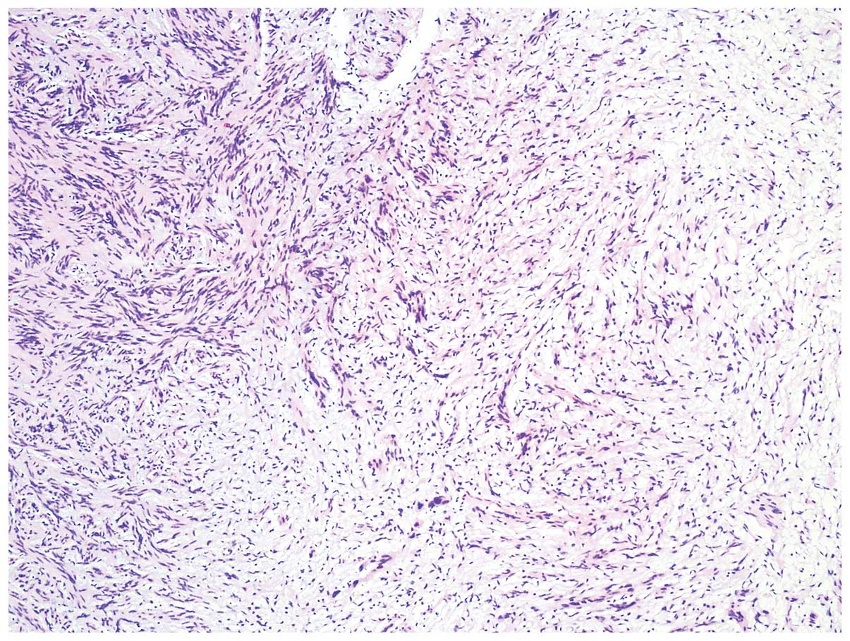
thyroid gland was performed. A detailed pathological examination
was conducted and the final diagnosis was benign PNSTs of the left
lobe of the thyroid gland. To date, the patient has been followed
up for 6 months and his thyroid hormone profile, other laboratory
tests and thyroid ultrasonography findings are normal.
Discussion
Primary PNSTs of the thyroid gland are classified
into MPNSTs and benign PNSTs. The benign PNSTs may be further
subclassified into neurofibromas and Schwannomas.
Neurofibromas mainly arise from peripheral or
cutaneous nerves, and usually occur sporadically, although they may
coexist with neurofibromatosis. Neurofibromas exhibit biallelic
inactivation of the NF1 gene, which encodes the neurofibromin
protein (4–6).
Schwannomas originate from neuronal sheath cells
(also known as Schwann cells), and are usually slow-growing tumors
presenting in the fourth to sixth decades of life. Schwannomas may
lead to pressure symptoms resulting from direct compression of
adjacent organs, such as the thyroid gland (7). As previously described, Schwannomas are
grouped into two histological types: Type A is characterized by
pallisading and spindle-shaped Schwann cells; and type B usually
exhibits a sparsely cellular pattern with cystic degeneration
(8).
The majority of patients with Schwannomas of the
thyroid gland only present with a painless, slowly-growing mass,
without any other symptoms (3). The
ultrasonography and computed tomography (CT) scans usually reveal a
well-delineated, solid nodule, wihtout involvement of the cervical
lymph nodes (3,8). The laboratory results are usually within
the normal range (3). It has been
reported in one case that fine-needle aspiration was helpful in
reaching a definitive diagnosis (9).
In the clinical setting, benign PNSTs should be
carefully differentiated from MPNSTs. MPNSTs are a group of
invasive tumors, which may result in a fatal outcome irrespective
of administering aggressive adjuvant therapies (3,10). MPNSTs
may efface the thyroid parenchyma in a fascicular pattern of
growth; they are characterized by neural-appearing cells, increased
cellularity, increased mitotic activity and focal necrosis
(3). The immunohistochemical staining
for S-100, CD34 and vimentin may be helpful in confirming the
diagnosis (3).
Since the majority of the PNSTs of the thyroid gland
are asymptomatic and fine-needle aspiration is not very efficient,
it is difficult to reach a definitive a diagnosis prior to surgical
removal (11,12). To the best of our knowledge, the
reported outcomes of these patients are satisfactory, with a low
recurrence rate (11–13).
In conclusion, PNSTs of the thyroid gland are rare
and generally asymptomatic. Although the majority of these tumors
are benign, surgical resection is required for the final diagnosis
and is the main therapeutic option for symptomatic patients.
References
|
1
|
Thompson LD, Wenig BM, Adair CF and
Heffess CS: Peripheral nerve sheath tumors of the thyroid gland, A
series of four cases and a review of the literature. Endocr Pathol.
7:309–318. 1996. View Article : Google Scholar : PubMed/NCBI
|
|
2
|
Kandil E, Khalek Abdel M, Abdullah O, Dali
D, Faruqui S, Khan A, Friedlander P, Jaffe BM and Crawford B:
Primary peripheral nerve sheath tumors of the thyroid gland.
Thyroid. 20:583–586. 2010. View Article : Google Scholar : PubMed/NCBI
|
|
3
|
Pallares J, Perez-Ruiz L, Ros S, Panades
MJ, Pardo-Mindan J, Lloreta J and Matias-Guiu X: Malignant
peripheral nerve sheath tumor of the thyroid A clinicopathological
and ultrastructural study of one case. Endocr Pathol. 15:167–174.
2004. View Article : Google Scholar : PubMed/NCBI
|
|
4
|
Baglaj MI, Markowska-Woyciechowska A,
Sawicz-Birkowska K and Dorobisz U: Primary neurilemmoma of the
thygland in a 12-year-old girl. J Pediatr Surg. 39:1418–1420. 2004.
View Article : Google Scholar : PubMed/NCBI
|
|
5
|
Aoki T, Kumeda S, Iwasa T, Inokawa K, Hori
T and Makiuchi M: Primary neurilemoma of the thyroid gland, Report
of a case. Surg Today. 23:265–268. 1993. View Article : Google Scholar : PubMed/NCBI
|
|
6
|
Delaney WE and Fry KE: Neurilemoma of the
thyroid gland. Ann Surg. 160:1014–1017. 1964. View Article : Google Scholar : PubMed/NCBI
|
|
7
|
Graceffa G, Cipolla C, Florena AM, Gentile
I, Pompei G and Latteri MA: Primary schwannoma of the thyroid gland
involving the isthmus, Report of a case. Surg Today. 43:106–109.
2013. View Article : Google Scholar : PubMed/NCBI
|
|
8
|
Sugita R, Nomura T and Yuda F: Primary
schwannoma of the thyroid gland, CT findings. AJR Am J Roentgenol.
171:528–529. 1998. View Article : Google Scholar : PubMed/NCBI
|
|
9
|
Jayaram G: Neurilemmoma (schwannoma) of
the thyroid diagnosed by fine needle aspiration cytology. Acta
Cytol. 43:743–744. 1999.PubMed/NCBI
|
|
10
|
Hruban RH, Shiu MH, Senie RT and Woodruff
JM: Malignant peripheral nerve sheath tumors of the buttock and
lower extremity. Histopathology. Cancer. 66:1253–1265. 1990.
View Article : Google Scholar : PubMed/NCBI
|
|
11
|
Kang GC, Soo KC and Lim DT: Extracranial
non-vestibular head and neck schwannomas, A ten-year experience.
Ann Acad Med Singapore. 36:233–238. 2007.PubMed/NCBI
|
|
12
|
Kar M, Deo SV, Shukla NK, Malik A,
DattaGupta S, Mohanti BK and Thulkar S: Malignant peripheral nerve
sheath tumors (MPNST) - clinicopathological study and treatment
outcome of twenty-four cases. World J Surg Oncol. 4:55–62. 2006.
View Article : Google Scholar : PubMed/NCBI
|
|
13
|
Gustafson LM, Liu JH, Rutter MJ, Stern Y
and Cotton RT: Primary neurilemoma of the thyroid gland, A case
report. Am J Otolaryngol. 22:84–86. 2001. View Article : Google Scholar : PubMed/NCBI
|