Spandidos Publications Logo
  • About
    • About Spandidos
    • Aims and Scopes
    • Abstracting and Indexing
    • Editorial Policies
    • Reprints and Permissions
    • Job Opportunities
    • Terms and Conditions
    • Contact
  • Journals
    • All Journals
    • Oncology Letters
      • Oncology Letters
      • Information for Authors
      • Editorial Policies
      • Editorial Board
      • Aims and Scope
      • Abstracting and Indexing
      • Bibliographic Information
      • Archive
    • International Journal of Oncology
      • International Journal of Oncology
      • Information for Authors
      • Editorial Policies
      • Editorial Board
      • Aims and Scope
      • Abstracting and Indexing
      • Bibliographic Information
      • Archive
    • Molecular and Clinical Oncology
      • Molecular and Clinical Oncology
      • Information for Authors
      • Editorial Policies
      • Editorial Board
      • Aims and Scope
      • Abstracting and Indexing
      • Bibliographic Information
      • Archive
    • Experimental and Therapeutic Medicine
      • Experimental and Therapeutic Medicine
      • Information for Authors
      • Editorial Policies
      • Editorial Board
      • Aims and Scope
      • Abstracting and Indexing
      • Bibliographic Information
      • Archive
    • International Journal of Molecular Medicine
      • International Journal of Molecular Medicine
      • Information for Authors
      • Editorial Policies
      • Editorial Board
      • Aims and Scope
      • Abstracting and Indexing
      • Bibliographic Information
      • Archive
    • Biomedical Reports
      • Biomedical Reports
      • Information for Authors
      • Editorial Policies
      • Editorial Board
      • Aims and Scope
      • Abstracting and Indexing
      • Bibliographic Information
      • Archive
    • Oncology Reports
      • Oncology Reports
      • Information for Authors
      • Editorial Policies
      • Editorial Board
      • Aims and Scope
      • Abstracting and Indexing
      • Bibliographic Information
      • Archive
    • Molecular Medicine Reports
      • Molecular Medicine Reports
      • Information for Authors
      • Editorial Policies
      • Editorial Board
      • Aims and Scope
      • Abstracting and Indexing
      • Bibliographic Information
      • Archive
    • World Academy of Sciences Journal
      • World Academy of Sciences Journal
      • Information for Authors
      • Editorial Policies
      • Editorial Board
      • Aims and Scope
      • Abstracting and Indexing
      • Bibliographic Information
      • Archive
    • International Journal of Functional Nutrition
      • International Journal of Functional Nutrition
      • Information for Authors
      • Editorial Policies
      • Editorial Board
      • Aims and Scope
      • Abstracting and Indexing
      • Bibliographic Information
      • Archive
    • International Journal of Epigenetics
      • International Journal of Epigenetics
      • Information for Authors
      • Editorial Policies
      • Editorial Board
      • Aims and Scope
      • Abstracting and Indexing
      • Bibliographic Information
      • Archive
    • Medicine International
      • Medicine International
      • Information for Authors
      • Editorial Policies
      • Editorial Board
      • Aims and Scope
      • Abstracting and Indexing
      • Bibliographic Information
      • Archive
  • Articles
  • Information
    • Information for Authors
    • Information for Reviewers
    • Information for Librarians
    • Information for Advertisers
    • Conferences
  • Language Editing
Spandidos Publications Logo
  • About
    • About Spandidos
    • Aims and Scopes
    • Abstracting and Indexing
    • Editorial Policies
    • Reprints and Permissions
    • Job Opportunities
    • Terms and Conditions
    • Contact
  • Journals
    • All Journals
    • Biomedical Reports
      • Information for Authors
      • Editorial Policies
      • Editorial Board
      • Aims and Scope
      • Abstracting and Indexing
      • Bibliographic Information
      • Archive
    • Experimental and Therapeutic Medicine
      • Information for Authors
      • Editorial Policies
      • Editorial Board
      • Aims and Scope
      • Abstracting and Indexing
      • Bibliographic Information
      • Archive
    • International Journal of Epigenetics
      • Information for Authors
      • Editorial Policies
      • Editorial Board
      • Aims and Scope
      • Abstracting and Indexing
      • Bibliographic Information
      • Archive
    • International Journal of Functional Nutrition
      • Information for Authors
      • Editorial Policies
      • Editorial Board
      • Aims and Scope
      • Abstracting and Indexing
      • Bibliographic Information
      • Archive
    • International Journal of Molecular Medicine
      • Information for Authors
      • Editorial Policies
      • Editorial Board
      • Aims and Scope
      • Abstracting and Indexing
      • Bibliographic Information
      • Archive
    • International Journal of Oncology
      • Information for Authors
      • Editorial Policies
      • Editorial Board
      • Aims and Scope
      • Abstracting and Indexing
      • Bibliographic Information
      • Archive
    • Medicine International
      • Information for Authors
      • Editorial Policies
      • Editorial Board
      • Aims and Scope
      • Abstracting and Indexing
      • Bibliographic Information
      • Archive
    • Molecular and Clinical Oncology
      • Information for Authors
      • Editorial Policies
      • Editorial Board
      • Aims and Scope
      • Abstracting and Indexing
      • Bibliographic Information
      • Archive
    • Molecular Medicine Reports
      • Information for Authors
      • Editorial Policies
      • Editorial Board
      • Aims and Scope
      • Abstracting and Indexing
      • Bibliographic Information
      • Archive
    • Oncology Letters
      • Information for Authors
      • Editorial Policies
      • Editorial Board
      • Aims and Scope
      • Abstracting and Indexing
      • Bibliographic Information
      • Archive
    • Oncology Reports
      • Information for Authors
      • Editorial Policies
      • Editorial Board
      • Aims and Scope
      • Abstracting and Indexing
      • Bibliographic Information
      • Archive
    • World Academy of Sciences Journal
      • Information for Authors
      • Editorial Policies
      • Editorial Board
      • Aims and Scope
      • Abstracting and Indexing
      • Bibliographic Information
      • Archive
  • Articles
  • Information
    • For Authors
    • For Reviewers
    • For Librarians
    • For Advertisers
    • Conferences
  • Language Editing
Login Register Submit
  • This site uses cookies
  • You can change your cookie settings at any time by following the instructions in our Cookie Policy. To find out more, you may read our Privacy Policy.

    I agree
Search articles by DOI, keyword, author or affiliation
Search
Advanced Search
presentation
Molecular and Clinical Oncology
Join Editorial Board Propose a Special Issue
Print ISSN: 2049-9450 Online ISSN: 2049-9469
Journal Cover
November-2016 Volume 5 Issue 5

Full Size Image

Sign up for eToc alerts
Recommend to Library

Journals

International Journal of Molecular Medicine

International Journal of Molecular Medicine

International Journal of Molecular Medicine is an international journal devoted to molecular mechanisms of human disease.

International Journal of Oncology

International Journal of Oncology

International Journal of Oncology is an international journal devoted to oncology research and cancer treatment.

Molecular Medicine Reports

Molecular Medicine Reports

Covers molecular medicine topics such as pharmacology, pathology, genetics, neuroscience, infectious diseases, molecular cardiology, and molecular surgery.

Oncology Reports

Oncology Reports

Oncology Reports is an international journal devoted to fundamental and applied research in Oncology.

Experimental and Therapeutic Medicine

Experimental and Therapeutic Medicine

Experimental and Therapeutic Medicine is an international journal devoted to laboratory and clinical medicine.

Oncology Letters

Oncology Letters

Oncology Letters is an international journal devoted to Experimental and Clinical Oncology.

Biomedical Reports

Biomedical Reports

Explores a wide range of biological and medical fields, including pharmacology, genetics, microbiology, neuroscience, and molecular cardiology.

Molecular and Clinical Oncology

Molecular and Clinical Oncology

International journal addressing all aspects of oncology research, from tumorigenesis and oncogenes to chemotherapy and metastasis.

World Academy of Sciences Journal

World Academy of Sciences Journal

Multidisciplinary open-access journal spanning biochemistry, genetics, neuroscience, environmental health, and synthetic biology.

International Journal of Functional Nutrition

International Journal of Functional Nutrition

Open-access journal combining biochemistry, pharmacology, immunology, and genetics to advance health through functional nutrition.

International Journal of Epigenetics

International Journal of Epigenetics

Publishes open-access research on using epigenetics to advance understanding and treatment of human disease.

Medicine International

Medicine International

An International Open Access Journal Devoted to General Medicine.

Journal Cover
November-2016 Volume 5 Issue 5

Full Size Image

Sign up for eToc alerts
Recommend to Library

  • Article
  • Citations
    • Cite This Article
    • Download Citation
    • Create Citation Alert
    • Remove Citation Alert
    • Cited By
  • Similar Articles
    • Related Articles (in Spandidos Publications)
    • Similar Articles (Google Scholar)
    • Similar Articles (PubMed)
  • Download PDF
  • Download XML
  • View XML
Case Report Open Access

Dysgerminoma developing from an ectopic ovary in a patient with WAGR syndrome: A case report

  • Authors:
    • Rie Miura
    • Yoshihito Yokoyama
    • Tatsuhiko Shigeto
    • Masayuki Futagami
    • Hideki Mizunuma
    • Akira Kurose
    • Kazushi Tsuruga
    • Shinya Sasaki
    • Kiminori Terui
    • Etsuro Ito
  • View Affiliations / Copyright

    Affiliations: Department of Obstetrics and Gynecology, Hirosaki University Graduate School of Medicine, Hirosaki, Aomori 036‑8562, Japan, Department of Anatomic Pathology, Hirosaki University Graduate School of Medicine, Hirosaki, Aomori 036‑8562, Japan, Department of Pediatrics, Hirosaki University Graduate School of Medicine, Hirosaki, Aomori 036‑8562, Japan
    Copyright: © Miura et al. This is an open access article distributed under the terms of Creative Commons Attribution License.
  • Pages: 503-506
    |
    Published online on: August 25, 2016
       https://doi.org/10.3892/mco.2016.1004
  • Expand metrics +
Metrics: Total Views: 0 (Spandidos Publications: | PMC Statistics: )
Metrics: Total PDF Downloads: 0 (Spandidos Publications: | PMC Statistics: )
Cited By (CrossRef): 0 citations Loading Articles...

This article is mentioned in:


Abstract

WAGR syndrome is caused by an 11p13 deletion and includes Wilms' tumor, aniridia, genitourinary anomalies and mental retardation. We encountered a case of a dysgerminoma originating in an ectopic ovary in a woman with WAGR syndrome. Our patient was a 24‑year‑old nulliparous woman who was diagnosed with WAGR syndrome. The patient had undergone left nephrectomy for a Wilms' tumor and postoperative chemotherapy at the age of 7 months. She also had a history of glaucoma surgery in both eyes, and was followed up at the Department of Pediatrics for diabetes mellitus, hypertension, liver dysfunction and hyperuricemia. The patient was investigated for oliguria and had elevated levels of blood urea nitrogen (45 mg/dl) and creatinine (5.4 mg/dl); she was admitted to the hospital with acute renal failure and a computed tomography scan revealed a pelvic tumor with a long axis of 10 cm that was obstructing the right ureter. Following insertion of a ureteral stent, the tumor was removed. The tumor had developed in the retroperitoneal space independent of the ovaries. The right adnexa were normal. The tumor was histopathologically diagnosed as dysgerminoma. Follicles were found in part of the tumor; it was thus hypothesized that the tumor developed from an ectopic ovary. The patient was administered etoposide after surgery, and has been recurrence‑free for 4 years since treatment.

Introduction

A congenital anomaly syndrome including Wilms' tumor, aniridia, genitourinary anomalies and mental retardation (WAGR syndrome) is a rare, sporadic genetic disorder characterized by a de novo deletion in the distal band of chromosome 11p13 (1). WAGR syndrome is often diagnosed when aniridia is clinically evident and a chromosomal analysis is performed. The precise incidence of WAGR syndrome remains unclear. The incidence of Wilms' tumor in Caucasians is 1/10,000, and a previous study reported that 0.75% of patients with Wilms' tumor have WAGR syndrome (2); thus, WAGR syndrome is an extremely rare condition. We encountered a case where a pelvic tumor in a female patient with WAGR syndrome was diagnosed as dysgerminoma. The tumor is considered to have developed from an ectopic ovary, which is defined as ovarian tissue found outside a normal ovary. An ectopic ovary is extremely rare, with an incidence of 1/93,000 (3). In addition, reports of an ovarian tumor forming from an ectopic ovary are also rare (4–6).

Case report

The present case involved a 24-year-old nulliparous woman with a height of 159 cm, weight of 89 kg and BMI of 35.2 kg/m2. Aniridia at birth led to the diagnosis of WAGR syndrome. At the age of 7 months, left nephrectomy and postoperative chemotherapy with vincristine and actinomycin D were performed to treat Wilms' tumor. At the age of 7 years, glaucoma surgery was performed. Since the age of 20 years, the patient has been receiving oral medication at the Department of Pediatrics of our hospital to treat diabetes mellitus, hypertension and hyperuricemia. The family history was unremarkable. The patient was investigated at the Department of Pediatrics for oliguria, and her blood pressure was 220/130 mmHg. Blood collected at the time was tested, revealing elevated levels of blood urea nitrogen (BUN; 45 mg/dl) and creatinine (Cr; 5.4 mg/dl). The patient was admitted to the hospital with renal hypertension and acute renal failure.

Blood test results upon admission revealed a white blood cell count of 6,100/µl, hemoglobin (Hb) level 10.7 g/dl, platelet count 279,000/µl, aspartate aminotransferase level 21 U/l, alanine aminotransferase level 34 U/l, lactate dehydrogenase 162 U/l, total cholesterol 214 mg/dl, triglycerides 298 mg/dl, BUN 45 mg/dl, Cr 5.4 mg/dl, uric acid 7.3 mg/dl, Na 144 mEq/l, K 4.1 mEq/l, Cl 104 mEq/l, Ca 9.1 mEq/l, blood glucose 101 mg/dl, HbA1c 
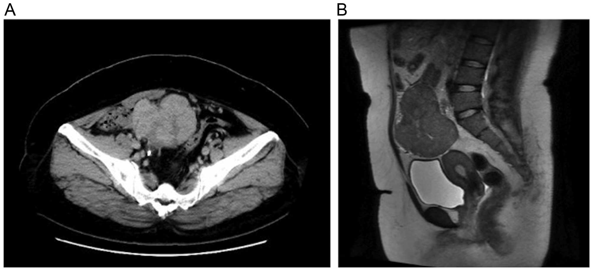
5.6% and C-reactive protein 2.52 mg/dl. A CT scan revealed a solid mass with a long axis of ~80 mm in the pelvis. There were signs of hydronephrosis in the right kidney due to ureteral compression by the mass, which was hypothesized to have caused postrenal failure. Placement of a ureteral stent in the right ureter alleviated hydronephrosis; the urinary output and blood pressure were normal. BUN and Cr returned to their previous levels and contrast-enhanced CT was performed. The scan revealed a solid mass with mild contrast enhancement that was heterogeneous (Fig. 1A). A tumor was hypothesized to have originated from the adnexa, so the patient was referred to the Department of Obstetrics and Gynecology. A magnetic resonance imaging scan revealed hypointensity on T1-weighted images and a mixture of mildly hypointense areas with some hyperintensity on T2-weighted images. A solid tumor without a cystic component was identified; thus, recurrence of Wilms' tumor, sex cord-stromal tumor and leiomyosarcoma were considered in the differential diagnosis (Fig. 1B). The levels of tumor markers were as follows: Carbohydrate antigen (CA) 125 10 IU/ml, CA19-9 4 U/ml, carcinoembryonic antigen 0.6 mg/ml, squamous cell carcinoma antigen 1.2 ng/ml, neuron-specific enolase 31.8 ng/ml, human chorionic gonadotropin <1.0 mIU/ml and α-fetoprotein 2.0 ng/ml.

Figure 1.

(A) A computed tomography scan revealed a solid mass with mild contrast enhancement that was heterogeneous. (B) A magnetic resonance imaging scan revealed a mixture of mildly hypointense areas with some hyperintensity on T2-weighted images.

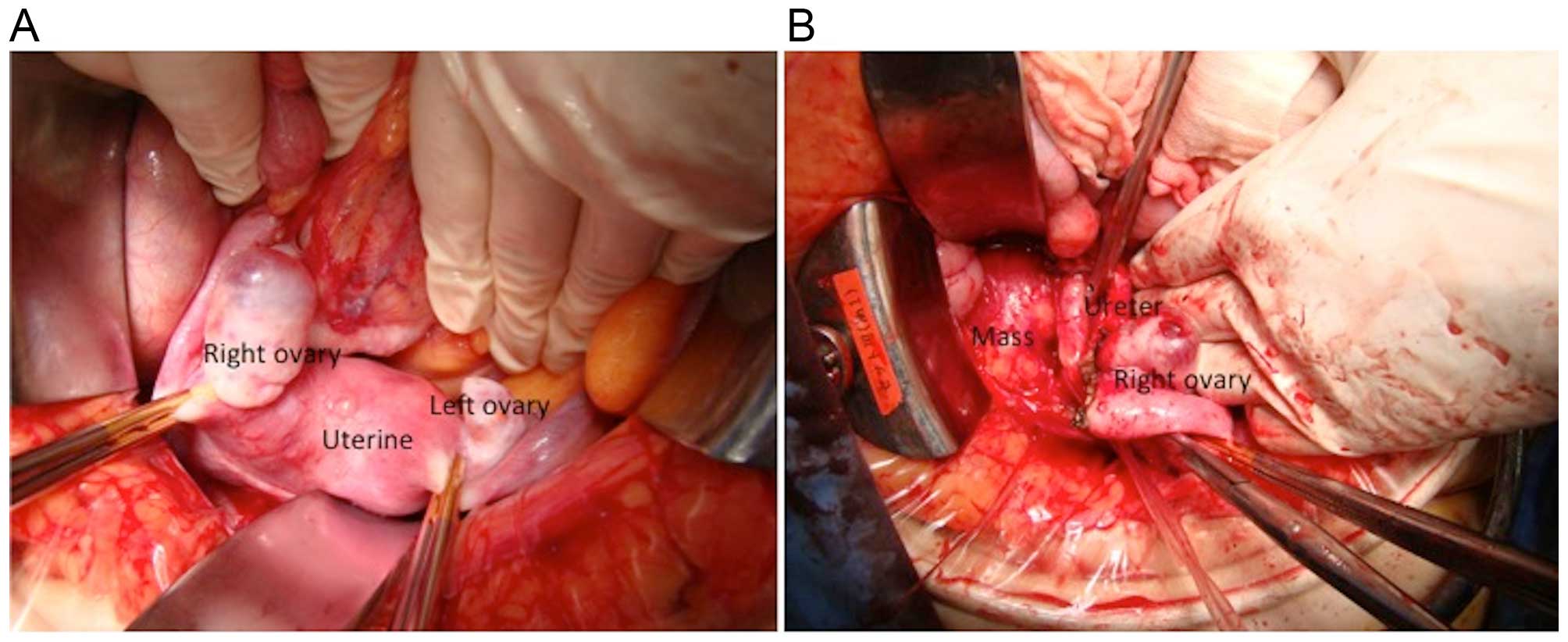
The surgical plan was to treat a pelvic tumor originating from the right adnexa. The left and right ovaries were normal (Fig. 2A); the tumor was located in the retroperitoneal space behind the right ovary (Fig. 2B), and was not connected to the ovary. The tumor was in contact with the right ureter, but ureteral involvement was not observed. The greater omentum and retroperitoneal space were dissected, the right adnexa was spared 
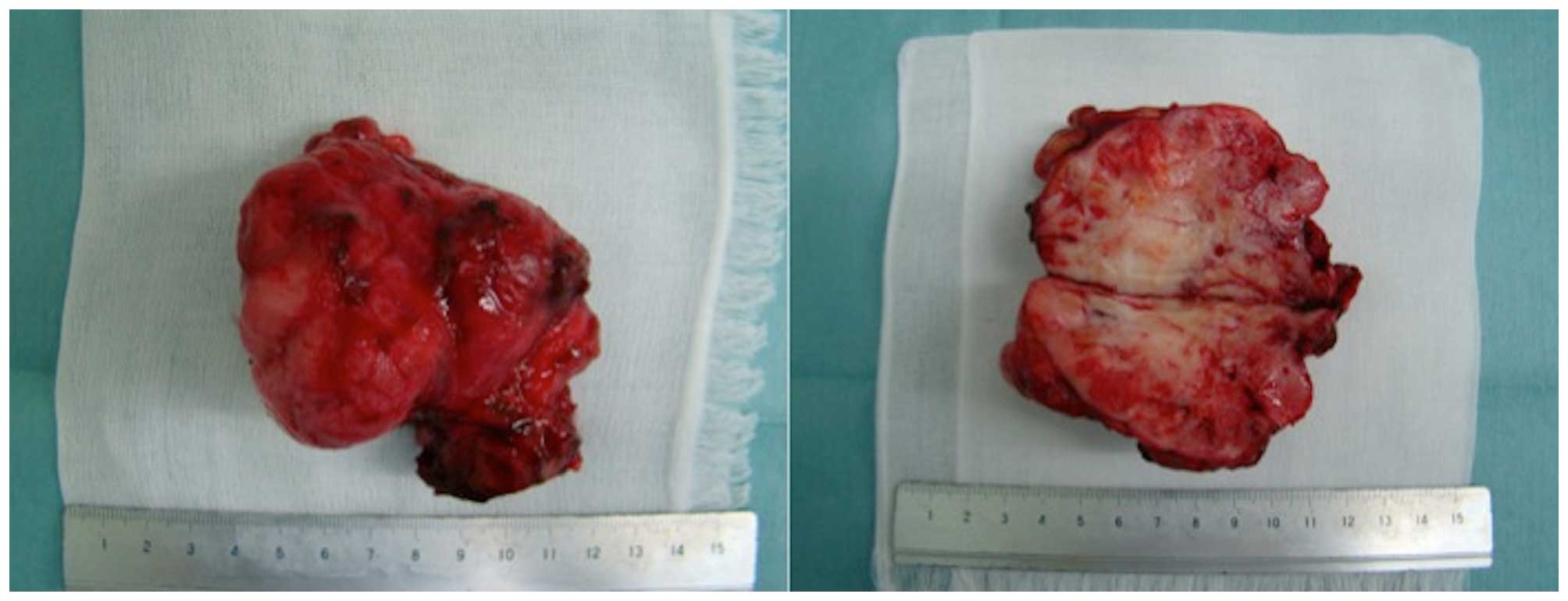
and only the tumor was removed. The resected specimen was a solid mass, which appeared whitish on cross sections (Fig. 3).

Figure 2.

Intraoperatively (A) the left and right ovaries were normal; (B) the tumor was located in the retroperitoneal space behind the right ovary.

Figure 3.

Resected specimen. The resected mass was solid and appeared whitish on cross sections.

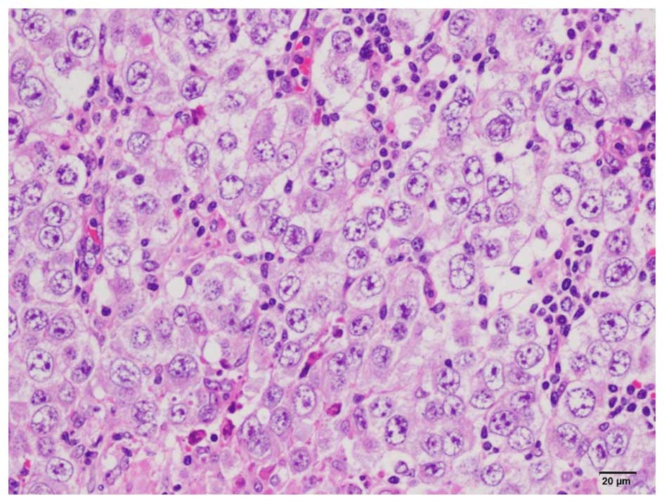
On histopathological examination, lymph node invasion was observed by semicircular tumor cells with large semicircular nuclei, and a ‘two-cell pattern’ (i.e., large tumor cells and small mature lymphocytes) was evident. The nucleus of the tumor cells contained 1–3 nucleoli (Fig. 
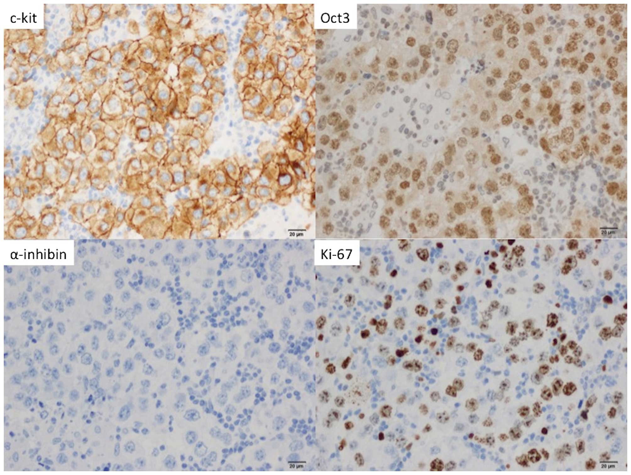
4). On immunostaining, the tumor cells stained positive for C-kit and Oct3, but negative for Wilms' tumor-1 and α-inhibin; 60% of the tumor cells stained positive for Ki-67 (Fig. 5). The final diagnosis was 
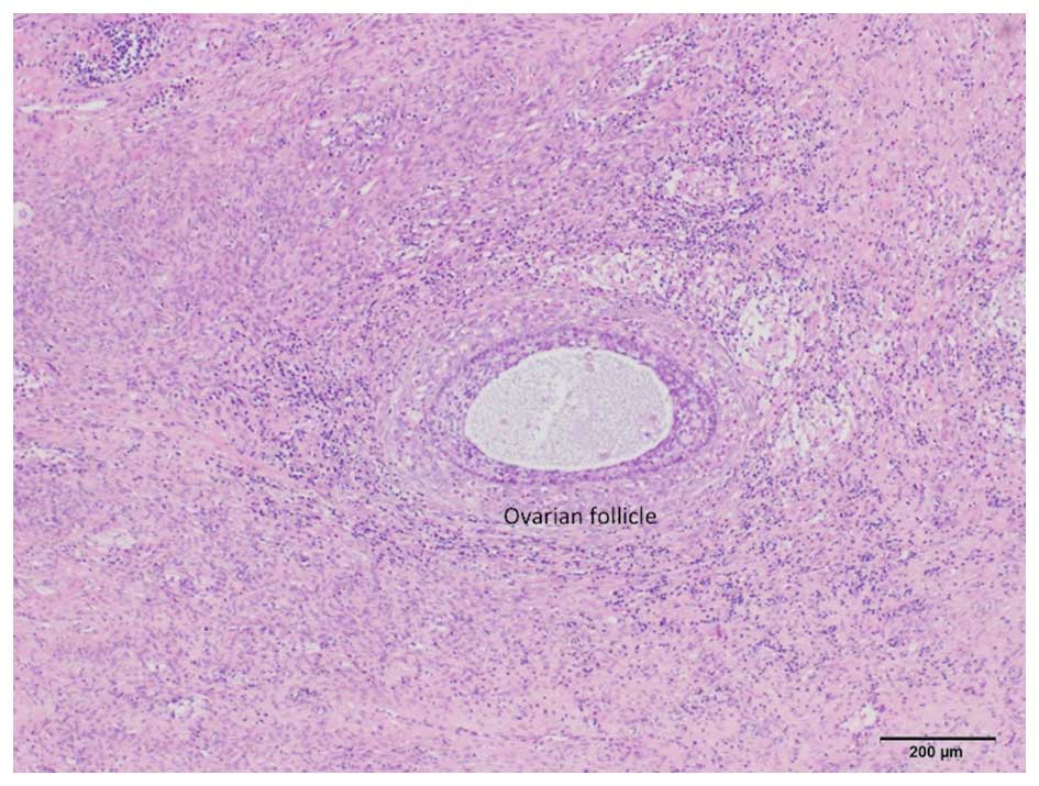
dysgerminoma. In addition, normal follicles were found in the tumor tissue (Fig. 6); thus, the dysgerminoma was hypothesized to have developed from an ectopic ovary.

Figure 4.

On histological examination, lymph node invasion from semicircular tumor cells with large semicircular nuclei. Large tumor cells and small mature lymphocytes were observed.

Figure 5.

On immunohistochemical staining, the tumor cells stained positive for C-kit and Oct3, but negative for α-inhibin. Furthermore, 60% of the tumor cells stained positive for Ki-67.

Figure 6.

Normal ovarian follicle identified in the tumor.

The patient's postoperative course was uneventful. There was no solitary kidney, multiple complications, or residual tumor, so she received 6 cycles of etoposide as postoperative treatment and has remained recurrence-free in the 3 years since the surgery.

The patient and her family provided their consent regarding the publication of the case details.

Discussion

Ectopic ovary describes the presence of ovarian tissue outside of a normal ovary. An ectopic ovary is extremely rare, with an incidence of 1/93,000 (3). Depending on their location, ectopic ovaries may be classified as ‘accessory’, which are adjacent or connected to a normal ovary, or ‘supernumerary’, which indicates a separate location. In 1959, Wharton et al reported 2 cases of supernumerary ovaries and 1 case of an accessory ovary (7). Since that time, <40 cases of supernumerary ovaries alone have been reported (8), with several reported cases of both supernumerary and accessory ovaries. Lachman et al contended that the principal causes of these conditions were i) ovarian tissue that was detached during pelvic surgery and implanted elsewhere, ii) ovarian tissue that was detached due to pelvic inflammatory disease and implanted elsewhere, or iii) abnormally located ovarian tissue due to an embryological anomaly (9). The rationale for this distinction is as follows: Of the reported cases of an ectopic ovary due to ovarian tissue that was detached during pelvic surgery or pelvic inflammatory disease and implanted elsewhere, ~50% involved salpingitis or a history of pelvic surgery or intra-abdominal surgery. The same literature described cases of an ectopic ovary due to developmental anomalies at the fetal stage. Primordial germ cells migrate from the posterior wall of the yolk sac into the hindgut epithelium and then to the urogenital ridge, where they differentiate into ovarian tissue. When an anomaly occurs during this process of cell migration, a congenital ectopic ovary is formed. Ectopic ovaries may be found at varying sites, including the perimetrium, intestinal tract, mesentery, retroperitoneal space and greater omentum (8). In addition, diagnosing an ectopic ovary prior to surgery is usually difficult. There are case reports where an ectopic ovary was suspected prior to surgery, when follicle stimulation with clomiphene citrate resulted in follicle development within a tumor (10). In reported cases of an ectopic ovary where a tumor has formed, that tumor is most often a mature teratoma, followed by serous or mucinous adenoma (8). In some cases of an ectopic ovary, a Brenner tumor (4), Wilms' tumor (5), or epithelial ovarian cancer (6) have been reported. To the best of our knowledge, there are currently no case reports of a dysgerminoma developing in an ectopic ovary, as in the present case.

It was previously reported that genitourinary or genetic anomalies are identified in ~11–36% of the cases of an ectopic ovary (5,9). In the aforementioned case of epithelial ovarian cancer, the patient had Mayer-Rokitansky-Kuster-Hauser syndrome. There are also case reports of an ectopic ovary and a unicorn uterus (10,11). In the aforementioned case of a Wilms' tumor, the patient had both congenital renal agenesis and an ectopic ovary. The patient in our case was diagnosed with WAGR syndrome and had a history of abdominal surgery for Wilms' tumor. The possibility that the surgery was responsible for separating ovarian tissue from an ovary is unlikely. We hypothesize that the ectopic ovary developed as part of the WAGR syndrome.

The ultimate diagnosis of an ectopic ovary is based on the presence of ovarian cortex, follicles, granulosa cells, or theca cells (i.e., tissue specific to the ovaries) in a resected tumor. In the present case, an initial histopathological examination led to the diagnosis of dysgerminoma, although its primary origin was not known. A more detailed examination revealed the presence of follicles in the tumor. If a tumor is large, the possibility that that tumor contains ovarian tissue may be overlooked. In cases of an abdominal mass of unknown origin, the possibility of a tumor developing from an ectopic ovary must be taken into consideration, and a detailed histological examination must be performed.

In conclusion, we herein present an extremely rare case of dysgerminoma developing in an ectopic ovary in a patient with WAGR syndrome. Identifying the presence of an ectopic ovary prior to surgery is difficult, but there are reports of a malignancy developing in patients with that condition. If this condition is suspected, or a retroperitoneal tumor of unknown origin is found in the abdomen, the possibility of an ectopic ovary must be kept in mind, and a detailed histological examination must be performed.

Acknowledgements

The present study was supported by a Grant-in-Aid for Cancer Research from the Ministry of Education, Culture, Sports, Science and Technology (Tokyo, Japan) (grant no. 20591935 to Dr Y. Yokoyama).

References

1 

Riccardi VM, Sujansky E, Smith AC and Francke U: Chromosomal imbalance in the Aniridia-Wilms' tumor association: 11p interstitial deletion. Pediatrics. 61:604–610. 1978.PubMed/NCBI

2 

Yi T, Weng J, Siwko S, Luo J, Li D and Liu M: LGR4/GPR48 inactivation leads to aniridia-genitourinary anomalies-mental retardation syndrome defects. J Biol Chem. 289:8767–8780. 2014. View Article : Google Scholar : PubMed/NCBI

3 

Watkins BP and Kothari SN: True ectopic ovary: A case and review. Arch Gynecol Obstet. 269:145–146. 2004. View Article : Google Scholar : PubMed/NCBI

4 

Heller DS, Harpaz N and Breakstone B: Neoplasm arising in ectopic ovaries: A case of brenner tumor in an accessory ovary. Int J of Gynecol Pathol. 9:185–189. 1990. View Article : Google Scholar

5 

Kini H, Baliga PB and Pai KG: Supernumerary ovary associated with Wilms' tumor. Pediatr Surg Int. 13:67–68. 1998. View Article : Google Scholar : PubMed/NCBI

6 

Bae HS, Ryu MJ, Kim IS, Kim SH and Song JY: Cancer of the supernumerary ovary in Mayer-Rokitansky-Küster-Hauser Syndrome: A case report. Oncol Lett. 5:598–600. 2013.PubMed/NCBI

7 

Wharton LR: Two cases of supernumerary ovary and one of accessory ovary with an analysis of previously reported cases. Am J Obstet Gynacol. 78:1101–1109. 1959.

8 

El-Gohary Y, Pagkratis S, Lee T and Scriven RJ: Supernumerary ovary presenting as a paraduodenal duplication cyst. J Pediatr Surg Case Rep. 3:316–319. 2015. View Article : Google Scholar

9 

Lachman MF and Berman MM: The ectopic ovary. A case report and review of the literature. Arch Pathol Lab Med. 115:233–235. 1991.PubMed/NCBI

10 

Uyar I, Gulhan I, Sipahi M, Hanhan HM and Ozeren M: Ectopic ovary confirmed by ovarian stimulation in a case of unicornuate uterus. Fertil Steril. 96:e122–e124. 2011. View Article : Google Scholar : PubMed/NCBI

11 

Ombelet W, Verswijvel G and de Jonge E: Ectopic ovary and unicornuate uterus. N Engl J Med. 348:667–668. 2003. View Article : Google Scholar : PubMed/NCBI

Related Articles

  • Abstract
  • View
  • Download
  • Twitter
Copy and paste a formatted citation
Spandidos Publications style
Miura R, Yokoyama Y, Shigeto T, Futagami M, Mizunuma H, Kurose A, Tsuruga K, Sasaki S, Terui K, Ito E, Ito E, et al: Dysgerminoma developing from an ectopic ovary in a patient with WAGR syndrome: A case report. Mol Clin Oncol 5: 503-506, 2016.
APA
Miura, R., Yokoyama, Y., Shigeto, T., Futagami, M., Mizunuma, H., Kurose, A. ... Ito, E. (2016). Dysgerminoma developing from an ectopic ovary in a patient with WAGR syndrome: A case report. Molecular and Clinical Oncology, 5, 503-506. https://doi.org/10.3892/mco.2016.1004
MLA
Miura, R., Yokoyama, Y., Shigeto, T., Futagami, M., Mizunuma, H., Kurose, A., Tsuruga, K., Sasaki, S., Terui, K., Ito, E."Dysgerminoma developing from an ectopic ovary in a patient with WAGR syndrome: A case report". Molecular and Clinical Oncology 5.5 (2016): 503-506.
Chicago
Miura, R., Yokoyama, Y., Shigeto, T., Futagami, M., Mizunuma, H., Kurose, A., Tsuruga, K., Sasaki, S., Terui, K., Ito, E."Dysgerminoma developing from an ectopic ovary in a patient with WAGR syndrome: A case report". Molecular and Clinical Oncology 5, no. 5 (2016): 503-506. https://doi.org/10.3892/mco.2016.1004
Copy and paste a formatted citation
x
Spandidos Publications style
Miura R, Yokoyama Y, Shigeto T, Futagami M, Mizunuma H, Kurose A, Tsuruga K, Sasaki S, Terui K, Ito E, Ito E, et al: Dysgerminoma developing from an ectopic ovary in a patient with WAGR syndrome: A case report. Mol Clin Oncol 5: 503-506, 2016.
APA
Miura, R., Yokoyama, Y., Shigeto, T., Futagami, M., Mizunuma, H., Kurose, A. ... Ito, E. (2016). Dysgerminoma developing from an ectopic ovary in a patient with WAGR syndrome: A case report. Molecular and Clinical Oncology, 5, 503-506. https://doi.org/10.3892/mco.2016.1004
MLA
Miura, R., Yokoyama, Y., Shigeto, T., Futagami, M., Mizunuma, H., Kurose, A., Tsuruga, K., Sasaki, S., Terui, K., Ito, E."Dysgerminoma developing from an ectopic ovary in a patient with WAGR syndrome: A case report". Molecular and Clinical Oncology 5.5 (2016): 503-506.
Chicago
Miura, R., Yokoyama, Y., Shigeto, T., Futagami, M., Mizunuma, H., Kurose, A., Tsuruga, K., Sasaki, S., Terui, K., Ito, E."Dysgerminoma developing from an ectopic ovary in a patient with WAGR syndrome: A case report". Molecular and Clinical Oncology 5, no. 5 (2016): 503-506. https://doi.org/10.3892/mco.2016.1004
Follow us
  • Twitter
  • LinkedIn
  • Facebook
About
  • Spandidos Publications
  • Careers
  • Cookie Policy
  • Privacy Policy
How can we help?
  • Help
  • Live Chat
  • Contact
  • Email to our Support Team