Spandidos Publications Logo
  • About
    • About Spandidos
    • Aims and Scopes
    • Abstracting and Indexing
    • Editorial Policies
    • Reprints and Permissions
    • Job Opportunities
    • Terms and Conditions
    • Contact
  • Journals
    • All Journals
    • Oncology Letters
      • Oncology Letters
      • Information for Authors
      • Editorial Policies
      • Editorial Board
      • Aims and Scope
      • Abstracting and Indexing
      • Bibliographic Information
      • Archive
    • International Journal of Oncology
      • International Journal of Oncology
      • Information for Authors
      • Editorial Policies
      • Editorial Board
      • Aims and Scope
      • Abstracting and Indexing
      • Bibliographic Information
      • Archive
    • Molecular and Clinical Oncology
      • Molecular and Clinical Oncology
      • Information for Authors
      • Editorial Policies
      • Editorial Board
      • Aims and Scope
      • Abstracting and Indexing
      • Bibliographic Information
      • Archive
    • Experimental and Therapeutic Medicine
      • Experimental and Therapeutic Medicine
      • Information for Authors
      • Editorial Policies
      • Editorial Board
      • Aims and Scope
      • Abstracting and Indexing
      • Bibliographic Information
      • Archive
    • International Journal of Molecular Medicine
      • International Journal of Molecular Medicine
      • Information for Authors
      • Editorial Policies
      • Editorial Board
      • Aims and Scope
      • Abstracting and Indexing
      • Bibliographic Information
      • Archive
    • Biomedical Reports
      • Biomedical Reports
      • Information for Authors
      • Editorial Policies
      • Editorial Board
      • Aims and Scope
      • Abstracting and Indexing
      • Bibliographic Information
      • Archive
    • Oncology Reports
      • Oncology Reports
      • Information for Authors
      • Editorial Policies
      • Editorial Board
      • Aims and Scope
      • Abstracting and Indexing
      • Bibliographic Information
      • Archive
    • Molecular Medicine Reports
      • Molecular Medicine Reports
      • Information for Authors
      • Editorial Policies
      • Editorial Board
      • Aims and Scope
      • Abstracting and Indexing
      • Bibliographic Information
      • Archive
    • World Academy of Sciences Journal
      • World Academy of Sciences Journal
      • Information for Authors
      • Editorial Policies
      • Editorial Board
      • Aims and Scope
      • Abstracting and Indexing
      • Bibliographic Information
      • Archive
    • International Journal of Functional Nutrition
      • International Journal of Functional Nutrition
      • Information for Authors
      • Editorial Policies
      • Editorial Board
      • Aims and Scope
      • Abstracting and Indexing
      • Bibliographic Information
      • Archive
    • International Journal of Epigenetics
      • International Journal of Epigenetics
      • Information for Authors
      • Editorial Policies
      • Editorial Board
      • Aims and Scope
      • Abstracting and Indexing
      • Bibliographic Information
      • Archive
    • Medicine International
      • Medicine International
      • Information for Authors
      • Editorial Policies
      • Editorial Board
      • Aims and Scope
      • Abstracting and Indexing
      • Bibliographic Information
      • Archive
  • Articles
  • Information
    • Information for Authors
    • Information for Reviewers
    • Information for Librarians
    • Information for Advertisers
    • Conferences
  • Language Editing
Spandidos Publications Logo
  • About
    • About Spandidos
    • Aims and Scopes
    • Abstracting and Indexing
    • Editorial Policies
    • Reprints and Permissions
    • Job Opportunities
    • Terms and Conditions
    • Contact
  • Journals
    • All Journals
    • Biomedical Reports
      • Information for Authors
      • Editorial Policies
      • Editorial Board
      • Aims and Scope
      • Abstracting and Indexing
      • Bibliographic Information
      • Archive
    • Experimental and Therapeutic Medicine
      • Information for Authors
      • Editorial Policies
      • Editorial Board
      • Aims and Scope
      • Abstracting and Indexing
      • Bibliographic Information
      • Archive
    • International Journal of Epigenetics
      • Information for Authors
      • Editorial Policies
      • Editorial Board
      • Aims and Scope
      • Abstracting and Indexing
      • Bibliographic Information
      • Archive
    • International Journal of Functional Nutrition
      • Information for Authors
      • Editorial Policies
      • Editorial Board
      • Aims and Scope
      • Abstracting and Indexing
      • Bibliographic Information
      • Archive
    • International Journal of Molecular Medicine
      • Information for Authors
      • Editorial Policies
      • Editorial Board
      • Aims and Scope
      • Abstracting and Indexing
      • Bibliographic Information
      • Archive
    • International Journal of Oncology
      • Information for Authors
      • Editorial Policies
      • Editorial Board
      • Aims and Scope
      • Abstracting and Indexing
      • Bibliographic Information
      • Archive
    • Medicine International
      • Information for Authors
      • Editorial Policies
      • Editorial Board
      • Aims and Scope
      • Abstracting and Indexing
      • Bibliographic Information
      • Archive
    • Molecular and Clinical Oncology
      • Information for Authors
      • Editorial Policies
      • Editorial Board
      • Aims and Scope
      • Abstracting and Indexing
      • Bibliographic Information
      • Archive
    • Molecular Medicine Reports
      • Information for Authors
      • Editorial Policies
      • Editorial Board
      • Aims and Scope
      • Abstracting and Indexing
      • Bibliographic Information
      • Archive
    • Oncology Letters
      • Information for Authors
      • Editorial Policies
      • Editorial Board
      • Aims and Scope
      • Abstracting and Indexing
      • Bibliographic Information
      • Archive
    • Oncology Reports
      • Information for Authors
      • Editorial Policies
      • Editorial Board
      • Aims and Scope
      • Abstracting and Indexing
      • Bibliographic Information
      • Archive
    • World Academy of Sciences Journal
      • Information for Authors
      • Editorial Policies
      • Editorial Board
      • Aims and Scope
      • Abstracting and Indexing
      • Bibliographic Information
      • Archive
  • Articles
  • Information
    • For Authors
    • For Reviewers
    • For Librarians
    • For Advertisers
    • Conferences
  • Language Editing
Login Register Submit
  • This site uses cookies
  • You can change your cookie settings at any time by following the instructions in our Cookie Policy. To find out more, you may read our Privacy Policy.

    I agree
Search articles by DOI, keyword, author or affiliation
Search
Advanced Search
presentation
Molecular and Clinical Oncology
Join Editorial Board Propose a Special Issue
Print ISSN: 2049-9450 Online ISSN: 2049-9469
Journal Cover
January-2017 Volume 6 Issue 1

Full Size Image

Sign up for eToc alerts
Recommend to Library

Journals

International Journal of Molecular Medicine

International Journal of Molecular Medicine

International Journal of Molecular Medicine is an international journal devoted to molecular mechanisms of human disease.

International Journal of Oncology

International Journal of Oncology

International Journal of Oncology is an international journal devoted to oncology research and cancer treatment.

Molecular Medicine Reports

Molecular Medicine Reports

Covers molecular medicine topics such as pharmacology, pathology, genetics, neuroscience, infectious diseases, molecular cardiology, and molecular surgery.

Oncology Reports

Oncology Reports

Oncology Reports is an international journal devoted to fundamental and applied research in Oncology.

Experimental and Therapeutic Medicine

Experimental and Therapeutic Medicine

Experimental and Therapeutic Medicine is an international journal devoted to laboratory and clinical medicine.

Oncology Letters

Oncology Letters

Oncology Letters is an international journal devoted to Experimental and Clinical Oncology.

Biomedical Reports

Biomedical Reports

Explores a wide range of biological and medical fields, including pharmacology, genetics, microbiology, neuroscience, and molecular cardiology.

Molecular and Clinical Oncology

Molecular and Clinical Oncology

International journal addressing all aspects of oncology research, from tumorigenesis and oncogenes to chemotherapy and metastasis.

World Academy of Sciences Journal

World Academy of Sciences Journal

Multidisciplinary open-access journal spanning biochemistry, genetics, neuroscience, environmental health, and synthetic biology.

International Journal of Functional Nutrition

International Journal of Functional Nutrition

Open-access journal combining biochemistry, pharmacology, immunology, and genetics to advance health through functional nutrition.

International Journal of Epigenetics

International Journal of Epigenetics

Publishes open-access research on using epigenetics to advance understanding and treatment of human disease.

Medicine International

Medicine International

An International Open Access Journal Devoted to General Medicine.

Journal Cover
January-2017 Volume 6 Issue 1

Full Size Image

Sign up for eToc alerts
Recommend to Library

  • Article
  • Citations
    • Cite This Article
    • Download Citation
    • Create Citation Alert
    • Remove Citation Alert
    • Cited By
  • Similar Articles
    • Related Articles (in Spandidos Publications)
    • Similar Articles (Google Scholar)
    • Similar Articles (PubMed)
  • Download PDF
  • Download XML
  • View XML
Article

Urachal carcinoma: Report of two cases and review of the literature

  • Authors:
    • Jing Quan
    • Xiang Pan
    • Lu Jin
    • Tao He
    • Jia Hu
    • Bentao Shi
    • Jian Peng
    • Zhebo Chen
    • Shangqi Yang
    • Xiangming Mao
    • Yongqing Lai
  • View Affiliations / Copyright

    Affiliations: Department of Urology, The First Clinical College, Anhui Medical University, Hefei, Anhui 230032, P.R. China, Department of Urology, Peking University Shenzhen Hospital, Shenzhen, Guangdong 518036, P.R. China
  • Pages: 101-104
    |
    Published online on: November 16, 2016
       https://doi.org/10.3892/mco.2016.1082
  • Expand metrics +
Metrics: Total Views: 0 (Spandidos Publications: | PMC Statistics: )
Metrics: Total PDF Downloads: 0 (Spandidos Publications: | PMC Statistics: )
Cited By (CrossRef): 0 citations Loading Articles...

This article is mentioned in:


Abstract

Urachal carcinoma is a rare tumor that most commonly occurs in ovaries and less often in the adnexal region and urinary system. We herein present two cases of urachal carcinoma: One case was a 32-year-old male patient who presented with painless hematuria with blood clots for 1 month, whereas the other case was a 50-year-old woman who presented with gross hematuria with mild dysuria, urgency and frequent urination for 1 year. Following surgical resection, the two patients were diagnosed with urachal adenocarcinoma (mixed type) and urachal mucinous adenocarcinoma, respectively, based on the histopathological examination. A review of previously published cases and relevant literature is also presented. The aim of the present study was to help understand this disease better, in order to reduce the rate of clinical and pathological misdiagnosis.

Introduction

Urachal carcinoma is a rare non-urothelial carcinoma, accounting for 0.01% of all malignancies and for 0.17–0.34% of all bladder tumors (1,2). Urachal carcinoma is more common among men, and the majority of the patients are aged >50 years (3). Although hematuria is the most common symptom, the disease is usually advanced when this symptom appears (4). As urachal carcinoma frequently invades the bladder at the dome or elsewhere along its midline, which is not easily detected during the early stages, it is often discovered late (5). Furthermore, there is currently no effective treatment for urachal carcinoma, leading to a poor prognosis. We herein report the cases of two patients with urachal carcinoma and urachal mucinous carcinoma. The relevant literature was also reviewed in order to help improve the diagnosis and treatment of this rare disease.

Case reports

Case 1

The patient was a 32-year-old man who noted painless hematuria with blood clots for 1 month. There were no obvious precipitating or alleviating factors. On physical examination, there were no positive physical findings and the patient experienced no other discomfort. The results of the laboratory and imaging examinations (hemogram, urinalysis, prothrombin time, activated partial thromboplastin time, liver and kidney function tests and chest X-ray) were normal. However, urinary tract ultrasonography revealed an abnormal mixed signal at the bottom of the bladder. On further cystoscopy, only a small blood clot was 
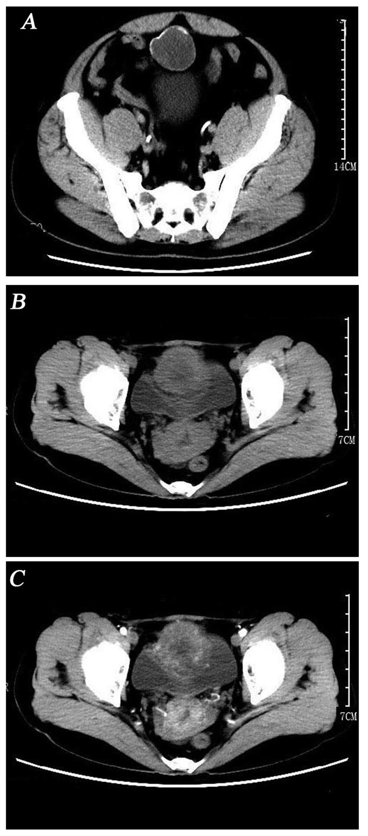
detected. However, computed tomography (CT) revealed a circular hypodense mass located at the top and anterior part of the bladder, sized ~44×43 mm, without enlarged lymph nodes or other sign of metastatic disease (Fig. 1A).

Figure 1.

(A) A circular hypodense mass sized ~44×43 mm was located at the top and anterior wall of the bladder, without accompanying enlarged lymph nodes or other sign of metastatic disease. (B) The cystic mass was sized ~57×58 mm and was located in the top and anterior wall of the bladder. (C) Enhanced scan showing heterogeneous enhancement of the mass. A cystic low-density mass of ~26×20 mm was located at the posterior wall of the cervix. Also, at the left attachment area, a 25×18 mm cystic low-density mass was observed.

Following the doctors' recommendation, the patient consented to radical resection of the urachal tumor and partial cystectomy in August, 2015. We found that the lower part of the tumor was connected to the anterior wall of the bladder, whereas the upper part of the tumor was connected with the umbilical region. The tumor was sized ~7×8 cm. On gross pathological examination, the resected specimen was grey-yellow and gray-red, with a diameter of 6 cm. A small area covered by mucosa was identified on the surface, sized ~2.6×1.8 cm. On cross-section, the 
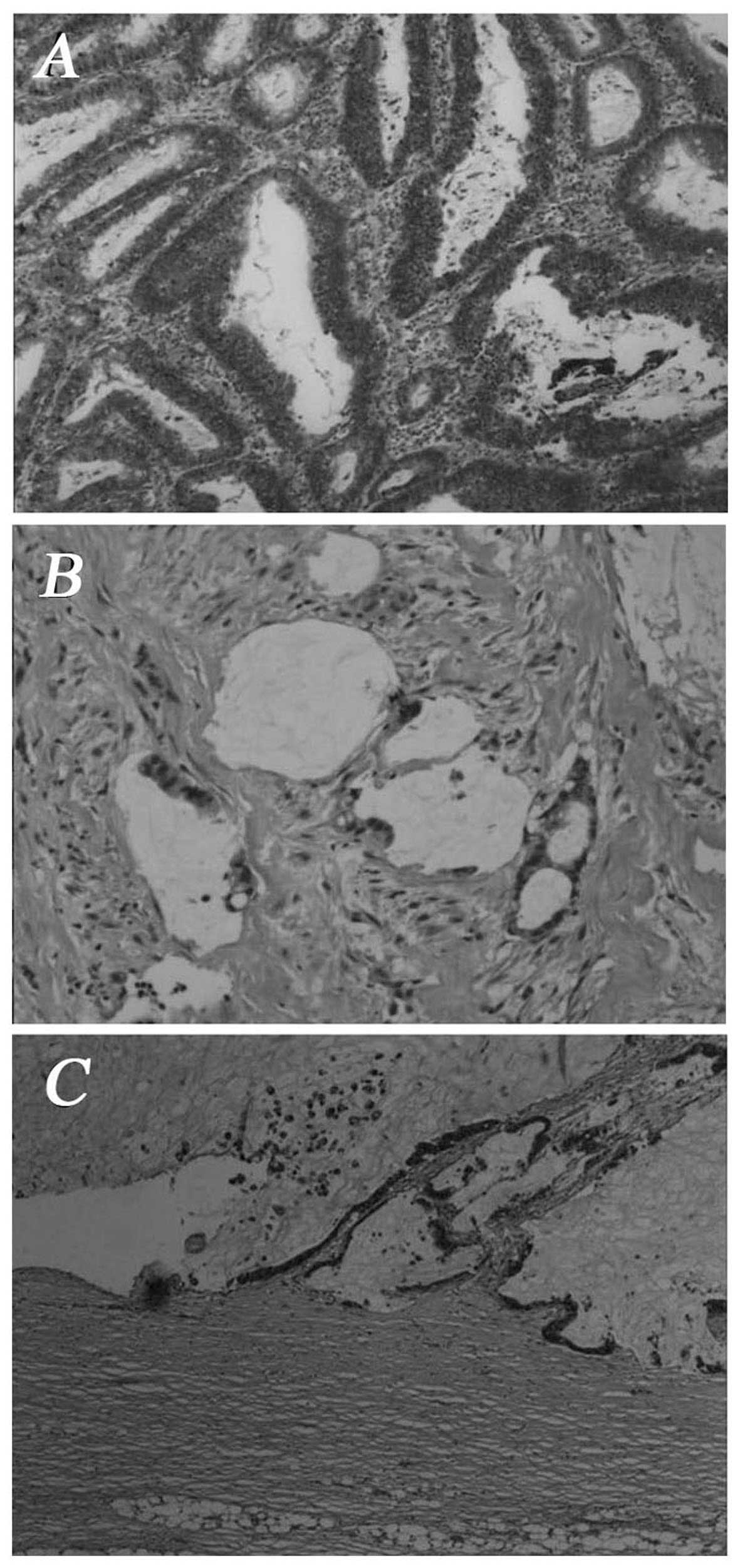
tumor included two jelly-like nodules; on microscopic examination, the nodules were cystides containing mucus; goblet cells were visible. Adenoid tumor cell formations were observed invading the smooth muscle of the bladder wall. The carcinoma tissue displayed tubular and alveolar cell structures (Fig. 2A and B). The pathological diagnosis was mixed-type urachal adenocarcinoma. Postoperatively, the patient recovered well. After 10 months of follow-up, the patient has no evidence of recurrence on laboratory and imaging examinations. Informed consent was obtained from the patient.

Figure 2.

(A) Some goblet cells are visible. The smooth muscle of the bladder wall was invaded by tumor cells arranged in adenoid formations. (B) The carcinoma tissue was composed of cells forming tubular and alveolar structures. (C) The carcinoma tissue was composed of neoplastic cells in tubular and alveolar formations, as well as single cells. The neoplastic cells exhibited heteromorphism and invasive growth.

Case 2

The patient was a 50-year-old woman who presented with gross hematuria with mild dysuria, urgency and frequent urination for 1 year. There was no other discomfort or obvious change. The patient had not received any treatment. The urinary tract ultrasonography revealed an abnormal echo in the bladder and routine urinalysis was positive for occult blood. Further magnetic resonance imaging revealed extensive thickening of the bladder wall and the possibility of malignant tumor of the bladder was considered. On cystoscopy, an irregular mass with eroded surface, sized ~6×5 cm, was identified at the top and anterior wall of the bladder. Following admission, blood routine examination (hemoglobin, 83g/l), urine routine examination (white blood cell count, 665.3/µl; red blood cell count, 4,524.8/µl) and measurement of tumor marker levels (carbohydrate antigen 19-9, 67.05 U/l; carcinoembryonic antigen, 7.46 ng/ml; pepsinogen I, 91.6 ng/ml) were performed. The findings on 3D-computed tomographyurography indicated that the cystic mass of the bladder wall was a malignant tumor originating in the bladder or urachus (Fig. 1B and C).

Following correction of the anemia, the patient consented to tumor resection and pelvic lymph node dissection in January, 2016. Intraoperatively, the tumor was located at the top of the bladder, was sized ~6×6 cm and had a cauliflower-like appearance. Grossly, the resected specimen was sized ~8.9×8.5×5.8 cm and included a mass 7.5×6.5×1.9 cm, solid, poorly circumscribed, gray-red and jelly-like, surrounded by grayish mucosa. On microscopic examination, the tumor was composed of neoplastic cells, single or arranged in tubular and alveolar formations. The neoplastic cells exhibited heteromorphism and invasive growth (Fig. 2C). The pathological diagnosis was urachal mucinous adenocarcinoma. Postoperatively, the patient recovered well. After 5 months of follow-up, the patient has no evidence of recurrence on laboratory and imaging examinations. Informed consent was obtained from the patient.

Discussion

The urachus is a canal between the allantois and the early fetal bladder. With the development of the fetus, the urachal lumen progressively disappears, but there remains a small fibromuscular cord connecting the dome of the bladder to the umbilicus, referred to as the median umbilical ligament. There are three distinct layers, an outer smooth muscle layer, an intermediate submucosal connective tissue layer and an inner luminal layer (6). The cells of these three layers, particularly the epithelial cells, may give rise to urachal carcinoma (3).

Primary urachal adenocarcinoma is a rare tumor, first described by Hue and Jacquin in 1863 (7). Approximately 70% of urachal adenocarcinomas are mucin-producing tumors and exhibit calcifications (4). Although hematuria is the most common symptom, the disease is usually advanced when this symptom appears. The common metastatic sites include the lymph nodes, peritoneum and lung. The urachal remnant from the bladder apex tumor towards the umbilicus is not always identified, but it is a crucial finding establishing the diagnosis. Thali-Schwab et al analyzed the results of 25 CT examinations of urachal adenocarcinomas and reported that calcification is the characteristic sign of urachal adenocarcinoma, particularly urachal mucinous adenocarcinoma (8). In the cases reported herein, calcifications were also present.

Urachal carcinomas were divided into five histological subtypes by Grignon et al (9) in 1991 as follows: Intestinal, mucinous, signet ring cell and mixed types (Table I). Molina et al conducted a retrospective study on urachal carcinoma including 49 patients and found that 89% were adenocarcinomas, whereas sarcomas and transitional cell carcinomas represented ~4%. Of the adenocarcinomas, 63.6% may produce mucin (10). The result is similar to those of Paner et al (11). Furthermore, Paner et al also reported that 85% of urachal adenocarcinomas express CDK2 and 50% express cytokeratin 7. In addition, they hypothesized that the expression of the Reg IV protein is associated with the production of mucin, as it is often observed in the mucinous and signet ring cell subtypes, as well as focally in the enteric subtype (11). At present, there are two comparative authoritative theories in diagnosis and staging, namely the Sheldon staging system (Table II) (12) and the Mayo staging system (Table III) (13). The Mayo Clinic conducted a retrospective study of 66 patients and, in cases with the same prediction of cancer-specific mortality, the new Mayo staging system was found to be simpler compared with the Sheldon system (13).

Table I.

Urachal cancer histological subtypes as defined by Grignon et al (9).

Table I.

Urachal cancer histological subtypes as defined by Grignon et al (9).

SubtypeDefinition
IntestinalArchitecture reminiscent of colon adenocarcinoma
MucinousCharacterized by a cell or group of cells in a matrix of extracellular mucin
Signet ringCarcinoma cells spread diffusely through the tissue
Unspecified adenocarcinomaPattern does not fit into any of the abovementioned categories
MixedCarcinoma exhibiting ≥2 of any of these patterns and none of those represents >75% of the material examined

Table II.

Urachal cancer staging system as defined by Sheldon et al (12).

Table II.

Urachal cancer staging system as defined by Sheldon et al (12).

StageDefinition
IUrachal cancer confined to urachal mucosa
IIUrachal cancer with invasion confined to urachus itself
IIIALocal urachal cancer extension to the bladder
IIIBLocal urachal cancer extension to the abdominal wall
IIICLocal urachal cancer extension to the peritoneum
IIIDLocal urachal cancer extension to viscera other than the bladder
IVAMetastatic urachal cancer to the lymph nodes
IVBMetastatic urachal cancer to distant sites

Table III.

Urachal cancer staging system as defined by the Mayo Clinic (13).

Table III.

Urachal cancer staging system as defined by the Mayo Clinic (13).

StageDefinition
ITumors confined to the urachus and or bladder
IITumors extending beyond the muscular layer of the urachus and/or the bladder
IIITumors infiltrating the regional lymph nodes
IVTumors infiltrating non regional lymph nodes or other distant sites

There is currently no effective treatment for this rare disease, and surgery is the main therapeutic option. In order to compare the prognosis of surgical and non-surgical treatment, Pinthus et al conducted a retrospective study including 40 patients with urachal adenocarcinoma and discovered that surgical treatment was associated with higher survival rates (14). There are currently two main surgical treatments, namely partial and radical cystectomy. When comparing partial with radical cystectomy, Bruins et al observed no significant differences in survival (15). However, the recurrence rate following partial cystectomy is higher compared with that for radical cystectomy (3). Thus, extensive tumor resection may be curative in the majority of non-metastatic urachal cancers (16). Furthermore, there is currently no definitive evidence regarding the curative effect of chemotherapy and radiotherapy.

In conclusion, urachal carcinoma is a rare type of cancer that is difficult to diagnose early. We herein present two cases of urachal carcinoma, with the aim to help further elucidate this disease and reduce the rate of clinical and pathological misdiagnosis.

Acknowledgements

The present study was supported by grants from the National Natural Science Foundation of China (no. 81101922), the Science and Technology Development Fund Project of Shenzhen (nos. JCY20130402114702124 and JCY20150403091443329) and funds from the Guangdong Key Medical Subject.

References

1 

Ravi R, Shrivastava BR, Chandrasekhar GM, Prahlad S, Balasubramanian KV and Mallikarjuna VS: Adenocarcinoma of the urachus. J Surg Oncol. 50:201–203. 1992. View Article : Google Scholar : PubMed/NCBI

2 

Chow YC, Lin WC, Tzen CY, Chow YK and Lo KY: Squamous cell carcinoma of the urachus. J Urol. 163:903–904. 2000. View Article : Google Scholar : PubMed/NCBI

3 

Gopalan A, Sharp DS, Fine SW, Tickoo SK, Herr HW, Reuter VE and Olgac S: Urachal carcinoma: A clinicopathologic analysis of 24 cases with outcome correlation. Am J Surg Pathol. 33:659–668. 2009. View Article : Google Scholar : PubMed/NCBI

4 

Takeuchi M, Matsuzaki K, Yoshida S, Nishitani H and Uehara H: Imaging findings of urachal mucinous cystadenocarcinoma associated with pseudomyxoma peritonei. Acta Radiol. 45:348–350. 2004. View Article : Google Scholar : PubMed/NCBI

5 

Siefker-Radtke A: Urachal adenocarcinoma: A clinician's guide for treatment. Semin Oncol. 39:619–624. 2012. View Article : Google Scholar : PubMed/NCBI

6 

Nix JT, Menville JG, Albert M and Wendt DL: Congenital patent urachus. J Urol. 79:264–273. 1958.PubMed/NCBI

7 

Hue L and Jacquin M: Colloid carcinoma of the umbilical and the anterior abdominal wall having invaded the urinary bladder. Union Med Seine-Inf Rouen. 6:4181863.(In French).

8 

Thali-Schwab CM, Woodward PJ and Wagner BJ: Computed tomographic appearance of urachal adenocarcinomas: Review of 25 cases. Eur Radiol. 15:79–84. 2005. View Article : Google Scholar : PubMed/NCBI

9 

Grignon DJ, Ro JY, Ayala AG, Johnson DE and Ordóñez NG: Primary adenocarcinoma of the urinary bladder. A clinicopathologic analysis of 72 cases. Cancer. 67:2165–2172. 1991. View Article : Google Scholar : PubMed/NCBI

10 

Molina JR, Quevedo JF, Furth AF, Richardson RL, Zincke H and Burch PA: Predictors of survival from urachal cancer: A Mayo Clinic study of 49 cases. Cancer. 110:2434–2440. 2007. View Article : Google Scholar : PubMed/NCBI

11 

Paner GP, McKenney JK, Barkan GA, Yao JL, Frankel WL, Sebo TJ, Shen SS and Jimenez RE: Immunohistochemical analysis in a morphologic spectrum of urachal epithelial neoplasms: Diagnostic implications and pitfalls. Am J Surg Pathol. 35:787–798. 2011. View Article : Google Scholar : PubMed/NCBI

12 

Sheldon CA, Clayman RV, Gonzalez R, Williams RD and Fraley EE: Malignant urachal lesions. J Urol. 131:1–8. 1984.PubMed/NCBI

13 

Ashley RA, Inman BA, Sebo TJ, Leibovich BC, Blute ML, Kwon ED and Zincke H: Urachal carcinoma: Clinicopathologic features and long-term outcomes of an aggressive malignancy. Cancer. 107:712–720. 2006. View Article : Google Scholar : PubMed/NCBI

14 

Pinthus JH, Haddad R, Trachtenberg J, Holowaty E, Bowler J, Herzenberg AM, Jewett M and Fleshner NE: Population based survival data on urachal tumors. J Urol. 175:2042–2047; discussion 2047. 2006. View Article : Google Scholar : PubMed/NCBI

15 

Bruins HM, Visser O, Ploeg M, Hulsbergen-van de Kaa CA, Kiemeney LA and Witjes JA: The clinical epidemiology of urachal carcinoma: Results of a large, population based study. J Urol. 188:1102–1107. 2012. View Article : Google Scholar : PubMed/NCBI

16 

Herr HW, Bochner BH, Sharp D, Dalbagni G and Reuter VE: Urachal carcinoma: Contemporary surgical outcomes. J Urol. 178:74–78; discussion 78. 2007. View Article : Google Scholar : PubMed/NCBI

Related Articles

  • Abstract
  • View
  • Download
  • Twitter
Copy and paste a formatted citation
Spandidos Publications style
Quan J, Pan X, Jin L, He T, Hu J, Shi B, Peng J, Chen Z, Yang S, Mao X, Mao X, et al: Urachal carcinoma: Report of two cases and review of the literature. Mol Clin Oncol 6: 101-104, 2017.
APA
Quan, J., Pan, X., Jin, L., He, T., Hu, J., Shi, B. ... Lai, Y. (2017). Urachal carcinoma: Report of two cases and review of the literature. Molecular and Clinical Oncology, 6, 101-104. https://doi.org/10.3892/mco.2016.1082
MLA
Quan, J., Pan, X., Jin, L., He, T., Hu, J., Shi, B., Peng, J., Chen, Z., Yang, S., Mao, X., Lai, Y."Urachal carcinoma: Report of two cases and review of the literature". Molecular and Clinical Oncology 6.1 (2017): 101-104.
Chicago
Quan, J., Pan, X., Jin, L., He, T., Hu, J., Shi, B., Peng, J., Chen, Z., Yang, S., Mao, X., Lai, Y."Urachal carcinoma: Report of two cases and review of the literature". Molecular and Clinical Oncology 6, no. 1 (2017): 101-104. https://doi.org/10.3892/mco.2016.1082
Copy and paste a formatted citation
x
Spandidos Publications style
Quan J, Pan X, Jin L, He T, Hu J, Shi B, Peng J, Chen Z, Yang S, Mao X, Mao X, et al: Urachal carcinoma: Report of two cases and review of the literature. Mol Clin Oncol 6: 101-104, 2017.
APA
Quan, J., Pan, X., Jin, L., He, T., Hu, J., Shi, B. ... Lai, Y. (2017). Urachal carcinoma: Report of two cases and review of the literature. Molecular and Clinical Oncology, 6, 101-104. https://doi.org/10.3892/mco.2016.1082
MLA
Quan, J., Pan, X., Jin, L., He, T., Hu, J., Shi, B., Peng, J., Chen, Z., Yang, S., Mao, X., Lai, Y."Urachal carcinoma: Report of two cases and review of the literature". Molecular and Clinical Oncology 6.1 (2017): 101-104.
Chicago
Quan, J., Pan, X., Jin, L., He, T., Hu, J., Shi, B., Peng, J., Chen, Z., Yang, S., Mao, X., Lai, Y."Urachal carcinoma: Report of two cases and review of the literature". Molecular and Clinical Oncology 6, no. 1 (2017): 101-104. https://doi.org/10.3892/mco.2016.1082
Follow us
  • Twitter
  • LinkedIn
  • Facebook
About
  • Spandidos Publications
  • Careers
  • Cookie Policy
  • Privacy Policy
How can we help?
  • Help
  • Live Chat
  • Contact
  • Email to our Support Team