Introduction
Adenocarcinoma of the renal pelvis is a rare
malignant type of renal tumor. Additionally, papillary
adenocarcinoma of the renal pelvis with renal calculus is rarely
reported in the literature. In the current study, a rare case
report of papillary adenocarcinoma of the renal pelvis with renal
calculus is presented, and the underlying clinical and pathological
factors that may have resulted in the misdiagnosis are
investigated. The aim of the present case study report was to focus
the attention of clinicians on this type of cancer by providing our
preliminary clinical experience in order to improve its diagnosis
and treatment.
Case report
All procedures in the present case report were
performed in accordance with the ethical standards of the
institutional review board and ethics committee of the Second
Affiliated Hospital of Nanchang University, Nanchang, China, and
with the 1964 Helsinki declaration. Written consent for using the
samples for research purposes was obtained from the patient
following surgery, and the ethics committee approved the consent
procedure.
A 65-year-old woman presented with 20 years' history
of renal calculus on the right side. The patient complained of
pain, particularly over a one month period. Additionally, the
patient had experienced a fever, with a temperature recorded as
high as 38.5°C for a week. The patient did not present with
observable symptoms, such as hematuria, urgency and urinary pain.
The patient was prescribed a course of penicillin as an
anti-inflammatory treatment in the local community hospital,
although the symptoms showed no indications of any improvement as a
consequence of the treatment.
The patient was referred to our urology facility,
and was thoroughly examined, including undergoing abdominal
computerized tomography (CT) using a Second Generation Dual-Source
CT Scanner (SOMATOM Definition Flash; Siemens Healthcare AG,
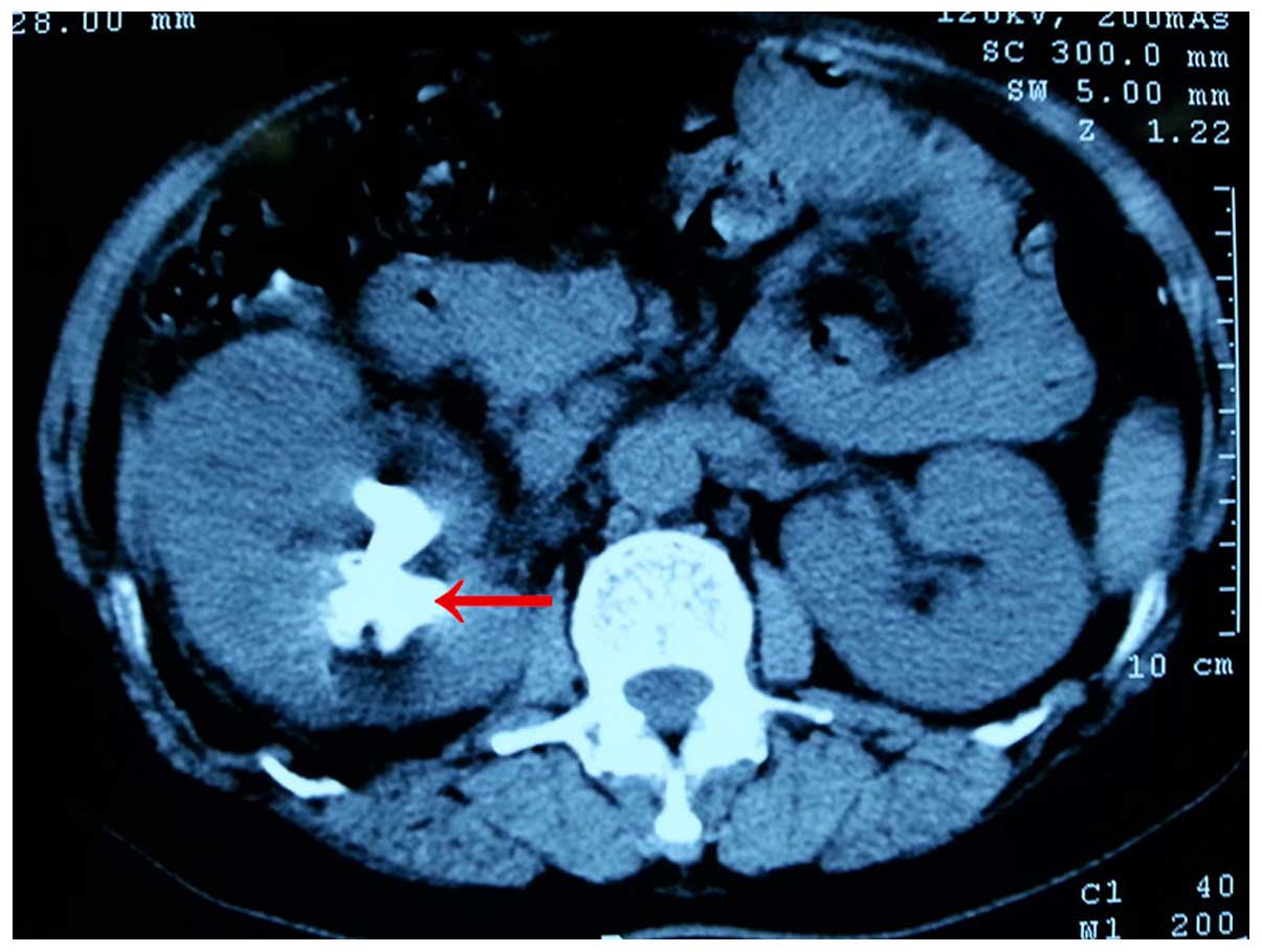
Munich, Germany), which revealed that the right kidney volume was
increased, with renal cortical thinning. The right renal pelvis
calyces were in a state of expansion and effusion. Several nodular
density shadows were detected on the right of the calyces, with a
clear border. The largest one measured ~2.6×2.5 cm (Fig. 1). The laboratory examinations revealed
that the patient had urinalysis, suggesting the presence of a
severe infection in the urinary tract, as did the blood cell
analysis. In order to block the development of septic shock, an
emergency percutaneous nephrostomy was performed. The stench of
milky white pus was observed in the drainage tube. A culture of
Escherichia coli bacteria was taken from the pus, which
proved to be sensitive to piperacillin treatment (2.5 mg, two
times/day). After 15 days' treatment of drainage and sensitive
antibiotics, the patient was offered nephroscope lithotripsy
through the former incision. However, a cauliflower-like neoplasm
was identified on the mucosa of the right mid-lower group calyces.
On the basis of the histopathological biopsy report, a diagnosis of
tubular papillary adenocarcinoma was made. Hematoxylin and eosin
(H&E) staining revealed features of the papillary
adenocarcinoma cells, including their adenoid arrangement, cellular
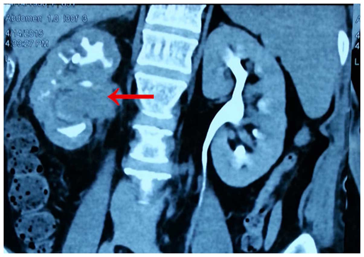
pleomorphism and invasive growth. Further coronal CT angiography
demonstrated a mass in the right renal pelvis (Fig. 2). After general anesthesia, the
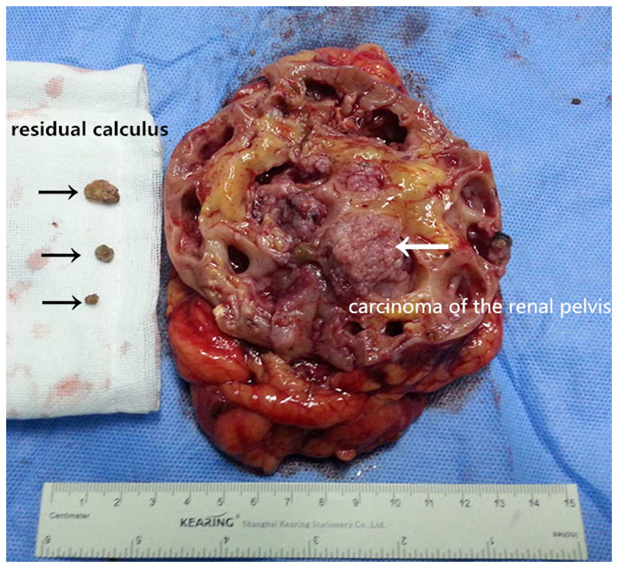
patient subsequently received radical nephroureterectomy. The gross
specimen of the removed mass is shown in Fig. 3.
For the immunohistochemical examination,
paraffin-embedded specimens were cut into 5 µm sections and mounted
onto poly-L-lysine-coated slides. The sections were deparaffinized,
rehydrated, and subsequently boiled for 10 min in 10 µmol/l citrate
buffer solution (pH 6.0) using a microwave oven. Endogenous
peroxidase was blocked with 0.3% hydrogen peroxide for 30 min, and
non-specific staining was blocked by treating the slides with 1%
fish-skin gelatin for 30 min at room temperature. Subsequently,
slides were incubated overnight with primary antibodies against
34Eβ12, CAM5.2 and cytokeratin 19 (1:100; Abcam, Cambridge, UK).
After washing with PBS, the slides were incubated with prediluted
secondary antibody (Abcam), followed by further incubation with
diaminobenzidine. Finally, the sections were counter-stained with
H&E stain and mounted. The immunohistochemical examination,
following the radical nephroureterectomy, revealed the tumor to be
a rare papillary adenocarcinoma. The patient returned to normal
following the surgery, and made an uneventful recovery. The patient
was normal at the 5-month follow-up stage.
Discussion
Tumors of the renal pelvis are uncommon in
clinicopathology, and constitute >5% of the malignancies arising
from the renal mass. Furthermore, adenocarcinoma accounts for an
extremely small percentage of neoplasms arising within the renal
pelvis, with a relative frequency of only 1% (1). Adenocarcinomas of the renal pelvis may
be subdivided into three major histological types: Tubulovillous
(71.5%), mucinous (21.5%) and papillary non-intestinal (7%).
Mucinous adenocarcinoma of the renal pelvis was first described in
1960 by Hasebe et al (2). They
are occasionally considered to arise in the foci of intestinal
metaplasia (2). According to the
statistics, a total of 51 cases were reported of adenocarcinoma of
the renal pelvis in the world by 1993, and reports of
adenocarcinoma have been predominantly recorded as being of the
mucinous type ever since (3,4). However, papillary adenocarcinoma of the
renal pelvis was described in Chinese journals, in 1987 by Fang
(5) and in 1997 by Kong et al
(6). Other reports of papillary
adenocarcinoma do exist in other language journals, although these
are rarer in occurrence. The finding, in 2005, that papillary
adenocarcinoma of the renal pelvis and ureter produce
carcinoembryonic antigen, carbohydrate antigen 19–9 and
carbohydrate antigen 125 in metastatic adenocarcinoma was reported
by Onishi et al (7). However,
in the present case study, the serum levels of the above tumor
biomarkers were normal, and none of the evidence from the imaging
suggested the presence of gastrointestinal tumors in the patient.
Immunohistochemical examination of the cytokeratin 34Eβ12(+), the
anticytokeratin CAM5.2(+) and cytokeratin 19(+) demonstrated that
the tumor originated in the glandular epithelium, and not in the
loops of Henle. Therefore, it was hypothesized that the patient in
the present case study only had primary papillary adenocarcinoma of
the renal pelvis, which may be associated with renal calculus and
chronic infection. This is in agreement with another study, which
elucidated that adenocarcinoma of the renal pelvis is associated
with lithiasis (7). Therefore, for
future reference, this clinical case study has indicated that,
firstly, a careful search for a papillary adenocarcinoma of the
renal pelvis should be excluded from metastasis of the digestive
tract. Subsequently, the possibility of long-term disease
associated with an infection of the kidney calculus should be
considered by screening the mucous membrane carcinoma of the renal
pelvis. Finally, a consensus on standardized surgery, postoperative
chemotherapy or radiotherapy of papillary adenocarcinoma in the
renal pelvis should be reached by global urology researchers for
the treatment of this disease.
Acknowledgements
We would like to thank Professor Zimin Shi and
Hongwei Huang at the Urology Department of the Second Affiliated
Hospital of Nanchang University, and Professor Liqing Wu at the
Pathology Department of the Second Affiliated Hospital of Nanchang
University, for their constant encouragement with this study and
for offering technology guidance.
References
|
1
|
Kaur G, Naik VR and Rahman MN: Mucinous
adenocarcinoma of the renal pelvis associated with lithiasis and
chronic gout. Singapore Med J. 45:125–126. 2004.PubMed/NCBI
|
|
2
|
Hasebe M, Serizawa S and Chino S: On a
case of papillary cystadenocarcinoma following malignant
degeneration of a papillary adenoma in the kidney pelvis. Yokohama
Med Bull. 11:491–500. 1960.PubMed/NCBI
|
|
3
|
Fareghi M, Mohammadi A and Madaen K:
Primary mucinous cystadenocarcinoma of renal pelvis: A case report.
Cases J. 2:93952009. View Article : Google Scholar : PubMed/NCBI
|
|
4
|
Xambre L, Cerqueira M, Cardoso A, Correia
T, Dias Macedo A, Carreira F and Galán T: Primary mucinous
adenocarcinoma of the renal pelvis-adicional case report. Actas
Urol Esp. 33:200–204. 2009.(In Spanish). View Article : Google Scholar : PubMed/NCBI
|
|
5
|
Fang G: Tow case of papillary
adenocarcinoma of renal pelvis with renal calculus. Guangxi
Medicine. 9:2161987.(In Chinese).
|
|
6
|
Kong X, Xia T, Pan B, Xue Z, Guo Y and Gu
F: A case of papillary adenocarcinoma of renal pelvis. Chin J
Pathol. 26:1271997.(In Chinese).
|
|
7
|
Onishi T, Franco OE, Shibahara T, Arima K
and Sugimura Y: Papillary adenocarcinoma of the renal pelvis and
ureter producing carcinoembryonic antigen, carbohydrate antigen
19–9 and carbohydrate antigen 125. Int J Urol. 12:214–216. 2005.
View Article : Google Scholar : PubMed/NCBI
|