Introduction
Mucinous adenocarcinoma of the uterine endometrium
accounts for 1–9% of all endometrial carcinomas (1). The diagnostic criteria of the World
Health Organization require that >50% of the neoplasm be
composed of mucinous cells. The prognosis of these tumors tends to
be relatively good (2). Mucinous
endometrial carcinomas display a variety of morphological
characteristics and may be subgrouped into endocervical and
intestinal subtypes (1). Abiko et
al, from our institution, documented the first case of mucinous
endometrial adenocarcinoma with gastric-type histology (3). This histological subtype, however, is a
rare malignant disease with only two case reports in the literature
(3,4).
Thus, the clinical behavior of this rare disease has not been fully
elucidated.
Gastric-type mucinous adenocarcinoma, also referred
to as minimal deviation adenocarcinoma or adenoma malignum,
accounts for <1% of adenocarcinomas originating in the uterine
cervix (5). Watery discharge is a
major symptom of gastric-type adenocarcinoma of the uterine cervix,
and macroscopic examination has revealed small cysts associated
with the tumor. Although gastric-type mucinous adenocarcinoma of
the uterine cervix exhibits a benign histological appearance, it is
typically associated with chemoresistance and poor prognosis
(6).
We herein describe an additional case of
gastric-type mucinous endometrial adenocarcinoma treated at our
institution. The clinical characteristics, symptoms, appearance on
magnetic resonance imaging (MRI) and prognosis are described, along
with a review of the relevant literature.
Case reports
Case 1
The first case involved a 72-year-old postmenopausal
woman (gravida 4, para 2) presenting with a primary complaint of
abnormal genital bleeding. The uterine cervical and endometrial
cytology were class V and suspected endometrioid adenocarcinoma,
respectively. Local examination indicated the presence of a dark
bloody secretion from the external orifice of the uterus.
Transvaginal ultrasonography revealed a walnut-sized tumor with an
irregular shape in the uterine cavity. MRI demonstrated a 4-cm mass
with low-intermediate intensity on T2-weighted images, and small
cysts with high intensity, suggesting mucin elements, were
identified in this tumor. The tumor invaded over half of the
myometrium (Fig. 1A). Enlargement of
a left obturator lymph node suggested the presence of metastasis.
The serum levels of carcinoembryonic antigen (CEA) and carbohydrate
antigen (CA)-125 were elevated (5.5 ng/ml, normal <5 ng/ml; and
40.3 U/ml, normal <35 U/ml, respectively). Hysteroscopy revealed
a polypoid tumor with atypical vessels in the uterine cavity.
Endometrial biopsy revealed a well-differentiated mucin-producing
adenocarcinoma, suggestive of gastric-type mucinous adenocarcinoma.
The patient underwent semiradical hysterectomy (type II radical
hysterectomy), bilateral salpingo-oophorectomy, pelvic
lymphadenectomy, para-aortic lymphadenectomy and partial
omentectomy. Intraoperative peritoneal washing cytology showed no
evidence of malignant cells. Macroscopically, the tumor appeared to
be whitish, sized 5.4 cm in the greatest dimension, arising from
the left wall and invading more than half of the myometrium,
without extension to the serosal surface (Fig. 1B). Microscopically, the tumor was
diagnosed as a mucinous adenocarcinoma with well-formed glands,
aggressively invading the myometrium, with a desmoplastic stromal
reaction and a partial microcystic elongated and fragmented pattern
(Fig. 1C). The carcinoma cells
exhibited abundant pale cytoplasm and minimal nuclear atypia, with
areas displaying fused glands and small clusters of cells with
ovoid or irregular nuclei. Multiple pelvic and para-aortic lymph
nodes metastases, as well as lymphovascular space invasion and
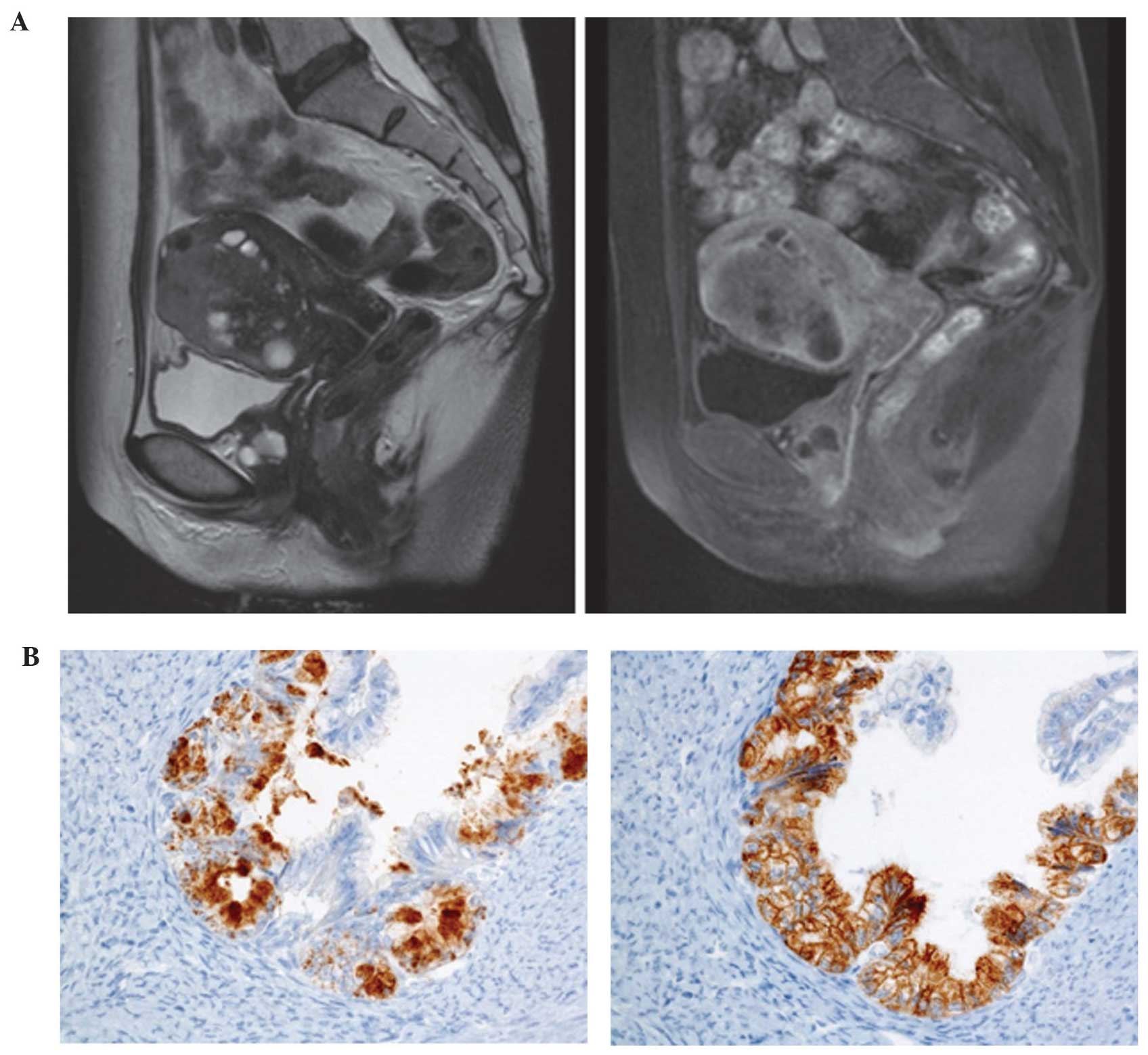
nerve involvement, were observed. Immunohistochemistry showed
positivity for MUC6, but negativity for HIK1083, p16 and p53
(Fig. 1D). The diagnosis was
confirmed as gastric-type mucinous adenocarcinoma of the uterine
corpus, pT1bN1M0, International Federation of Gynecology and
Obstetrics (FIGO) stage IIIC2. Therefore, postoperative adjuvant
chemotherapy with paclitaxel and carboplatin was administered;
however, 2 months after adjuvant chemotherapy, recurrence at the
para-aortic and pelvic lymph nodes was observed on a computed
tomography (CT) scan. The cancer cells then spread to the lungs and
the patient succumbed to the disease 6 months after recurrence to
the para-aortic and pelvic lymph nodes.
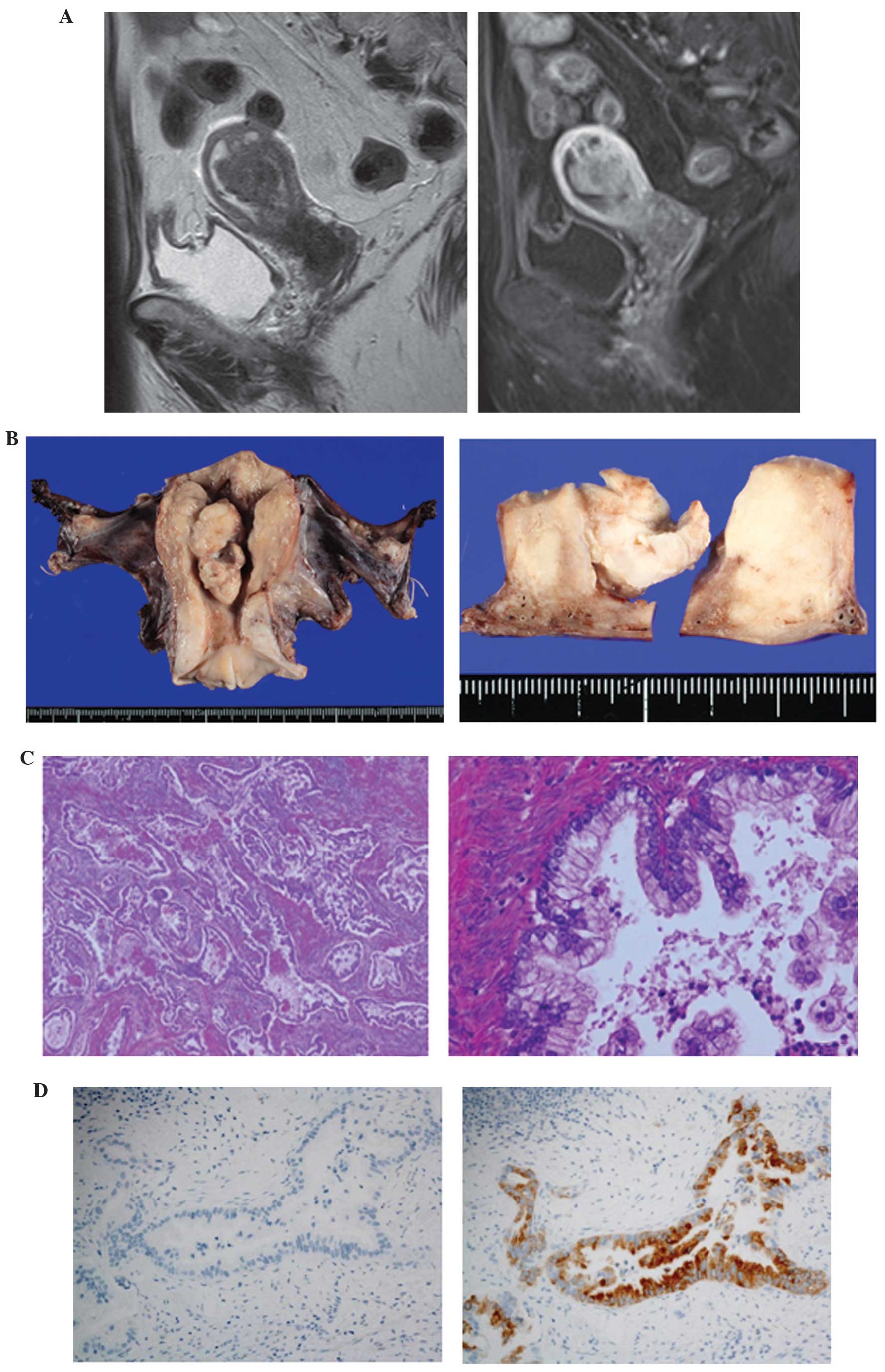 | Figure 1.Magnetic resonance imaging (MRI) and
pathological findings in case 1. (A) T2-weighted MRI scan showing a
tumor with intermediate intensity (left panel). Enhanced
T1-weighted images showing that the tumor invaded over half the
thickness of the myometrium (right panel). (B) Macroscopically, the
tumor was whitish, sized 5.4 cm, arising from the left wall and
invading over half of the myometrium. (C) The microscopic findings
included a diffusely infiltrating mucinous adenocarcinoma with
well-formed glands, aggressively invading the myometrium, along
with a desmoplastic stromal reaction; (left panel) magnification,
×100; right panel magnification, ×400. (D) On immunohistochemistry,
the tumor cells were positive for MUC6 (right panel), but negative
for HIK1083 (left panel); magnification, ×400. |
Case 2
The second case of gastric-type mucinous
adenocarcinoma of the uterine corpus in our institution has been
previously reported (3). The patient
was a 56-year-old postmenopausal woman with primary complaints of
abnormal genital bleeding and vaginal discharge, who was diagnosed
with high-grade adenocarcinoma of the uterine corpus by endometrial
biopsy prior to surgery (3).
Preoperative MRI revealed a solid tumor with multiple cysts
exhibiting non-enhanced high intensity on T2-weighted images,
suggesting mucin elements. The tumor invaded deep into the
myometrium (Fig. 2A). A CT scan
revealed para-aortic lymph node metastases and multiple small lung
nodules, suggestive of lung metastases. The patient underwent total
abdominal hysterectomy, bilateral salpingo-oophorectomy, pelvic
lymphadenectomy, para-aortic lymphadenectomy and omentectomy.
Gastric-type mucinous adenocarcinoma of the uterine corpus was
diagnosed based on the pathological findings, which included
well-formed glands composed of carcinoma cells with abundant clear
or pale eosinophilic cytoplasm. Although nuclear anaplasia and
architectural abnormalities were minimal, certain areas of the
tumor displayed fused glands, carcinoma cells with nuclear atypia
characterized by heterogeneous nuclear size and shape, and a
desmoplastic stromal reaction. Endometrial hyperplasia was not
observed. Immunohistochemistry was positive for MUC6, HIK1083,
CA19-9 and CEA, but negative for MUC2, estrogen receptor, p16 and
vimentin, suggesting a gastric phenotype (Fig. 2B). The pathological stage was
pT1cN0M1. Although the patient underwent postoperative adjuvant
chemotherapy with TS-1 and cisplatin (commonly used for gastric
cancer), the disease progressed. The patient succumbed to the
disease with peritonitis carcinomatosa and carcinomatous
lymphangiomatosis 2 years and 4 months after surgery.
Discussion
Gastric-type mucinous adenocarcinoma is a newly
characterized variant of mucinous adenocarcinoma with a
gynecological origin. Gastric-type adenocarcinoma of the uterine
cervix, also referred to as minimal deviation mucinous
adenocarcinoma or adenoma malignum, represents ~1–3% of all
endocervical adenocarcinomas (6,7). Kojima
et al (6) defined mucinous
adenocarcinoma with gastric differentiation using the following
criteria: i) Clear or pale eosinophilic cytoplasm, ii) voluminous
cytoplasm and iii) distinct cell borders. The immunophenotype of
these neoplasms is characterized by positivity for HIK1083 and/or
MUC6, partial positivity for p53, and negativity for p16 (5). This type of adenocarcinoma of the
uterine cervix is considered to be associated with poor prognosis
due to its more aggressive clinical behavior compared with the
usual types of adenocarcinoma of the uterine cervix (6,8). However,
among mucinous adenocarcinomas of the uterine corpus, only two
cases displaying the gastric phenotype have been previously
reported in the English literature (3,4).
Therefore, the biological characteristics of this type of tumor
have not been fully elucidated, owing to its rarity.
The findings of the two previously reported cases,
in addition to our case, are summarized in Table I. Gotoh et al (4) reported a case of synchronous mucinous
adenocarcinoma of the endometrium and mucinous cystadenoma
involving both ovaries in a 31-year-old woman with primary
infertility. The three cases exhibited morphology, immunophenotype
and clinical characteristics that are similar to those of
gastric-type adenocarcinoma originating from the uterine cervix.
All the patients presented with abnormal genital bleeding, and the
primary complaint in case 2 in the present report involved vaginal
discharge. The typical primary symptom of gastric-type
adenocarcinoma of the uterine cervix is watery discharge, whereas
gastric-type endometrial cancer may be suspected if abnormal
genital bleeding is also a main symptom.
 | Table I.Reported cases of gastric-type
mucinous neoplasm of the uterine corpus. |
Table I.
Reported cases of gastric-type
mucinous neoplasm of the uterine corpus.
| Author, year
(Refs.) | FIGO stage | Symptom | MRI appearance | Immunophenotype | Co-existence of
atypical endometrial hyperplasia | Primary
treatment | PFS (months) | OS (months) | Follow-up |
|---|
| Gotoh et al,
2004 (4) | IA | Atypical
bleeding | Solid, no cysts |
HIK1083+ | Yes | TAH, BSO | 6 | 6 | NED |
| Abiko et al,
2010 (3) | IVB | Vaginal discharge and
abnormal genital bleeding | Solid with cysts | HIK1083+
MUC6+ CEA+ CA19–9+ p53+
(partially) MUC2− p16−
vimentin− | No | TAH, BSO, PeN, PAN,
OM | 0 | 28 | DOD |
| Hino et al,
this case | IIIC2 | Abnormal genital
bleeding | Solid, no cysts | MUC6+
HIK1083− p53− p16− | No | Semi-RH, BSO, PeN,
PAN, OM | 2 | 14 | DOD |
Kido et al (9)
reported that tiny cysts associated with tumors were observed in
80% of the cases of gastric-type endocervical adenocarcinoma on
MRI. The tumors in cases 1 and 2 displayed cystic regions on MRI,
while in the case described by Gotoh et al (4), the small mass did not include cystic
cavities. Thus, this type of endometrial cancer exhibits a cystic
form in large tumors, such as gastric-type adenocarcinoma of the
uterine cervix, when examined using MRI. However, cysts are not
always present in small tumors. Histopathologically, the tumor is a
low-grade adenocarcinoma, while immunohistochemistry for HIK1083 or
MUC6 was positive in all three cases, indicating the gastric
phenotype. There was no co-existing endometrial hyperplasia or
endometrioid adenocarcinoma in our two cases, suggesting that these
cancers were estrogen-independent. Case 2, which was FIGO stage
IVB, presented with distant metastases at diagnosis, and the tumor
was resistant to the chemotherapy commonly used for gastric cancer.
Case 1, which was FIGO stage IIIC2, also exhibited chemoresistance
to paclitaxel and carboplatin, which is generally an effective
regimen for gynecological cancers. Both patients eventually
succumbed to the disease.
In our institution, the 5-year overall survival rate
of patients diagnosed with stage III endometrial cancer between
2004 and 2011 was ~80% (unpublished data). The clinical behaviors
of the cancer in our two cases indicates that gastric-type mucinous
adenocarcinoma of the uterine endometrium exhibits an aggressive
clinical behavior, even if the morphological findings are those of
low-grade adenocarcinoma. The case reported by Gotoh et al,
however, exhibited concomitant atypical endometrial hyperplasia and
was stage IA (4). In that case,
following complete resection, the prognosis was good, as the
patient was reported to remain alive and cancer-free. Thus,
gastric-type endometrial adenocarcinoma may be curable if complete
resection of the tumor is feasible.
Nucci et al (10) suggested that architectural complexity
alone is often the only diagnostic factor for endometrial mucinous
adenocarcinoma, as several cases exhibit only mild-to-moderate
nuclear atypia, thus highlighting the difficulty in differentially
diagnosing gastric-type mucinous adenocarcinoma from the
endocervical subtype. The reported frequency of mucinous
endometrial adenocarcinoma that shows a relatively favorable
prognosis ranges from <1 to 9% of all endometrial cancers
(1,11,12). As
two cases of gastric-type endometrial adenocarcinoma were diagnosed
in our institution over a period of 7 years, the frequency of
gastric-type endometrial mucinous adenocarcinoma is possibly higher
than expected. Although the new concept of gastric-type mucinous
adenocarcinoma of the uterine corpus is not widely recognized,
owing to its rarity among mucinous endometrial adenocarcinomas,
gastric-type mucinous adenocarcinoma should be distinguished from
other mucinous endometrial carcinomas, considering its aggressive
clinical behavior. The diagnosis should be confirmed by
immunohistochemistry in order to select the optimal treatment.
In summary, gastric-type adenocarcinoma of the
uterine corpus is a relatively rare malignant disease. The
incidence of gastric-type endometrial adenocarcinoma, however, is
possibly higher than expected. The symptoms include abnormal
genital bleeding and watery discharge. Gastric-type endometrial
adenocarcinoma is identified as a solid, irregularly shaped tumor
on MRI. Cases with large masses may display cystic cavities in the
tumor, similar to gastric-type adenocarcinoma originating from the
uterine cervix. These tumors exhibit an aggressive clinical
behavior and poor prognosis, owing to chemoresistance. A better
understanding of this disease is crucial in order to improve
diagnosis and develop appropriate therapeutic strategies.
References
|
1
|
Ross JC, Eifel PJ, Cox RS, Kempson RL and
Hendrickson MR: Primary mucinous adenocarcinoma of the endometrium.
A clinicopathologic and histochemical study. Am J Surg Pathol.
7:715–729. 1983. View Article : Google Scholar : PubMed/NCBI
|
|
2
|
Creasman WT, Morrow CP, Bundy BN, Homesley
HD, Graham JE and Heller PB: Surgical pathologic spread patterns of
endometrial cancer. A Gynecologic Oncology Group Study. Cancer.
60(Suppl 8): S2035–S2041. 1987. View Article : Google Scholar
|
|
3
|
Abiko K, Baba T, Ogawa M, Mikami Y, Koyama
T, Mandai M and Konishi I: Minimal deviation mucinous
adenocarcinoma (‘adenoma malignum’) of the uterine corpus. Pathol
Int. 60:42–47. 2010. View Article : Google Scholar : PubMed/NCBI
|
|
4
|
Gotoh T, Hayashi N, Takeda S, Itoyama S,
Takano M and Kikuchi Y: Synchronous mucinous adenocarcinoma of the
endometrium and mucinous cystadenoma of bilateral ovaries
presenting during fertility therapy. Int J Gynecol Cancer.
14:169–171. 2004. View Article : Google Scholar : PubMed/NCBI
|
|
5
|
Mikami Y and McCluggage WG: Endocervical
glandular lesions exhibiting gastric differentiation: An emerging
spectrum of benign, premalignant and malignant lesions. Adv Anat
Pathol. 20:227–237. 2013. View Article : Google Scholar : PubMed/NCBI
|
|
6
|
Kojima A, Mikami Y, Sudo T, Yamaguchi S,
Kusanagi Y, Ito M and Nishimura R: Gastric morphology and
immunophenotype predict poor outcome in mucinous adenocarcinoma of
the uterine cervix. Am J Surg Pathol. 31:664–672. 2007. View Article : Google Scholar : PubMed/NCBI
|
|
7
|
Silverberg SG and Hurt WG: Minimal
deviation adenocarcinoma (‘adenoma malignum’) of the cervix: A
reappraisal. Am J Obstet Gynecol. 121:971–975. 1975. View Article : Google Scholar : PubMed/NCBI
|
|
8
|
Gilks CB, Young RH, Aguirre P, DeLellis RA
and Scully RE: Adenoma malignum (minimal deviation adenocarcinoma)
of the uterine cervix. A clinicopathological and
immunohistochemical analysis of 26 cases. Am J Surg Pathol.
13:717–729. 1989. View Article : Google Scholar : PubMed/NCBI
|
|
9
|
Kido A, Mikami Y, Koyama T, Kataoka M,
Shitano F, Konishi I and Togashi K: Magnetic resonance appearance
of gastric-type adenocarcinoma of the uterine cervix in comparison
with that of usual-type endocervical adenocarcinoma: A pitfall of
newly described unusual subtype of endocervical adenocarcinoma. Int
J Gynecol Cancer. 24:1474–1479. 2014. View Article : Google Scholar : PubMed/NCBI
|
|
10
|
Nucci MR, Prasad CJ, Crum CP and Mutter
GL: Mucinous endometrial epithelial proliferations: A morphologic
spectrum of changes with diverse clinical significance. Mod Pathol.
12:1137–1142. 1999.PubMed/NCBI
|
|
11
|
Fujiwara M and Longacre TA: Low-grade
mucinous adenocarcinoma of the uterine corpus: A rare and
deceptively bland form of endometrial carcinoma. Am J Surg Pathol.
35:537–544. 2011. View Article : Google Scholar : PubMed/NCBI
|
|
12
|
Plataniotis G and Castiglione M: ESMO
Guidelines Working Group: Endometrial cancer: ESMO Clinical
Practice Guidelines for diagnosis, treatment and follow-up. Ann
Oncol. 21(Suppl 5): v41–v45. 2010. View Article : Google Scholar : PubMed/NCBI
|