Spandidos Publications Logo
  • About
    • About Spandidos
    • Aims and Scopes
    • Abstracting and Indexing
    • Editorial Policies
    • Reprints and Permissions
    • Job Opportunities
    • Terms and Conditions
    • Contact
  • Journals
    • All Journals
    • Oncology Letters
      • Oncology Letters
      • Information for Authors
      • Editorial Policies
      • Editorial Board
      • Aims and Scope
      • Abstracting and Indexing
      • Bibliographic Information
      • Archive
    • International Journal of Oncology
      • International Journal of Oncology
      • Information for Authors
      • Editorial Policies
      • Editorial Board
      • Aims and Scope
      • Abstracting and Indexing
      • Bibliographic Information
      • Archive
    • Molecular and Clinical Oncology
      • Molecular and Clinical Oncology
      • Information for Authors
      • Editorial Policies
      • Editorial Board
      • Aims and Scope
      • Abstracting and Indexing
      • Bibliographic Information
      • Archive
    • Experimental and Therapeutic Medicine
      • Experimental and Therapeutic Medicine
      • Information for Authors
      • Editorial Policies
      • Editorial Board
      • Aims and Scope
      • Abstracting and Indexing
      • Bibliographic Information
      • Archive
    • International Journal of Molecular Medicine
      • International Journal of Molecular Medicine
      • Information for Authors
      • Editorial Policies
      • Editorial Board
      • Aims and Scope
      • Abstracting and Indexing
      • Bibliographic Information
      • Archive
    • Biomedical Reports
      • Biomedical Reports
      • Information for Authors
      • Editorial Policies
      • Editorial Board
      • Aims and Scope
      • Abstracting and Indexing
      • Bibliographic Information
      • Archive
    • Oncology Reports
      • Oncology Reports
      • Information for Authors
      • Editorial Policies
      • Editorial Board
      • Aims and Scope
      • Abstracting and Indexing
      • Bibliographic Information
      • Archive
    • Molecular Medicine Reports
      • Molecular Medicine Reports
      • Information for Authors
      • Editorial Policies
      • Editorial Board
      • Aims and Scope
      • Abstracting and Indexing
      • Bibliographic Information
      • Archive
    • World Academy of Sciences Journal
      • World Academy of Sciences Journal
      • Information for Authors
      • Editorial Policies
      • Editorial Board
      • Aims and Scope
      • Abstracting and Indexing
      • Bibliographic Information
      • Archive
    • International Journal of Functional Nutrition
      • International Journal of Functional Nutrition
      • Information for Authors
      • Editorial Policies
      • Editorial Board
      • Aims and Scope
      • Abstracting and Indexing
      • Bibliographic Information
      • Archive
    • International Journal of Epigenetics
      • International Journal of Epigenetics
      • Information for Authors
      • Editorial Policies
      • Editorial Board
      • Aims and Scope
      • Abstracting and Indexing
      • Bibliographic Information
      • Archive
    • Medicine International
      • Medicine International
      • Information for Authors
      • Editorial Policies
      • Editorial Board
      • Aims and Scope
      • Abstracting and Indexing
      • Bibliographic Information
      • Archive
  • Articles
  • Information
    • Information for Authors
    • Information for Reviewers
    • Information for Librarians
    • Information for Advertisers
    • Conferences
  • Language Editing
Spandidos Publications Logo
  • About
    • About Spandidos
    • Aims and Scopes
    • Abstracting and Indexing
    • Editorial Policies
    • Reprints and Permissions
    • Job Opportunities
    • Terms and Conditions
    • Contact
  • Journals
    • All Journals
    • Biomedical Reports
      • Information for Authors
      • Editorial Policies
      • Editorial Board
      • Aims and Scope
      • Abstracting and Indexing
      • Bibliographic Information
      • Archive
    • Experimental and Therapeutic Medicine
      • Information for Authors
      • Editorial Policies
      • Editorial Board
      • Aims and Scope
      • Abstracting and Indexing
      • Bibliographic Information
      • Archive
    • International Journal of Epigenetics
      • Information for Authors
      • Editorial Policies
      • Editorial Board
      • Aims and Scope
      • Abstracting and Indexing
      • Bibliographic Information
      • Archive
    • International Journal of Functional Nutrition
      • Information for Authors
      • Editorial Policies
      • Editorial Board
      • Aims and Scope
      • Abstracting and Indexing
      • Bibliographic Information
      • Archive
    • International Journal of Molecular Medicine
      • Information for Authors
      • Editorial Policies
      • Editorial Board
      • Aims and Scope
      • Abstracting and Indexing
      • Bibliographic Information
      • Archive
    • International Journal of Oncology
      • Information for Authors
      • Editorial Policies
      • Editorial Board
      • Aims and Scope
      • Abstracting and Indexing
      • Bibliographic Information
      • Archive
    • Medicine International
      • Information for Authors
      • Editorial Policies
      • Editorial Board
      • Aims and Scope
      • Abstracting and Indexing
      • Bibliographic Information
      • Archive
    • Molecular and Clinical Oncology
      • Information for Authors
      • Editorial Policies
      • Editorial Board
      • Aims and Scope
      • Abstracting and Indexing
      • Bibliographic Information
      • Archive
    • Molecular Medicine Reports
      • Information for Authors
      • Editorial Policies
      • Editorial Board
      • Aims and Scope
      • Abstracting and Indexing
      • Bibliographic Information
      • Archive
    • Oncology Letters
      • Information for Authors
      • Editorial Policies
      • Editorial Board
      • Aims and Scope
      • Abstracting and Indexing
      • Bibliographic Information
      • Archive
    • Oncology Reports
      • Information for Authors
      • Editorial Policies
      • Editorial Board
      • Aims and Scope
      • Abstracting and Indexing
      • Bibliographic Information
      • Archive
    • World Academy of Sciences Journal
      • Information for Authors
      • Editorial Policies
      • Editorial Board
      • Aims and Scope
      • Abstracting and Indexing
      • Bibliographic Information
      • Archive
  • Articles
  • Information
    • For Authors
    • For Reviewers
    • For Librarians
    • For Advertisers
    • Conferences
  • Language Editing
Login Register Submit
  • This site uses cookies
  • You can change your cookie settings at any time by following the instructions in our Cookie Policy. To find out more, you may read our Privacy Policy.

    I agree
Search articles by DOI, keyword, author or affiliation
Search
Advanced Search
presentation
Molecular and Clinical Oncology
Join Editorial Board Propose a Special Issue
Print ISSN: 2049-9450 Online ISSN: 2049-9469
Journal Cover
July-2016 Volume 5 Issue 1

Full Size Image

Sign up for eToc alerts
Recommend to Library

Journals

International Journal of Molecular Medicine

International Journal of Molecular Medicine

International Journal of Molecular Medicine is an international journal devoted to molecular mechanisms of human disease.

International Journal of Oncology

International Journal of Oncology

International Journal of Oncology is an international journal devoted to oncology research and cancer treatment.

Molecular Medicine Reports

Molecular Medicine Reports

Covers molecular medicine topics such as pharmacology, pathology, genetics, neuroscience, infectious diseases, molecular cardiology, and molecular surgery.

Oncology Reports

Oncology Reports

Oncology Reports is an international journal devoted to fundamental and applied research in Oncology.

Experimental and Therapeutic Medicine

Experimental and Therapeutic Medicine

Experimental and Therapeutic Medicine is an international journal devoted to laboratory and clinical medicine.

Oncology Letters

Oncology Letters

Oncology Letters is an international journal devoted to Experimental and Clinical Oncology.

Biomedical Reports

Biomedical Reports

Explores a wide range of biological and medical fields, including pharmacology, genetics, microbiology, neuroscience, and molecular cardiology.

Molecular and Clinical Oncology

Molecular and Clinical Oncology

International journal addressing all aspects of oncology research, from tumorigenesis and oncogenes to chemotherapy and metastasis.

World Academy of Sciences Journal

World Academy of Sciences Journal

Multidisciplinary open-access journal spanning biochemistry, genetics, neuroscience, environmental health, and synthetic biology.

International Journal of Functional Nutrition

International Journal of Functional Nutrition

Open-access journal combining biochemistry, pharmacology, immunology, and genetics to advance health through functional nutrition.

International Journal of Epigenetics

International Journal of Epigenetics

Publishes open-access research on using epigenetics to advance understanding and treatment of human disease.

Medicine International

Medicine International

An International Open Access Journal Devoted to General Medicine.

Journal Cover
July-2016 Volume 5 Issue 1

Full Size Image

Sign up for eToc alerts
Recommend to Library

  • Article
  • Citations
    • Cite This Article
    • Download Citation
    • Create Citation Alert
    • Remove Citation Alert
    • Cited By
  • Similar Articles
    • Related Articles (in Spandidos Publications)
    • Similar Articles (Google Scholar)
    • Similar Articles (PubMed)
  • Download PDF
  • Download XML
  • View XML
Case Report

Evaluation of a tuberculous abscess on the right side of the diaphragm with contrast-enhanced computed tomography: A case report

  • Authors:
    • Peng Dong
    • Jing‑Jing Chen
    • Xi‑Zhen Wang
  • View Affiliations / Copyright

    Affiliations: Medical Imaging Center, The Affiliated Hospital of Weifang Medical University, Weifang, Shandong 261031, P.R. China, Department of Radiology, The Affiliated Hospital of Qingdao University School of Medicine, Qingdao, Shandong 266000, P.R. China
  • Pages: 210-212
    |
    Published online on: April 20, 2016
       https://doi.org/10.3892/mco.2016.863
  • Expand metrics +
Metrics: Total Views: 0 (Spandidos Publications: | PMC Statistics: )
Metrics: Total PDF Downloads: 0 (Spandidos Publications: | PMC Statistics: )
Cited By (CrossRef): 0 citations Loading Articles...

This article is mentioned in:


Abstract

We herein investigate the case of a patient with a tuberculous diaphragmatic abscess confirmed by pathology. The patient underwent plain computed tomography (CT) examination of the chest and contrast‑enhanced abdominal CT examination. The abscess appeared as hypodense mass with thick and irregular wall, which was enhanced on the contrast‑enhanced CT images. The shape of the mass resembled an irregular double convex lens. No enlarged lymph nodes were detected on the CT images. The presence of a tuberculous diaphragmatic abscess should be suspected in patients with a diaphragmatic hypodense mass with enhanced thick walls, even when there is absence of enlarged lymph nodes on the CT images.

Introduction

Mycobacterium tuberculosis (TB) infection remains a challenge to human health (1,2). Although abdominal TB is uncommon, the peritoneum is commonly involved in patients with abdominal TB infection (3,4). Charoensak et al (4) reported that certain cases with tuberculous peritonitis may mimic other diseases, which represents a diagnostic challenge for clinicians.

A few cases with intraperitoneal tuberculous abscesses have been previously reported, whereas diaphragmatic tuberculous abscess (DTA) is particularly rare (4–6). The aim of this case report was to gain some experience regarding the computed tomography (CT) findings in this DTA case, which was pathologically confirmed, with the hope that it will be contributory to the comprehensive CT diagnosis of DTAs.

Case report

The present study was reviewed and approved by the Institutional Review Board of The Affiliated Hospital of Weifang Medical University. The patient provided written informed concent was provided to the publication of this case.

A 22-year-old male patient presented with night sweats, easy fatigability and obscure right upper abdominal pain for 60 days. The clinical examination of the patient did not identify an abdominal palpable mass, and there was no history of lung TB.

Plain CT scanning of the chest was performed using the GE Bright Speed 16-detector spiral CT scanner (GE Medical Systems, Chicago, IL, USA) with the following parameters: 120 KV, 130 mAs and 5 mm slice width. Contrast-enhanced abdominal CT scanning was performed using the Siemens Somatom Sensation Cardiac 64-slice spiral CT scanner (Siemens, Munich, Germany) with the following parameters: 120 KV, 200–390 mAs and 5 mm slice width. The patient received intravenous Ultravist (300 mgI/ml; Bayer Schering Pharma AG, Berlin, Germany) administered as a bolus, at an injection rate of 3.0 ml/sec. Oral contrast material (water) was administered.

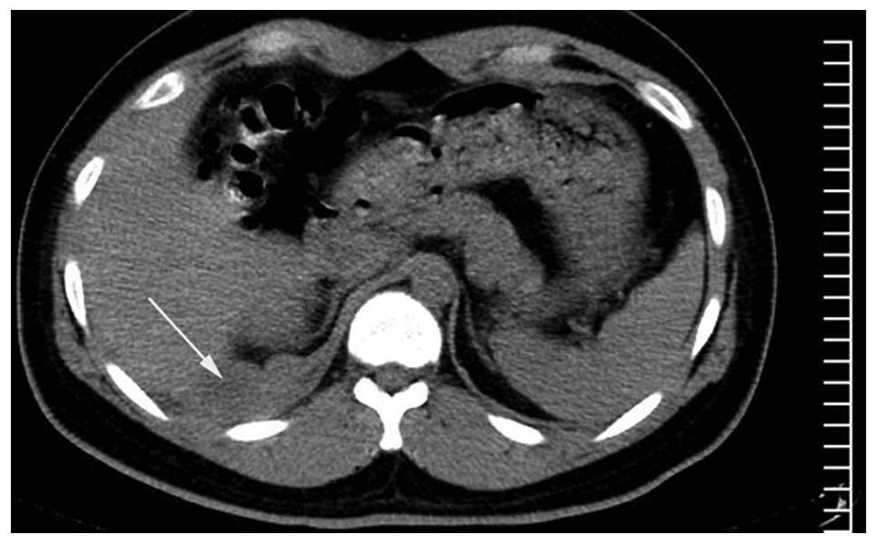
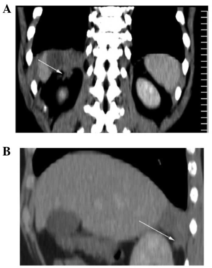
The CT images revealed a hypodense diaphragmatic mass, sized 6.0×4.5×3.5 cm, pushing on the parenchyma of the liver. The shape of the mass resembled an irregular double convex lens based on the right side of the diaphragm. The wall of the mass exhibited irregular thickening with enhancement. Gas collection or 
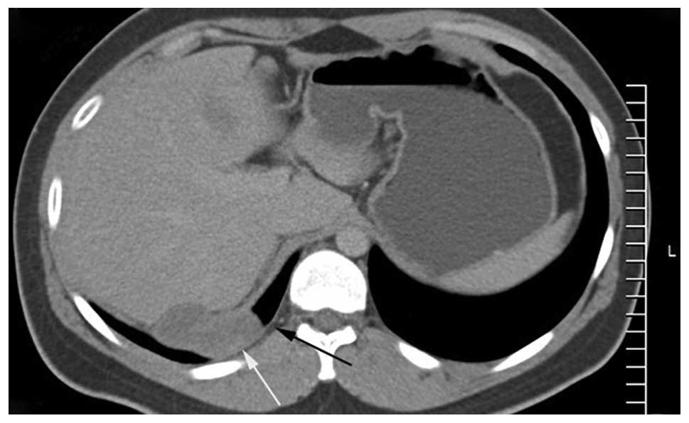
calcification were not detected within the mass (Figs. 1–3). The right side of the diaphragm was discontinuous. The right retroperitoneal space and the adjacent pleura were invaded, with marginally increased thickness of the subpleural fatty tissue 
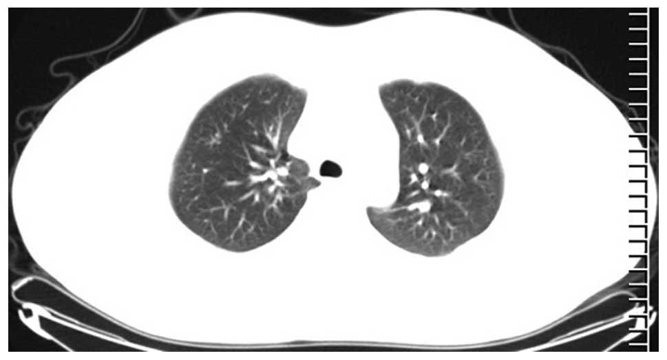
(Fig. 4). No enlarged lymph nodes were detected in the mediastinum, root of the mesentery or upper para-aortic region. No tuberculous lesion was detected on the bilateral lung fields (Fig. 5).

Figure 1.

Axial plain computed tomography image showing a hypodense diaphragmatic mass (white arrow) pushing on the hepatic parenchyma. The shape of the mass resembled an irregular double convex lens.

Figure 3.

(A) Coronal and (B) sagittal contrast-enhanced computed tomography images showing invasion of the right retroperitoneal space (white arrows).

Figure 4.

Axial computed tomography image showing invasion of the adjacent pleura (white arrow). The subpleural fatty tissue was thicker compared with that of the contralateral side (black arrow). No enlarged lymph nodes were identified.

Figure 5.

Axial computed tomography image did not reveal tuberculous lesions of the bilateral lung field.

Surgery was performed. The mass involved the right side of the diaphragm and adhered to the liver. The small and large bowel were normal. The pathological diagnosis was that of caseous TB. After being treated with anti-tuberculosis drugs, the patient fully recovered and was discharged from hospital.

Discussion

Tuberculous lesions are associated with a variable spectrum of medical imaging manifestations, and they may mimic other diseases (6–9). It is important to familiarize with the CT manifestations of tuberculous lesions to ensure accurate CT diagnosis.

Abdominal TB commonly spreads or disseminates by the following routes: i) Intestinal TB spreading to the lymph nodes; ii) hematogenous dissemination; and iii) direct extension of adjacent tuberculous lesions (8). Only a small proportion of patients with abdominal TB also suffer from pulmonary TB (7). It has been reported that multifocal TB may occur secondary to lympho-hematogenous dissemination after the initial infection (10). In our patient, the adjacent pleura and retroperitoneal space were invaded, whereas no enlarged lymph nodes or other tuberculous lesions were detected. This indicates that the spreading route of this case of DTA may be hematogenous dissemination.

The reasons for the rarity of skeletal muscle TB may be as follows: i) High levels of lactic acid; ii) not enough reticuloendothelial tissue; iii) not enough lymphatic tissue; and iv) rich blood supply (11,12). Thus, the abovementioned reasons may help to explain the rarity of the DTA.

In the present study, the wall of the DTA was irregularly thickened with permanent enhancement, which was similar to the cases reported in the literature (6). The right retroperitoneal space displayed fibrous strands neighboring the mass, which also conformed to inflammatory changes. In this case, the shape of the abscess (irregular double convex lens), the simultaneous involvement of the pleural cavity and retroperitoneal space with a discontinuous diaphragm on CT images may help with the localization and diagnosis.

Differential diagnosis

Most inflammations of the diaphragm result from direct extension from adjacent lesions (13), whereas lung cancer may directly invade the diaphragm (14). In this case, no lesions were detected within the lung field.

Malignant pleural mesothelioma may involve the diaphragm, and trans-diaphragmatic extension may be detected on CT examination (15). A rind-like pleura, with a thickness of >10 mm is crucial for the diagnosis of malignant pleural mesothelioma (16). Increased thickness of the subpleural fatty tissue was the sign for the diagnosis of benign pleural lesion on CT images (16). In the present case, the subpleural fatty tissue adjacent to the diaphragmatic mass was thicker compared with that of the contralateral side, which conformed to the diagnosis of a benign pleural lesion.

It may be difficult to distinguish diaphragmatic from abdominal tumors, particularly in the case of tumors arising from the right side of the diaphragm (17). In the present case, no abnormal signs were detected within the liver parenchyma and peritoneum.

Primary tumors of the diaphragm are rare, and the most common benign lesions are diaphragmatic cysts (18). The CT density of the diaphragmatic mesothelial cyst is similar to that of water, and the margin is well-defined without enhancement (19), which was different from the findings in this case.

In summary, DTA, despite its rarity, should be suspected in patients with a diaphragmatic hypodense mass exhibiting enhanced thick wall, even in the absence of enlarged lymph nodes on CT images.

References

1 

Lee WK, Van Tonder F, Tartaglia CJ, Dagia C, Cazzato RL, Duddalwar VA and Chang SD: CT appearances of abdominal tuberculosis. Clin Radiol. 67:596–604. 2012. View Article : Google Scholar : PubMed/NCBI

2 

LoBue PA, Enarson DA and Thoen TC: Tuberculosis in humans and its epidemiology, diagnosis and treatment in the United States. Int J Tuberc Lung Dis. 14:1226–1232. 2010.PubMed/NCBI

3 

Khan R, Abid S, Jafri W, Abbas Z, Hameed K and Ahmad Z: Diagnostic dilemma of abdominal tuberculosis in non-HIV patients: An ongoing challenge for physicians. World J Gastroenterol. 12:6371–6375. 2006. View Article : Google Scholar : PubMed/NCBI

4 

Charoensak A, Nantavithya P and Apisarnthanarak P: Abdominal CT findings to distinguish between tuberculous peritonitis and peritoneal carcinomatosis. J Med Assoc Thai. 95:1449–1456. 2012.PubMed/NCBI

5 

Dong P, Chen JJ, Wang XZ and Wang YQ: Intraperitoneal tuberculous abscess: Computed tomography features. World J Radiol. 7:286–293. 2015.PubMed/NCBI

6 

Dong P, Wang B and Sun YQ: Tuberculous abscess in the hepatoduodenal ligament: Evaluation with contrast-enhanced computed tomography. World J Gastroenterol. 14:2284–2287. 2008. View Article : Google Scholar : PubMed/NCBI

7 

Akhan O and Pringot J: Imaging of abdominal tuberculosis. Eur Radiol. 12:312–323. 2002. View Article : Google Scholar : PubMed/NCBI

8 

Yang ZG, Min PQ, Sone S, He ZY, Liao ZY, Zhou XP, Yang GQ and Silverman PM: Tuberculosis versus lymphomas in the abdominal lymph nodes: Evaluation with contrast-enhanced CT. AJR Am J Roentgenol. 172:619–623. 1999. View Article : Google Scholar : PubMed/NCBI

9 

Yang GY, Zhao D, Zhang WZ, Meng J, Li J, Li XH and Wan HF: Role of ultrasound evaluation for the diagnosis and monitoring of thyroid tuberculosis: A case report and review of the literature. Oncol Lett. 9:227–230. 2015.PubMed/NCBI

10 

Al-Tawfiq JA: Multifocal systemic tuberculosis: The many faces of an old nemesis. Med Sci Monit. 13:CS56–CS60. 2007.PubMed/NCBI

11 

Chewoolkar V, Bichile L and Patel H: Pyomyositis with multifocal osteomyelitis - an uncommon presentation of skeletal tuberculosis. J Assoc Physicians India. 57:7062009.PubMed/NCBI

12 

Neogi DS, Bandekar SM and Chawla L: Skeletal muscle tuberculosis simultaneously involving multiple sites. J Pediatr Orthop B. 22:167–169. 2013. View Article : Google Scholar : PubMed/NCBI

13 

Ye Y, Yang Z, Li H, Deng W, Li Y and Guo Y: MDCT features and anatomic-pathology in right thoracic-abdominal junctional region diseases. Sheng Wu Yi Xue Gong Cheng Xue Za Zhi. 28:255–259. 2011.(In Chinese). PubMed/NCBI

14 

Yokoi K, Tsuchiya R, Mori T, Nagai K, Furukawa T, Fujimura S, Nakagawa K and Ichinose Y: Results of surgical treatment of lung cancer involving the diaphragm. J Thorac Cardiovasc Surg. 120:799–805. 2000. View Article : Google Scholar : PubMed/NCBI

15 

Nickell LT Jr, Lichtenberger JP III, Khorashadi L, Abbott GF and Carter BW: Multimodality imaging for characterization, classification and staging of malignant pleural mesothelioma. Radiographics. 34:1692–1706. 2014. View Article : Google Scholar : PubMed/NCBI

16 

Metintas M, Ucgun I, Elbek O, Erginel S, Metintas S, Kolsuz M, Harmanci E, Alatas F, Hillerdal G, Ozkan R and Kaya T: Computed tomography features in malignant pleural mesothelioma and other commonly seen pleural diseases. Eur J Radiol. 41:1–9. 2002. View Article : Google Scholar : PubMed/NCBI

17 

Asahi Y, Kamiyama T, Nakanishi K, Yokoo H, Tahara M, Usui A, Funakoshi T, Sato M, Sasaki A, Matsuno Y, et al: Chondroma of the diaphragm mimicking a giant liver tumor with calcification: Report of a case. Surg Today. 44:2361–2365. 2014. View Article : Google Scholar : PubMed/NCBI

18 

Kim MP and Hofstetter WL: Tumors of the diaphragm. Thorac Surg Clin. 19:521–529. 2009. View Article : Google Scholar : PubMed/NCBI

19 

Esparza Estaún J, González Alfageme A and Sáenz Bañuelos J: Radiological appearance of diaphragmatic mesothelial cysts. Pediatr Radiol. 33:855–858. 2003. View Article : Google Scholar : PubMed/NCBI

Related Articles

  • Abstract
  • View
  • Download
  • Twitter
Copy and paste a formatted citation
Spandidos Publications style
Dong P, Chen JJ and Wang XZ: Evaluation of a tuberculous abscess on the right side of the diaphragm with contrast-enhanced computed tomography: A case report. Mol Clin Oncol 5: 210-212, 2016.
APA
Dong, P., Chen, J., & Wang, X. (2016). Evaluation of a tuberculous abscess on the right side of the diaphragm with contrast-enhanced computed tomography: A case report. Molecular and Clinical Oncology, 5, 210-212. https://doi.org/10.3892/mco.2016.863
MLA
Dong, P., Chen, J., Wang, X."Evaluation of a tuberculous abscess on the right side of the diaphragm with contrast-enhanced computed tomography: A case report". Molecular and Clinical Oncology 5.1 (2016): 210-212.
Chicago
Dong, P., Chen, J., Wang, X."Evaluation of a tuberculous abscess on the right side of the diaphragm with contrast-enhanced computed tomography: A case report". Molecular and Clinical Oncology 5, no. 1 (2016): 210-212. https://doi.org/10.3892/mco.2016.863
Copy and paste a formatted citation
x
Spandidos Publications style
Dong P, Chen JJ and Wang XZ: Evaluation of a tuberculous abscess on the right side of the diaphragm with contrast-enhanced computed tomography: A case report. Mol Clin Oncol 5: 210-212, 2016.
APA
Dong, P., Chen, J., & Wang, X. (2016). Evaluation of a tuberculous abscess on the right side of the diaphragm with contrast-enhanced computed tomography: A case report. Molecular and Clinical Oncology, 5, 210-212. https://doi.org/10.3892/mco.2016.863
MLA
Dong, P., Chen, J., Wang, X."Evaluation of a tuberculous abscess on the right side of the diaphragm with contrast-enhanced computed tomography: A case report". Molecular and Clinical Oncology 5.1 (2016): 210-212.
Chicago
Dong, P., Chen, J., Wang, X."Evaluation of a tuberculous abscess on the right side of the diaphragm with contrast-enhanced computed tomography: A case report". Molecular and Clinical Oncology 5, no. 1 (2016): 210-212. https://doi.org/10.3892/mco.2016.863
Follow us
  • Twitter
  • LinkedIn
  • Facebook
About
  • Spandidos Publications
  • Careers
  • Cookie Policy
  • Privacy Policy
How can we help?
  • Help
  • Live Chat
  • Contact
  • Email to our Support Team