Spandidos Publications Logo
  • About
    • About Spandidos
    • Aims and Scopes
    • Abstracting and Indexing
    • Editorial Policies
    • Reprints and Permissions
    • Job Opportunities
    • Terms and Conditions
    • Contact
  • Journals
    • All Journals
    • Oncology Letters
      • Oncology Letters
      • Information for Authors
      • Editorial Policies
      • Editorial Board
      • Aims and Scope
      • Abstracting and Indexing
      • Bibliographic Information
      • Archive
    • International Journal of Oncology
      • International Journal of Oncology
      • Information for Authors
      • Editorial Policies
      • Editorial Board
      • Aims and Scope
      • Abstracting and Indexing
      • Bibliographic Information
      • Archive
    • Molecular and Clinical Oncology
      • Molecular and Clinical Oncology
      • Information for Authors
      • Editorial Policies
      • Editorial Board
      • Aims and Scope
      • Abstracting and Indexing
      • Bibliographic Information
      • Archive
    • Experimental and Therapeutic Medicine
      • Experimental and Therapeutic Medicine
      • Information for Authors
      • Editorial Policies
      • Editorial Board
      • Aims and Scope
      • Abstracting and Indexing
      • Bibliographic Information
      • Archive
    • International Journal of Molecular Medicine
      • International Journal of Molecular Medicine
      • Information for Authors
      • Editorial Policies
      • Editorial Board
      • Aims and Scope
      • Abstracting and Indexing
      • Bibliographic Information
      • Archive
    • Biomedical Reports
      • Biomedical Reports
      • Information for Authors
      • Editorial Policies
      • Editorial Board
      • Aims and Scope
      • Abstracting and Indexing
      • Bibliographic Information
      • Archive
    • Oncology Reports
      • Oncology Reports
      • Information for Authors
      • Editorial Policies
      • Editorial Board
      • Aims and Scope
      • Abstracting and Indexing
      • Bibliographic Information
      • Archive
    • Molecular Medicine Reports
      • Molecular Medicine Reports
      • Information for Authors
      • Editorial Policies
      • Editorial Board
      • Aims and Scope
      • Abstracting and Indexing
      • Bibliographic Information
      • Archive
    • World Academy of Sciences Journal
      • World Academy of Sciences Journal
      • Information for Authors
      • Editorial Policies
      • Editorial Board
      • Aims and Scope
      • Abstracting and Indexing
      • Bibliographic Information
      • Archive
    • International Journal of Functional Nutrition
      • International Journal of Functional Nutrition
      • Information for Authors
      • Editorial Policies
      • Editorial Board
      • Aims and Scope
      • Abstracting and Indexing
      • Bibliographic Information
      • Archive
    • International Journal of Epigenetics
      • International Journal of Epigenetics
      • Information for Authors
      • Editorial Policies
      • Editorial Board
      • Aims and Scope
      • Abstracting and Indexing
      • Bibliographic Information
      • Archive
    • Medicine International
      • Medicine International
      • Information for Authors
      • Editorial Policies
      • Editorial Board
      • Aims and Scope
      • Abstracting and Indexing
      • Bibliographic Information
      • Archive
  • Articles
  • Information
    • Information for Authors
    • Information for Reviewers
    • Information for Librarians
    • Information for Advertisers
    • Conferences
  • Language Editing
Spandidos Publications Logo
  • About
    • About Spandidos
    • Aims and Scopes
    • Abstracting and Indexing
    • Editorial Policies
    • Reprints and Permissions
    • Job Opportunities
    • Terms and Conditions
    • Contact
  • Journals
    • All Journals
    • Biomedical Reports
      • Information for Authors
      • Editorial Policies
      • Editorial Board
      • Aims and Scope
      • Abstracting and Indexing
      • Bibliographic Information
      • Archive
    • Experimental and Therapeutic Medicine
      • Information for Authors
      • Editorial Policies
      • Editorial Board
      • Aims and Scope
      • Abstracting and Indexing
      • Bibliographic Information
      • Archive
    • International Journal of Epigenetics
      • Information for Authors
      • Editorial Policies
      • Editorial Board
      • Aims and Scope
      • Abstracting and Indexing
      • Bibliographic Information
      • Archive
    • International Journal of Functional Nutrition
      • Information for Authors
      • Editorial Policies
      • Editorial Board
      • Aims and Scope
      • Abstracting and Indexing
      • Bibliographic Information
      • Archive
    • International Journal of Molecular Medicine
      • Information for Authors
      • Editorial Policies
      • Editorial Board
      • Aims and Scope
      • Abstracting and Indexing
      • Bibliographic Information
      • Archive
    • International Journal of Oncology
      • Information for Authors
      • Editorial Policies
      • Editorial Board
      • Aims and Scope
      • Abstracting and Indexing
      • Bibliographic Information
      • Archive
    • Medicine International
      • Information for Authors
      • Editorial Policies
      • Editorial Board
      • Aims and Scope
      • Abstracting and Indexing
      • Bibliographic Information
      • Archive
    • Molecular and Clinical Oncology
      • Information for Authors
      • Editorial Policies
      • Editorial Board
      • Aims and Scope
      • Abstracting and Indexing
      • Bibliographic Information
      • Archive
    • Molecular Medicine Reports
      • Information for Authors
      • Editorial Policies
      • Editorial Board
      • Aims and Scope
      • Abstracting and Indexing
      • Bibliographic Information
      • Archive
    • Oncology Letters
      • Information for Authors
      • Editorial Policies
      • Editorial Board
      • Aims and Scope
      • Abstracting and Indexing
      • Bibliographic Information
      • Archive
    • Oncology Reports
      • Information for Authors
      • Editorial Policies
      • Editorial Board
      • Aims and Scope
      • Abstracting and Indexing
      • Bibliographic Information
      • Archive
    • World Academy of Sciences Journal
      • Information for Authors
      • Editorial Policies
      • Editorial Board
      • Aims and Scope
      • Abstracting and Indexing
      • Bibliographic Information
      • Archive
  • Articles
  • Information
    • For Authors
    • For Reviewers
    • For Librarians
    • For Advertisers
    • Conferences
  • Language Editing
Login Register Submit
  • This site uses cookies
  • You can change your cookie settings at any time by following the instructions in our Cookie Policy. To find out more, you may read our Privacy Policy.

    I agree
Search articles by DOI, keyword, author or affiliation
Search
Advanced Search
presentation
Molecular and Clinical Oncology
Join Editorial Board Propose a Special Issue
Print ISSN: 2049-9450 Online ISSN: 2049-9469
Journal Cover
July-2016 Volume 5 Issue 1

Full Size Image

Sign up for eToc alerts
Recommend to Library

Journals

International Journal of Molecular Medicine

International Journal of Molecular Medicine

International Journal of Molecular Medicine is an international journal devoted to molecular mechanisms of human disease.

International Journal of Oncology

International Journal of Oncology

International Journal of Oncology is an international journal devoted to oncology research and cancer treatment.

Molecular Medicine Reports

Molecular Medicine Reports

Covers molecular medicine topics such as pharmacology, pathology, genetics, neuroscience, infectious diseases, molecular cardiology, and molecular surgery.

Oncology Reports

Oncology Reports

Oncology Reports is an international journal devoted to fundamental and applied research in Oncology.

Experimental and Therapeutic Medicine

Experimental and Therapeutic Medicine

Experimental and Therapeutic Medicine is an international journal devoted to laboratory and clinical medicine.

Oncology Letters

Oncology Letters

Oncology Letters is an international journal devoted to Experimental and Clinical Oncology.

Biomedical Reports

Biomedical Reports

Explores a wide range of biological and medical fields, including pharmacology, genetics, microbiology, neuroscience, and molecular cardiology.

Molecular and Clinical Oncology

Molecular and Clinical Oncology

International journal addressing all aspects of oncology research, from tumorigenesis and oncogenes to chemotherapy and metastasis.

World Academy of Sciences Journal

World Academy of Sciences Journal

Multidisciplinary open-access journal spanning biochemistry, genetics, neuroscience, environmental health, and synthetic biology.

International Journal of Functional Nutrition

International Journal of Functional Nutrition

Open-access journal combining biochemistry, pharmacology, immunology, and genetics to advance health through functional nutrition.

International Journal of Epigenetics

International Journal of Epigenetics

Publishes open-access research on using epigenetics to advance understanding and treatment of human disease.

Medicine International

Medicine International

An International Open Access Journal Devoted to General Medicine.

Journal Cover
July-2016 Volume 5 Issue 1

Full Size Image

Sign up for eToc alerts
Recommend to Library

  • Article
  • Citations
    • Cite This Article
    • Download Citation
    • Create Citation Alert
    • Remove Citation Alert
    • Cited By
  • Similar Articles
    • Related Articles (in Spandidos Publications)
    • Similar Articles (Google Scholar)
    • Similar Articles (PubMed)
  • Download PDF
  • Download XML
  • View XML
Case Report

Curative resection for leiomyosarcoma of the descending mesocolon with metachronous liver metastasis: A case report and literature review

  • Authors:
    • Takaharu Kato
    • Hiroshi Noda
    • Iku Abe
    • Sergio Alonso
    • Nobu Yokoyama
    • Toshiki Rikiyama
  • View Affiliations / Copyright

    Affiliations: Department of Surgery, Saitama Medical Center, Jichi Medical University, Saitama, Saitama 330‑8503, Japan, Institute of Predictive and Personalized Medicine of Cancer (IMPPC), Health Sciences Research Institute Germans Trias i Pujol (IGTP), Campus Can Ruti, 08916 Badalona, Barcelona, Spain
  • Pages: 53-56
    |
    Published online on: April 25, 2016
       https://doi.org/10.3892/mco.2016.874
  • Expand metrics +
Metrics: Total Views: 0 (Spandidos Publications: | PMC Statistics: )
Metrics: Total PDF Downloads: 0 (Spandidos Publications: | PMC Statistics: )
Cited By (CrossRef): 0 citations Loading Articles...

This article is mentioned in:


Abstract

Mesenteric leiomyosarcoma is a rare disease with poor prognosis. Previously, mesenteric leiomyosarcoma was not differentiated from gastrointestinal stromal tumor (GIST), which is the most common mesenchymal tumor of the gastrointestinal tract, and several cases of GIST may have been misclassified as mesenteric leiomyosarcoma. Thus, the actual clinicopathological characteristics of mesenteric leiomyosarcomas remain unclear. We herein describe a case of leiomyosarcoma arising in the descending mesocolon in a patient who developed metachronous liver metastasis. A 76‑year‑old woman reported a mass in her left upper abdomen. Computed tomography imaging revealed a low‑density tumor adjacent to the descending colon. The patient underwent surgery and the tumor was resected along with part of the descending colon. Immunohistochemical differential diagnosis revealed that the tumor was positive for smooth muscle actin and desmin, and negative for CD117 (c‑KIT) and S‑100, which are characteristic of gastrointestinal leiomyosarcoma. A single liver metastasis developed 24 months after the operation. The patient underwent curative resection of the metastatic lesion. Sixteen months following surgery for the liver metastasis and 40 months after the initial removal of the primary lesion, the patient remains disease‑free. The prognosis of leiomyosarcoma remains poor and standardized chemotherapy for this rare disease has not yet been established. Early diagnosis and surgical removal of the tumor is the only potentially curative option for liver metastasis of mesenteric leiomyosarcoma.

Introduction

Mesenteric leiomyosarcoma is a rare disease with poor prognosis. Since 1998, it is acknowledged that the proper diagnosis of leiomyosarcoma requires immunohistochemical (IHC) analysis to differentiate it from gastrointestinal stromal tumor (GIST) (1), which is the most common mesenchymal tumor of the gastrointestinal tract. Several GISTs diagnosed before 1998 were misclassified as leiomyosarcomas; therefore, the frequency of leiomyosarcoma may have been previously overestimated. The clinical and pathological characteristics of mesenteric leiomyosarcoma have not been clearly determined. In this report, we present the case of a patient who underwent curative surgery for leiomyosarcoma arising in the descending mesocolon and for a metachronous liver metastasis, together with the clinicopathological findings and a literature review.

Case report

A 76-year-old woman noticed a mass in her left upper abdomen and consulted her local clinic, where a 12-cm abdominal tumor was detected by ultrasonographic evaluation. The patient was referred to the Saitama Medical Center, Jichi Medical University (Saitama, Japan) for further investigation and treatment. The patient's past medical history included hepatitis C infection, an ovarian cyst and appendicitis. The cyst and the appendix were surgically resected at the age of 4 and 34 years, respectively. The patient had a family history of cancer, as her mother had developed colorectal cancer. Physical examination revealed a palpable mass, sized ~5 cm, in the left upper abdomen, which was elastic hard and movable. No lymph nodes were palpable. The patient did not suffer from nausea. The tumor markers were marginally elevated (carcinoembryonic antigen, 5.6 ng/ml and carbohydrate antigen 19-9, 22.2 U/ml). Interleukin-2 receptor level was within the normal 
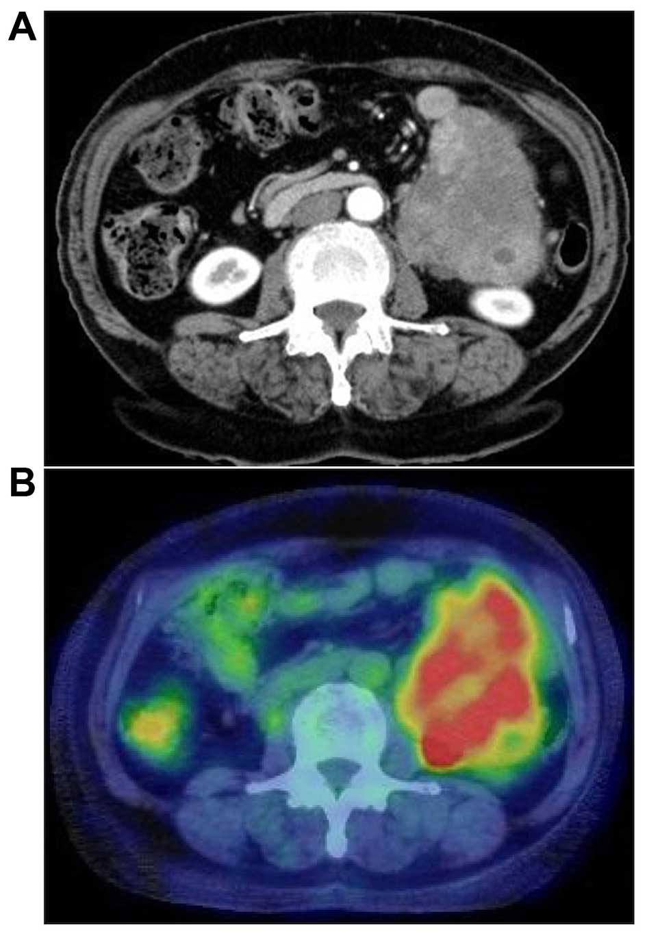
range. Abdominal computed tomography (CT) revealed a 13-cm tumor, heterogeneously enhanced, located in the abdominal cavity between the tail of the pancreas and the anterior surface of the left kidney. The margin with the surrounding organs was clear, except for the small intestine. There was no evidence of ascites, liver metastasis, or enlarged lymph nodes (Fig. 1A). The tumor was fluorodeoxyglucose (FDG)-avid on FDG-positron emission tomography (PET)/CT (Fig. 1B). Colonoscopy revealed no evidence of a tumor in the descending colon. The patient was diagnosed with a tumor arising in the retroperitoneal or descending mesocolon and underwent surgery in June, 2012. Intraoperatively, the tumor was located in the descending mesocolon, it was movable and easily excised from the left psoas muscle and urinary duct. However, the tumor invaded the mesenteric vein; therefore, en bloc removal of 
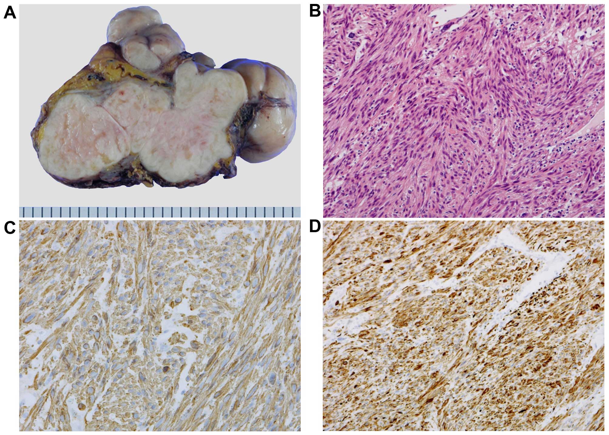
the descending colon was performed. Macroscopically, the size of the tumor was 14×9×9 cm, it weighed 500 g, its surface was smooth and elastic hard, and the cut surface was solid and white (Fig. 2A). The histological findings included high cellularity, spindle-shaped cells with blunted hyperchromatic nuclei, exhibiting a trabecular proliferation pattern that indicated differentiation towards smooth muscle (Fig. 2B). IHC analysis revealed that the tumor was positive for smooth muscle actin and desmin (Fig. 2C and D) and negative for CD117 (c-KIT) and S-100. From these findings, the final diagnosis was confirmed as leiomyosarcoma of the descending mesocolon. The patient's postoperative course was uncomplicated and she was discharged from the hospital 11 days after surgery. She was followed up every 6 months by blood tests and imaging studies [CT, magnetic resonance imaging (MRI) and ultrasonography] and did not receive any adjuvant 
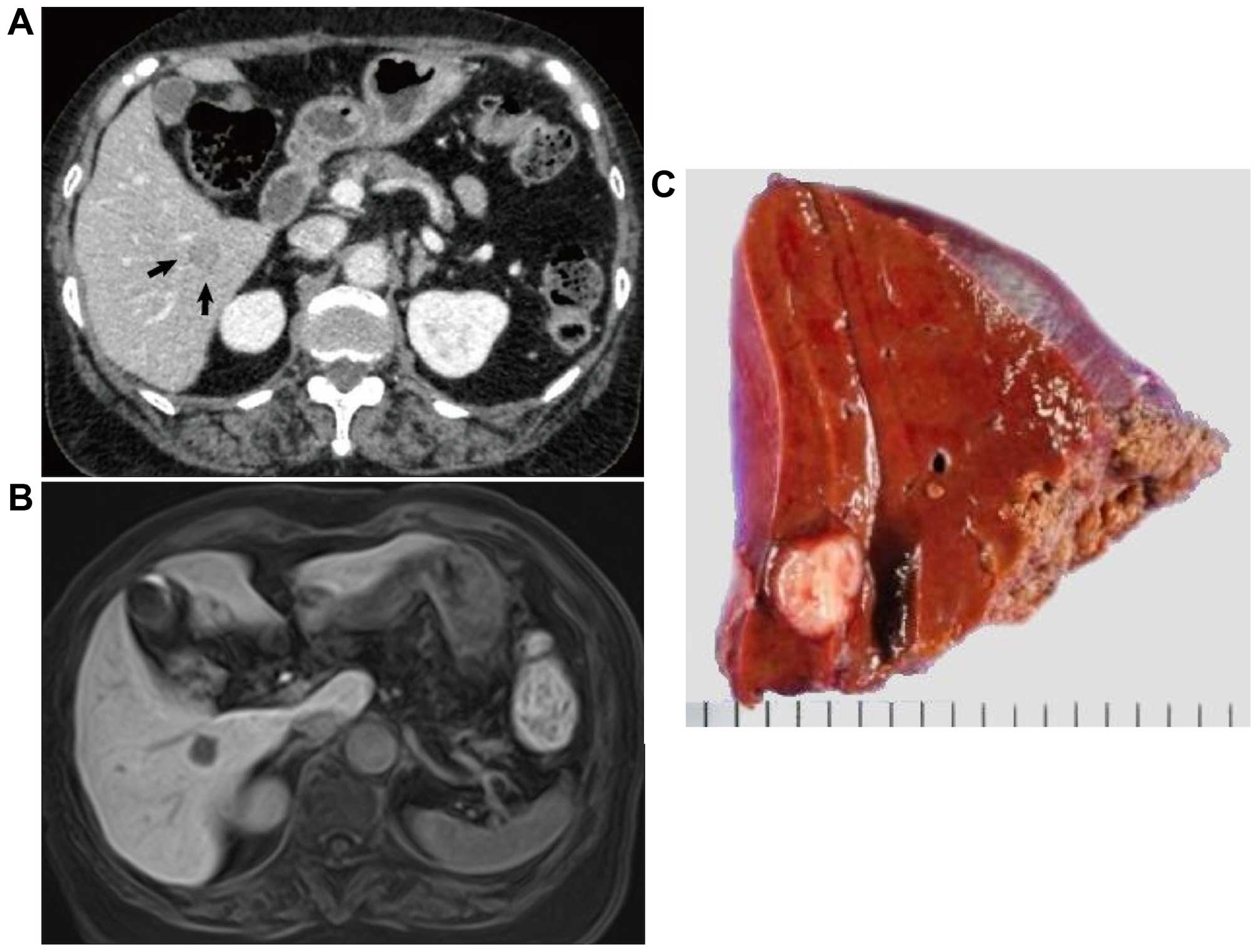
chemotherapy. A tumor at segment VI in the right lobe of the liver was detected 24 months after the surgery. The tumor size was ~15 mm and marginally enhanced on CT (Fig. 3A). The tumor was also detected as a hypointense lesion on gadolinium ethoxybenzyl diethylenetriamine pentaacetic acid-enhanced MRI with a T1-weighted image of the hepatobiliary phase (Fig. 3B), but not detected on FDGPET/CT. The patient underwent partial liver resection to remove the tumor. The tumor was measured to be 12×10 mm (Fig. 3C) and was diagnosed as a metastatic lesion from the leiomyosarcoma of the descending mesocolon by pathological examination. At the most recent follow-up, 16 months after the surgery for liver metastasis and 40 months after the primary surgery, the patient remains disease-free.

Figure 1.

(A) Abdominal computed tomography (CT) revealed an irregular mass enhanced heterogeneously in the left lower abdomen, sized 9×7 cm. (B) A fluorodeoxyglucose (FDG) positron emission tomography/CT fusion image revealed a tumor in the left lower abdomen with significantly increased FDG uptake (maximum standardized uptake value = 5.6).

Figure 2.

(A) The resected tumor was solid and white-yellowish, sized 140×90×90 mm, containing several lobules. The cut surface was white and elastic hard. (B) The histological examination of the resected specimen revealed atypical spindle cell proliferation, indicating differentiation towards smooth muscle (hematoxylin-eosin staining; magnification, ×200). On immunohistochemistry, the tumor cells stained positive for (C) α smooth muscle actin and (D) desmin (magnification, ×200), but negative for CD117 (c-KIT) and S-100 (not shown).

Figure 3.

(A) Abdominal computed tomography revealed a 15-mm tumor at segment VI in the liver (arrows). (B) The tumor was detected as a hypointense lesion on gadolinium ethoxybenzyl diethylenetriamine pentaacetic acid-enhanced magnetic resonance imaging with a T1-weighted image of the hepatobiliary phase at segment VI of the liver. (C) The resected specimen from the liver exhibited a white nodular tumor, sized 10×10 mm.

Discussion

We herein report a case who underwent surgery for primary leiomyosarcoma of the descending mesocolon and a single metachronous liver metastasis. Mesenteric leiomyosarcoma is a rare malignancy. The first mesenteric leiomyosarcoma case in the literature was reported by Derechin et al (1) in 1956. Proper diagnosis of leiomyosarcoma requires IHC to differentiate this type of lesion from GIST, the most common mesenchymal tumor of the gastrointestinal tract. However, older reports on mesenteric smooth muscle tumors cannot be included in the current classification, since, at the time, the studies did not differentiate between leiomyosarcoma and GIST. Thus, several GISTs may have been misclassified as leiomyosarcomas; therefore, the frequency of leiomyosarcoma may have been previously overestimated. We searched the PubMed database using the key words ‘leiomyosarcoma AND (mesentery OR mesenteric)’, excluding articles that were published prior to 1998, which was the year the GIST concept was introduced (2). A total of 163 articles were initially identified and we eliminated reports that did not apply IHC for the differential diagnosis of GIST or reports that were not written in English. Including our case, only 13 patients have been reported to be diagnosed with mesenteric leiomyosarcoma (Table I). The median age of the patients was 62 years (range, 13–82 years) and they included 6 men and 7 women. The most common site developing leiomyosarcoma was the mesentery of the small intestine (6 cases) followed by the mesocolon (4 cases) and mesorectum (1 case). The remaining 2 cases were described as just mesentery, with no more specific location information. Of the 12 cases, 11 had follow-up information, with a median of 18 months (range, 3–59 months). Surgical resection with a wide margin of normal tissue is the most effective treatment strategy for mesenteric leiomyosarcoma (3). All 13 cases received surgery for the primary lesion; 6 patients succumbed to the disease during the follow-up period (3–58 months). No patient was reported to survive for >59 months. Of the patients who succumbed to the disease, 1 died with the originally diagnosed leiomyosarcoma (no information on surgical intervention was reported); 4 had developed liver metastasis, with (n=3) or without (n=1) local recurrence after surgical resection of the primary lesion; and 1 patient succumbed to lung metastasis after removal of the primary lesion (Table I). Liver is the most common organ in which metastatic lesions developed among these cases. Of note, all the patients who were followed up after surgery for >6 months developed liver metastasis synchronously (n=2) or metachronously (n=6). One case reported by Miettinen et al (4) developed a liver metastasis at an undetermined time after the operation and during the12-month follow-up. The median interval for developing liver metastasis after primary surgery was 20 months (range, 0–48 months). The majority of these liver metastases were multiple, with 1 case developing metastases both in the liver and lung. Only 2 patients, including the one reported in this study, developed single liver metastasis (5). Three cases, including the patient reported in our study, underwent surgery for liver metastasis (6), and 3 cases received chemotherapy for the metastatic lesions (5,7). There is no mention of surgical intervention or chemotherapy treatment for the secondary liver lesions in the remaining 3 cases. Some reports demonstrated that doxorubicin-containing chemotherapy improved the outcome of patients with metastatic leiomyosarcoma (8,9). Although these reports did not include patients with mesenteric leiomyosarcoma, due to its rarity, doxorubicin-containing chemotherapy may also be effective in reducing the incidence of metastasis or recurrence of mesenteric leiomyosarcoma. However, a standardized chemotherapy regimen for metastasis or recurrence of this type of tumor is not yet established. In our case, 40 months after the initial removal of the primary lesion, the patient remains disease-free. The favorable postoperative course over an extended period of time was achieved by curative resection of both the primary tumor and the metachronous liver metastasis, without adjuvant chemotherapy. Albeit the current literature is limited due to the low incidence of mesenteric leiomyosarcomas, the data reveal a high incidence of liver metastasis in these patients. We recommend periodic monitoring using imaging techniques following surgical resection, as it would be highly beneficial in the clinical management of mesenteric leiomyosarcoma patients, facilitating early detection and curative resection of metachronous liver metastases. Further studies are required to elucidate the clinical and pathological characteristics of mesenteric leiomyosarcoma patients.

Table I.

Reported cases of mesenteric leiomyosarcoma studied by IHC for differential diagnosis of GIST.

Table I.

Reported cases of mesenteric leiomyosarcoma studied by IHC for differential diagnosis of GIST.

First author (year)Age/genderSymptomsSize (mm)Location (mesentery)Operative procedureOutcome (months)Cause of deathDistant metastasisInterval between primary and metastatic lesion (months)ChemotherapyRefs.
Miettinen (1999)41/MN.M.N.M.N.M.N.M.Alive (12)–LiverN.M.N.M.(4)
Miettinen (1999)86/MN.M.N.M.N.M.N.M.Dead (3)Present diseaseN.M.–N.M.(4)
Fukanaga (2004)62/FMass140Sigmoid colonExtirpationDead (10)Liver metastasisLiver0No(10)
Simonovich (2006)82/FMass, pain110Small intestineResection with small intestineAlive (24)–Liver24No(3)
Iwasaki (2010)13/MMass100Small intestineResection with small intestineAlive (3)–No––(11)
Koczkowska (2013)62/F–78Small intestineExtirpationDead (24)Liver metastasis, local recurrenceLiver, local0Yes(7)
Koczkowska (2013)46/FMassN.M.Sigmoid colonResection with colon and uterusDead (58)Liver metastasis, local recurrenceLiver, local48Yes(7)
Mizobe (2013)65/MMass, pain200Ascending colonIleocecal resectionDead (18)Liver metastasis, local recurrenceLiver, local11Yes(5)
Dasgupta (2016)62/FFullness220Small intestineResection with small intestineAlive (6)–No––(12)
Sidhic (2015)33/MPain150Small intestineResection with small intestineAlive (0)–No––(13)
Hamed (2015)49/FN.M.N.M.Small intestineResection with small intestineAlive (59)–Liver29N.M.(6)
Hamed (2015)46/MN.M.N.M.RectumResection with rectumDead (26)Lung metastasisLiver, lung16N.M.(6)
Present case76/FMass140Descending colonResection with colonAlive (40)–Liver24No

[i] M, male; F, female; N.M, not mentioned.

Glossary

Abbreviations

Abbreviations:

GIST

gastrointestinal stromal tumor

IHC

immunohistochemical

CT

computed tomography

FDGPET

fluorodeoxyglucose positron emission tomography

MRI

magnetic resonance imaging

References

1 

Derechin W and Wolfe S: Leiomyosarcoma of the mesentery. Can Med Assoc J. 75:1028–1029. 1956.PubMed/NCBI

2 

Hirota S, Isozaki K, Moriyama Y, Hashimoto K, Nishida T, Ishiguro S, Kawano K, Hanada M, Kurata A, Takeda M, et al: Gain-of-function mutations of c-kit in human gastrointestinal stromal tumors. Science. 279:577–580. 1998. View Article : Google Scholar : PubMed/NCBI

3 

Simonovich CJ, Hardman JM, Navin JJ, Jacobs J and Fergusson N: An unusual abdominal tumor - leiomyosarcoma of the mesentery: A case report. Hawaii Med J. 65:18–20. 2006.PubMed/NCBI

4 

Miettinen M, Monihan JM, Sarlomo-Rikala M, Kovatich AJ, Carr NJ, Emory TS and Sobin LH: Gastrointestinal stromal tumors/smooth muscle tumors (GISTs) primary in the omentum and mesentery: clinicopathologic and immunohistochemical study of 26 cases. Am J Surg Pathol. 23:1109–1118. 1999. View Article : Google Scholar : PubMed/NCBI

5 

Mizobe T, Akagi Y, Ishikawa H, Shiratsuchi I, Oka Y, Kinugasa T, Ohshima K, Setojima K and Shirouzu K: Gemcitabine with paclitaxel therapy against mesocolic leiomyosarcoma: A case report. Anticancer Res. 33:2929–2933. 2013.PubMed/NCBI

6 

Hamed MO, Roberts KJ, Merchant W and Lodge JP: Contemporary management and classification of hepatic leiomyosarcoma. HPB (Oxford). 17:362–367. 2015. View Article : Google Scholar : PubMed/NCBI

7 

Koczkowska M, Lipska BS, Grzeszewska J, Limon J, Biernat W and Jassem J: Primary leiomyosarcoma of the mesentery in two sisters: Clinical and molecular characteristics. Pol J Pathol. 64:59–63. 2013. View Article : Google Scholar : PubMed/NCBI

8 

Maki RG, Wathen JK, Patel SR, Priebat DA, Okuno SH, Samuels B, Fanucchi M, Harmon DC, Schuetze SM, Reinke D, et al: Randomized phase II study of gemcitabine and docetaxel compared with gemcitabine alone in patients with metastatic soft tissue sarcomas: Results of sarcoma alliance for research through collaboration study 002 (corrected). J Clin Oncol. 25:2755–2763. 2007. View Article : Google Scholar : PubMed/NCBI

9 

Penel N, Italiano A, Isambert N, Bompas E, Bousquet G and Duffaud F: French Sarcoma Group (Groupe Sarcome Français/Groupe d'Etude des Tumeurs Osseuses): Factors affecting the outcome of patients with metastatic leiomyosarcoma treated with doxorubicin-containing chemotherapy. Ann Oncol. 21:1361–1365. 2010. View Article : Google Scholar : PubMed/NCBI

10 

Fukunaga M: Neuron-specific enolase-producing leiomyosarcoma of the mesentery. APMIS. 112:105–108. 2004. View Article : Google Scholar : PubMed/NCBI

11 

Iwasaki M, Kitaguchi K and Kobayashi H: Mesenteric leiomyosarcoma in a 13-year-old boy. J Pediatr Surg. 45:1893–1895. 2010. View Article : Google Scholar : PubMed/NCBI

12 

Dasgupta S, Chakrabarti S, Ghosh S and Das S: Ileal Mesenteric Leiomyosarcoma-Report of a Rare Neoplasm. J Gastrointest Cancer. 47:114–117. 2016. View Article : Google Scholar : PubMed/NCBI

13 

Sidhic AK, Ranjith M, Ali KP and Tej PR: Leiomyosarcoma of the mesentry, a rare mesentric tumour. Int J Surg Case Rep. 7C:58–60. 2015. View Article : Google Scholar : PubMed/NCBI

Related Articles

  • Abstract
  • View
  • Download
  • Twitter
Copy and paste a formatted citation
Spandidos Publications style
Kato T, Noda H, Abe I, Alonso S, Yokoyama N and Rikiyama T: Curative resection for leiomyosarcoma of the descending mesocolon with metachronous liver metastasis: A case report and literature review. Mol Clin Oncol 5: 53-56, 2016.
APA
Kato, T., Noda, H., Abe, I., Alonso, S., Yokoyama, N., & Rikiyama, T. (2016). Curative resection for leiomyosarcoma of the descending mesocolon with metachronous liver metastasis: A case report and literature review. Molecular and Clinical Oncology, 5, 53-56. https://doi.org/10.3892/mco.2016.874
MLA
Kato, T., Noda, H., Abe, I., Alonso, S., Yokoyama, N., Rikiyama, T."Curative resection for leiomyosarcoma of the descending mesocolon with metachronous liver metastasis: A case report and literature review". Molecular and Clinical Oncology 5.1 (2016): 53-56.
Chicago
Kato, T., Noda, H., Abe, I., Alonso, S., Yokoyama, N., Rikiyama, T."Curative resection for leiomyosarcoma of the descending mesocolon with metachronous liver metastasis: A case report and literature review". Molecular and Clinical Oncology 5, no. 1 (2016): 53-56. https://doi.org/10.3892/mco.2016.874
Copy and paste a formatted citation
x
Spandidos Publications style
Kato T, Noda H, Abe I, Alonso S, Yokoyama N and Rikiyama T: Curative resection for leiomyosarcoma of the descending mesocolon with metachronous liver metastasis: A case report and literature review. Mol Clin Oncol 5: 53-56, 2016.
APA
Kato, T., Noda, H., Abe, I., Alonso, S., Yokoyama, N., & Rikiyama, T. (2016). Curative resection for leiomyosarcoma of the descending mesocolon with metachronous liver metastasis: A case report and literature review. Molecular and Clinical Oncology, 5, 53-56. https://doi.org/10.3892/mco.2016.874
MLA
Kato, T., Noda, H., Abe, I., Alonso, S., Yokoyama, N., Rikiyama, T."Curative resection for leiomyosarcoma of the descending mesocolon with metachronous liver metastasis: A case report and literature review". Molecular and Clinical Oncology 5.1 (2016): 53-56.
Chicago
Kato, T., Noda, H., Abe, I., Alonso, S., Yokoyama, N., Rikiyama, T."Curative resection for leiomyosarcoma of the descending mesocolon with metachronous liver metastasis: A case report and literature review". Molecular and Clinical Oncology 5, no. 1 (2016): 53-56. https://doi.org/10.3892/mco.2016.874
Follow us
  • Twitter
  • LinkedIn
  • Facebook
About
  • Spandidos Publications
  • Careers
  • Cookie Policy
  • Privacy Policy
How can we help?
  • Help
  • Live Chat
  • Contact
  • Email to our Support Team