Spandidos Publications Logo
  • About
    • About Spandidos
    • Aims and Scopes
    • Abstracting and Indexing
    • Editorial Policies
    • Reprints and Permissions
    • Job Opportunities
    • Terms and Conditions
    • Contact
  • Journals
    • All Journals
    • Oncology Letters
      • Oncology Letters
      • Information for Authors
      • Editorial Policies
      • Editorial Board
      • Aims and Scope
      • Abstracting and Indexing
      • Bibliographic Information
      • Archive
    • International Journal of Oncology
      • International Journal of Oncology
      • Information for Authors
      • Editorial Policies
      • Editorial Board
      • Aims and Scope
      • Abstracting and Indexing
      • Bibliographic Information
      • Archive
    • Molecular and Clinical Oncology
      • Molecular and Clinical Oncology
      • Information for Authors
      • Editorial Policies
      • Editorial Board
      • Aims and Scope
      • Abstracting and Indexing
      • Bibliographic Information
      • Archive
    • Experimental and Therapeutic Medicine
      • Experimental and Therapeutic Medicine
      • Information for Authors
      • Editorial Policies
      • Editorial Board
      • Aims and Scope
      • Abstracting and Indexing
      • Bibliographic Information
      • Archive
    • International Journal of Molecular Medicine
      • International Journal of Molecular Medicine
      • Information for Authors
      • Editorial Policies
      • Editorial Board
      • Aims and Scope
      • Abstracting and Indexing
      • Bibliographic Information
      • Archive
    • Biomedical Reports
      • Biomedical Reports
      • Information for Authors
      • Editorial Policies
      • Editorial Board
      • Aims and Scope
      • Abstracting and Indexing
      • Bibliographic Information
      • Archive
    • Oncology Reports
      • Oncology Reports
      • Information for Authors
      • Editorial Policies
      • Editorial Board
      • Aims and Scope
      • Abstracting and Indexing
      • Bibliographic Information
      • Archive
    • Molecular Medicine Reports
      • Molecular Medicine Reports
      • Information for Authors
      • Editorial Policies
      • Editorial Board
      • Aims and Scope
      • Abstracting and Indexing
      • Bibliographic Information
      • Archive
    • World Academy of Sciences Journal
      • World Academy of Sciences Journal
      • Information for Authors
      • Editorial Policies
      • Editorial Board
      • Aims and Scope
      • Abstracting and Indexing
      • Bibliographic Information
      • Archive
    • International Journal of Functional Nutrition
      • International Journal of Functional Nutrition
      • Information for Authors
      • Editorial Policies
      • Editorial Board
      • Aims and Scope
      • Abstracting and Indexing
      • Bibliographic Information
      • Archive
    • International Journal of Epigenetics
      • International Journal of Epigenetics
      • Information for Authors
      • Editorial Policies
      • Editorial Board
      • Aims and Scope
      • Abstracting and Indexing
      • Bibliographic Information
      • Archive
    • Medicine International
      • Medicine International
      • Information for Authors
      • Editorial Policies
      • Editorial Board
      • Aims and Scope
      • Abstracting and Indexing
      • Bibliographic Information
      • Archive
  • Articles
  • Information
    • Information for Authors
    • Information for Reviewers
    • Information for Librarians
    • Information for Advertisers
    • Conferences
  • Language Editing
Spandidos Publications Logo
  • About
    • About Spandidos
    • Aims and Scopes
    • Abstracting and Indexing
    • Editorial Policies
    • Reprints and Permissions
    • Job Opportunities
    • Terms and Conditions
    • Contact
  • Journals
    • All Journals
    • Biomedical Reports
      • Information for Authors
      • Editorial Policies
      • Editorial Board
      • Aims and Scope
      • Abstracting and Indexing
      • Bibliographic Information
      • Archive
    • Experimental and Therapeutic Medicine
      • Information for Authors
      • Editorial Policies
      • Editorial Board
      • Aims and Scope
      • Abstracting and Indexing
      • Bibliographic Information
      • Archive
    • International Journal of Epigenetics
      • Information for Authors
      • Editorial Policies
      • Editorial Board
      • Aims and Scope
      • Abstracting and Indexing
      • Bibliographic Information
      • Archive
    • International Journal of Functional Nutrition
      • Information for Authors
      • Editorial Policies
      • Editorial Board
      • Aims and Scope
      • Abstracting and Indexing
      • Bibliographic Information
      • Archive
    • International Journal of Molecular Medicine
      • Information for Authors
      • Editorial Policies
      • Editorial Board
      • Aims and Scope
      • Abstracting and Indexing
      • Bibliographic Information
      • Archive
    • International Journal of Oncology
      • Information for Authors
      • Editorial Policies
      • Editorial Board
      • Aims and Scope
      • Abstracting and Indexing
      • Bibliographic Information
      • Archive
    • Medicine International
      • Information for Authors
      • Editorial Policies
      • Editorial Board
      • Aims and Scope
      • Abstracting and Indexing
      • Bibliographic Information
      • Archive
    • Molecular and Clinical Oncology
      • Information for Authors
      • Editorial Policies
      • Editorial Board
      • Aims and Scope
      • Abstracting and Indexing
      • Bibliographic Information
      • Archive
    • Molecular Medicine Reports
      • Information for Authors
      • Editorial Policies
      • Editorial Board
      • Aims and Scope
      • Abstracting and Indexing
      • Bibliographic Information
      • Archive
    • Oncology Letters
      • Information for Authors
      • Editorial Policies
      • Editorial Board
      • Aims and Scope
      • Abstracting and Indexing
      • Bibliographic Information
      • Archive
    • Oncology Reports
      • Information for Authors
      • Editorial Policies
      • Editorial Board
      • Aims and Scope
      • Abstracting and Indexing
      • Bibliographic Information
      • Archive
    • World Academy of Sciences Journal
      • Information for Authors
      • Editorial Policies
      • Editorial Board
      • Aims and Scope
      • Abstracting and Indexing
      • Bibliographic Information
      • Archive
  • Articles
  • Information
    • For Authors
    • For Reviewers
    • For Librarians
    • For Advertisers
    • Conferences
  • Language Editing
Login Register Submit
  • This site uses cookies
  • You can change your cookie settings at any time by following the instructions in our Cookie Policy. To find out more, you may read our Privacy Policy.

    I agree
Search articles by DOI, keyword, author or affiliation
Search
Advanced Search
presentation
Molecular and Clinical Oncology
Join Editorial Board Propose a Special Issue
Print ISSN: 2049-9450 Online ISSN: 2049-9469
Journal Cover
July-2016 Volume 5 Issue 1

Full Size Image

Sign up for eToc alerts
Recommend to Library

Journals

International Journal of Molecular Medicine

International Journal of Molecular Medicine

International Journal of Molecular Medicine is an international journal devoted to molecular mechanisms of human disease.

International Journal of Oncology

International Journal of Oncology

International Journal of Oncology is an international journal devoted to oncology research and cancer treatment.

Molecular Medicine Reports

Molecular Medicine Reports

Covers molecular medicine topics such as pharmacology, pathology, genetics, neuroscience, infectious diseases, molecular cardiology, and molecular surgery.

Oncology Reports

Oncology Reports

Oncology Reports is an international journal devoted to fundamental and applied research in Oncology.

Experimental and Therapeutic Medicine

Experimental and Therapeutic Medicine

Experimental and Therapeutic Medicine is an international journal devoted to laboratory and clinical medicine.

Oncology Letters

Oncology Letters

Oncology Letters is an international journal devoted to Experimental and Clinical Oncology.

Biomedical Reports

Biomedical Reports

Explores a wide range of biological and medical fields, including pharmacology, genetics, microbiology, neuroscience, and molecular cardiology.

Molecular and Clinical Oncology

Molecular and Clinical Oncology

International journal addressing all aspects of oncology research, from tumorigenesis and oncogenes to chemotherapy and metastasis.

World Academy of Sciences Journal

World Academy of Sciences Journal

Multidisciplinary open-access journal spanning biochemistry, genetics, neuroscience, environmental health, and synthetic biology.

International Journal of Functional Nutrition

International Journal of Functional Nutrition

Open-access journal combining biochemistry, pharmacology, immunology, and genetics to advance health through functional nutrition.

International Journal of Epigenetics

International Journal of Epigenetics

Publishes open-access research on using epigenetics to advance understanding and treatment of human disease.

Medicine International

Medicine International

An International Open Access Journal Devoted to General Medicine.

Journal Cover
July-2016 Volume 5 Issue 1

Full Size Image

Sign up for eToc alerts
Recommend to Library

  • Article
  • Citations
    • Cite This Article
    • Download Citation
    • Create Citation Alert
    • Remove Citation Alert
    • Cited By
  • Similar Articles
    • Related Articles (in Spandidos Publications)
    • Similar Articles (Google Scholar)
    • Similar Articles (PubMed)
  • Download PDF
  • Download XML
  • View XML
Case Report Open Access

Giant hydronephrosis due to ureteropelvic junction obstruction: A rare case report, and a review of the literature

  • Authors:
    • Qi‑Fei Wang
    • Guang Zeng
    • Lin Zhong
    • Quan‑Lin Li
    • Xiang‑Yu Che
    • Tao Jiang
    • Zhi‑Wei Zhang
    • Wei Zheng
    • Qi‑Zhen Tang
    • Feng Chen
    • Ke‑Nan Wang
  • View Affiliations / Copyright

    Affiliations: Department of Urology, The First Affiliated Hospital of Dalian Medical University, Dalian, Liaoning 116000, P.R. China, Training Office of Graduate School of Dalian Medical University, Dalian, Liaoning 116044, P.R. China, Department of Pathology, The First Affiliated Hospital of Dalian Medical University, Dalian, Liaoning 116000, P.R. China
    Copyright: © Wang et al. This is an open access article distributed under the terms of Creative Commons Attribution License.
  • Pages: 19-22
    |
    Published online on: April 26, 2016
       https://doi.org/10.3892/mco.2016.876
  • Expand metrics +
Metrics: Total Views: 0 (Spandidos Publications: | PMC Statistics: )
Metrics: Total PDF Downloads: 0 (Spandidos Publications: | PMC Statistics: )
Cited By (CrossRef): 0 citations Loading Articles...

This article is mentioned in:


Abstract

The hydronephrotic kidney, resulting from a ureteropelvic junction obstruction (UPJO), presents commonly as a clinical condition, with the presence of usually no more than 1‑2 liters in the collecting system, but a very small number of cases of giant hydronephroses (GHs) has been reported in adults. A GH is defined as the adult renal pelvis containing >1 liter of urine, or at least 1.5% of the body weight. In the majority of cases, the range of the hydronephrotic kidney remains restricted to the renal area. However, the patient described in the present case report had a range for the hydronephrotic kidney which occupied almost the entire abdominal cavity (~24 l), and cases such as these are rarely presented; therefore the aim of the present case study was to document a clear case of GH resulting from UPJO, also accompanied by a review of the current literature.

Case report

An 18-year-old woman patient was admitted to hospital with complaints of abdominal distension, and the patient had experienced intermittent mild abdominal pain since birth. The abdominal girth had slowly increased in diameter over the previous 18 years, and the patient's general condition of health was poor, presenting symptoms including fatigue, fever, nausea, vomiting, decreased appetite and weight loss. No specific urinary or bowel 
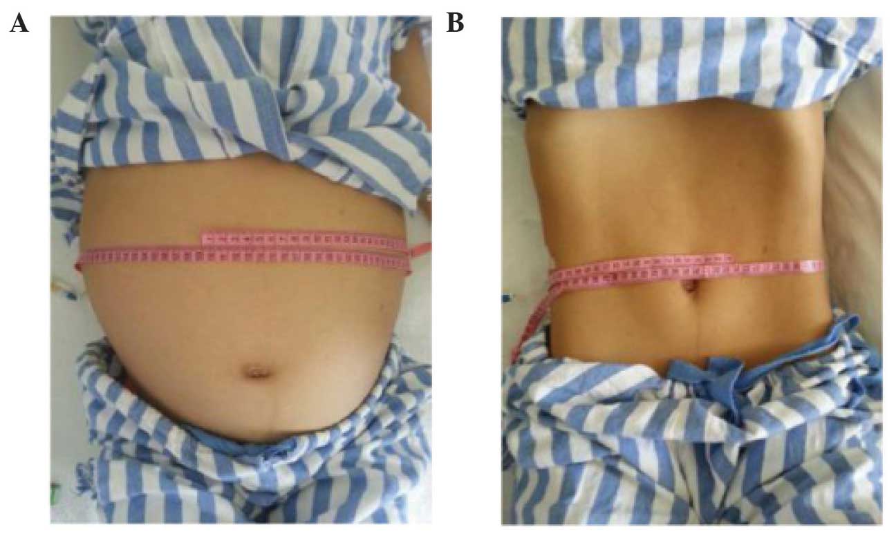
dysfunction was identified. Clinically, the diagnosis of a giant hydronephrose (GH) was made, and upon physical examination, an extremely distended abdomen was revealed, with a palpable mass in the all-flank region (Fig. 1A). Bowel sounds could not be auscultated, and neither was there any shifting dullness. An examination of the other systems failed to disclose any noteworthy phenomena.

Figure 1.

(A) Preoperative photograph of the patient showing the grossly distended abdomen caused by GH of the right kidney. (B) Postoperative photograph of the patient following nephrectomy of the affected side.

The blood laboratory analyses revealed a concentration of blood urea nitrogen of 30.1 mg/dl, and a level of creatinine of 1.56 mg/dl. The level of hemoglobin was 12 g/dl, and the hematocrit was determined to be 45.7%, whereas tumor markers were all revealed to be in normal range, The results of the urine analysis revealed a red blood cell count of 32/high power field (HPF) a white blood cell count of 6/HPF and a yeast cell count of 5/HPF. Blood sugar, phosphate, liver function, serum calcium, serum amylase and electrolyte analyses all yielded results within the normal limits. A diagnostic aspiration performed from the area of abdominal swelling revealed the presence of urine. Ultrasonography revealed the presence of a massive hypoechoic lesion occupying almost the entire abdomen, which exerted pressure on the bowel on the left-hand side. The left kidney appeared normal, although the right kidney could not be identified. A cystic lesion revealed multiple septations, which were likely to have arisen from the 
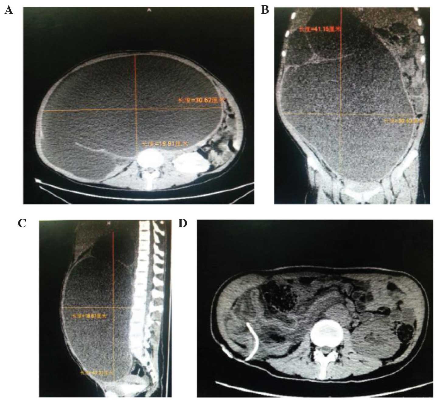
right kidney. A computerized tomography (CT) scan revealed a large abdominal mass of water density in the all-abdomen area, which comprised a huge cystic mass (41.5×30.5×30.5 cm; Fig. 2A–C) that, in the right kidney, occupied almost the entire abdominal cavity. This mass had displaced the small intestine, colon, pancreas, spleen and the left kidney. After admission, the cystic mass was punctured, and a total of ~24 l urinal fluid was drained by urethral catheterization over a period of 8 days (3 liters each day). There was an improvement in the general condition of the patient: The distended abdomen decreased in size (Fig. 1B), the level of urea declined to 1.42 g/l, and the level of creatinine was 63 mg/l. The results of subsequent laboratory analyses and urine culture were normal. A subsequent CT scan revealed an almost 
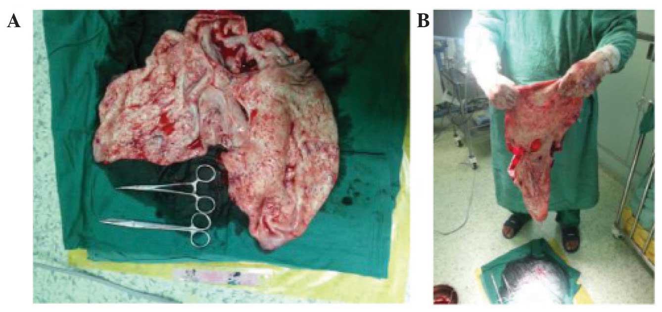
complete disappearance of the hydronephrosis (Fig. 2D). A right nephrectomy was performed: When an incision was made in the right lumbar region, the cyst presented as a retroperitoneal soft cystic dilatation mass, lacking in definition, measuring almost 30×20×15 cm (Fig. 3), which was able to adhere to adjacent organs, including the liver and the pelvic cavity, lifting the 
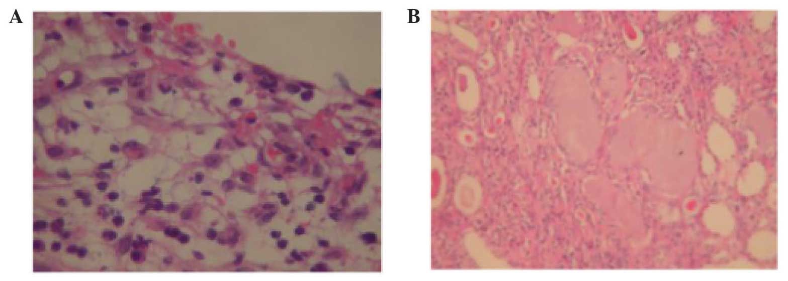
cecum and the ascending colon. The ureter was normal, and ureteropelvic junction obstruction (UPJO) was established as the cause of GH. The pathological tissue sections are shown in Fig. 4.

Figure 2.

(A) CT scan of a GH of the right kidney (30.6×19.9 cm; transverse section). (B) CT scan of a GH of the right kidney (41.1×30.5 cm; coronal section). (C) CT scan of a GH of the right kidney (40.0×18.6 cm; vertical plane). (D) CT scan of a GH of the right kidney following drainage by urethral catheterization. CT, computerised tomography; GH, giant hydronephrosis.

Figure 3.

(A) Close-up photograph of the cystic dilatation kidney following surgery. (B) Gross photograph of the cystic dilatation kidney following surgery.

Figure 4.

(A) Pathological section from the right kidney (H&E staining, magnification, ×400). (B) Pathological section from the right kidney (H&E staining, magnification, ×200). H&E, hematoxylin and eosin.

Discussion

GH is a rare urological condition, which occurs in patients of all ages, defined as the presence of >1 liter of fluid in the collecting system (1). It is considered to develop gradually over a long period of time. Symptomatic nephrolithiasis and hydronephrosis more frequently present as clinical conditions, and GH is a rare clinical phenomenon. Although numerous cases of GH have been reported in the literature, only a few of them contained > 2 liters of fluid (Table I). In the present case report, the hydronephrotic kidney contained 24 l urine, and GHs seldom fill the entire abdomenal space, as had occurred in our patient. The most common cause of GH, as detailed in the literature, is a condition caused by UPJO, although stone disease, trauma, renal ectopy and ureteral tumors have also been reported (2). In the present study, UPJO was the cause of GH.

Table I.

List of documented cases of GHs reported in the literature of the present study.

Table I.

List of documented cases of GHs reported in the literature of the present study.

Patient no.YearAge/genderRefs.Size of mass (cm)/quantity of fluid (ml)Initial symptomsTreatmentCause of GH
1200965/F(1)30×20×25/15,000Fatigue, feverNephrectomyObstruction
2200378/F(2)35×30×25/30,000Nausea, vomit, fatigue, fever, weight lossPuncture/drainage, nephrectomyTumor
3201242/M(3)44×32×30/20,000Weight lossNephrectomyBladder neck obstruction
4201382/M(4)30×21×10/4,500FeverNephrectomyTumor
5200945/M(5)30×20×20/15,000Anemia, liver dysfunctionNephrectomyStone
6201483/M(6)20×16×22/4,000Fatigue, nausea, vomit, weight lossPuncture/drain nephrectomyUreteral stone

[i] M, male; F, female; GH, giant hydronephrosis.

The clinical symptoms of GH are non-specific, and may present with vague symptoms, including an increased abdominal girth due to the presence of a mass in the flank. GH is a slowly progressive disease, and a huge abdominal mass or distended abdomen may cause pain, and be symptomatic of hematuria, recurrent urinary tract infection or other symptoms or complications described in the literature, including nausea, fatigue or dyspepsia, urinary tract infection, loss of weight, renal insufficiency, gross hematuria resulting from trauma in the area, the compression of surrounding structures, or even rupture of the kidney (3). Ultrasonography and CT scans have facilitated the diagnosis of hydronephrosis, and a case of GH may be defined as the presence of hydronephrosis occupying a hemiabdomen, which meets, or extends beyond, the midline, and which extends at least five or six vertebral bodies in length (4). However, in a number of cases, a differential diagnosis between GH and other cystic formations proves to be difficult. Therefore, as demonstrated in the present case report, an accurate diagnosis of GH in individual cases remains challenging. Contrast-enhanced CT of the abdomen and the pelvis provides the ‘gold standard’ for an accurate diagnosis of GH, and other useful diagnostic imaging techniques include abdominal radiography and intravenous urography. The ideal treatment for GH is nephrectomy, and this is the procedure of choice; other treatment options in a functional kidney include percutaneous nephrostomy, reduction pyeloplasty with nephropexy calycoureterostomy, and calycocystostomy (5). In spite of the widespread use of prenatal ultrasound and the development of new diagnostic techniques, GH is encountered in all age groups. A puncture/drainage procedure may be performed in cases where the condition of the patient does not allow other treatments to be performed, or where hemodynamic changes may occur following a sudden abdominal decompression (6). In the present case study, the patient underwent a right nephrectomy and was discharged on postoperative day 8. In conclusion, GH is a rare condition, which is associated with the occurrence of cystic abdominal masses, and the puncture/drainage procedure or nephrectomy provides the most suitable method for surgical intervention.

References

1 

Vishwanath M, Pattanshetti MK, Swamy SI, Godhi AS and Metgud SC: Giant hydronephrosis. Indian J Surg. 72:359–360. 2009.

2 

Schrader AJ, Anderer G, von Knobloch R, Heidenreich A and Hofmann R: Giant hydronephrosis mimicking progressive malignancy. BMC Urol. 3:42003. View Article : Google Scholar : PubMed/NCBI

3 

Tazi MF, Riyach O, Ahallal Y, Mellas S, Khallouk A, El Fassi MJ and Farih MH: Giant urinary bladder and bilateral giant hydronephrosis due to bladder neck obstruction: One case report and literature review. Case Rep Urol. 2012:8175192012.PubMed/NCBI

4 

Mediavilla E, Ballestero R, Correas MA and Gutierrez JL: About a case report of giant hydronephrosis. Case Rep Urol. 2013:2579692013.PubMed/NCBI

5 

Wu CC and Sun GH: Giant hydronephrosis. Mayo Clin Proc. 84:9542009. View Article : Google Scholar : PubMed/NCBI

6 

Golcuk Y, Ozsarac M, Eseroglu E and Yuksel MB: Giant hydronephrosis. West J Emerg Med. 15:3562014. View Article : Google Scholar : PubMed/NCBI

Related Articles

  • Abstract
  • View
  • Download
  • Twitter
Copy and paste a formatted citation
Spandidos Publications style
Wang QF, Zeng G, Zhong L, Li QL, Che XY, Jiang T, Zhang ZW, Zheng W, Tang QZ, Chen F, Chen F, et al: Giant hydronephrosis due to ureteropelvic junction obstruction: A rare case report, and a review of the literature. Mol Clin Oncol 5: 19-22, 2016.
APA
Wang, Q., Zeng, G., Zhong, L., Li, Q., Che, X., Jiang, T. ... Wang, K. (2016). Giant hydronephrosis due to ureteropelvic junction obstruction: A rare case report, and a review of the literature. Molecular and Clinical Oncology, 5, 19-22. https://doi.org/10.3892/mco.2016.876
MLA
Wang, Q., Zeng, G., Zhong, L., Li, Q., Che, X., Jiang, T., Zhang, Z., Zheng, W., Tang, Q., Chen, F., Wang, K."Giant hydronephrosis due to ureteropelvic junction obstruction: A rare case report, and a review of the literature". Molecular and Clinical Oncology 5.1 (2016): 19-22.
Chicago
Wang, Q., Zeng, G., Zhong, L., Li, Q., Che, X., Jiang, T., Zhang, Z., Zheng, W., Tang, Q., Chen, F., Wang, K."Giant hydronephrosis due to ureteropelvic junction obstruction: A rare case report, and a review of the literature". Molecular and Clinical Oncology 5, no. 1 (2016): 19-22. https://doi.org/10.3892/mco.2016.876
Copy and paste a formatted citation
x
Spandidos Publications style
Wang QF, Zeng G, Zhong L, Li QL, Che XY, Jiang T, Zhang ZW, Zheng W, Tang QZ, Chen F, Chen F, et al: Giant hydronephrosis due to ureteropelvic junction obstruction: A rare case report, and a review of the literature. Mol Clin Oncol 5: 19-22, 2016.
APA
Wang, Q., Zeng, G., Zhong, L., Li, Q., Che, X., Jiang, T. ... Wang, K. (2016). Giant hydronephrosis due to ureteropelvic junction obstruction: A rare case report, and a review of the literature. Molecular and Clinical Oncology, 5, 19-22. https://doi.org/10.3892/mco.2016.876
MLA
Wang, Q., Zeng, G., Zhong, L., Li, Q., Che, X., Jiang, T., Zhang, Z., Zheng, W., Tang, Q., Chen, F., Wang, K."Giant hydronephrosis due to ureteropelvic junction obstruction: A rare case report, and a review of the literature". Molecular and Clinical Oncology 5.1 (2016): 19-22.
Chicago
Wang, Q., Zeng, G., Zhong, L., Li, Q., Che, X., Jiang, T., Zhang, Z., Zheng, W., Tang, Q., Chen, F., Wang, K."Giant hydronephrosis due to ureteropelvic junction obstruction: A rare case report, and a review of the literature". Molecular and Clinical Oncology 5, no. 1 (2016): 19-22. https://doi.org/10.3892/mco.2016.876
Follow us
  • Twitter
  • LinkedIn
  • Facebook
About
  • Spandidos Publications
  • Careers
  • Cookie Policy
  • Privacy Policy
How can we help?
  • Help
  • Live Chat
  • Contact
  • Email to our Support Team