Introduction
The syndrome of combined pulmonary fibrosis and
emphysema (CPFE) has been suggested to be a clinically important
phenotype of pulmonary fibrosis (1).
Cottin et al (1) defined CPFE
as a distinct condition, with characteristic features in the chest
computed tomography (CT) image that include emphysema in the upper
pulmonary region concomitant with parenchymal fibrosis in the lower
region. The syndrome is characterized by sub-normal spirometry,
severely impaired gas exchange, high prevalence of pulmonary
hypertension and poor survival (2–4). Mejía
et al (3) reported that
survival of CPFE patients was poorer compared with that of patients
with idiopathic pulmonary fibrosis (IPF) alone and that pulmonary
hypertension in CPFE patients was an independent predictor of
mortality. Previous studies have indicated that CPFE patients have
a significantly increased risk of lung cancer compared with
patients with chronic obstructive pulmonary disease (COPD) or IPF
alone (1,4,5).
Post-operative acute exacerbation (AE) of interstitial lung disease
in patients with lung cancer is associated with a high mortality
rate (43.9%) (6). However, the
clinical features and prognosis of patients with lung cancer and
CPFE have remained elusive. The present study performed a
retrospective analysis to determine the prevalence, clinical
characteristics and outcome of patients with surgically resected
lung cancer and CPFE. In addition, differences between the clinical
features and outcomes of patients with lung cancer and CPFE, solely
IPF and solely emphysema were assessed, and the impact of
post-operative AE on patient survival was determined.
Patients and methods
Patient data
Of 831 consecutive patients with primary lung cancer
who underwent surgical resection at Toho University School of
Medicine between June 2004 to December 2014, 23 patients with CPFE
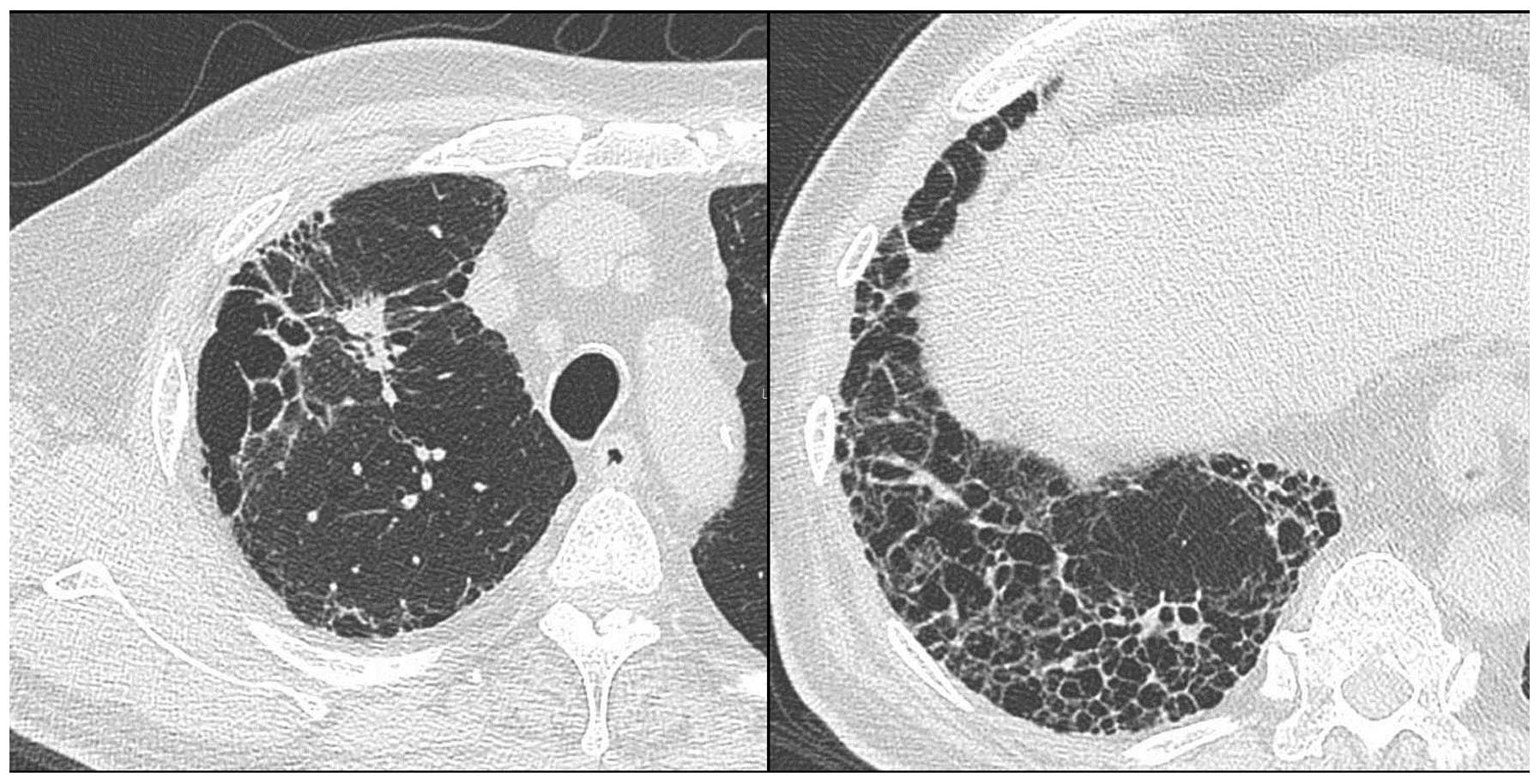
(2.8%) were retrospectively reviewed by the present study. CPFE was
diagnosed by chest high-resolution computed tomography (HRCT)
images showing emphysema involving >25% of both upper lobes and
a usual interstitial pneumonia (UIP) pattern of fibrosis in the
lower lobes (Fig. 1). Histological
examination of resected specimens and/or diagnosis of IPF based on
a high-resolution CT scan showing honeycombing (multiple
equal-sized cystic lesions of 2–10 mm with a thick wall) in the
sub-pleural area of both lung fields according to the American
Thoracic Society/European Respiratory Society International
Consensus Statement on IPF were used to confirm the presumptive
diagnosis based on the UIP pattern (7). AE was diagnosed according to the
guidelines of the Japanese Respiratory Society (8) based on the following criteria fulfilled
over one month: i) Increased respiratory distress; ii) fibrosis,
newly developed ground glass opacity and an infiltrative shadow in
the HRCT scan; and iii) a decrease in arterial oxygen pressure of
more than 10 Torr under constant oxygenation conditions. Acute
deterioration triggered by anti-cancer drug (carboplatin/etoposide,
carboplatin/paclitaxel/bevacizumab, gefitinib) and AE as natural
courses of IPF (16 months after the surgery) were excluded from
post-operative AE. The cumulative amount of cigarette consumption
was expressed as pack years, calculated as the number of cigarette
packs consumed per day multiplied by the number of years of
smoking. For analysis, patients in the CPFE group were further
stratified into a group which developed post-operative AE (n=3) and
another group without AE (n=20). For comparison, patients with IPF
other than CPFE were selected from the abovementioned cohort as the
solely IPF group (n=9), which was further divided into an IPF with
post-operative AE group (n=4) and one group without AE (n=5).
Furthermore, 35 patients with COPD and emphysema observed by chest
HRCT were defined as the solely emphysema group with adjustment of
the pathological stage. The present study was approved by the local
Ethics committee of Toho University Omori Medical Center (assurance
no. 26-232).
Measurement of serum marker
levels
As indicators of UIP, the serum levels of Krebs von
den Lungen-6 (KL-6) and surfactant protein-D (SP-D) were determined
prior to surgery. Serum KL-6 (normal levels, <500 U/ml) was
measured using an enzyme-linked immunosorbent assay (ELISA; ELTEST
KL-6 kit; Eisai Co., Tokyo, Japan) and serum SP-D (normal levels,
<110 ng/ml) was measured using an ELISA kit (SP-D kit YAMASA EIA
II, Yamasa Corp., Tokyo, Japan).
Chest CT
Chest CT was performed using a helical CT scanner
(Aquilion 16; Toshiba, Tokyo, Japan). Routine scanning of the
entire lung was performed with a slice thicknesses of 5–10 mm,
followed by HRCT imaging at full inspiration with 1–2-mm sections
(120 kVp; 300 mA; pitch, 1.0). For all patients, HRCT images were
captured with a window setting appropriate for the lungs (window
level from −600 Hounsfield units; width from 1,600 Hounsfield
units).
Pulmonary function testing
Spirometry and measurement of the diffusing capacity
of the lung for carbon monoxide (DLco) were performed using a
pulmonary function test system (Chestac-33; CHEST Co. Ltd., Tokyo,
Japan). The DLco was measured using the single-breath technique.
The pulmonary function tests were performed by two technicians
according to the recommendations of the American Thoracic Society
(9).
Statistical analysis
All statistical analyses were performed using the
JMP version 10.0.0 statistical software package (SAS Institute,
Cary, NC, USA). Comparison of categorical and dichotomous variables
was performed using Pearson's χ2 test or Fisher's exact
test. Analysis of variance with life tables and Kaplan-Meier curves
were used for the analyses of overall survival. Differences between
two groups were analyzed using the log-rank test. P<0.05 was
considered to indicate a statistically significant difference.
Results
Patient characteristics
The characteristics of the patients are shown in
Table I. All patients in the CPFE or
solely IPF groups were male, while five patients in the emphysema
group were female (14%). The results of the pulmonary function
tests, including the percent vital capacity (%VC) or forced
expiratory volume in one second as percent of forced vital capacity
(FEV1.0%) of the patients in the CPFE group were almost
normal, while patients in the solely IPF group had a slightly
decreased %VC and patients in the solely emphysema had a decreased
FEV1.0% and %FEV1.0. However, the percent
predicted DLco (%DLco) of the patients in the CPFE group was
noticeably low. The predominant histology of lung tumors in the
CPFE group was squamous-cell carcinoma and the majority of patients
had advanced-stage disease. No statistically significant
differences were observed in the mean age, smoking status, %VC,
FEV1.0%, %DLco, type of surgical procedure, histological
type and pathological stage of lung cancer between patients in the
CPFE with AE group and those in the CPFE without AE group (Table II).
 | Table I.Patient characteristics. |
Table I.
Patient characteristics.
| Characteristic | CPFE (n=23) | Solely IPF (n=9) | Solely Em (n=35) | P-value (CPFE vs.
IPF) | P-value (CPFE vs.
Em) |
|---|
| Age (years), mean ±
SD | 69.4±6.7 | 72.1±6.8 | 70.3±6.7 | 0.323 |
0.649 |
| Male/female | 23/0 | 9/0 | 30/5 | – |
0.146 |
| Smoking status |
|
|
|
|
|
|
Current/former/never | 6/17/0 | 3/6/0 | 13/20/2 | 0.685 |
0.206 |
| Pack
years | 50±27 | 37±20 | 53±23 | 0.201 |
0.707 |
| Pulmonary
function |
|
|
|
|
|
| %VC
(%) | 102±19 | 89±21 | 98±18 | 0.114 |
0.409 |
|
FEV1.0% (%) | 72±10 | 82±8 | 59±9 | 0.009 | <0.001 |
|
%FEV1.0 (%) | 103±21 | 103±27 | 80±21 | 0.992 | <0.001 |
| %DLco
(%) | 68±19 | 73±16 | 83±20 | 0.494 |
0.009 |
| KL-6 (U/ml) | 673±393 | 821±241 | – | 0.309 | – |
| SP-D (ng/ml) | 135±90 | 165±73 | – | 0.397 | – |
| Surgical
procedure |
|
|
|
|
|
|
Lobectomy/limited | 20/3 | 8/1 | 33/2 | 1.000 |
0.376 |
| Histology |
|
|
|
|
|
|
Sq/Ad/other | 13/5/5 | 3/4/2 | 20/13/2 | 0.398 |
0.135 |
| Pathological
stage |
|
|
|
|
|
|
I/II/III | 6/10/7 | 2/4/3 | 9/15/11 | 0.971 |
0.997 |
| Table II.Characteristics of CPFE with AE and
without AE groups. |
Table II.
Characteristics of CPFE with AE and
without AE groups.
| Characteristic | AE (n=3) | Without AE
(n=20) | P-value |
|---|
| Age (years), mean ±
SD | 66.7±8.6 | 69.9±6.6 | 0.459 |
| Male/female | 3/0 | 20/0 | – |
| Smoking status |
|
|
|
|
Former/current | 3/0 | 14/6 | 0.539 |
| Pack
years |
44±17 |
51±28 | 0.687 |
| Pulmonary
function |
|
|
|
| %VC
(%) | 95±10 | 103±20 | 0.504 |
|
%FEV1.0 (%) | 96±12 | 104±22 | 0.558 |
|
FEV1.0% (%) | 77±13 |
71±10 | 0.345 |
| %DLco
(%) | 65±3 | 69±21 | 0.761 |
| KL-6 (U/ml) | 486±149 | 706±416 | 0.386 |
| SP-D (ng/ml) | 134±55 | 135±96 | 0.984 |
| Surgical
procedure |
|
|
|
|
Lobectomy/limited | 2/1 | 18/2 | 0.320 |
| Histology |
|
|
|
|
Sq/Ad/other | 2/0/1 | 11/5/4 | 0.439 |
| Pathological
stage |
|
|
|
|
I/II/III | 2/0/1 | 4/10/6 | 0.109 |
Survival
Survival in the CPFE and solely IPF groups is
significantly lower compared with that in the solely emphysema
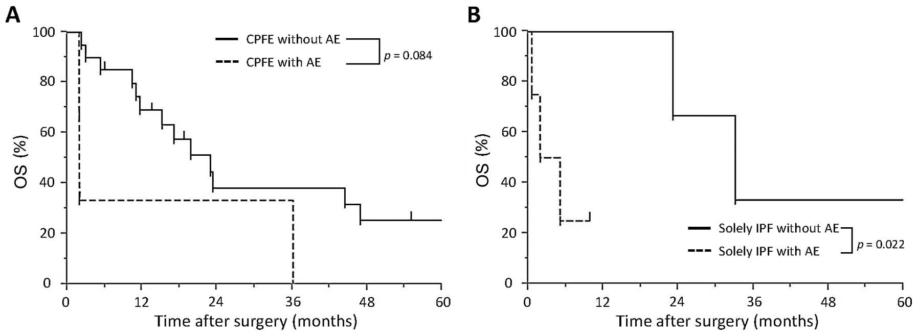
group, while AE does not affect survival in the CPFE group. The 3-
and 5-year survival rate in the CPFE group was 38 and 22%,
respectively. The Kaplan-Meier survival curves for patients in the
CPFE group showed no significant differences between patients with
AE and those without AE (P=0.084; Fig.
2A). The 3-year survival rate in the CPFE with AE group was 33%
and that in the CPFE without AE group was 38%. The survival time in
the solely IPF with AE group was significantly shorter compared
with that in the solely IPF without AE group (5.0 vs. 24.4 month
P=0.022, log-rank test; Fig. 2B). The
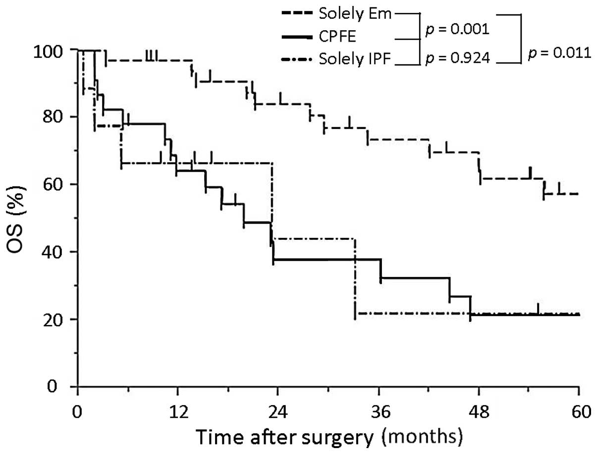
5-year survival rate of patients in the CPFE, solely IPF and solely
emphysema groups was 22, 22 and 58%, respectively. Furthermore, the
CPFE and solely IPF groups showed a significantly shorter survival
than the solely emphysema group (P=0.001 and 0.011, respectively;
Fig. 3); however, no significant
difference was determined between the CPFE and solely IPF group
(P=0.924. In Tables III and
IV, data on the fatal cases in the
CPFE with AE and CPFE without AE groups are summarized,
respectively. In the CPFE with AE group, two patients succumbed to
post-operative AE after 2 months and one patient succumbed after 36
months (Table III). In the CPFE
without AE group, four patients succumbed to lung cancer and two
succumbed to other cancer types. Furthermore, the cause of
mortality was chemotherapy-induced AE in three patients
(carboplatin/etoposide, carboplatin/paclitaxel/bevacizumab,
gefitinib), pneumonia in another three patients and myocardial
infarction in one patient (Table
IV).
| Table III.Fatal cases in the combined pulmonary
fibrosis and emphysema with post-operative acute exacerbation
group. |
Table III.
Fatal cases in the combined pulmonary
fibrosis and emphysema with post-operative acute exacerbation
group.
| Age (years) | Surgical
procedure | Stage | Histology | Trigger of AE | Time between
surgery and AE (months) | Survival
(months) | Cause of
mortality |
|---|
| 59 | Lobectomy | pIB | AdSq | Surgery | 0.3 | 36 | AE |
| 65 | Limited | pIA | Sq | Surgery | 1.4 | 2 | AE |
| 76 | Lobectomy | pIIIA | Sq | Surgery | 0.2 | 2 | AE |
| Table IV.Fatal cases in the combined pulmonary
fibrosis and emphysema group without post-operative acute
exacerbation. |
Table IV.
Fatal cases in the combined pulmonary
fibrosis and emphysema group without post-operative acute
exacerbation.
| Age (years) | Surgical
procedure | Stage | Histology | Recurrent site | Survival
(months) | Cause of
mortality |
|---|
| 63 | Lobectomy | pIIIA | Sq | Lung | 29 | Lung cancer |
| 65 | Lobectomy | pIIB | Sq | Mediastinum | 47 | Lung cancer |
| 68 | Lobectomy | pIB | Sm | Lymph node | 23 | Lung cancer |
| 81 | Lobectomy | pIIB | Ad | Mediastinum | 17 | Lung cancer |
| 64 | Lobectomy | pIA | Sm | – | 11 | AE |
| 65 | Lobectomy | pIB | Ad | – | 44 | AE |
| 71 | Lobectomy | pIIA | Ad | Pleural
dissemination | 2 | AE |
| 61 | Lobectomy | pIIB | Sq | – | 12 | Pneumonia |
| 61 | Lobectomy | pIIB | Sq | – | 13 | Pneumonia |
| 69 | Lobectomy | pIIIA | Ad | – | 10 | Pneumonia |
| 61 | Lobectomy | pIIIA | Sq | – | 23 | Other cancer
type |
| 73 | Limited | pIIIA | Sq | – | 16 | Other cancer
type |
| 75 | Limited | pIB | Sm | – | 4 | Myocardial
infarction |
Discussion
As first defined by Cottin et al (1) in 2005, CPFE is a well-defined syndrome
characterized by upper-lobe-predominant emphysema and
lower-lobe-predominant fibrosis observed by chest HRCT. The median
survival of CPFE patients has been reported to range from 22 months
to 8 years and the 5-year survival rate is 22–80% (1,4,10–12). Mejía
et al (3) reported that 31/110
patients with IPF (28.2%) had emphysema and that their mortality
rate was higher compared with that of patients with IPF alone.
Previous studies have shown that the risk of primary
lung cancer is significantly increased in patients with CPFE.
Kitaguchi et al (2) reported
that in a cohort with CPFE, the prevalence of lung cancer was
significantly increased compared with that in a cohort with COPD
alone (46.8 vs. 7.3%; P<0.01), and Kurashima et al
(11) identified a significantly
increased rate of lung cancer-associated mortality among patients
with CPFE (12 of 36 mortalities, 33.3%) compared with that in
patients with IPF alone (8 of 66 mortalities, 12.1%; P=0.0097).
Sugino et al (4) suggested
that the following factors or diagnostic errors may account for the
differential outcomes of CPFE and IPF alone: i) The presence of
primary lung cancer, ii) disease severity based on distributions or
extension of fibrosis and/or emphysema, iii) possible confusion
between chronic fibrotic interstitial pneumonia and fibrotic
non-specific interstitial pneumonia, iv) the difficulty in
radiological discrimination between emphysema with non-specific
fibrosis and IPF (for example, wall thickening due to emphysematous
may be mistaken for honeycomb cysts). The study also reported that
patients with CPFE had a poor outcome even in patients without the
complication of lung cancer at the initial visit, with median
survival time of 28 months in their subgroup analysis (4). However, these findings suggested that
CPFE is associated with a poor prognosis, which was in accordance
with the present study, which reported poor 3- and 5-year survival
rates of 38 and 22%, respectively for resected CPFE patients with
lung cancer. Of note, a number of CPFE patients in the present
study succumbed to AE or lung cancer recurrence. However, as shown
in Table I, the pulmonary function of
these patients was almost normal, except for the DLco results. CPFE
may be difficult to detect by the pulmonary function tests that are
usually performed for patients. DLco is important to detect CPFE
and to consider the potential risk of chemotherapy for the patients
with CPFE.
AE is an important negative prognostic factor for
surgically resected lung cancer patients with interstitial lung
disease (ILD). Sato et al (6)
reported that AE occurred in 9.3% of surgically resected lung
cancer patients with ILD, accounting for a mortality rate of 43.9%.
They also identified the following independent risk factors for AE:
Surgical procedures (wedge resection, lobectomy/segmentectomy,
bilobectomy/pneumonectomy), male gender, history of exacerbation,
pre-operative steroid use, elevated serum KL-6 levels (≥1,000
U/ml), appearance of UIP in the CT spectrum and reduced percentage
of predicted vital capacity. Although two of the patients in the
present study matched the risk factors of male gender and CT
findings, it was not possible to determine whether these or any
other factors were associated with CPFE due to the low patient
number. In the present study, only three cases with post-operative
AE were included and no statistically significant differences in
clinicopathological parameters or survival rates between patients
with AE and those without AE were identified. Previous studies have
reported that surgically resected lung cancer patients with IPF who
developed post-operative AE had a poorer survival compared with
those without AE (13–16), which was in line with the findings of
the present study regarding the solely IPF with AE and the solely
IPF without AE groups. The finding that AE did not affect survival
in CPFE patients in the present study may be due to inclusion of
cases of small-cell carcinoma in the CPFE without AE group, in
addition to the small sample size. Therefore, additional analysis
is required, including the comparison of the molecular
characteristics of lung cancer patients with CPFE and those with
lung cancer and IPF.
The present study had several limitations. There
were intrinsic limitations associated with the data, as they were
collected and reviewed retrospectively. In addition, the study
cohort was small, and further studies on larger populations are
required to validate the results.
In conclusion, the present study revealed that lung
cancer patients with CPFE had poor survival, which was comparable
with that of lung cancer patients with IPF. Numerous patients with
lung cancer and CPFE presented with post-operative recurrence of
lung cancer or developed AE or pneumonia. The present study
contributed to the current understanding of the clinical and
pathophysiological features as well as the outcome of lung cancer
patients with CPFE, which requires to be confirmed and further
elucidated by larger-scale studies.
Acknowledgements
The present study was supported in part by
Grants-in-aid for Scientific Research [nos. (C) 15K10272 and
26462140] from the Japanese Ministry of Education, Culture, Sports,
Science and Technology, as well as by a Grant-in-aid for Project
Research from Toho University School of Medicine (no. 26-14) and
the Research Promotion Grant from Toho University Graduate School
of Medicine (no. 15-02).
References
|
1
|
Cottin V, Nunes H, Brillet PY, Delaval P,
Devouassoux G, Tillie-Leblond I, Israel-Biet D, Court-Fortune I,
Valeyre D and Cordier JF: Groupe d'Etude et de Recherche sur les
Maladies Orphelines Pulmonaires (GERM O P): Combined pulmonary
fibrosis and emphysema: A distinct underrecognised entity. Eur
Respir J. 26:586–593. 2005. View Article : Google Scholar : PubMed/NCBI
|
|
2
|
Kitaguchi Y, Fujimoto K, Hanaoka M,
Kawakami S, Honda T and Kubo K: Clinical characteristics of
combined pulmonary fibrosis and emphysema. Respirology. 15:265–271.
2010. View Article : Google Scholar : PubMed/NCBI
|
|
3
|
Mejía M, Carrillo G, Rojas-Serrano J,
Estrada A, Suárez T, Alonso D, Barrientos E, Gaxiola M, Navarro C
and Selman M: Idiopathic pulmonary fibrosis and emphysema:
Decreased survival associated with severe pulmonary arterial
hypertension. Chest. 136:10–15. 2009. View Article : Google Scholar : PubMed/NCBI
|
|
4
|
Sugino K, Ishida F, Kikuchi N, Hirota N,
Sano G, Sato K, Isobe K, Sakamoto S, Takai Y and Homma S:
Comparison of clinical characteristics and prognostic factors of
combined pulmonary fibrosis and emphysema versus idiopathic
pulmonary fibrosis alone. Respirology. 19:239–245. 2014. View Article : Google Scholar : PubMed/NCBI
|
|
5
|
Dai H, Liu J, Liang L, Ban C, Jiang J, Liu
Y, Ye Q and Wang C: Lung cancer risk among patients with combined
pulmonary fibrosis and emphysema. Respirology. 19:707–713. 2014.
View Article : Google Scholar : PubMed/NCBI
|
|
6
|
Sato T, Teramukai S, Kondo H, Watanabe A,
Ebina M, Kishi K, Fujii Y, Mitsudomi T, Yoshimura M, Maniwa T, et
al: Impact and predictors of acute exacerbation of interstitial
lung diseases after pulmonary resection for lung cance. J Thorac
Cardiovasc Surg. 147:1604–1611. 2014. View Article : Google Scholar : PubMed/NCBI
|
|
7
|
Raghu G, Collard HR, Egan JJ, Martinez FJ,
Behr J, Brown KK, Colby TV, Cordier JF, Flaherty KR, Lasky JA, et
al: An official ATS/ERS/JRS/ALAT statement: Idiopathic pulmonary
fibrosis: Evidence-based guidelines for diagnosis and management.
Am J Respir Crit Care Med. 183:788–824. 2011. View Article : Google Scholar : PubMed/NCBI
|
|
8
|
Akira M, Hamada H, Sakatani M, Kobayashi
C, Nishioka M and Yamamoto S: CT findings during phase of
accelerated deterioration in patients with idiopathic pulmonary
fibrosis. AJR Am J Roentgenol. 168:79–83. 1997. View Article : Google Scholar : PubMed/NCBI
|
|
9
|
Single breath carbon monoxide diffusing
capacity (transfer factor): Recommendations for a standard
technique. Statement of the American thoracic society. Am Rev
Respir Dis. 136:1299–1307. 1987. View Article : Google Scholar : PubMed/NCBI
|
|
10
|
Akagi T, Matsumoto T, Harada T, Tanaka M,
Kuraki T, Fujita M and Watanabe K: Coexistent emphysema delays the
decrease of vital capacity in idiopathic pulmonary fibrosis. Respir
Med. 103:1209–1215. 2009. View Article : Google Scholar : PubMed/NCBI
|
|
11
|
Kurashima K, Takayanagi N, Tsuchiya N,
Kanauchi T, Ueda M, Hoshi T, Miyahara Y and Sugita Y: The effect of
emphysema on lung function and survival in patients with idiopathic
pulmonary fibrosis. Respirology. 15:843–848. 2010. View Article : Google Scholar : PubMed/NCBI
|
|
12
|
Ryerson CJ, Hartman T, Elicker BM, Ley B,
Lee JS, Abbritti M, Jones KD, King TE Jr, Ryu J and Collard HR:
Clinical features and outcomes in combined pulmonary fibrosis and
emphysema in idiopathic pulmonary fibrosis. Chest. 144:234–240.
2013. View Article : Google Scholar : PubMed/NCBI
|
|
13
|
Chiyo M, Sekine Y, Iwata T, Tatsumi K,
Yasufuku K, Iyoda A, Otsuji M, Yoshida S, Shibuya K, Iizasa T, et
al: Impact of interstitial lung disease on surgical morbidity and
mortality for lung cancer: Analyses of short-term and long-term
outcomes. J Thorac Cardiovasc Surg. 126:1141–1146. 2003. View Article : Google Scholar : PubMed/NCBI
|
|
14
|
Kumar P, Goldstraw P, Yamada K, Nicholson
AG, Wells AU, Hansell DM, Dubois RM and Ladas G: Pulmonary fibrosis
and lung cancer: Risk and benefit analysis of pulmonary resection.
J Thorac Cardiovasc Surg. 125:1321–1327. 2003. View Article : Google Scholar : PubMed/NCBI
|
|
15
|
Koizumi K, Hirata T, Hirai K, Mikami I,
Okada D, Yamagishi S, Kawashima T, Kinoshita H, Enomoto Y, Nakajima
Y and Shimizu K: Surgical treatment of lung cancer combined with
interstitial pneumonia: The effect of surgical approach on
postoperative acute exacerbation. Ann Thorac Cardiovasc Surg.
10:340–346. 2004.PubMed/NCBI
|
|
16
|
Watanabe A, Kawaharada N and Higami T:
Postoperative acute exacerbation of IPF after lung resection for
primary lung cancer. Pulm Med. 2011:9603162011. View Article : Google Scholar : PubMed/NCBI
|