Open Access

Clinical and imaging characteristics of 53 ulcers of post‑radiation nasopharyngeal necrosis in patients with nasopharyngeal carcinoma

  • Authors:
    • Fengqin Yan
    • Zhimin Ye
    • Fangzheng Wang
    • Lei Wang
    • Weiyang Li
    • Zhenfu Fu
  • View Affiliations

  • Published online on: July 27, 2016     https://doi.org/10.3892/mco.2016.968
  • Pages: 351-356
  • Copyright: © Yan et al. This is an open access article distributed under the terms of Creative Commons Attribution License.

Metrics: Total Views: 0 (Spandidos Publications: | PMC Statistics: )
Total PDF Downloads: 0 (Spandidos Publications: | PMC Statistics: )


Abstract

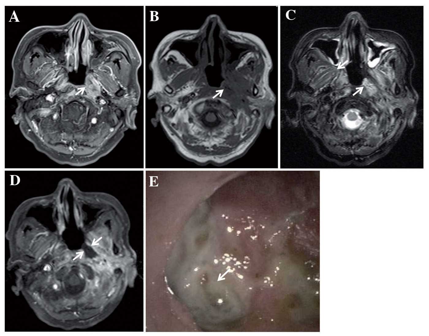
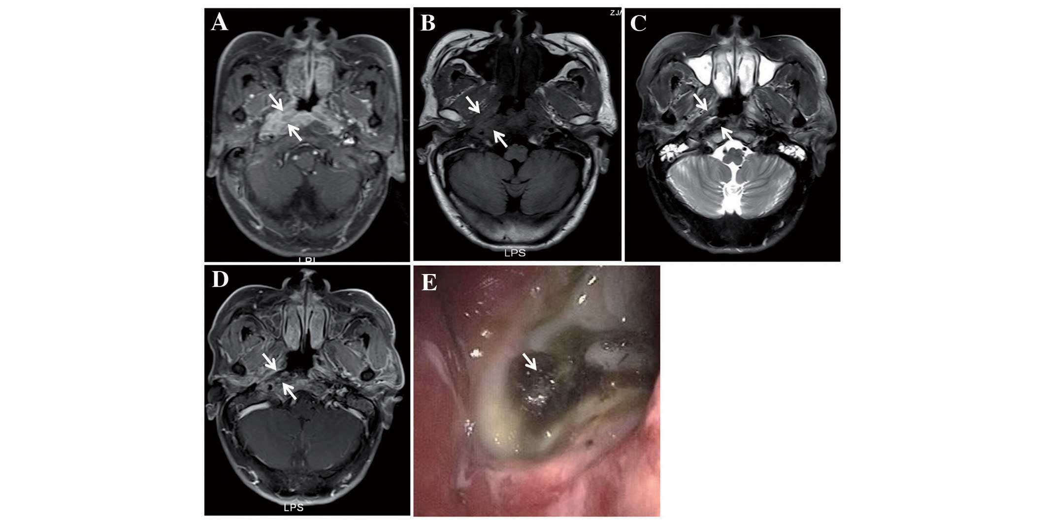
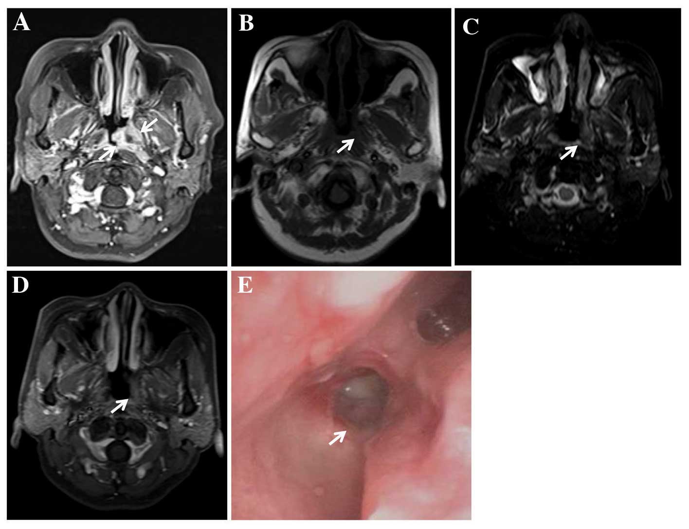
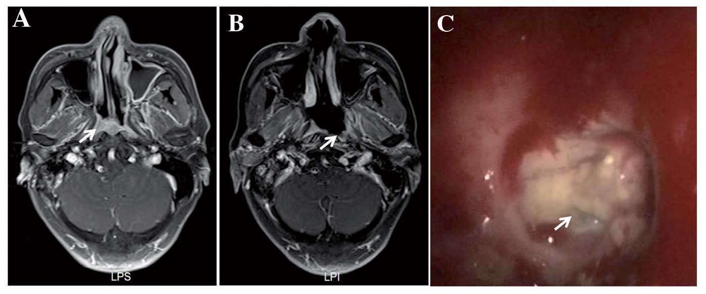
It is widely accepted that a mucosal ulcer induced by radiation (RIMU) is the predominant type of post-radiation nasopharyngeal ulcer in patients with nasopharyngeal carcinoma (NPC) who underwent radiotherapy (RT); however, another type of ulcer, an ulcer of post‑radiation nasopharyngeal necrosis (UPRNN), has rarely been reported for patients with NPC. In the present study, the clinical and imaging features of 53 patients who were treated at the Zhejiang Provincial Cancer Center (Zhejiang Cancer Hospital, Hangzhou, China) between March 2009 and December 2015, and who were diagnosed with UPRNN, were reviewed. The clinical factors, laboratory examinations, magnetic resonance imaging (MRI) features and endoscopic findings were analysed. A UPRNN has its characteristic imaging features and ulcer locations at the primary tumour bed, which are different from a traditional RIMU. In the retrospective analysis of the clinical factors, a tumour (T) 3/4 stage, with invasion of muscular tissue, poor response of neoadjuvant chemotherapy and anaemia during the RT, may be associated with the occurrence of a UPRNN. To evaluate the severity, a UPRNN was divided into three grades according to the invasion depth of the ulcer based on its appearance in MRI, and the subsequent treatment and prognosis varied according to the severity of the UPRNN. In conclusion, a UPRNN has its clinical features and characteristic MRI appearances, and the occurrence of a UPRNN may be associated with several clinical factors.
View Figures
View References

Related Articles

Journal Cover

October-2016
Volume 5 Issue 4

Print ISSN: 2049-9450
Online ISSN:2049-9469

Sign up for eToc alerts

Recommend to Library

Copy and paste a formatted citation
x
Spandidos Publications style
Yan F, Ye Z, Wang F, Wang L, Li W and Fu Z: Clinical and imaging characteristics of 53 ulcers of post‑radiation nasopharyngeal necrosis in patients with nasopharyngeal carcinoma. Mol Clin Oncol 5: 351-356, 2016
APA
Yan, F., Ye, Z., Wang, F., Wang, L., Li, W., & Fu, Z. (2016). Clinical and imaging characteristics of 53 ulcers of post‑radiation nasopharyngeal necrosis in patients with nasopharyngeal carcinoma. Molecular and Clinical Oncology, 5, 351-356. https://doi.org/10.3892/mco.2016.968
MLA
Yan, F., Ye, Z., Wang, F., Wang, L., Li, W., Fu, Z."Clinical and imaging characteristics of 53 ulcers of post‑radiation nasopharyngeal necrosis in patients with nasopharyngeal carcinoma". Molecular and Clinical Oncology 5.4 (2016): 351-356.
Chicago
Yan, F., Ye, Z., Wang, F., Wang, L., Li, W., Fu, Z."Clinical and imaging characteristics of 53 ulcers of post‑radiation nasopharyngeal necrosis in patients with nasopharyngeal carcinoma". Molecular and Clinical Oncology 5, no. 4 (2016): 351-356. https://doi.org/10.3892/mco.2016.968