Spandidos Publications Logo
  • About
    • About Spandidos
    • Aims and Scopes
    • Abstracting and Indexing
    • Editorial Policies
    • Reprints and Permissions
    • Job Opportunities
    • Terms and Conditions
    • Contact
  • Journals
    • All Journals
    • Oncology Letters
      • Oncology Letters
      • Information for Authors
      • Editorial Policies
      • Editorial Board
      • Aims and Scope
      • Abstracting and Indexing
      • Bibliographic Information
      • Archive
    • International Journal of Oncology
      • International Journal of Oncology
      • Information for Authors
      • Editorial Policies
      • Editorial Board
      • Aims and Scope
      • Abstracting and Indexing
      • Bibliographic Information
      • Archive
    • Molecular and Clinical Oncology
      • Molecular and Clinical Oncology
      • Information for Authors
      • Editorial Policies
      • Editorial Board
      • Aims and Scope
      • Abstracting and Indexing
      • Bibliographic Information
      • Archive
    • Experimental and Therapeutic Medicine
      • Experimental and Therapeutic Medicine
      • Information for Authors
      • Editorial Policies
      • Editorial Board
      • Aims and Scope
      • Abstracting and Indexing
      • Bibliographic Information
      • Archive
    • International Journal of Molecular Medicine
      • International Journal of Molecular Medicine
      • Information for Authors
      • Editorial Policies
      • Editorial Board
      • Aims and Scope
      • Abstracting and Indexing
      • Bibliographic Information
      • Archive
    • Biomedical Reports
      • Biomedical Reports
      • Information for Authors
      • Editorial Policies
      • Editorial Board
      • Aims and Scope
      • Abstracting and Indexing
      • Bibliographic Information
      • Archive
    • Oncology Reports
      • Oncology Reports
      • Information for Authors
      • Editorial Policies
      • Editorial Board
      • Aims and Scope
      • Abstracting and Indexing
      • Bibliographic Information
      • Archive
    • Molecular Medicine Reports
      • Molecular Medicine Reports
      • Information for Authors
      • Editorial Policies
      • Editorial Board
      • Aims and Scope
      • Abstracting and Indexing
      • Bibliographic Information
      • Archive
    • World Academy of Sciences Journal
      • World Academy of Sciences Journal
      • Information for Authors
      • Editorial Policies
      • Editorial Board
      • Aims and Scope
      • Abstracting and Indexing
      • Bibliographic Information
      • Archive
    • International Journal of Functional Nutrition
      • International Journal of Functional Nutrition
      • Information for Authors
      • Editorial Policies
      • Editorial Board
      • Aims and Scope
      • Abstracting and Indexing
      • Bibliographic Information
      • Archive
    • International Journal of Epigenetics
      • International Journal of Epigenetics
      • Information for Authors
      • Editorial Policies
      • Editorial Board
      • Aims and Scope
      • Abstracting and Indexing
      • Bibliographic Information
      • Archive
    • Medicine International
      • Medicine International
      • Information for Authors
      • Editorial Policies
      • Editorial Board
      • Aims and Scope
      • Abstracting and Indexing
      • Bibliographic Information
      • Archive
  • Articles
  • Information
    • Information for Authors
    • Information for Reviewers
    • Information for Librarians
    • Information for Advertisers
    • Conferences
  • Language Editing
Spandidos Publications Logo
  • About
    • About Spandidos
    • Aims and Scopes
    • Abstracting and Indexing
    • Editorial Policies
    • Reprints and Permissions
    • Job Opportunities
    • Terms and Conditions
    • Contact
  • Journals
    • All Journals
    • Biomedical Reports
      • Information for Authors
      • Editorial Policies
      • Editorial Board
      • Aims and Scope
      • Abstracting and Indexing
      • Bibliographic Information
      • Archive
    • Experimental and Therapeutic Medicine
      • Information for Authors
      • Editorial Policies
      • Editorial Board
      • Aims and Scope
      • Abstracting and Indexing
      • Bibliographic Information
      • Archive
    • International Journal of Epigenetics
      • Information for Authors
      • Editorial Policies
      • Editorial Board
      • Aims and Scope
      • Abstracting and Indexing
      • Bibliographic Information
      • Archive
    • International Journal of Functional Nutrition
      • Information for Authors
      • Editorial Policies
      • Editorial Board
      • Aims and Scope
      • Abstracting and Indexing
      • Bibliographic Information
      • Archive
    • International Journal of Molecular Medicine
      • Information for Authors
      • Editorial Policies
      • Editorial Board
      • Aims and Scope
      • Abstracting and Indexing
      • Bibliographic Information
      • Archive
    • International Journal of Oncology
      • Information for Authors
      • Editorial Policies
      • Editorial Board
      • Aims and Scope
      • Abstracting and Indexing
      • Bibliographic Information
      • Archive
    • Medicine International
      • Information for Authors
      • Editorial Policies
      • Editorial Board
      • Aims and Scope
      • Abstracting and Indexing
      • Bibliographic Information
      • Archive
    • Molecular and Clinical Oncology
      • Information for Authors
      • Editorial Policies
      • Editorial Board
      • Aims and Scope
      • Abstracting and Indexing
      • Bibliographic Information
      • Archive
    • Molecular Medicine Reports
      • Information for Authors
      • Editorial Policies
      • Editorial Board
      • Aims and Scope
      • Abstracting and Indexing
      • Bibliographic Information
      • Archive
    • Oncology Letters
      • Information for Authors
      • Editorial Policies
      • Editorial Board
      • Aims and Scope
      • Abstracting and Indexing
      • Bibliographic Information
      • Archive
    • Oncology Reports
      • Information for Authors
      • Editorial Policies
      • Editorial Board
      • Aims and Scope
      • Abstracting and Indexing
      • Bibliographic Information
      • Archive
    • World Academy of Sciences Journal
      • Information for Authors
      • Editorial Policies
      • Editorial Board
      • Aims and Scope
      • Abstracting and Indexing
      • Bibliographic Information
      • Archive
  • Articles
  • Information
    • For Authors
    • For Reviewers
    • For Librarians
    • For Advertisers
    • Conferences
  • Language Editing
Login Register Submit
  • This site uses cookies
  • You can change your cookie settings at any time by following the instructions in our Cookie Policy. To find out more, you may read our Privacy Policy.

    I agree
Search articles by DOI, keyword, author or affiliation
Search
Advanced Search
presentation
Molecular and Clinical Oncology
Join Editorial Board Propose a Special Issue
Print ISSN: 2049-9450 Online ISSN: 2049-9469
Journal Cover
April-2017 Volume 6 Issue 4

Full Size Image

Sign up for eToc alerts
Recommend to Library

Journals

International Journal of Molecular Medicine

International Journal of Molecular Medicine

International Journal of Molecular Medicine is an international journal devoted to molecular mechanisms of human disease.

International Journal of Oncology

International Journal of Oncology

International Journal of Oncology is an international journal devoted to oncology research and cancer treatment.

Molecular Medicine Reports

Molecular Medicine Reports

Covers molecular medicine topics such as pharmacology, pathology, genetics, neuroscience, infectious diseases, molecular cardiology, and molecular surgery.

Oncology Reports

Oncology Reports

Oncology Reports is an international journal devoted to fundamental and applied research in Oncology.

Experimental and Therapeutic Medicine

Experimental and Therapeutic Medicine

Experimental and Therapeutic Medicine is an international journal devoted to laboratory and clinical medicine.

Oncology Letters

Oncology Letters

Oncology Letters is an international journal devoted to Experimental and Clinical Oncology.

Biomedical Reports

Biomedical Reports

Explores a wide range of biological and medical fields, including pharmacology, genetics, microbiology, neuroscience, and molecular cardiology.

Molecular and Clinical Oncology

Molecular and Clinical Oncology

International journal addressing all aspects of oncology research, from tumorigenesis and oncogenes to chemotherapy and metastasis.

World Academy of Sciences Journal

World Academy of Sciences Journal

Multidisciplinary open-access journal spanning biochemistry, genetics, neuroscience, environmental health, and synthetic biology.

International Journal of Functional Nutrition

International Journal of Functional Nutrition

Open-access journal combining biochemistry, pharmacology, immunology, and genetics to advance health through functional nutrition.

International Journal of Epigenetics

International Journal of Epigenetics

Publishes open-access research on using epigenetics to advance understanding and treatment of human disease.

Medicine International

Medicine International

An International Open Access Journal Devoted to General Medicine.

Journal Cover
April-2017 Volume 6 Issue 4

Full Size Image

Sign up for eToc alerts
Recommend to Library

  • Article
  • Citations
    • Cite This Article
    • Download Citation
    • Create Citation Alert
    • Remove Citation Alert
    • Cited By
  • Similar Articles
    • Related Articles (in Spandidos Publications)
    • Similar Articles (Google Scholar)
    • Similar Articles (PubMed)
  • Download PDF
  • Download XML
  • View XML
Case Report

Renal cell carcinoma presenting as a simple renal cyst: A case report

  • Authors:
    • Yanlan Yu
    • Liang Ma
    • Zhenghui Wang
    • Zhigen Zhang
  • View Affiliations / Copyright

    Affiliations: Department of Urology, Sir Run‑Run Shaw Hospital, College of Medicine, Zhejiang University, Hangzhou, Zhejiang 310016, P.R. China
  • Pages: 550-552
    |
    Published online on: February 24, 2017
       https://doi.org/10.3892/mco.2017.1173
  • Expand metrics +
Metrics: Total Views: 0 (Spandidos Publications: | PMC Statistics: )
Metrics: Total PDF Downloads: 0 (Spandidos Publications: | PMC Statistics: )
Cited By (CrossRef): 0 citations Loading Articles...

This article is mentioned in:


Abstract

Cases of renal cell carcinoma (RCC) presenting as a simple cyst are extremely rare. We herein report the case of a patient with RCC diagnosed as a simple renal cyst preoperatively. A 39-year-old female patient presented with abdominal pain for 3 months. Ultrasonography and contrast‑enhanced computed tomography revealed a simple cyst in the left kidney. The patient underwent laparoscopic decortication of the renal cyst. Biochemical analysis of the cystic fluid revealed unusually low levels of potassium, sodium, calcium and glucose, and the histological examination of the floor of the cyst indicated malignancy. Laparoscopic nephrectomy was performed 20 days later and the pathological examination confirmed the diagnosis of RCC of the clear cell type. At the 2‑year follow‑up, the patient remained well and recurrence‑free on imaging. The aim of the present study was to emphasize the importance of recognizing that RCC may occur in what appears to be a simple renal cyst based on imaging results. Biochemical analysis of the cystic fluid may help identify the presence of malignancy.

Introduction

Simple renal cysts are the most common renal masses in the urinary tract. The majority of these cysts are benign, asymptomatic, and are usually treated conservatively. The major clinical concern is distinguishing simple renal cysts from complex cysts that may harbor malignancy. Recent advances in radiology have improved the clinical approach to asymptomatic renal masses, including benign cysts and renal cell carcinoma (RCC), by ultrasound (US), computed tomography (CT) or magnetic resonance imaging. Clinically, the classification of renal cystic tumors is based on the Bosniak classification system (1,2). In Bosniak category I, the incidence of malignancy is very low. In categories II and III, a certain extent of malignant cystic disease is included and adequate treatment may be required. Renal cysts with thickened, irregular or smooth walls or septa and enhancement following contrast injection, favor malignancy.

Case reports of RCCs presenting as simple cysts are extremely rare. It is difficult to determine preoperatively whether or not a cyst is malignant. Although cytology and serum tumor markers have also been investigated, their diagnostic value is limited to cases of renal malignancy (3). We herein describe the case of a patient with transformation of a simple renal cyst to RCC.

Case report

A 39-year-old female patient with left flank pain for 3 months was referred to the Sir Run-Run Shaw Hospital (Hangzhou, China). The patient had no underlying predisposing conditions or syndromes, such as a history of trauma, chronic renal disease, or tuberous sclerosis. There was also no family history of renal cysts, RCC, or renal malformations. The results of the 
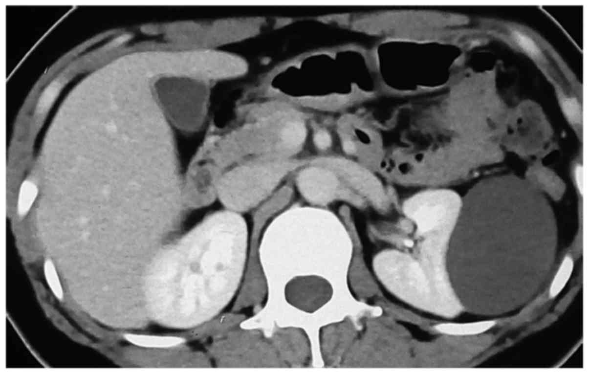
laboratory investigations were normal. US examination revealed a 70×60-mm cyst in the upper pole of the left kidney and an abdominal contrast-enhanced CT scan showed a simple cyst in the lateral aspect of the left kidney (Fig. 1). The density of the cyst contents was similar to that of water and there was no enhancement. The patient underwent laparoscopic decortication of the cyst. Under laparoscopic visualization, the cystic fluid was aspirated by a syringe and immediately sent for biochemical analysis. The cystic fluid was clear and yellow, and the biochemical analysis showed that the aspirated fluid contained extremely low levels of potassium, sodium, calcium and glucose (Table I). This was quite different from the contents of regular cysts, which usually have a 
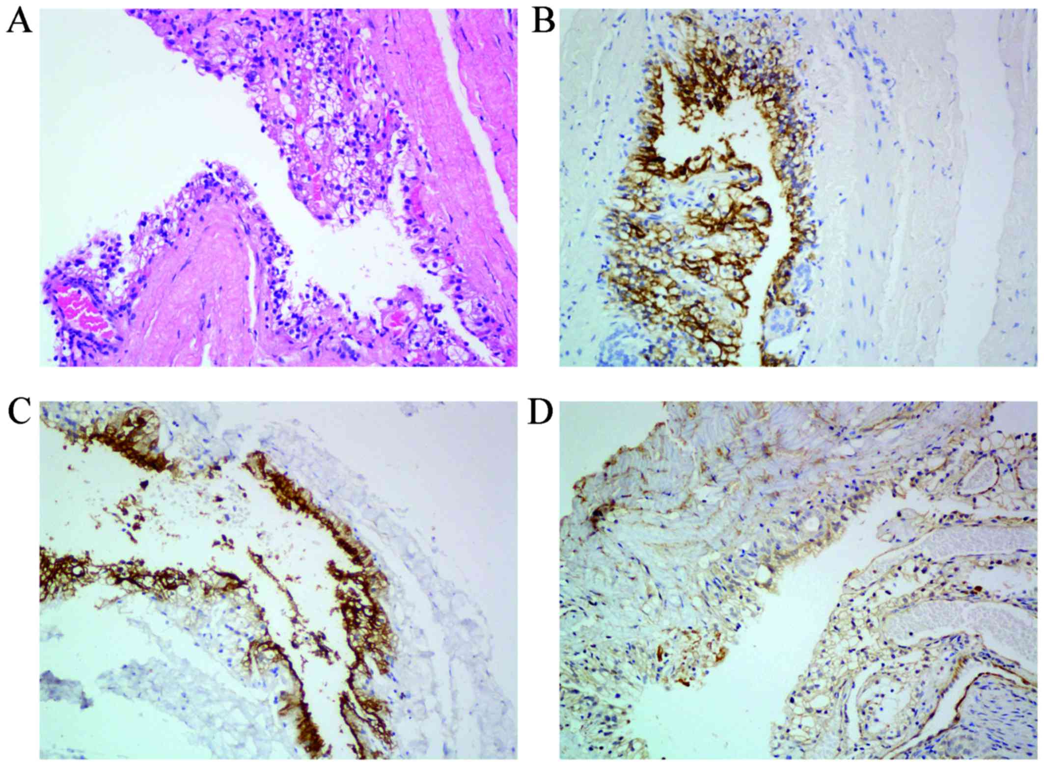
consistency similar to that of serum. The cyst wall was excised and sent for pathological evaluation and the histological examination of the floor of the cyst indicated malignancy. Macroscopically, the external surface of the cyst was smooth, but small excrescences were observed in the inner wall of the cyst. On immunostaining, the tumor cells stained positive for RCC marker, CD10 and vimentin, and negative for cytokeratin 7 (Fig. 2). Laparoscopic nephrectomy was performed 20 days after decortication and the pathological examination confirmed the diagnosis of RCC of the clear cell type. The postoperative course was uneventful. The patient was followed up by chest X-ray, liver function tests, abdominal CT scans and physical examination every 3 months for the first year and every 6 months thereafter. There has been no evidence of local recurrence or metastatic disease to date.

Figure 1.

Abdominal computed tomography scan with contrast showing a simple cyst sized 7×6×6 cm in the left kidney, with a density of 2 Hounsfied units.

Figure 2.

Histological findings of the tumor. (A) Hematoxylin and eosin staining of the tumor showing clear cells with relative small nuclei forming alveolar structures; (B) imunohistochemical staining for renal cell carcinoma marker; (C) imunohistochemical staining for CD10; (D) Imunohistochemical staining for vimentin (all magnifications, ×200).

Table I.

Biochemical analysis of the cystic fluid.

Table I.

Biochemical analysis of the cystic fluid.

Potassium (mmol/l)Sodium (mmol/l)Chlorine (mmol/l)Calcium (mmol/l)Blood urea nitrogen (mmol/l)Creatinine (µmol/l)Glucose (mmol/l)
<1.00<100<500.872.40221.94

Discussion

RCC presenting as a simple renal cyst is extremely rare and only few cases have been reported to date in the English literature. Lin et al (4) reported a patient with flank pain who was initially diagnosed with a simple cyst and was treated by ultrasound-guided aspiration, but the pain developed again 3 weeks later and ultrasound examination indicated a recurrent sizeable cyst. Nephrectomy was then performed and pathological examination confirmed the diagnosis of papillary RCC. Visapää et al (5) evaluated 482 patients who underwent partial or radical nephrectomy for kidney tumors and demonstrated an association between simple renal cysts and papillary RCC. Sakai et al (6) reported the case of a simple renal cyst that developed into a septated renal cyst and finally, to cystic RCC of the clear cell type, over a period of 6 years. That case demonstrated the natural history of malignant transformation from a simple renal cyst, and emphasized that careful follow-up of renal cysts, particularly of complicated cysts, is mandatory for successful treatment of RCC. Furthermore, in the Japanese literature, Takao et al (7) also reported a cystic RCC diagnosed as a simple cyst preoperatively.

Diagnosing a simple renal cyst is generally not difficult. The Bosniak classification system has been accepted as a method for diagnosing and determining the management of cystic renal masses, which was designed to analyze the morphology of cystic masses based solely on CT findings. However, it is difficult to determine preoperatively whether or not a cyst is malignant. Our patient, who initially presented with a simple cyst on CT imaging, apparently classified as Bosniak category I, was pathologically diagnosed with RCC. This may be due to the small size of the malignant tissue inside the cyst, which cannot be detected by imaging examinations alone.

The management options for renal cysts include radiological surveillance, ultrasound- or CT-guided cyst aspiration or biopsy and laparoscopic exploration or open surgical exploration, with or without radical or partial nephrectomy. Cyst aspiration for cytology may be helpful in the management of a patient only if malignant cells are clearly seen. Radical or partial nephrectomy may result in overtreatment, as nephrectomies often prove unnecessary. Omachi et al (8) reviewed the characteristics of intracystic fluid associated with RCC from the Japanese literature and found that cytological examination was negative for tumor cells in the majority of these cases, and 70% was hemorrhagic intracystic fluid. CT-guided biopsy may also be difficult and occasionally leads to misdiagnosis (9,10).

In the present case, the renal lesion was found to be a simple cyst on ultrasound and CT examination, classified as Bosniak category I. The possibility of malignancy appeared to be extremely low, but finally pathology demonstrated RCC. Interestingly, the biochemical analysis of the aspiration fluid revealed that it contained low levels of potassium, sodium, calcium and glucose, which is unusual in a simple renal cyst. In the case reported by Lin et al (4) the cystic RCC also had low glucose (0.333 mmol/l), and increased lactate dehydrogenase (2,361 U/l) levels. In our institute, the cystic fluid is routinely tested in all patients who undergo laparoscopic decortication, in order to detect whether the cyst communicates with the renal pelvis, and we observed that the consistency of the contents of simple renal cysts is almost the same as that of the serum (data not shown). The change of biochemical consistency and protein levels may be associated with the microenvironment changes in malignancy.

Therefore, it is crucial that we recognize the possibility of RCC appearing as a simple renal cyst. Biochemical analysis of the cystic fluid may help identify the presence malignancy and we recommend that biochemical analysis of the cystic fluid be routinely performed, either by ultrasound- or CT-guided cyst aspiration or during laparoscopic procedures. If suspicious, achieving a rapid pathological diagnosis by biopsy of the cyst wall is necessary. If the presence of malignancy is pathologically confirmed, an immediate partial or radical nephrectomy may be performed under the same anesthetic as the laparoscopic procedure.

References

1 

Bosniak MA: The current radiological approach to renal cysts. Radiology. 158:1–10. 1986. View Article : Google Scholar : PubMed/NCBI

2 

Israel GM and Bosniak MA: An update of the Bosniak renal cyst classification system. Urology. 66:484–488. 2005. View Article : Google Scholar : PubMed/NCBI

3 

Li G, Bilal I, Gentil-Perret A, Feng G, Zhao A, Peoc'h M, Genin C, Tostain J and Gigante M: CA9 as a molecular marker for differential diagnosis of cystic renal tumors. Urol Oncol. 30:463–468. 2012. View Article : Google Scholar : PubMed/NCBI

4 

Lin CJ, Chen YC, Chen HH, Wu CJ and Hsu JM: Renal cell carcinoma presenting as a huge simple renal cyst. Med Oncol. 25:104–106. 2008. View Article : Google Scholar : PubMed/NCBI

5 

Visapää H, Glucker E, Haukka J, Taari K and Nisen H: Papillary renal cell cancer is strongly associated with simple renal cysts. Urol Int. 91:269–272. 2013. View Article : Google Scholar : PubMed/NCBI

6 

Sakai N, Kanda F, Kondo K, Fukuoka H and Tanaka T: Sonographically detected malignant transformation of a simple renal cyst. Int J Urol. 8:23–25. 2001. View Article : Google Scholar : PubMed/NCBI

7 

Takao T, Gotoh T, Takada S and Sugao H: Cystic renal cell carcinoma diagnosed as a simple cyst preoperatively with incidental renal tumor: A case report. Hinyokika Kiyo. 45:339–342. 1999.(In Japanese). PubMed/NCBI

8 

Omachi T, Sakamoto W, Kishimoto T, Kawano M, Oyama A, Kamizuru M, Maekawa M, Hagihara S and Nakamura K: A case of renal cyst associated with renal cell carcinoma-characteristics of intracystic fluid associated with renal cell carcinoma from Japanese reports. Hinyokika Kiyo. 38:323–326. 1992.(In Japanese). PubMed/NCBI

9 

Siegel CL: Accuracy of diagnosis by guided biopsy of renal mass lesions classified indeterminate by imaging studies. J Urol. 165:322–323. 2001. View Article : Google Scholar : PubMed/NCBI

10 

Kadekawa K, Miyazato M, Saito S, Morozumi M, Matsuzaki A, Yoshimi N and Sugaya K: Renal cell carcinoma originating in a renal cyst in a 12-year-old girl. J Pediatr Surg. 44:e5–e7. 2009. View Article : Google Scholar : PubMed/NCBI

Related Articles

  • Abstract
  • View
  • Download
  • Twitter
Copy and paste a formatted citation
Spandidos Publications style
Yu Y, Ma L, Wang Z and Zhang Z: Renal cell carcinoma presenting as a simple renal cyst: A case report. Mol Clin Oncol 6: 550-552, 2017.
APA
Yu, Y., Ma, L., Wang, Z., & Zhang, Z. (2017). Renal cell carcinoma presenting as a simple renal cyst: A case report. Molecular and Clinical Oncology, 6, 550-552. https://doi.org/10.3892/mco.2017.1173
MLA
Yu, Y., Ma, L., Wang, Z., Zhang, Z."Renal cell carcinoma presenting as a simple renal cyst: A case report". Molecular and Clinical Oncology 6.4 (2017): 550-552.
Chicago
Yu, Y., Ma, L., Wang, Z., Zhang, Z."Renal cell carcinoma presenting as a simple renal cyst: A case report". Molecular and Clinical Oncology 6, no. 4 (2017): 550-552. https://doi.org/10.3892/mco.2017.1173
Copy and paste a formatted citation
x
Spandidos Publications style
Yu Y, Ma L, Wang Z and Zhang Z: Renal cell carcinoma presenting as a simple renal cyst: A case report. Mol Clin Oncol 6: 550-552, 2017.
APA
Yu, Y., Ma, L., Wang, Z., & Zhang, Z. (2017). Renal cell carcinoma presenting as a simple renal cyst: A case report. Molecular and Clinical Oncology, 6, 550-552. https://doi.org/10.3892/mco.2017.1173
MLA
Yu, Y., Ma, L., Wang, Z., Zhang, Z."Renal cell carcinoma presenting as a simple renal cyst: A case report". Molecular and Clinical Oncology 6.4 (2017): 550-552.
Chicago
Yu, Y., Ma, L., Wang, Z., Zhang, Z."Renal cell carcinoma presenting as a simple renal cyst: A case report". Molecular and Clinical Oncology 6, no. 4 (2017): 550-552. https://doi.org/10.3892/mco.2017.1173
Follow us
  • Twitter
  • LinkedIn
  • Facebook
About
  • Spandidos Publications
  • Careers
  • Cookie Policy
  • Privacy Policy
How can we help?
  • Help
  • Live Chat
  • Contact
  • Email to our Support Team