Introduction
Gastric cancer is the third most commonly diagnosed
cancer worldwide, and a leading cause of cancer-related mortality.
The most common metastatic sites of gastric cancer are the lymph
nodes, liver and peritoneum, while it may be associated with
multiple sites of metastasis. Subcutaneous metastasis from gastric
cancer is a rare manifestation, with a reported incidence of
0.8–1.0% (1–3). We herein report a case of subcutaneous
metastasis from gastric cancer in a patient who underwent surgical
resection and systemic chemotherapy.
Case report
A 59-year-old man was referred to the Kochi Medical
School Hospital (Nankoku, Japan) in February, 2016, due to left
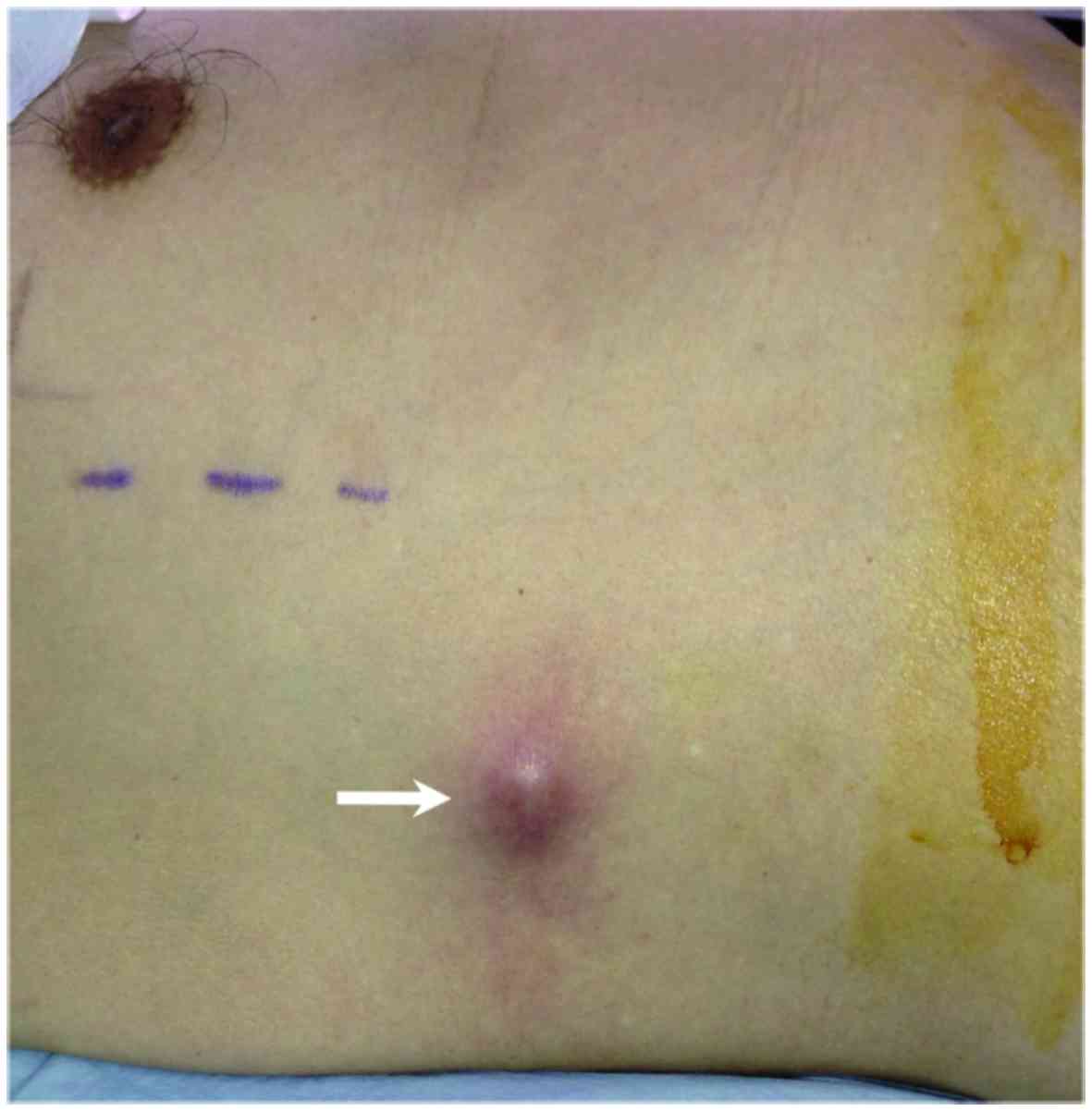
shoulder pain. Physical examination revealed a nodular painful mass
lesion in the subcutaneous tissue of the right chest wall,
measuring ~2 cm in diameter (Fig.
1). X-ray and magnetic resonance imaging revealed an osteolytic
tumor in the proximal region of the left humerus.
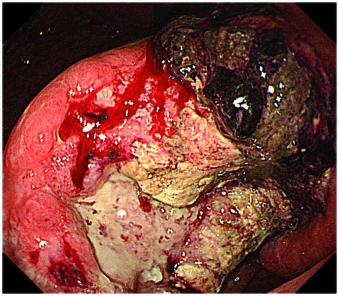
Esophagogastroduodenoscopy revealed a large ulcerated tumor in the
lower gastric body near the lesser curvature, and biopsy specimens
from the gastric and humeral lesions revealed poorly differentiated
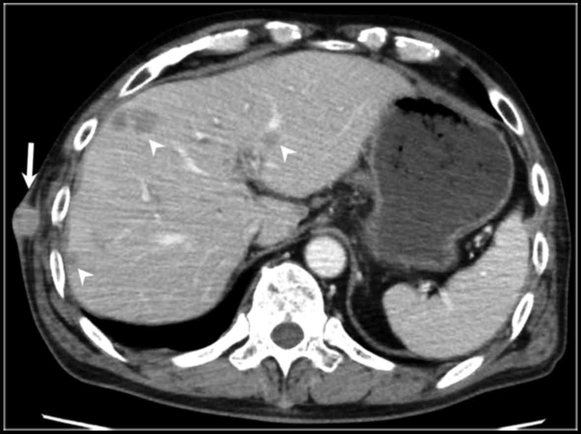
adenocarcinoma (Fig. 2). Abdominal
computed tomography examination revealed multiple low-density
lesions in the liver and a well-defined, 2.2-cm mass in the
subcutaneous tissue of the right chest wall (Fig. 3).
The diagnosis was advanced gastric cancer with
metastases to the liver, bone and skin, and systemic chemotherapy
with radiotherapy were recommended for the bone metastasis. The
patient also underwent distal gastrectomy to control bleeding from
the primary gastric cancer lesion due to unresolved anemia, not
improving by blood transfusion, and a low hemoglobin level (7.3
g/dl; normal range, 13.7–16.8 g/dl). The subcutaneous tumor in the
right chest wall was resected at the same time, in order to
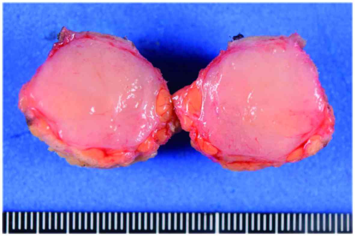
alleviate the pain caused by the mass. Macroscopic examination of
the resected specimen revealed a well-circumscribed, solid tumor,
measuring 2.2×2.1 cm, with a firm consistency (Fig. 4). Microscopic examination revealed
infiltration of the subcutaneous tissue by poorly differentiated
adenocarcinoma cells. The patient received chemotherapy with
oxaliplatin and oral fluoropyrimidine (S-1). However, the liver
metastases and peritoneal carcinomatosis progressed, and the
patient succumbed to the disease 6 months after the diagnosis.
Discussion
The most commonly reported origins of cutaneous
metastasis are lung, breast and colon cancer, melanoma, squamous
cell carcinoma of the oral cavity and renal cell carcinoma
(2,4). Lymphatic and hematogenous spread have
been suggested as possible mechanisms for metastasis to the skin as
well as to other organs (4,5), although most metastases of primary
cancer are generally considered to progress, in a stepwise manner,
from locoregional to distant lesions (6).
Cutaneous metastasis of cancer generally appears
late in the course of the disease. Therefore, the mean survival
time of patients with skin metastases is only a few months and, in
cases of metastatic gastric tumors, treatment usually consists of
systemic therapy rather than surgery (6). However, as in the present case,
surgical resection of metastatic skin tumors is occasionally
undertaken as palliative treatment to improve the patient's quality
of life by controlling severe symptoms such as pain and
hemorrhage.
References
|
1
|
Kawai S, Nishida T, Hayashi Y, Ezaki H,
Yamada T, Shinzaki S, Miyazaki M, Nakai K, Yakushijin T, Watabe K,
et al: Choroidal and cutaneous metastasis from gastric
adenocarcinoma. World J Gastroenterol. 19:1485–1488. 2013.
View Article : Google Scholar : PubMed/NCBI
|
|
2
|
Hu SC, Chen GS, Wu CS, Chai CY, Chen WT
and Lan CC: Rates of cutaneous metastases from different internal
malignancies: Experience from a Taiwanese medical center. J Am Acad
Dermatol. 60:379–387. 2009. View Article : Google Scholar : PubMed/NCBI
|
|
3
|
Kairouani M, Perrin J, Dietemann-Barabinot
A, Diab R and Ruck S: Cutaneous metastasis revealing a relapse of
gastric linitis: Another case. Int J Surg Case Rep. 4:185–187.
2013. View Article : Google Scholar : PubMed/NCBI
|
|
4
|
Alcaraz I, Cerroni L, Rütten A, Kutzner H
and Requena L: Cutaneous metastases from internal malignancies: A
clinicopathologic and immunohistochemical review. Am J
Dermatopathol. 34:347–393. 2012. View Article : Google Scholar : PubMed/NCBI
|
|
5
|
Avgerinou G, Flessas I, Hatziolou E,
Zografos G, Nitsios I, Zagouri F, Katsambas A and Kanitakis J:
Cutaneous metastasis of signet-ring gastric adenocarcinoma to the
breast with unusual clinicopathological features. Anticancer Res.
31:2373–2378. 2011.PubMed/NCBI
|
|
6
|
Namikawa T and Hanazaki K:
Clinicopathological features and treatment outcomes of metastatic
tumors in the stomach. Surg Today. 44:1392–1399. 2014. View Article : Google Scholar : PubMed/NCBI
|