Spandidos Publications Logo
  • About
    • About Spandidos
    • Aims and Scopes
    • Abstracting and Indexing
    • Editorial Policies
    • Reprints and Permissions
    • Job Opportunities
    • Terms and Conditions
    • Contact
  • Journals
    • All Journals
    • Oncology Letters
      • Oncology Letters
      • Information for Authors
      • Editorial Policies
      • Editorial Board
      • Aims and Scope
      • Abstracting and Indexing
      • Bibliographic Information
      • Archive
    • International Journal of Oncology
      • International Journal of Oncology
      • Information for Authors
      • Editorial Policies
      • Editorial Board
      • Aims and Scope
      • Abstracting and Indexing
      • Bibliographic Information
      • Archive
    • Molecular and Clinical Oncology
      • Molecular and Clinical Oncology
      • Information for Authors
      • Editorial Policies
      • Editorial Board
      • Aims and Scope
      • Abstracting and Indexing
      • Bibliographic Information
      • Archive
    • Experimental and Therapeutic Medicine
      • Experimental and Therapeutic Medicine
      • Information for Authors
      • Editorial Policies
      • Editorial Board
      • Aims and Scope
      • Abstracting and Indexing
      • Bibliographic Information
      • Archive
    • International Journal of Molecular Medicine
      • International Journal of Molecular Medicine
      • Information for Authors
      • Editorial Policies
      • Editorial Board
      • Aims and Scope
      • Abstracting and Indexing
      • Bibliographic Information
      • Archive
    • Biomedical Reports
      • Biomedical Reports
      • Information for Authors
      • Editorial Policies
      • Editorial Board
      • Aims and Scope
      • Abstracting and Indexing
      • Bibliographic Information
      • Archive
    • Oncology Reports
      • Oncology Reports
      • Information for Authors
      • Editorial Policies
      • Editorial Board
      • Aims and Scope
      • Abstracting and Indexing
      • Bibliographic Information
      • Archive
    • Molecular Medicine Reports
      • Molecular Medicine Reports
      • Information for Authors
      • Editorial Policies
      • Editorial Board
      • Aims and Scope
      • Abstracting and Indexing
      • Bibliographic Information
      • Archive
    • World Academy of Sciences Journal
      • World Academy of Sciences Journal
      • Information for Authors
      • Editorial Policies
      • Editorial Board
      • Aims and Scope
      • Abstracting and Indexing
      • Bibliographic Information
      • Archive
    • International Journal of Functional Nutrition
      • International Journal of Functional Nutrition
      • Information for Authors
      • Editorial Policies
      • Editorial Board
      • Aims and Scope
      • Abstracting and Indexing
      • Bibliographic Information
      • Archive
    • International Journal of Epigenetics
      • International Journal of Epigenetics
      • Information for Authors
      • Editorial Policies
      • Editorial Board
      • Aims and Scope
      • Abstracting and Indexing
      • Bibliographic Information
      • Archive
    • Medicine International
      • Medicine International
      • Information for Authors
      • Editorial Policies
      • Editorial Board
      • Aims and Scope
      • Abstracting and Indexing
      • Bibliographic Information
      • Archive
  • Articles
  • Information
    • Information for Authors
    • Information for Reviewers
    • Information for Librarians
    • Information for Advertisers
    • Conferences
  • Language Editing
Spandidos Publications Logo
  • About
    • About Spandidos
    • Aims and Scopes
    • Abstracting and Indexing
    • Editorial Policies
    • Reprints and Permissions
    • Job Opportunities
    • Terms and Conditions
    • Contact
  • Journals
    • All Journals
    • Biomedical Reports
      • Information for Authors
      • Editorial Policies
      • Editorial Board
      • Aims and Scope
      • Abstracting and Indexing
      • Bibliographic Information
      • Archive
    • Experimental and Therapeutic Medicine
      • Information for Authors
      • Editorial Policies
      • Editorial Board
      • Aims and Scope
      • Abstracting and Indexing
      • Bibliographic Information
      • Archive
    • International Journal of Epigenetics
      • Information for Authors
      • Editorial Policies
      • Editorial Board
      • Aims and Scope
      • Abstracting and Indexing
      • Bibliographic Information
      • Archive
    • International Journal of Functional Nutrition
      • Information for Authors
      • Editorial Policies
      • Editorial Board
      • Aims and Scope
      • Abstracting and Indexing
      • Bibliographic Information
      • Archive
    • International Journal of Molecular Medicine
      • Information for Authors
      • Editorial Policies
      • Editorial Board
      • Aims and Scope
      • Abstracting and Indexing
      • Bibliographic Information
      • Archive
    • International Journal of Oncology
      • Information for Authors
      • Editorial Policies
      • Editorial Board
      • Aims and Scope
      • Abstracting and Indexing
      • Bibliographic Information
      • Archive
    • Medicine International
      • Information for Authors
      • Editorial Policies
      • Editorial Board
      • Aims and Scope
      • Abstracting and Indexing
      • Bibliographic Information
      • Archive
    • Molecular and Clinical Oncology
      • Information for Authors
      • Editorial Policies
      • Editorial Board
      • Aims and Scope
      • Abstracting and Indexing
      • Bibliographic Information
      • Archive
    • Molecular Medicine Reports
      • Information for Authors
      • Editorial Policies
      • Editorial Board
      • Aims and Scope
      • Abstracting and Indexing
      • Bibliographic Information
      • Archive
    • Oncology Letters
      • Information for Authors
      • Editorial Policies
      • Editorial Board
      • Aims and Scope
      • Abstracting and Indexing
      • Bibliographic Information
      • Archive
    • Oncology Reports
      • Information for Authors
      • Editorial Policies
      • Editorial Board
      • Aims and Scope
      • Abstracting and Indexing
      • Bibliographic Information
      • Archive
    • World Academy of Sciences Journal
      • Information for Authors
      • Editorial Policies
      • Editorial Board
      • Aims and Scope
      • Abstracting and Indexing
      • Bibliographic Information
      • Archive
  • Articles
  • Information
    • For Authors
    • For Reviewers
    • For Librarians
    • For Advertisers
    • Conferences
  • Language Editing
Login Register Submit
  • This site uses cookies
  • You can change your cookie settings at any time by following the instructions in our Cookie Policy. To find out more, you may read our Privacy Policy.

    I agree
Search articles by DOI, keyword, author or affiliation
Search
Advanced Search
presentation
Molecular and Clinical Oncology
Join Editorial Board Propose a Special Issue
Print ISSN: 2049-9450 Online ISSN: 2049-9469
Journal Cover
May-2017 Volume 6 Issue 5

Full Size Image

Sign up for eToc alerts
Recommend to Library

Journals

International Journal of Molecular Medicine

International Journal of Molecular Medicine

International Journal of Molecular Medicine is an international journal devoted to molecular mechanisms of human disease.

International Journal of Oncology

International Journal of Oncology

International Journal of Oncology is an international journal devoted to oncology research and cancer treatment.

Molecular Medicine Reports

Molecular Medicine Reports

Covers molecular medicine topics such as pharmacology, pathology, genetics, neuroscience, infectious diseases, molecular cardiology, and molecular surgery.

Oncology Reports

Oncology Reports

Oncology Reports is an international journal devoted to fundamental and applied research in Oncology.

Experimental and Therapeutic Medicine

Experimental and Therapeutic Medicine

Experimental and Therapeutic Medicine is an international journal devoted to laboratory and clinical medicine.

Oncology Letters

Oncology Letters

Oncology Letters is an international journal devoted to Experimental and Clinical Oncology.

Biomedical Reports

Biomedical Reports

Explores a wide range of biological and medical fields, including pharmacology, genetics, microbiology, neuroscience, and molecular cardiology.

Molecular and Clinical Oncology

Molecular and Clinical Oncology

International journal addressing all aspects of oncology research, from tumorigenesis and oncogenes to chemotherapy and metastasis.

World Academy of Sciences Journal

World Academy of Sciences Journal

Multidisciplinary open-access journal spanning biochemistry, genetics, neuroscience, environmental health, and synthetic biology.

International Journal of Functional Nutrition

International Journal of Functional Nutrition

Open-access journal combining biochemistry, pharmacology, immunology, and genetics to advance health through functional nutrition.

International Journal of Epigenetics

International Journal of Epigenetics

Publishes open-access research on using epigenetics to advance understanding and treatment of human disease.

Medicine International

Medicine International

An International Open Access Journal Devoted to General Medicine.

Journal Cover
May-2017 Volume 6 Issue 5

Full Size Image

Sign up for eToc alerts
Recommend to Library

  • Article
  • Citations
    • Cite This Article
    • Download Citation
    • Create Citation Alert
    • Remove Citation Alert
    • Cited By
  • Similar Articles
    • Related Articles (in Spandidos Publications)
    • Similar Articles (Google Scholar)
    • Similar Articles (PubMed)
  • Download PDF
  • Download XML
  • View XML
Case Report

Chondroblastic osteosarcoma arising in the maxilla mimicking the radiographic and histological characteristics of cemento-osseous lesions: A case report

  • Authors:
    • Bin‑Bin Li
    • Jian‑Yun Zhang
    • Yan Gao
  • View Affiliations / Copyright

    Affiliations: Department of Oral Pathology, Peking University School and Hospital of Stomatology, Beijing 100081, P.R. China
  • Pages: 733-736
    |
    Published online on: April 10, 2017
       https://doi.org/10.3892/mco.2017.1219
  • Expand metrics +
Metrics: Total Views: 0 (Spandidos Publications: | PMC Statistics: )
Metrics: Total PDF Downloads: 0 (Spandidos Publications: | PMC Statistics: )
Cited By (CrossRef): 0 citations Loading Articles...

This article is mentioned in:


Abstract

Osteosarcomas of the jaw are comparatively rare and represent only 2-10% of all osteosarcomas. We herein present a rare case of an osteosarcoma exhibiting the radiographic and histological characteristics of cemento‑osseous lesions in the alveolar ridge of the maxilla. A 53-year-old male patient presented with the complaint of gradual swelling of the left maxilla over 4 years. Radiography revealed an ill‑defined radioopaque mass, intimately associated with the apices of the involved teeth, without a periosteal reaction. Microscopically, a cementicle‑like structure was identified in the alveolar bone. In addition, the lesion exhibited typical characteristics of chondroblastic osteosarcoma in the body of the maxilla. The tumor contained abundant osteoid and cartilage intimately associated with anaplastic tumor cells. The cartilage displayed malignant‑appearing cells in lacunae, and there was crowding at the periphery of the lobule where the spindle cells formed sheets. The differential diagnosis included primary osteosarcoma, concurrent cemento‑osseous dysplasia and osteosarcoma, or a secondary osteosarcoma based on a pre‑existing cemento‑osseous lesion. The presence of the cementicle‑like structure in the alveolar bone and the involvement of the periodontal ligament and alveolar bone proper were unique in our case. The general invasive growth pattern and the abundance of the irregular tumor bone helped establish the diagnosis of primary osteosarcoma. This case may represent evidence of the pathogenesis of primary osteosarcoma in the jaw.

Introduction

Osteosarcoma is a type of malignant bone tumor in which the neoplastic cells produce osteoid or bone (1). Osteosarcomas of the jaw are rare and represent only 2–10% of all osteosarcomas. Osteosarcoma of the head and neck region is the most common primary malignant bone tumor, representing 23% of all head and neck malignancies (2). Osteosarcoma arises more frequently in the maxilla compared with the mandible (3). Unlike the hallmark clinical presentation of pain in lesions of the long bones, the most characteristic symptom of jaw osteosarcoma is swelling. Similar with osteosarcoma of long bones, the dominant histological variant of osteosarcoma is the osteoblastic type, followed by the chondroblastic and fibroblastic types (4). However, the pathologist may encounter a potential diagnostic pitfall when an osteosarcoma of the jaw resembles a cemento-osseous lesion. We herein present a case of maxillary osteosarcoma mimicking the histological and radiographic characteristics of cemento-osseous lesions.

Case report

A 53-year-old male patient presented in May, 2014 with gradual swelling of the left side of the face over a period of 4 years. The patient's medical history was non-contributory. The physical examination revealed an uncircumscribed swelling of the 
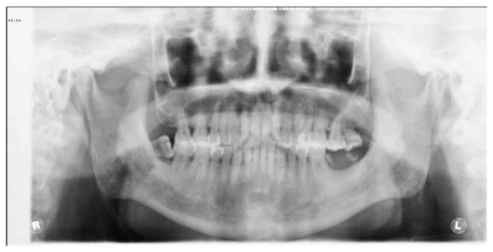
left maxilla, with normal color of the overlying skin. A panoramic radiograph revealed an ill-defined radiopaque mass at the apices of the roots of the left maxillary bone, from the incisor to the second bicuspid (Fig. 1). The mass was unilateral and closely associated with the teeth roots; however, there was no root resorption in the involved teeth. 
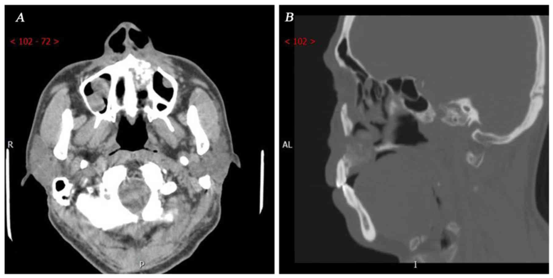
Further computed tomography examination revealed that the size of the mass was 2.3×2×1.7 cm. No periosteal reaction was observed, and no soft tissue extension component beyond the area of the cancellous and/or cortical bone destruction was identified radiographically. The lesion involved the left wall of the incisive canal and the left hard palate, resulting in a discontinuous labial and palatal alveolar bone plate (Fig. 2A). The corresponding nasal bottom and left wall of the maxillary sinus were eroded (Fig. 2B). Given its association with the teeth root and the diffuse borders, the most likely clinical diagnosis was a malignant tumor originating from cementum. The tumor was treated by radical resection of the left maxillary bone.

Figure 1.

Panoramic X-ray revealed a radiopaque lesion adjacent to the roots of the teeth from the left maxillary incisor to the second bicuspid. The border of the mass was diffuse and irregular. Moreover, the porosity in the surrounding bone indicated an invasive process.

Figure 2.

Computed tomography (CT) images. (A) Axial CT scan, soft tissue algorithm, showing an osteoblastic destructive mass extending in the nasal cavity and maxillary sinus, without periosteal reaction or soft tissue extension. (B) Multiplanar reformatted sagittal CT scan, bone algorithm, showing a destructive mass extending from the left alveolar ridge into the nasal cavity, with osteoid calcification.

The specimens were fixed in 10% buffered formalin, dehydrated through graded concentrations of ethanol and embedded in paraffin wax. The paraffin block was then cut in 4-mm sections that were stained with haematoxylin and eosin (H&E).

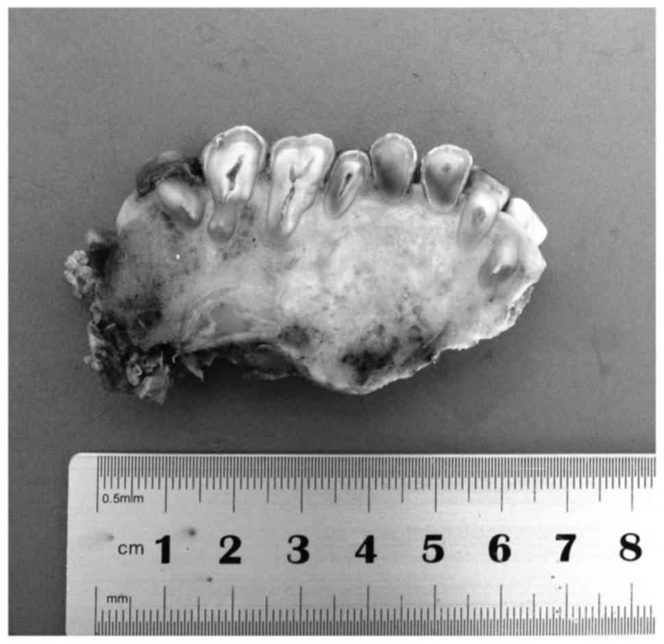
Grossly, the tumor involved alveolar bone as well as bone from the body of the maxilla, from the first incisor to the proximal side of the first molar (Fig. 3). All the roots of the involved teeth were partially embedded in the tumor. The tumor appeared as pale, without clear borders from the surrounding bone tissue. The sectional area of the mass 
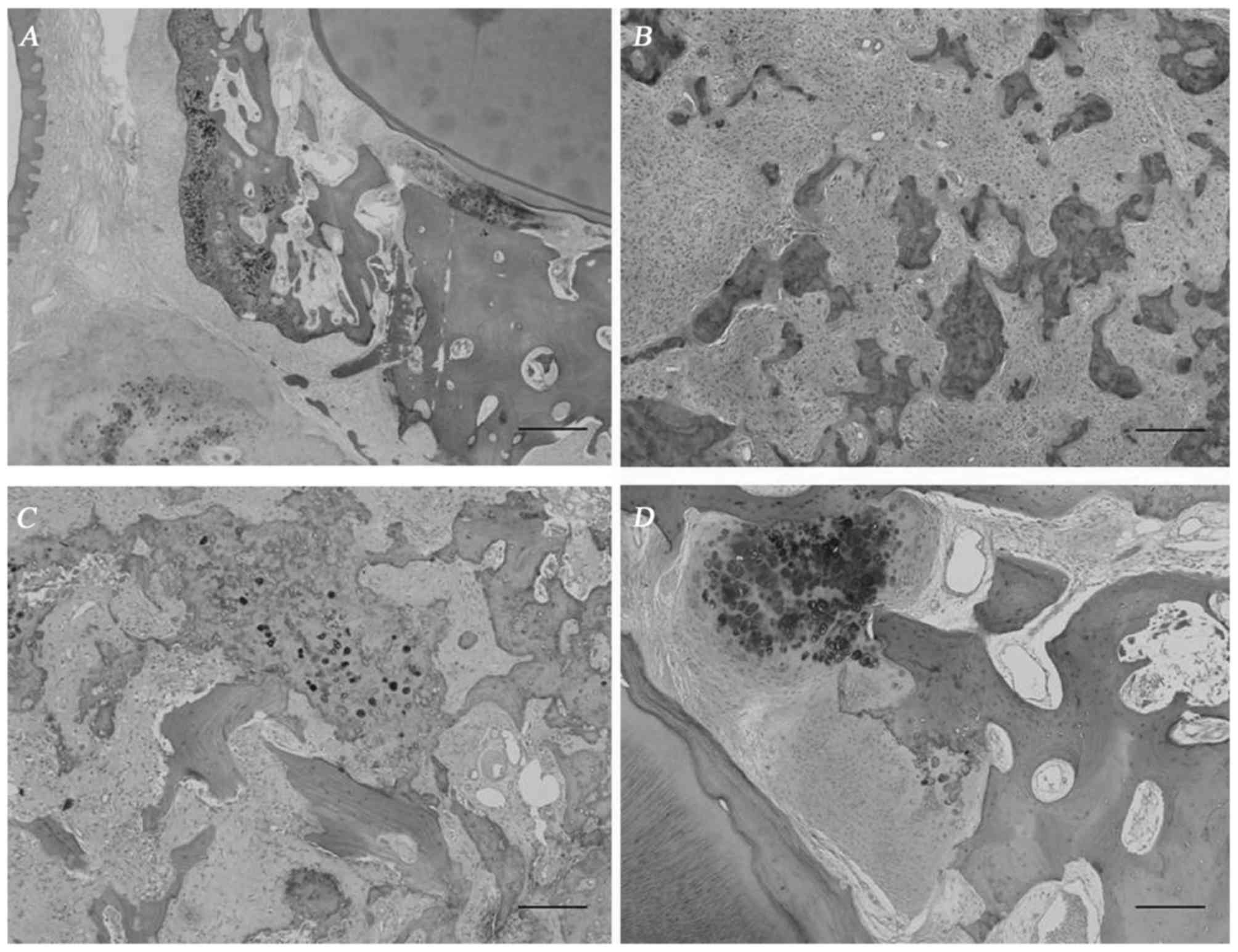
was 3.4×3 cm. Microscopically, the mass displayed a different appearance in the alveolar bone and in the body of the jaw. In the alveolar bone part, the tumor infiltrated the periodontal ligament and alveolar bone proper (Fig. 4A). Extensive cementicle-like structure and deposits of hypercellular cartilage were observed in the alveolar bone (Fig. 4B). However, this cementum-like material was intimately associated with the pleomorphic and hyperchromatic spindle cell component, and it infiltrated the trabeculae of the medullary bone (Fig. 4C), suggesting a malignant tumor. Interestingly, a continuous transition from the trabecular alveolar bone to the tumor was observed at the bony side of the periodontal ligament, strongly supporting the hypothesis that the tumor originated from bone, not from the periodontal ligament, and suggesting that the tumor would differentiate towards osteoid and not towards cementum-like material.

Figure 3.

Macroscopic view of the surgical specimen of the left maxillary bone from the palatal view. The mass was pale, diffuse and irregular, and it involved a larger part of the bone than what appeared on radiographic images, including the alveolar ridge and the body of the maxilla from the first incisor to the first molar. On sectioning, the teeth roots were partially embedded in the tumor.

Figure 4.

The morphology of the mass in the alveolar part of the left maxillary bone resembled a cemento-osseous lesion. (A) Interface of the tooth apex and the tumor. Low-power view of the tumor involving alveolar bone and periodontal ligament. (B) Abundant cementicle-like structures were formed among highly atypical spindle cells. Matrix was present between the tumor cells. (C) The tumor cells permeated between the preexisting trabeculae of the medullary bone. (D) The tumor was located on the side of alveolar bone, rather than the side of the cementum, which indicated its osteogenic nature. Hematoxylin and eosin staining; magnification, ×10.

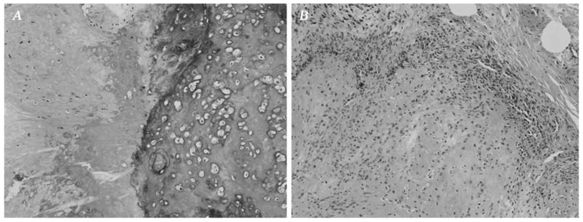
In the bone of the maxilla, the mass exhibited the morphological characteristics of a typical chondroblastic osteosarcoma. The tumor contained a relatively equal distribution of abnormal osteoid and chondroid, intimately associated with anaplastic tumor cells (Fig. 5A). Islands of osteoid were observed within the cartilage. The tumor bone exhibited a woven or basket weave pattern and was strongly hematoxylinophilic, unlike the uniform lamellar pattern of normal bone. The cartilage displayed malignant-appearing cells in lacunae, and there was crowding at the periphery of the lobule, where spindle cells were arranged in sheets (Fig. 5B).

Figure 5.

The histological appearance of the tumor in body of the left maxillary bone suggested a typical chondroblastic osteosarcoma. (A) The tumor contained deposits of hypercellular cartilage and abnormal osteoid and bone. (B) There was crowding at the periphery of the lobule, where sheets of atypical, pleomorphic and hyperchromatic sarcomatous cells were formed. Hematoxylin and eosin staining; magnification, ×10.

Discussion

Osteosarcoma is characterized by the osteoid production by tumor cells. Osteosarcoma of the jaw is associated with certain specific characteristics, such as a higher prevalence in the maxilla and male predominance. Wang et al reported that all maxillary de novo osteosarcomas arose from the alveolar ridge (5). The mass in the present case involved both the alveolar ridge and the body of the maxilla, possibly due to long-term growth leading to extension of the mass from the alveolar ridge to the body.

The differential diagnosis between lesions with hard tissue formation may be problematic in the oral area. These lesions are associated with the formation of bone, cartilage or cementum. Among these, osteosarcoma may arise de novo, may be synchronous multicentric (6), metastatic (7), or appear in benign precursor lesions, which may include cemento-osseous dysplasia (8) or ossifying fibroma (9). Cemento-osseous lesions are lesions of the jaw closely associated with the apices of the teeth that contain amorphous spherical calcifications resembling an aberrant form of cementum.

Specifically for this case, the differential diagnosis among primary osteosarcoma, concurrent cemento-osseous dysplasia and secondary malignant tumor in a background of cemento-osseous dysplasia or ossifying fibroma, may be difficult radiographically as well as histopathologically. Generally, the suspected clinical diagnosis was a cementum-originating tumor due to its association with the roots of the teeth and absence of periosteal reaction. The pathological appearance in the alveolar bone was confusing due to the presence of a cementicle-like structure. However, the typical manifestations in the body of the maxilla provided strong evidence for the diagnosis of primary chondroblastic osteosarcoma.

The mechanisms underlying the formation of bone, cartilage, or cementum in the oral area are helpful for understanding the nature of the lesions involving hard tissue formation, and may provide clues for differential diagnosis. Cells forming bone in the jaw may be divided into three types: Osteoblasts lying on the bone surface, osteogenic fibroblasts in the periodontal ligament and periosteum, and non-specific fibroblasts stimulated to differentiate into osteogenic cells after degeneration (10). The first two cell types are associated with the origin of jaw osteosarcoma, whereas the osteogenic cells in the periodontal ligament produce cementum (11). Therefore, the tumor cells responsible for hard tissue formation in osteosarcoma, cemento-osseous dysplasia or ossifying fibroma, may share a common origin. This may explain the presence of the cementicle-like structure in this osteosarcoma. However, cementum is a bone-like tissue that covers the root of the tooth. Cementocytes are similar to osteocytes, but their cell processes tend to be orientated in one direction, namely towards the periodontal ligament, rather than equally around the cell body, as in osteocytes. Therefore, what was referred to as a cementicle-like structure in this case was in fact abnormal osteoid structure of osteosarcoma. Osteoid displays a woven or mat-like appearance, unlike the more orderly longitudinal fiber array found in collagen.

References

1 

Sato K and Unni KK: Malignant tumors of bone and cartilagePathology and Genetics of Head and Neck Tumors: World Health Organization Classification of Tumors. Barnes L, Eveson JW, Reichart P and Sidransky D: IARC Press; Lyon: pp. 7–52. 2005

2 

Nthumba PM: Osteosarcoma of the jaws: A review of literature and a case report on synchronous multicentric osteosarcomas. World J Surg Oncol. 10:2402012. View Article : Google Scholar : PubMed/NCBI

3 

Forteza G, Colmenero B and López-Barea F: Osteogenic sarcoma of maxilla and mandible. Oral Surg Oral Med Oral Pathol. 62:179–184. 1986. View Article : Google Scholar : PubMed/NCBI

4 

Paparella ML, Olvi LG, Brandizzi D, Keszler A, Santini-Araujo E and Cabrini RL: Osteosarcoma of the jaw: An analysis of a series of 74 cases. Histopathology. 63:551–557. 2013.PubMed/NCBI

5 

Wang S, Shi H and Yu Q: Osteosarcoma of the jaws: Demographic and CT imaging features. Dentomaxillofac Radiol. 41:37–42. 2012. View Article : Google Scholar : PubMed/NCBI

6 

Jia S and Li B: Osteosarcoma of the Jaws: Case report on synchronous multicentric osteosarcomas. J Clin Diagn Res. 8:ZD01–ZD03. 2014.PubMed/NCBI

7 

Carnelio S, Pai K, Rao N, Solomon M and Ahasan A: Metastatic osteosarcoma to the maxilla: A case report and a review of the literature. Quintessence Int. 33:397–399. 2002.PubMed/NCBI

8 

Olusanya AA, Adeyemi BF and Adisa AO: Concurrent cemento-osseous dysplasia and osteogenic sarcoma: Report of two cases. Case Rep Med. 2012:1805612012.PubMed/NCBI

9 

Koury ME, Regezi JA, Perrott DH and Kaban LB: ‘Atypical’ fibro-osseous lesions: Diagnostic challenges and treatment concepts. Int J Oral Maxillofac Surg. 24:162–169. 1995. View Article : Google Scholar : PubMed/NCBI

10 

Matsuzaka K, Shimono M, Uchiyama T, Noma H and Inoue T: Lesions related to the formation of bone, cartilage or cementum arising in the oral area: A statistical study and review of the literature. Bull Tokyo Dent Coll. 43:173–180. 2002. View Article : Google Scholar : PubMed/NCBI

11 

Roguljic H, Matthews BG, Yang W, Cvija H, Mina M and Kalajzic I: In vivo identification of periodontal progenitor cells. J Dent Res. 92:709–715. 2013. View Article : Google Scholar : PubMed/NCBI

Related Articles

  • Abstract
  • View
  • Download
  • Twitter
Copy and paste a formatted citation
Spandidos Publications style
Li BB, Zhang JY and Gao Y: Chondroblastic osteosarcoma arising in the maxilla mimicking the radiographic and histological characteristics of cemento-osseous lesions: A case report. Mol Clin Oncol 6: 733-736, 2017.
APA
Li, B., Zhang, J., & Gao, Y. (2017). Chondroblastic osteosarcoma arising in the maxilla mimicking the radiographic and histological characteristics of cemento-osseous lesions: A case report. Molecular and Clinical Oncology, 6, 733-736. https://doi.org/10.3892/mco.2017.1219
MLA
Li, B., Zhang, J., Gao, Y."Chondroblastic osteosarcoma arising in the maxilla mimicking the radiographic and histological characteristics of cemento-osseous lesions: A case report". Molecular and Clinical Oncology 6.5 (2017): 733-736.
Chicago
Li, B., Zhang, J., Gao, Y."Chondroblastic osteosarcoma arising in the maxilla mimicking the radiographic and histological characteristics of cemento-osseous lesions: A case report". Molecular and Clinical Oncology 6, no. 5 (2017): 733-736. https://doi.org/10.3892/mco.2017.1219
Copy and paste a formatted citation
x
Spandidos Publications style
Li BB, Zhang JY and Gao Y: Chondroblastic osteosarcoma arising in the maxilla mimicking the radiographic and histological characteristics of cemento-osseous lesions: A case report. Mol Clin Oncol 6: 733-736, 2017.
APA
Li, B., Zhang, J., & Gao, Y. (2017). Chondroblastic osteosarcoma arising in the maxilla mimicking the radiographic and histological characteristics of cemento-osseous lesions: A case report. Molecular and Clinical Oncology, 6, 733-736. https://doi.org/10.3892/mco.2017.1219
MLA
Li, B., Zhang, J., Gao, Y."Chondroblastic osteosarcoma arising in the maxilla mimicking the radiographic and histological characteristics of cemento-osseous lesions: A case report". Molecular and Clinical Oncology 6.5 (2017): 733-736.
Chicago
Li, B., Zhang, J., Gao, Y."Chondroblastic osteosarcoma arising in the maxilla mimicking the radiographic and histological characteristics of cemento-osseous lesions: A case report". Molecular and Clinical Oncology 6, no. 5 (2017): 733-736. https://doi.org/10.3892/mco.2017.1219
Follow us
  • Twitter
  • LinkedIn
  • Facebook
About
  • Spandidos Publications
  • Careers
  • Cookie Policy
  • Privacy Policy
How can we help?
  • Help
  • Live Chat
  • Contact
  • Email to our Support Team