Introduction
The tight junction protein claudin-1 is an integral
part of the epithelial and endothelial tight junction complex,
which executes signal transduction pathways and cellular transport
functions (1,2). A total of 24 tissue-specific claudin
subtypes have been identified (3).
Loss of tight junction complex integrity serves a role in
tumorigenesis of solid tumors, and it has been hypothesized that
claudin-1 can act tissue-specifically as a tumor suppressor or as
an oncoprotein (4). Thus, claudin-1
can be either downregulated or upregulated according to the
specific tumor entity (2,5). Claudins are crucial for cell migration,
invasion and metastasis; this was demonstrated in a xenograft
colorectal cancer mouse model, where tumor growth and metastasis
were linked to E-Cadherin expression and β-catenin/T-cell factor
(TCF) signaling cascade (5). In
basal-like high-grade ductal breast cancer, colorectal cancer,
gastric cancer, melanoma and cervical cancer, the protein
expression of claudin-1 was enhanced, whereas in hepatocellular
carcinoma, esophageal cancer and prostate cancer, claudin-1
expression was reduced (5). Initial
attempts have been made to introduce pre-clinical tests for an
anti-claudin antibody-based anti-tumor-therapy targeting the second
extracellular domain of claudin-3 and −4 which is overexpressed in
pancreatic, prostate, breast and ovarian cancer. The antibodies
could carry radionuclides, toxins or the Clostridium
perfringens enterotoxin (CPE). CPE binding to the extracellular
domain of claudin-3 or −4 interferes with the membrane-pore complex
and causes cell lysis via the disruption of the cellular osmotic
equilibrium (2).
In cervical cancer claudin-1 expression is elevated
according to the stage of invasiveness. In vitro studies
have demonstrated that claudin-1 is involved in the tumorigenesis
of cervical cancer (3,6,7). In
cervical intraepithelial neoplasia, claudin-1 expression levels
correlate with p16INK4a, increasing with the severity of
cervical dysplasia (8). However, a
correlation of claudin-1 expression with certain
clinicopathological parameters, and a possible prognostic value for
claudin-1 in cervical cancer, has yet to be demonstrated. In the
present study, claudin-1 expression in cervical cancer tissues was
analyzed and cross-referenced with clinicopathological data in
order to elucidate the impact of claudin-1 on cervical cancer
prognosis.
Patients and methods
Patients
Tissue samples were obtained from the resected
tumors of 106 patients with early squamous cervical cancer,
collected consecutively at the University Medical Center
Schleswig-Holstein, Campus Lübeck (Lübeck, Germany) between January
2003 and November 2011. The patients (mean age, 52.86 years; range,
25–79 years) underwent a radical hysterectomy and ≥1 lymph node was
removed for the detection of tumor cells. Patients who received
neoadjuvant therapies were excluded from the study prior to sample
collection. The Ethics Committee of the University of Lübeck
approved the study protocol. All patients provided written informed
consent to allow retrospective data analysis on the basis of an
anonymized dataset.
Histochemical analysis
Details of the histopathological diagnosis, FIGO
stage, patient characteristics and adjuvant therapies were
retrieved from hospital records, along with follow-up data.
Cervical cancer tissue and corresponding peritumoral stromal tissue
samples were used to construct tissue microarrays (TMAs), as
described previously (9). Biopsies
were procured from paraffinized tissue blocks and cores were
inserted into a standard-sized recipient array block. The TMAs were
constructed using a semi-automated arrayer (TMArrayer™; Pathology
Devices, Inc., Westminster, MD, USA). Four replicate cores were
obtained from each sample. In order to include all samples, three
TMA blocks were constructed. TMA sections (2 µm) were mounted on
glass slides, dewaxed in xylene and rehydrated through graded
ethanol to water. Antigens were retrieved by microwaving the
sections in 10 mM citrate buffer (pH 6.0) for 20 min at 800 W.
Blocking was performed using antibody dilution buffer (DCS
Innovative Diagnostik-Systeme; Dr. Christian Sartori GmbH &
Co., KG, Hamburg, Germany) at room temperature for 15 min. The
rabbit polyclonal claudin-1 antibody (cat. no. RB-9209; Thermo
Fisher Scientific, Inc., Waltham, MA, USA) was diluted 1:100 in the
same buffer and incubated with the sections for 60 min at room
temperature. For the negative control, the primary antibody was
replaced with PBS. A goat anti-rabbit secondary antibody (cat. no.
DAB-088153; dilution, 1:2,500; Dianova GmbH, Hamburg, Germany) was
subsequently used for 1 h at room temperature. For the detection of
immunoreactivity, the Dako REAL™ Detection System Alkaline
Phosphatase/RED (Dako; Agilent Technologies GmbH, Waldbronn,
Germany) was used, according to the manufacturer's protocol. Then,
the sections were counterstained with Hematoxylin Solution, Gill
No. 3 (Sigma-Aldrich; Merck KGaA, Darmstadt, Germany). IHC was
assessed using a Leica DM5000B light microscope (Leica Microsystems
GmbH, Wetzlar, Germany) by a pathologist in a blinded manner
according to the immunoreactivity score (IRS) established by
Remmele and Stegner (10). The IRS
combines a score for staining intensity from 0–3 (0, no color
reaction; 1, mild; 2, moderate; 3, intense) multiplied with the
score for the percentage of positive cells from 0–4 (0, no positive
cells; 1, <10%; 2, 10–50%; 3, 51–80%; 4, >80%). The
combination results in the IRS, ranging from 0–12.
Statistical analysis
Statistical analysis was conducted using SPSS
software version 22 (IBM Corp., Armonk, NY, USA) and graphs were
created in Microsoft Excel 2013 (Microsoft Corporation, Redmond,
WA, USA). Continuous data are presented as the means ± standard
deviation. The patients' age at diagnosis is reported together with
the age-range. Categorical and nominal data are presented as
absolute and relative frequencies. The Student's independent
samples t-test, Mann-Whitney U-test and Kruskal-Wallis test, were
used to identify differences of claudin-1 expression between
subgroups defined by clinicopathological characteristics. The
Chi-squared and Fisher's exact tests were used to evaluate
differences regarding molecular markers and clinicopathological
characteristics between patients with and without claudin-1
expression. The differences in prognosis between cervical cancer
patients with and without claudin-1 expression were analyzed using
the log-rank test, and Kaplan-Meier survival curves were plotted.
P<0.05 was considered to indicate a statistically significant
result.
Results
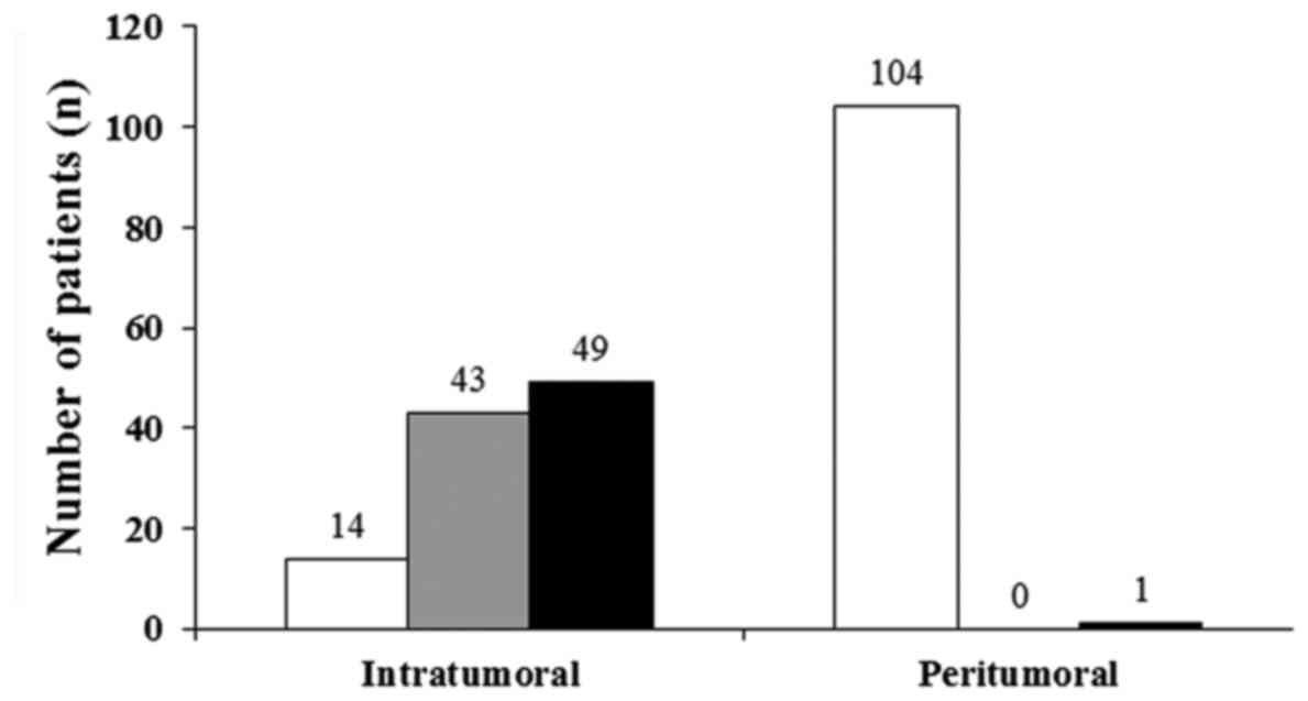
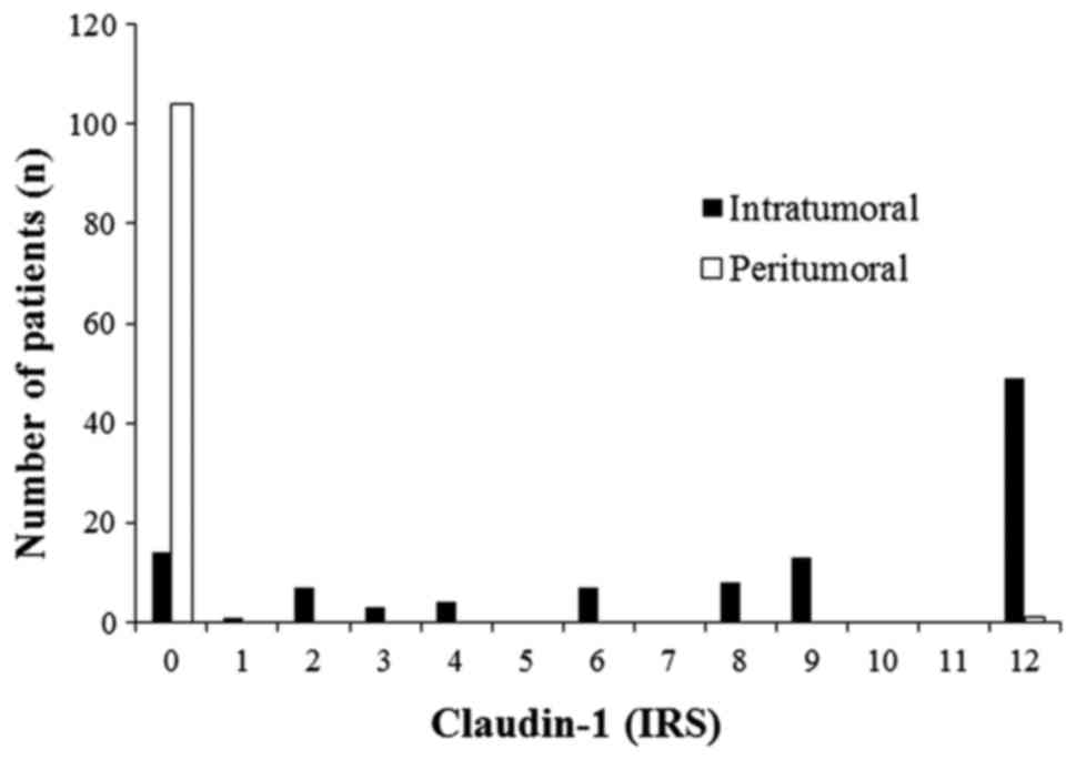
In the patient cohort, the protein expression of
claudin-1 was significantly higher in cervical cancer tissues,
compared with in the peritumoral environment (P<0.001;
Mann-Whitney U-test). In 104/106 (98%) cases, staining for
claudin-1 was not detected in cervical tissue of the peritumoral
environment (IRS=0). In cervical tumor tissues claudin-1 was
overexpressed to differing extents in 92/106 cases (87%; Figs. 1 and 2). Overall, 49 cervical cancer tissue
samples exhibited a maximum claudin-1 expression of IRS=12.
No correlation was observed between claudin-1
expression levels and tumor size (T), lymphovascular space invasion
(L), distant metastasis (M), grade (G), vessel invasion (V) or FIGO
stage (Table I). Regarding lymph
node metastasis (N), claudin-1 positive cervical cancer tissue
samples (IRS=1-12) had an increased frequency of lymph node
involvement in comparison with claudin-1 negative cervical cancer
tissues (IRS=0; 7.1 vs. 28.3%; Table
II). However, this observation was not significant (P=0.110;
two-tailed Fisher's Exact test), which may be partly due to the
small amount of claudin-1 negative cancer cases among all 106
samples (n=14).
 | Table I.Clinical and pathological parameters
and expression of claudin-1 (associations not significant). |
Table I.
Clinical and pathological parameters
and expression of claudin-1 (associations not significant).
| Patients | Total (106 patients)
n (%) | Claudin-1 negative
(IRS, 0; n=14) n (%) | Claudin-1 positive
(IRS ≥1; =92) n (%) |
|---|
| Age, mean ± SD
(range) | 52.86±12.48
(25–79) | 50±13.96 (25–74) | 53.3±12.26
(26–79) |
| Type |
|
|
|
| T0 | 2 (1.9) | 0 | 2 (2.2) |
| T1 | 64 (61) | 10 (76.9) | 54 (58.7) |
| T2 | 35 (33.3) | 3 (23.1) | 32 (34.8) |
| T3 | 2 (1.9) | 0 | 2 (2.2) |
| T4 | 2 (1.9) | 0 | 2 (2.2) |
| N | – |
|
|
| N0 | 79 (74.5) | 13 (92.9) | 66 (71.7) |
| N≤1 | 27 (25.5) | 1 (7.1) | 26 (28.3) |
| M |
|
|
|
| M0 | 102 (96.2) | 13 (92.9) | 89 (96.7) |
| M≤1 | 4 (3.8) | 1 (7.1) | 3 (3.3) |
| Hemangiosis |
|
|
|
|
Negative | 48 (98) | 4 (100) | 44 (97.8) |
|
Positive | 1 (2) | 0 | 1 (2.2) |
|
Unknown | 57 |
| – |
| Lymphangiosis |
|
|
|
|
Negative | 40 (67.8) | 4 (66.7) | 36 (67.9) |
|
Positive | 19 (32.2) | 2 (33.3) | 17 (32.1) |
|
Unknown | 47 |
|
|
| Grading |
|
|
|
| G1 | 2 (1.9) | 0 | 2 (2.2) |
| G2 | 48 (46.6) | 3 (23.1) | 45 (50) |
| G3 | 53 (51.5) | 10 (76.9) | 43 (47.8) |
| Gx | 3 |
|
|
| FIGO |
|
|
|
| FIGO
I | 64 (60.4) | 10 (71.4) | 54 (58.7) |
| FIGO
II | 36 (34) | 4 (28.6) | 32 (34.8) |
| FIGO
III | 4 (3.8) | 0 | 4 (4.3) |
| FIGO
IV | 2 (1.9) | 0 | 2 (2.2) |
| Smoking habits |
|
|
|
|
Smokers | 46 (43.4) | 6 (42.9) | 40 (43.5) |
| Table II.Claudin-1 expression in IRS and lymph
node metastases (no significant correlation). |
Table II.
Claudin-1 expression in IRS and lymph
node metastases (no significant correlation).
| Lymph node stage (n,
%) | Claudin-1 negative
(IRS=0) (%) | Claudin-1 positive
(IRS=1.12) (%) | Total (%) |
|---|
| N0 | 13 (92.9) | 66 (71.7) | 79 (74.5) |
| N1 | 1 (7.1) | 26 (28.3) | 27 (25.5) |
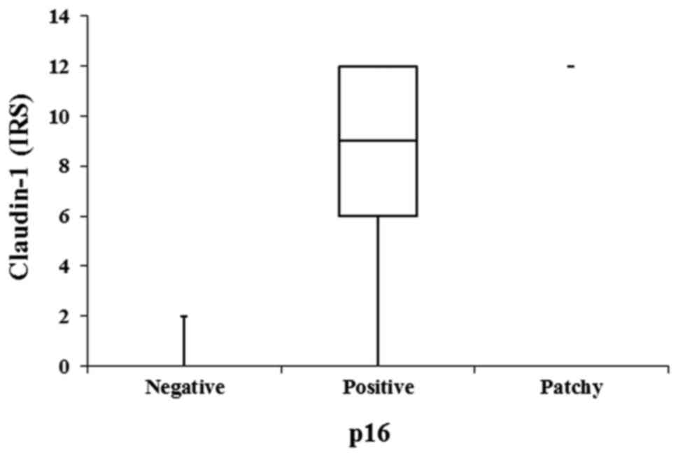
Claudin-1 expression was not significantly
associated with p53, epidermal growth factor receptor, the
proliferation-associated antigen detected with the antibody
Molecular Immunology Borstel-1 (MIB-1) or CD-3 either in cancer
tissues or in the peritumoral stroma. However, a positive
correlation was observed between claudin-1 and p16 expression in
cancer tissues (P=0.002; Kruskal-Wallis test; Fig. 3).
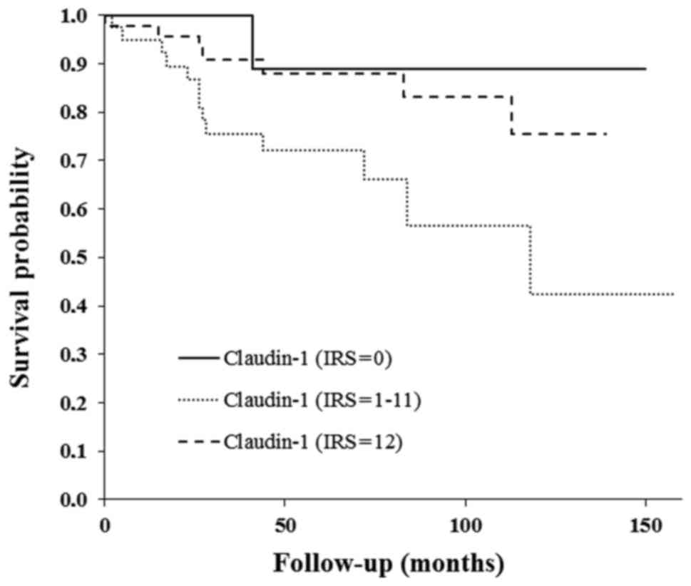
Follow-up data from ≤10 years was available for 98
patients; all were included in the survival analysis. The overall
survival curve indicated a worse prognosis for claudin-1-positive
patients, as compared with for claudin-1-negative patients
(Fig. 4). However, this difference
was not determined to be statistically significant.
Discussion
A significantly higher expression level of claudin-1
was identified in squamous cervical cancer tissues obtained from
the patient collective, as compared with in the peritumoral stroma,
as determined by immunohistochemistry. This observation is
concordant with the results of prior studies (3,11).
Dysregulation of claudin-1 has previously been demonstrated to be
involved in tumorigenesis and progression in various malignant
entities, including cervical cancer (2,5). The
pivotal role of claudin-1 in tumorigenesis has been hypothesized in
the context of the pre-cancerous cervical lesions, intraepithelial
neoplasia (3). Certain studies have
proposed claudin-1 to be a tumor marker or a marker of
pre-cancerous lesions equivalent to p16INK4a (8). However, the findings of the current
study did not determine any significant association between the
claudin-1 expression levels in the cervical cancer patient cohort
and the tumor size, FIGO stage or grade. Thus, it is hypothesized
that claudin-1 overexpression may be an early event in cervical
cancer tumorigenesis, and that each specific cervical cancer tissue
is defined by a characteristic grade of claudin-1 expression.
Notably, higher levels of claudin-1 expression were identified in
patients with lymph node metastases (not statistically
significant). Further studies with larger patient cohorts are
necessary to elucidate whether claudin-1 serves a role in lymph
node metastasis. A recent publication by Zhang et al
(11) corroborated this observation:
The authors revealed correlation between claudin-1 overexpression
in cervical cancer and lymph node metastasis (P<0.05). However,
Zhang et al (11) analyzed a
relatively small patient collective (73 patients). In contrast to
the present study, the authors used an IHC scoring system based on
the percentage of stained cells. Furthermore, the authors opted to
use cervical tissues obtained from healthy females as the controls.
In the current study, peritumoral cervical tissues were used as the
controls in order to exclude other possible confounders that may
interfere with claudin-1 expression (e.g. immunologic mechanisms
due to infection). IRS scoring according to Remmele and Stegner
(10) offers an IHC scoring system
that considers the quantity as well as the quality of cell
staining. Furthermore, a significant correlation was demonstrated
between claudin-1 expression and p16 expression in cervical cancer
tissues, data that has not yet been published. However, one study
with >350 patients reported a correlation between
p16INK4a and claudin-1 expression in pre-invasive
cervical lesions (8). Consequently
claudin-1 could serve as a marker for pre-invasive lesions and
early invasive cervical carcinoma. Further studies are required to
investigate the course of claudin-1 expression in pre-invasive
lesions and consecutive invasive lesions.
The overall survival curve indicated a worse
prognosis for claudin-1-positive patients when compared with
claudin-1-negative patients (Fig.
4). However, this observation is not significant, which could
be attributed to the small number of patients involved who had
claudin-1-negative tumors (n=14 with IRS=0). Only one of these
patients succumbed to the disease within the follow-up time. In the
current patient collective, those with moderate claudin-1
expression had the worst prognosis. These data must be interpreted
with care due to the small patient cohort, and further studies are
required to clarify whether claudin-1 expression has a prognostic
impact on cervical cancer. Zhang et al (11) also reported on the anti-apoptotic and
invasive impact of claudin-1 in SiHa cells via the loss of
E-cadherin and increased vimentin (11). Claudin-1 is a promising molecular
marker in squamous cervical cancer. Its potential as a diagnostic,
prognostic and therapeutic marker must be analyzed in further in
vitro and in vivo studies.
References
|
1
|
Cunniffe C, Ryan F, Lambkin H and Brankin
B: Expression of tight and adherens junction proteins in cervical
neoplasia. Br J Biomed Sci. 69:147–153. 2012.PubMed/NCBI
|
|
2
|
Kominsky SL: Claudins: Emerging targets
for cancer therapy. Expert Rev Mol Med. 8:1–11. 2006. View Article : Google Scholar : PubMed/NCBI
|
|
3
|
Szabó I, Kiss A, Schaff Z and Sobel G:
Claudins as diagnostic and prognostic markers in gynecological
cancer. Histol Histopathol. 24:1607–1615. 2009.PubMed/NCBI
|
|
4
|
Swisshelm K, Macek R and Kubbies M: Role
of claudins in tumorigenesis. Adv Drug Deliv Rev. 57:919–928. 2005.
View Article : Google Scholar : PubMed/NCBI
|
|
5
|
Kwon MJ: Emerging roles of claudins in
human cancer. Int J Mol Sci. 14:18148–18180. 2013. View Article : Google Scholar : PubMed/NCBI
|
|
6
|
Cunniffe C, Brankin B, Lambkin H and Ryan
F: The role of claudin-1 and claudin-7 in cervical tumorigenesis.
Anticancer Res. 34:2851–2857. 2014.PubMed/NCBI
|
|
7
|
Lee JW, Lee SJ, Seo J, Song SY, Ahn G,
Park CS, Lee JH, Kim BG and Bae DS: Increased expressions of
claudin-1 and claudin-7 during the progression of cervical
neoplasia. Gynecol Oncol. 97:53–59. 2005. View Article : Google Scholar : PubMed/NCBI
|
|
8
|
Benczik M, Galamb Á, Koiss R, Kovács A,
Járay B, Székely T, Szekerczés T, Schaff Z, Sobel G and Jeney C:
Claudin-1 as a biomarker of cervical cytology and histology. Pathol
Oncol Res. 22:179–188. 2016. View Article : Google Scholar : PubMed/NCBI
|
|
9
|
Oberländer M, Alkemade H, Bünger S, Ernst
F, Thorns C, Braunschweig T and Habermann JK: A ‘waterfall’
transfer-based workflow for improved quality of tissue microarray
construction and processing in breast cancer research. Pathol Oncol
Res. 20:719–726. 2014. View Article : Google Scholar : PubMed/NCBI
|
|
10
|
Remmele W and Stegner HE: Recommendation
for uniform definition of an immunoreactive score (IRS) for
immunohistochemical estrogen receptor detection (ER-ICA) in breast
cancer tissue. Pathologe. 8:138–140. 1987.(In German). PubMed/NCBI
|
|
11
|
Zhang WN, Li W, Wang XL, Hu Z, Zhu D, Ding
WC, Liu D, Li KZ, Ma D and Wang H: CLDN1 expression in cervical
cancer cells is related to tumor invasion and metastasis.
Oncotarget. 7:87449–87461. 2016.PubMed/NCBI
|