Introduction
Meningeal carcinomatosis (MC) is defined as the
spread of tumor cells within the leptomeninges and the subarachnoid
space (1). MC is diagnosed in 1–5%
of patients with solid tumors, 5–15% of patients with leukemia and
lymphoma, and 1–2% of patients with primary brain tumors (2). Among solid tumors, breast cancer
(12–35%), lung cancer (10–26%) and melanoma (5–25%) represent the
most common causative cancers resulting in MC (2–6). The
prognosis from initial diagnosis as MC in solid tumors is ~6–8
weeks. MC-directed treatment may contribute to improved survival of
a few months (1). We herein present
a rare case of MC from bladder cancer with lymph node and bone
metastases. The case is reviewed based on pathological examination
at autopsy.
Case report
A 66-year-old Japanese male patient visited a local
hospital with complaints of asymptomatic gross hematuria in January
2014. The patient had a medical history of diabetes mellitus and a
smoking history of >40 years (unspecified smoking amount). The
urine cytological examination detected urothelial cancer cells;
however, the patient refused further examination or treatment at
that time. In April 2015, the patient experienced pain on urination
and visited the hospital again. A computed tomography (CT) scan
revealed a thickened anterior bladder wall, which was suspicious
for bladder cancer; no metastases were identified. Following
transurethral resection of the bladder tumor, the pathological
diagnosis was high-grade muscle-invasive urothelial carcinoma and
the patient was referred to the Saitama Medical University
International Medical Center (Hidaka, Japan) to receive radical
treatment in June 2015 (SIMC-Uro #8526: a unique non-sequential
patient control number in the Department of Uro-Oncology, Saitama
Medical University International Medical Center). No obvious
abnormalities were found on physical examination during the first
visit. Laboratory examination revealed slight elevation of the
serum C-reactive protein levels (0.444 mg/dl, normal range:
0.000–0.250 mg/dl) and positive for urothelial cancer cells in
urine cytology, without any abnormal findings in particular. The
patient declined neoadjuvant chemotherapy, and radical cystectomy
with pelvic lymphadenectomy was performed. The pathological
diagnosis was high-grade urothelial carcinoma, pT2b (Fig. 1A) N2 (2 of 7 pelvic lymph nodes), and
lymphatic vessel invasion (Fig. 1B).
Starting at 2 months after radical cystectomy, two cycles of
adjuvant systemic chemotherapy with gemcitabine (1,000
mg/m2 on days 1, 8 and 15) and cisplatin (70
mg/m2 on day 2) (GC chemotherapy) were administered.
Follow-up consisted of physical examination, routine blood work,
and chest and abdominal CT imaging every 3 months. At 15 months
after radical cystectomy, despite the lack of symptoms, CT imaging
revealed multiple enlarged lymph nodes [including mediastinal
(Fig. 1E) and lung hilar nodes] and
osteosclerotic changes in multiple bones, while a bone scan
revealed multiple systemic hot spots (Fig. 1F). In addition, there was a sudden
increase in serum alkaline phosphatase (ALP; 3,943 U/l, normal
range: 104–338 U/l) and lactate dehydrogenase (LDH; 573 U/l, normal
range: 106–211 U/l) levels compared with previous data (Fig. 1I). For multiple lymph node and bone
metastases following radical cystectomy with adjuvant chemotherapy,
salvage GC chemotherapy (at the same dosage as previous adjuvant GC
chemotherapy) and denosumab (120 mg, once monthly) were
administered. CT imaging and bone scan after 3 months of GC
chemotherapy revealed that lymph node and bone metastases had
slightly shrunk (images not shown). Furthermore, the serum ALP and
LDH had decreased to normal levels (Fig.
1I); therefore, continuous systemic treatment was
scheduled.
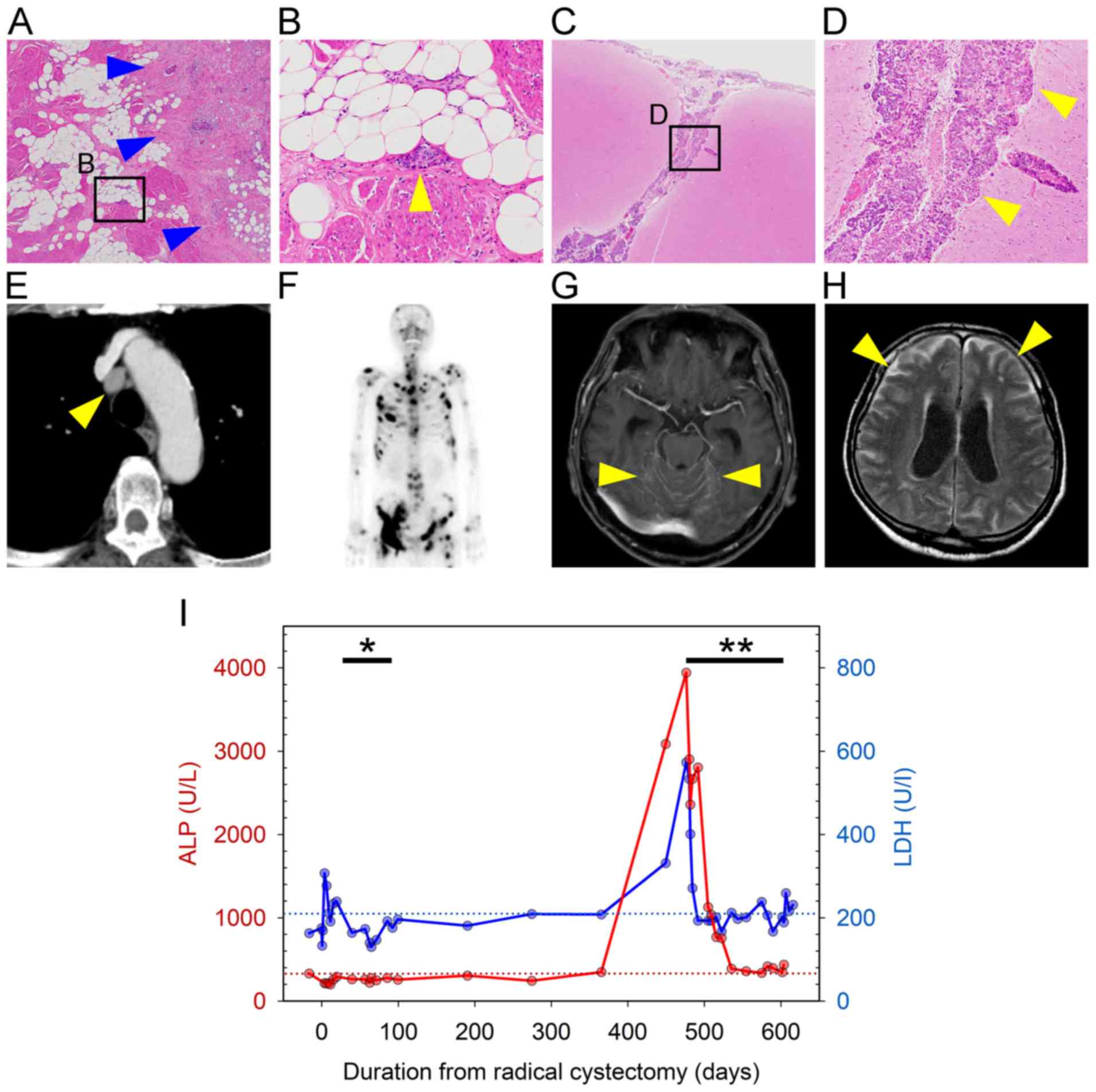 | Figure 1.Pathological and radiographic images,
and the change of laboratory data during the clinical course. (A
and B) Histopathological images of the bladder following radical
cystectomy (A, magnification, ×20; B, ×400 magnification of the
square in A). Tumor cells infiltrated the bladder muscle layer
(blue arrowheads in A), and invaded lymphatic vessels (yellow
arrowhead in B). (C and D) Pathological images of brain at autopsy
(C, magnification, ×20; D, ×400 magnification of the square in C).
Tumor cells infiltrated the subarachnoid space and cerebral sulcus
(arrowheads). (E) Chest computed tomography images with enhancement
showing lymph node metastasis in the mediastinum (arrowhead). (F)
Bone scan image at 15 months after cystectomy showing multiple bone
metastases. (G and H) Brain magnetic resonance imaging at diagnosis
of meningeal carcinomatosis (G, post-contrast T1-weighted image; H,
FLAIR image). The arrowheads indicate (G) abnormal enhancement of
the brainstem surface and cerebellar fissure and (H) diffuse
abnormal high signal intensity in the subarachnoid space;
hydrocephalus is also observed. (I) Change in laboratory data
during the clinical course of the disease. Red and blue lines show
changes in serum ALP and LDH levels, respectively. Red and blue
dotted lines show the upper limit of normal for serum ALP (338 U/l)
and LDH levels (211 U/l), respectively. *, Duration of adjuvant
chemotherapy; **, duration of salvage chemotherapy. ALP, alkaline
phosphatase; LDH, lactate dehydrogenase. |
However, 26 days after the initiation of the 4th
cycle of chemotherapy, the patient was admitted for a thorough
examination due to rapidly worsening nausea and lower limb
weakness. Spinal and brain magnetic resonance (MR) imaging with
gadolinium revealed diffuse enhancement on the surface of spinal
cord, cerebrum and brainstem. In particular, the cerebellar
fissures were markedly enhanced (Fig.
1G). On fluid-attenuated inversion recovery images, diffuse
abnormal signal intensity was observed in the subarachnoid space
(Fig. 1H). These results, even
without examination of the cerebrospinal fluid, were sufficient to
diagnose MC. Treatment, including whole-brain radiotherapy for MC,
was scheduled; however, the patient's condition rapidly
deteriorated and he succumbed to the disease 14 days after the
diagnosis of MC.
An autopsy was performed to determine the cause of
his rapid deterioration and death following the initial diagnosis
of MC. The autopsy revealed that tumor cells, which were similar to
the bladder cancer cells observed following cystectomy, diffusely
infiltrated the subarachnoid space of the cerebrum, cerebellum,
brainstem and spinal cord (Fig. 1C and
D). Mediastinal and lung hilar lymph node metastases, as well
as multiple bone metastases, were also found; however, there were
no other findings, such as infection, allergy, metabolic disorders,
or cardiac disease. Therefore, MC from bladder cancer was
definitively diagnosed as the direct cause of death.
Discussion
To the best of our knowledge, 33 cases of MC from
bladder cancer, including the present case, have been reported to
date (Table I) (7–27). In
the majority of the cases, the histology of bladder cancer was
transitional cell or urothelial carcinoma; however, MC from
small-cell neuroendocrine carcinoma or adenocarcinoma was also
reported (15,17).
 | Table I.Reported cases of meningeal
carcinomatosis from bladder cancer. |
Table I.
Reported cases of meningeal
carcinomatosis from bladder cancer.
| Case no. | Authors | Journal | Year | Reference no. | Histology of bladder
cancer | Status of primary
lesion or other metastatic sites of onset of MC | Systemic
chemotherapy | Intrathecal
therapy | RT | Survival (days) | Patient status |
|---|
| 1 | Hust and Pfitzer | Acta Cytol | 1982 | (7) | TCC | Progressive | – | – | – | 16 | Deceased |
| 2 |
|
|
|
| TCC | MC diagnosed at first
visit. Bladder cancer and other metastases diagnosed at
autopsy | – | – | – | NN | Deceased |
| 3 | Mandell et
al | Urology | 1985 | (8) | Poorly differentiated
epidermoid Ca | Stable | – | MTX | CS | NN | NN |
| 4 | Hussein et
al | J Neurooncol | 1989 | (9) | TCC | Stable | – | MTX | – | 150 | Deceased |
| 5 | Bishop et
al | Urology | 1990 | (10) | TCC | Stable | – | MTX | WB | 35 | Deceased |
| 6 |
|
|
|
| TCC | Stable | – | – | – | 20 | Deceased |
| 7 | Raghavan and
Chye | Br J Urol | 1991 | (11) | TCC | Stable | MVAC | MTX | – | 134 | Deceased |
| 8 | Eng et al | Cancer | 1993 | (12) | TCC | Stable | – | – | – | 25 | Deceased |
| 9 |
|
|
|
| TCC | Stable | – | MTX | SP | 90 | Deceased |
| 10 | Santarossa et
al | J Neurooncol | 1997 | (13) | TCC | Stable | – | MTX | WB | 270 | Deceased |
| 11 | Cozzarini et
al | Cancer Invest | 1999 | (14) | TCC | Stable | – | MTX | – | NN | Deceased |
| 12 |
|
|
|
| TCC | Stable | – | MTX | – | NN | Deceased |
| 13 | Isaka et
al | Brain Tumor
Pathol | 2002 | (15) | Small-cell
neuroendocrine Ca | Progressive | – | MTX | – | 26 | Deceased |
| 14 | Bruna et
al | J Neurooncol | 2003 | (16) | TCC | MC diagnosed at
first visit. | – | MTX | – | 66 | Deceased |
| 15 | Sugimori et
al | Neuropathology | 2005 | (17) | AdenoCa | MC diagnosed at
first visit. Bladder cancer and adrenal gland metastasis diagnosed
at autopsy | – | – | – | 6 | Deceased |
| 16 | Uncu et
al | Tumori | 2010 | (18) | TCC | Progressive | – | MTX | WB | 65 | Deceased |
| 17 | Bowen et
al | South Med J | 2010 | (19) | TCC | Stable | – | – | WB | NN | NN |
| 18 | Butchart et
al | Case Rep Oncol | 2010 | (20) | TCC | Stable | – | – | – | 30 | Deceased |
| 19 | Zada and Chen | Neurosurgery | 2010 | (21) | TCC | NN | – | Yes (drug not
reported) | – | NN | NN |
| 20 | Kastritis et
al | J BUON | 2011 | (22) | TCC | Progressive | – | MTX | – | 9 | Deceased |
| 21 | Tadepalli et
al | Case Rep Oncol | 2011 | (23) | TCC | Stable | – | MTX | WB | 270 | Alive |
| 22 | Molek et
al | Diagn
Cytopathol | 2013 | (24) | Plasmacythoid
UC | Bladder cancer,
bone metastases and MC diagnosed at first visit | – | – | – | 10 | Deceased |
| 23 | Yust-Katz et
al | Med Oncol | 2013 | (25) | NN | NN | NN | NN | NN | 56 | Deceased |
| 24 | Yust-Katz et
al | Med Oncol | 2013 | (25) | NN | NN | NN | NN | NN | 273 | Deceased |
| 25 | Yust-Katz et
al | Med Oncol | 2013 | (25) | NN | NN | NN | NN | NN | 98 | Deceased |
| 26 | Yust-Katz et
al | Med Oncol | 2013 | (25) | NN | NN | NN | NN | NN | 14 | Deceased |
| 27 | Yust-Katz et
al | Med Oncol | 2013 | (25) | NN | NN | NN | NN | NN | 42 | Deceased |
| 28 | Yust-Katz et
al | Med Oncol | 2013 | (25) | NN | NN | NN | NN | NN | 21 | Deceased |
| 29 | Yust-Katz et
al | Med Oncol | 2013 | (25) | NN | NN | NN | NN | NN | 154 | Deceased |
| 30 | Yust-Katz et
al | Med Oncol | 2013 | (25) | NN | NN | NN | NN | NN | 28 | Deceased |
| 31 | Swallow et
al | Can Urol Assoc
J | 2015 | (26) | UC | Stable | – | – | – | 8 | Deceased |
| 32 | Teyssonneau et
al | BMC Cancer | 2017 | (27) | UC | Progressive | GC | – | – | 1500 | Alive |
| 33 | Present case |
| 2018 |
| UC | Stable | – | – | – | 14 | Deceased |
MC is diagnosed in ~10% of patients with metastatic
cancer during the clinical course. Among those cases, it occurs in
<10% at the time of diagnosis, and in ~20% at the time of first
progression after initial treatment (1). Among all cases of MC from bladder
cancer, including the present case, 24 cases were described in
detail at the onset of MC (Table I).
Even in non-muscle-invasive bladder cancer, a case of MC with
multiple bone metastases following transurethral resection with
intravesical Bacillus Calmette-Guerin instillation was reported
(case no. 32, Table I) (27). MC had already developed at initial
diagnosis of bladder cancer in 4 cases (16.7%). Among the 20 cases
with MC developing during treatment for bladder cancer, 4 (16.7%)
had synchronously progressive primary or other metastatic sites,
while 16 (66.6%) did not.
There is no established treatment for MC from solid
tumors, including breast cancer, lung cancer and melanoma, as it is
such a rare occurrence. Generally, systemic chemotherapy based on
the primary tumor, intrathecal therapy and radiotherapy are
implemented. The aim in the majority of the cases is to improve or
maintain the patient's quality of life, and to prevent or delay
neurological deterioration, as notable prolongation of survival is
rare (1). Among 24 cases that
received treatment for MC from bladder cancer (Table I), systemic chemotherapy, intrathecal
therapy and radiotherapy were administered to 2 (8.3%), 14 (58.3%)
(single-agent methotrexate in 13 and unknown in 1) and 7 (29.2%)
(whole-brain radiotherapy in 5, craniospinal radiotherapy in 1 and
spinal radiotherapy in 1) cases, respectively. The remaining 9
cases (37.5%) did not receive any treatment for MC due to rapid
disease progression. The prognosis of MC from bladder cancer is
very poor [median survival time from the diagnosis of MC, 35 days
(interquartile range: 16–134 days)], although there was one case
with a survival duration of ~50 months (case no. 32, Table I). Other cases with relatively long
survival were also reported: Case no. 7, treated by systemic
chemotherapy [methotrexate, vinblastine, doxorubicin and cisplatin
(MVAC)] and intrathecal methotrexate (survival time, 134 days)
(11), and case no. 21, treated with
intrathecal methotrexate and whole-brain radiotherapy (survival
time: 270 days) (23).
Methotrexate is one of the key drugs for the
treatment of MC, along with cytarabine or
thiotriethylenephosphoramide, and it is administered as systemic
and/or intrathecal chemotherapy (1).
In systemic chemotherapy for bladder cancer, methotrexate is widely
used in the MVAC regimen as the first-line therapy, as well as in
the GC regimen (28). In the present
case, although the GC regimen was selected as salvage chemotherapy,
the oncological outcome may have been different by reducing a risk
of the onset of MC if the MVAC regimen, including methotrexate
(which penetrates cerebrospinal fluid relatively easily), had been
deployed.
In MC from other cancers, the effectiveness of
molecular targeted therapy or immune checkpoint inhibitors, such as
bevacizumab combined with etoposide and cisplatin for breast cancer
(29), intrathecal trastuzumab for
human EGFR-related 2 (HER2) positive breast cancer (30) and nivolumab for lung adenocarcinoma
(31), was reported. The efficacy of
an immune checkpoint inhibitor in bladder cancer has also been
reported (32). Although it is
unclear whether these drugs are effective for MC from bladder
cancer, a promising strategy for MC may be expected to be designed
in the future.
MC from solid tumors, including bladder cancer, may
become more common, as cancer patients survive longer due to the
more effective treatments (25). In
the present case, the MR imaging findings matched the pathological
findings on autopsy. Cerebrospinal imaging is not a practice
routinely recommended for bladder cancer; however, it is necessary
to perform this as soon as possible when the patient has symptoms
that suggest meningitis, even if the primary lesion or other
metastatic sites remain stable during systemic treatment.
Acknowledgements
The authors would like to thank the pathologist Dr
Mika Sakaki (Department of Diagnostic Pathology, Saitama Medical
University International Medical Center) for her valuable support
in the autopsy.
Funding
No funding was received.
Availability of data and materials
Not applicable.
Ethics approval and consent to
participate
Not applicable.
Patient consent for publication
Opt-out consent was applied in this case report.
Authors' contributions
YU, SS and GK planned the case study, and collected
the data and the images of the case in addition to producing the
draft of the manuscript. KN and MO for urology, YO and AU for
diagnostic radiology, and MY for diagnostic pathology critically
revised this manuscript for important intellectual content. All
authors read and approved the final manuscript to be published.
Competing interests
All authors declare that they have no competing
interests to disclose.
References
|
1
|
Le Rhun E, Weller M, Brandsma D, Van den
Bent M, de Azambuja E, Henriksson R, Boulanger T, Peters S, Watts
C, Wick W, et al EANO Executive Board and ESMO Guidelines
Committee, : EANO-ESMO Clinical Practice Guidelines for diagnosis,
treatment and follow-up of patients with leptomeningeal metastasis
from solid tumours. Ann Oncol. 28 (Suppl 4):iv84–iv99. 2017.
View Article : Google Scholar : PubMed/NCBI
|
|
2
|
Chamberlain MC: Leptomeningeal metastasis.
Curr Opin Oncol. 22:627–635. 2010. View Article : Google Scholar : PubMed/NCBI
|
|
3
|
Roth P and Weller M: Management of
neoplastic meningitis. Chin Clin Oncol. 4:262015.PubMed/NCBI
|
|
4
|
Le Rhun E, Taillibert S and Chamberlain
MC: Carcinomatous meningitis: Leptomeningeal metastases in solid
tumors. Surg Neurol Int. 4 (Suppl 4):S265–S288. 2013. View Article : Google Scholar : PubMed/NCBI
|
|
5
|
Gleissner B and Chamberlain MC: Neoplastic
meningitis. Lancet Neurol. 5:443–452. 2006. View Article : Google Scholar : PubMed/NCBI
|
|
6
|
van der Ree TC, Dippel DW, Avezaat CJ,
Sillevis Smitt PA, Vecht CJ and van den Bent MJ: Leptomeningeal
metastasis after surgical resection of brain metastases. J Neurol
Neurosurg Psychiatry. 66:225–227. 1999. View Article : Google Scholar : PubMed/NCBI
|
|
7
|
Hust MH and Pfitzer P: Cerebrospinal fluid
and metastasis of transitional cell carcinoma of the bladder. Acta
Cytol. 26:217–223. 1982.PubMed/NCBI
|
|
8
|
Mandell S, Wernz J, Morales P, Weinberg H
and Steinfeld A: Carcinomatous meningitis from transitional cell
carcinoma of bladder. Urology. 25:520–521. 1985. View Article : Google Scholar : PubMed/NCBI
|
|
9
|
Hussein AM, Savaraj N, Feun LG, Ganjei P
and Donnelly E: Carcinomatous meningitis from transitional cell
carcinoma of the bladder: Case report. J Neurooncol. 7:255–260.
1989. View Article : Google Scholar : PubMed/NCBI
|
|
10
|
Bishop JR Jr, Moul JW, Maldonado L and
McLeod DG: Transitional cell carcinomatous meningitis after M-VAC
(methotrexate, vinblastine, doxorubicin, and cisplatin)
chemotherapy. Urology. 36:373–377. 1990. View Article : Google Scholar : PubMed/NCBI
|
|
11
|
Raghavan D and Chye RW: Treatment of
carcinomatous meningitis from transitional cell carcinoma of the
bladder. Br J Urol. 67:438–440. 1991. View Article : Google Scholar : PubMed/NCBI
|
|
12
|
Eng C, Cunningham D, Quade BJ, Schwamm L,
Kantoff PW and Skarin AT: Meningeal carcinomatosis from
transitional cell carcinoma of the bladder. Cancer. 72:553–557.
1993. View Article : Google Scholar : PubMed/NCBI
|
|
13
|
Santarossa S, Vaccher E, Balestreri L,
Volpe R and Tirelli U: Solitary meningeal recurrence in a patient
with transitional cell carcinoma of the bladder with locally bulky
disease at presentation. J Neurooncol. 35:141–143. 1997. View Article : Google Scholar : PubMed/NCBI
|
|
14
|
Cozzarini C, Reni M, Mangili F, Baldoli
MC, Galli L and Bolognesi A: Meningeal carcinomatosis from
transitional cell carcinoma of the bladder: Report of two cases and
review of the literature. Cancer Invest. 17:402–407. 1999.
View Article : Google Scholar : PubMed/NCBI
|
|
15
|
Isaka T, Maruno M, Sato M, Kinoshita M,
Nishida T, Kiyohara H and Yoshimine T: Brain metastasis from
small-cell neuroendocrine carcinoma of the urinary bladder: A case
report. Brain Tumor Pathol. 19:117–122. 2002. View Article : Google Scholar : PubMed/NCBI
|
|
16
|
Bruna J, Rojas-Marcos I, Martínez-Yelamos
S, Català I, Vidaller A, Galán C, Krupinski J and Rubio F:
Meningeal carcinomatosis as the first manifestation of a
transitional cell carcinoma of the bladder. J Neurooncol. 63:63–67.
2003. View Article : Google Scholar : PubMed/NCBI
|
|
17
|
Sugimori K, Kobayashi K, Hayashi M, Sakai
N, Sasaki M and Koshino Y: Leptomeningeal carcinomatosis from
urinary bladder adenocarcinoma: A clinicopathological case study.
Neuropathology. 25:89–94. 2005. View Article : Google Scholar : PubMed/NCBI
|
|
18
|
Uncu D, Arpaci F, Beyzadeoglu M, Gunal A,
Surenkok S, Ozturk M and Ozet A: Meningeal carcinomatosis: An
extremely rare involvement of urinary bladder carcinoma. Tumori.
96:352–354. 2010. View Article : Google Scholar : PubMed/NCBI
|
|
19
|
Bowen CD, Von Burton G, Bargen RC, Madonia
P, Zhang S, Toledo EG, Zweig R and Pant C: Carcinomatous meningitis
secondary to transitional cell bladder cancer. South Med J.
103:809–812. 2010. View Article : Google Scholar : PubMed/NCBI
|
|
20
|
Butchart C, Dahle-Smith A, Bissett D,
Mackenzie JM and Williams DJ: Isolated Meningeal Recurrence of
Transitional Cell Carcinoma of the Bladder. Case Rep Oncol.
3:171–175. 2010. View Article : Google Scholar : PubMed/NCBI
|
|
21
|
Zada G and Chen TC: A novel method for
administering intrathecal chemotherapy in patients with
leptomeningeal metastases and shunted hydrocephalus: case report.
Neurosurgery. 67:onsE306–307. 2010.PubMed/NCBI
|
|
22
|
Kastritis E, Zagouri F, Dimopoulos MA and
Papadimitriou CA: Carcinomatous meningitis from transitional cell
carcinoma of the urinary bladder. J BUON. 16:373–374.
2011.PubMed/NCBI
|
|
23
|
Tadepalli S, Coleman T, Hacket LA and
Liles GB: Carcinomatous meningitis: The natural history of
successfully treated metastatic bladder cancer. Case Rep Oncol.
4:406–412. 2011. View Article : Google Scholar : PubMed/NCBI
|
|
24
|
Molek KR, Seili-Bekafigo I, Štemberger C,
Jonjić N, Đordević G and Duletić-Načinović A: Plasmacytoid
urothelial carcinoma - diagnostic challenge in cytology. Diagn
Cytopathol. 41:369–373. 2013. View
Article : Google Scholar : PubMed/NCBI
|
|
25
|
Yust-Katz S, Mathis S and Groves MD:
Leptomeningeal metastases from genitourinary cancer: The University
of Texas MD Anderson Cancer Center experience. Med Oncol.
30:4292013. View Article : Google Scholar : PubMed/NCBI
|
|
26
|
Swallow TW, Mabbutt S and Bell CR: Muscle
invasive bladder cancer culminating with leptomeningeal
carcinomatosis. Can Urol Assoc J. 9:E903–E904. 2015. View Article : Google Scholar : PubMed/NCBI
|
|
27
|
Teyssonneau D, Daste A, Dousset V,
Hoepffner JL, Ravaud A and Gross-Goupil M: Metastatic non-muscle
invasive bladder cancer with meningeal carcinomatosis: Case report
of an unexpected response. BMC Cancer. 17:3232017. View Article : Google Scholar : PubMed/NCBI
|
|
28
|
Alfred Witjes J, Lebret T, Compérat EM,
Cowan NC, De Santis M, Bruins HM, Hernández V, Espinós EL, Dunn J,
Rouanne M, et al: Updated 2016 EAU Guidelines on Muscle-invasive
and Metastatic Bladder Cancer. Eur Urol. 71:462–475. 2017.
View Article : Google Scholar : PubMed/NCBI
|
|
29
|
Wu PF, Lin CH, Kuo CH, Chen WW, Yeh DC,
Liao HW, Huang SM, Cheng AL and Lu YS: A pilot study of bevacizumab
combined with etoposide and cisplatin in breast cancer patients
with leptomeningeal carcinomatosis. BMC Cancer. 15:2992015.
View Article : Google Scholar : PubMed/NCBI
|
|
30
|
Zagouri F, Sergentanis TN, Bartsch R,
Berghoff AS, Chrysikos D, de Azambuja E, Dimopoulos MA and Preusser
M: Intrathecal administration of trastuzumab for the treatment of
meningeal carcinomatosis in HER2-positive metastatic breast cancer:
A systematic review and pooled analysis. Breast Cancer Res Treat.
139:13–22. 2013. View Article : Google Scholar : PubMed/NCBI
|
|
31
|
Gion M, Remon J, Caramella C, Soria JC and
Besse B: Symptomatic leptomeningeal metastasis improvement with
nivolumab in advanced non-small cell lung cancer patient. Lung
Cancer. 108:72–74. 2017. View Article : Google Scholar : PubMed/NCBI
|
|
32
|
Massari F, Di Nunno V, Cubelli M, Santoni
M, Fiorentino M, Montironi R, Cheng L, Lopez-Beltran A, Battelli N
and Ardizzoni A: Immune checkpoint inhibitors for metastatic
bladder cancer. Cancer Treat Rev. 64:11–20. 2018. View Article : Google Scholar : PubMed/NCBI
|


