Introduction
Adrenal myelolipoma is a rare mesenchymal tumour
with benign biological behaviour that is mainly composed of mature
adipose and myeloid tissue (1). Its
pathogenesis is largely unknown, but infection, inflammation,
necrosis, stressful lifestyle and an unbalanced diet are known risk
factors; adrenal myelolipoma is also often associated with obesity,
high levels of blood lipids, hypertension, diabetes and Cushing's
disease (2). The two sexes are
equally affected, mainly between the fifth and seventh decades of
life (3). The most common site is
the right adrenal gland (4), but
other sites, such as the presacral area, spleen, stomach, lung,
liver, retroperitoneum and testis, are also reported (5).
Adrenal myelolipoma is generally non-secretory and
its diagnosis is most commonly incidental: It currently represents
10–15% of incidental adrenal masses discovered on imaging
investigations, such as ultrasonography, computed tomography (CT)
and magnetic resonance imaging (MRI). Myelolipoma is usually
asymptomatic, but it may occasionally be associated with necrosis,
rupture and haemorrhage, causing abdominal pain (6). Due to the absence of symptoms and its
small size (usually <4 cm), the management of myelolipoma is
usually conservative. Surgical treatment is recommended when the
mass is symptomatic, or grows quickly or to a size of >6 cm
(6). Adrenalectomy must be performed
in case of malignant or potentially malignant tumors (1). Myelolipoma is defined as ‘giant’ when
its greatest diameter is >10 cm (2), which is a rare clinical occurrence.
Open radical surgery is the standard treatment of choice for giant
myelolipomas, while the minimally invasive approach has been used
in only a few cases (7). We herein
present the case of a patient with an 18-cm adrenal myelolipoma who
was treated by robotic partial adrenalectomy. To the best of our
knowledge, this is the largest adrenal myelolipoma treated with
robotic surgery reported in the literature to date.
Case report
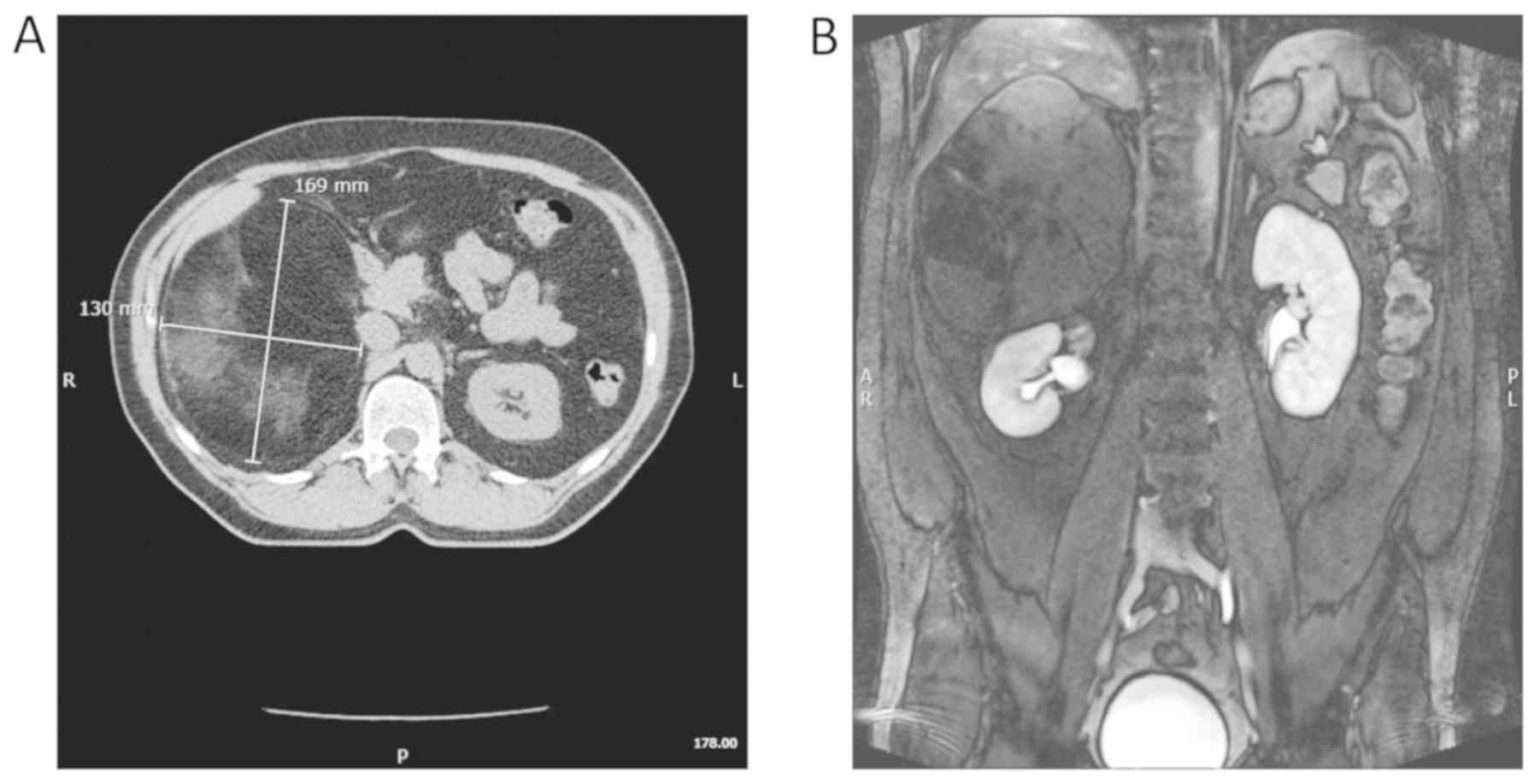
A 55-year-old male patient underwent an abdominal CT
scan during follow-up after radical prostatectomy. The examination
revealed a right adrenal mass, sized 16×13 cm, homogeneous,
hypodense (<-30 HU), imprinting on the right liver lobe and
dislocating the right kidney downwards, the vena cava, duodenum and
pancreas medially, and the ascending colon and gallbladder
anteriorly. Following administration of contrast medium, the
adrenal lesion exhibited poor enhancement with a rapid washout. The
abdominal MRI confirmed the presence of a right adrenal lesion,
sized 16×13 cm. Ηyperintense on T1-weighted, isointense on
T2-weighted and hypointense on fat-suppression images, without
significant contrast enhancement, characteristics suggestive of a
myelolipoma (Fig. 1). The patient
did not report any symptoms, but exhibited a deformation of the
right abdominal contour. Staging workup excluded other adrenal
diseases.
The patient did not have other major comorbidities,
and had a Charlson Comorbidity index score of 3 and an Eastern
Cooperative Oncology Group performance status score of 0.
Due to the benign characteristics of the mass, it
was decided to perform transperitoneal robotic partial
adrenalectomy in order to preserve adrenal healthy tissue and
maintain functional adrenal integrity in a young patient undergoing
surgery for a benign lesion.
Written informed consent was obtained from the
patient prior to surgery.
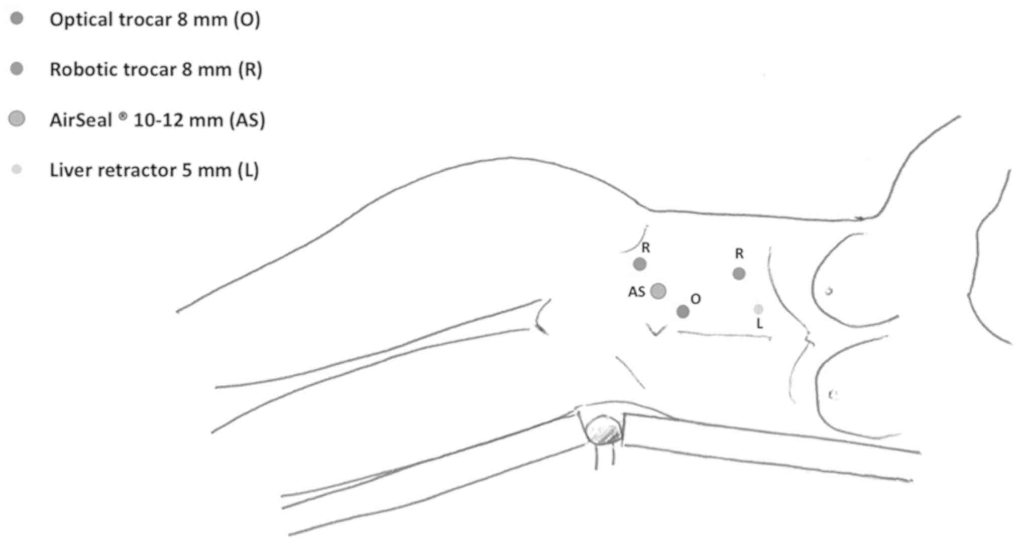
The operation was performed by a robotic-skilled
urologist, using the DaVinci® Model Xi Surgical System
(Intuitive Surgical, Inc., Sunnyvale, CA, USA) in the Tertiary Care
Hospital Santa Maria di Terni (Terni, Italy). The patient was
placed in a left flank position with a 45° side tilt. The optical
trocar was inserted on the intersection between the transumbilical
and periumbilical lines. Two more robotic trocars and two trocars
for the assistant were inserted (Fig.
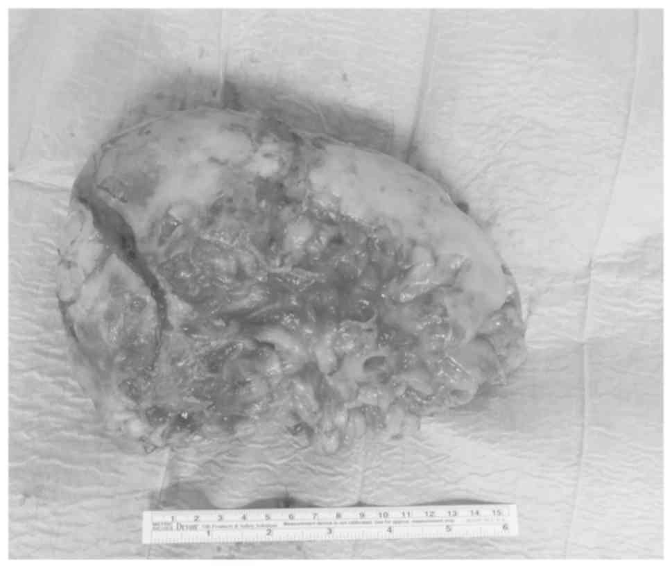
2). The triangular hepatic ligament was dissected to mobilize
the liver, followed by enucleation of the mass (Fig. 3). The operative time (OT) and
estimated blood loss (EBL) were 205 min and 100 ml, respectively.
No intra- or postoperative complications occurred. On the first
postoperative day, the nasogastric tube was removed and the patient
was mobilized; the time to flatus was 36 h, oral feeding was
initiated 3 days after surgery and the length of hospitalization
was 4 days. The histological examination confirmed the diagnosis of
adrenal myelolipoma, sized 18×11.5×6 cm. Follow-up examinations
included an abdominal ultrasound and CT scan at 6 and 12 months
after surgery, respectively. At 12 months of follow-up no
recurrence had occurred.
A systematic bibliography search up to January 2018
was conducted through PubMed, Web of Science and Scopus. One of the
authors (AP) independently performed online bibliographic research
in order to identify titles and abstracts of interest. All titles
and abstracts were assessed to select those focusing on minimally
invasive adrenalectomy for sizeable masses. Subsequently, the full
text of the selected articles was independently screened by two
authors (ES and JARV) for eligibility (Table I).
 | Table I.Cases of mini-invasive surgery for
adrenal masses. |
Table I.
Cases of mini-invasive surgery for
adrenal masses.
| Study ID | Year | Technique | No. of patients | Tumour size (cm) | Refs. |
|---|
| Molnar, et
al | 2017 | R | 1 | 4.6×4.1 | (5) |
| Economopoulos, et
al | 2017 | L/R | 415/353 | 2.8–6.2/2.57–6.5
(mean) | (21) |
| Campos Arbulù, et
al | 2017 | L | 1 | 14 | (17) |
| Burttet, et
al | 2017 | R | 1 | 8.3 | (22) |
| Morelli, et
al | 2016 | L/R | 41/41 | 4.7/4.9 (mean) | (16) |
| Teo and Lim | 2016 | L/R | 263/569 | 1-15/1-13.9 | (7) |
| Deniwar, et
al | 2015 | R | 1 | 3.9 | (23) |
| Akarsu, et
al | 2014 | R | 8 | 2-9 | (24) |
| Brandao, et
al | 2013 | L/R | 323/277 |
3.78±1.06–3.86±1.32 | (25) |
| Yates, et
al | 2010 | R | 1 | 1.5 | (26) |
Discussion
Adrenal myelolipoma is a rare benign tumour with a
prevalence of 0.08–0.2% (3) that
consists of mature adipose and hematopoietic tissue (8). Myelolipoma was first described by
Gierke in 1905, and was named ‘formations myelolipomatoses’ by
Oberling in 1929 (9). In the
majority of the cases, it is incidentally diagnosed during clinical
workup for other reasons. Myelolipoma mainly presents with
abdominal discomfort and pain due to necrosis, rupture,
haemorrhage, or even haemorrhagic shock (6). The mean size at the time of diagnosis
is ~4 cm, while myelolipomas >10 cm in greatest diameter are
defined as ‘giant’ (2). The
management of smaller, asymptomatic, slow-growing lesions is
conservative, and patients are monitored by abdominal CT or MRI
annually or biannually (2). Surgical
treatment is recommended for asymptomatic tumours >6 cm in
diameter, for symptomatic lesions and for those exhibiting fast
growth (6). In the AACE/AAES Adrenal
Incidentaloma Guidelines, resection of myelolipoma may be
considered under certain conditions, such as the presence of
clinical symptoms or a diameter of >4 cm (10). Based on the preoperative imaging
examinations, partial adrenalectomy was considered to be the best
choice in the present case.
An open or a minimally invasive approach is selected
according to the size of the lesion and the skills of the surgeon.
The open approach includes lumbar, subcostal, posterior or
transabdominal laparotomy, and is usually reserved for giant masses
or as an emergency intervention in cases with rupture and
haemorrhage.
The advantages of the laparoscopic compared with the
traditional approach are less pain, faster recovery and shorter
hospitalization, and it is usually reserved for smaller
myelolipomas (11,12). Robotic surgery is being increasingly
used in different common surgical procedures, although its
applicability in adrenal surgery remains debated upon (13,14). A
magnified 3D view of the operative field, the absence of tremor,
wristed instruments and improved ergonomics are considered
invaluable advantages over the laparoscopic approach.
Giant myelolipomas are associated with more
intraoperative complications, such as bleeding, capsular breach and
a higher risk of local recurrence (9,15).
Morelli et al reported better results with the robotic vs.
the laparoscopic approach in patients with large adrenal tumours
(>6 cm in diameter). The OT for lesions sized >6 cm was
shorter in the robotic group (171.3 vs. 260.0 min, P=0.002). There
were no significant differences between the two groups regarding
the rate of conversion, intra- and postoperative complications
(P=0.49, 0.95 and 0.96, respectively) (16).
However, only few cases of minimally invasive
adrenalectomy for giant adrenal myelolipomas have been reported to
date (Table I). Campos Arbulú et
al described a laparoscopic adrenalectomy for a 14-cm
myelolipoma; no complications occurred and the length of
hospitalization was 48 h (17).
Undre et al performed robotic adrenalectomy for a 13.9-cm
adrenal myelolipoma. The OT was 155 min and the EBL was 300 ml. No
complications occurred and the length of hospitalization was 4 days
(18). Brunaud et al
performed 100 robotic adrenalectomies; tumour capsule rupture
occurred in only 1 patient (1%) with a 7.5-cm lesion (19). There was 1 case of conversion to
laparoscopy due to camera malfunction, in 4 cases open conversion
was necessary due to bleeding and difficulty to identify the
adrenal vein in 1 patient with a 7-cm mass. Of note, all open
conversions occurred in patients with high BMI (≥29
kg/m2, 75%) and large tumours (≥6 cm, 50%) (15). Agcaoglu et al performed 24
robotic and 38 laparoscopic adrenalectomies. Tumour size was
similar in both groups (6.5±0.4, range 5-10.2 cm for the robotic
group; 6.2±0.3, range 5–15 cm for the laparoscopic group) as were
age and sex. The robotic group had lower BMI (27.1±0.8 vs. 30.2±0.9
kg/m2). The robotic approach was associated with a
shorter OT (159 vs. 187 min; P=0.043) and lower EBL (87 vs. 167 ml;
P=0.147). The conversion rate was markedly higher in the
laparoscopic group (11 vs. 4%) and the causes of conversion in the
laparoscopic group included bleeding from the renal vein or the
adrenal gland, adhesion to the vena cava and difficulty with the
dissection plane; in the robotic group, the main cause of
conversion was the adherence of the tumour to the renal hilum
(20). To the best of our knowledge,
the case presented herein is the largest giant myelolipoma treated
by robotic approach that has been reported in the literature to
date.
A robotic partial adrenalectomy was performed, with
enucleation of the mass. No intra- or postoperative complications
occurred. The EBL was 100 ml, which was comparable with previous
reported cases (7,21,25). The
choice of partial adrenalectomy for a benign tumour was based on
finding an avascular plane between the tumour capsule and the
healthy surrounding adrenal tissue, in order to significantly
reduce intraoperative bleeding and preserve healthy adrenal tissue.
The robotic approach allowed a safer preservation of the adrenal
vessels due to the magnified 3D vision and the wristed instruments
that permitted more accurate dissection. Furthermore, no capsular
effraction was observed. Several studies demonstrated the
feasibility and safety of robotic partial adrenalectomy, with
satisfactory oncological as well as functional results (9). In the present case, the functional
outcome was optimal, as the patient did not require steroid
supplementation.
In conclusion, the standard treatment for giant
myelolipoma is laparotomic adrenalectomy. However, while the
minimally invasive techniques were initially used for smaller
lesions, the improved laparoscopic and robotic instrumentations and
the increasing experience of the surgeons have led to the wider
application of the minimally invasive approach in more complex
cases.
We consider the transperitoneal robotic approach to
be a safe strategy for giant adrenal masses due to the better
visualization of the operative field and more accurate dissection,
decreasing the risk of capsular effraction. Moreover, the
enucleation of the mass decreases the risk of bleeding, which is
the most common cause of conversion, and enables preservation of
adrenal functional healthy tissue.
Acknowledgements
Not applicable.
Funding
No funding was received.
Availability of data and materials
All the necessary information on the present case
report is provided in this manuscript.
Authors' contributions
AP independently performed online bibliographic
research in order to identify titles and abstracts of interest. GC
and EM were responsible for the conception and design. AB, GC, ES,
AT and JDV participated in either drafting or critically revising
the article for important intellectual content. All authors have
approved the final version of the manuscript to be published and
agree to be accountable for all aspects of the work in ensuring
that questions related to the accuracy or integrity of any part of
the work are appropriately investigated and resolved.
Ethics approval and consent to
participate
The study has been approved by an Internal Board
Committee and written informed consent was obtained from the
patient prior to surgery.
Patient consent for publication
The patient provided written consent regarding the
publication of the case details and associated images.
Competing interests
The authors declare that they have no competing
interests to disclose.
Glossary
Abbreviations
Abbreviations:
|
CT
|
computed tomography
|
|
MRI
|
magnetic resonance imaging
|
|
OT
|
operative time
|
|
EBL
|
estimated blood loss
|
References
|
1
|
Lam AK: Update on adrenal tumours in 2017
World Health Organization (WHO) of endocrine tumours. Endocr
Pathol. 28:213–227. 2017. View Article : Google Scholar : PubMed/NCBI
|
|
2
|
Bokhari MR and Bhimji S: Adrenal,
myelolipoma. StatPearls NCBI Bookshelf. 2017.
|
|
3
|
Al-Bahri S, Tariq A, Lowentritt B and
Nasrallah DV; Hindawi Publishing Corporation, : Giant bilateral
adrenal myelolipoma with congenital adrenal hyperplasia. Case Rep
Surg. 2014:7281982014.PubMed/NCBI
|
|
4
|
Zhao J, Sun F, Jing X, Zhou W, Huang X,
Wang H, Zhu Y, Yuan F and Shen Z: The diagnosis and treatment of
primary adrenal lipomatous tumours in Chinese patients: A 31-year
follow-up study. Can Urol Assoc J. 8:E132–E136. 2014. View Article : Google Scholar : PubMed/NCBI
|
|
5
|
Molnar C, Lata L, Pisica R, Russu C,
Gherghinescu M, Molnar C, Borda A, Butiurca VO, Suciu BA and
Copotoiu C: Anterior transabdominal laparoscopic adrenalectomy,
without ligatures, for a symptomatic right adrenal myelolipoma with
intratumorally haemorrhage. Chirurgia (Bucur). 112:58–62. 2017.
View Article : Google Scholar : PubMed/NCBI
|
|
6
|
Ramirez M and Misra S: Adrenal
myelolipoma: To operate or not? A case report and review of the
literature. Int J Surg Case Rep. 5:494–496. 2014. View Article : Google Scholar : PubMed/NCBI
|
|
7
|
Teo XL and Lim SK: Robotic assisted
adrenalectomy: is it ready for prime time? Investig Clin Urol. 57
(Suppl 2):S130–S146. 2016. View Article : Google Scholar : PubMed/NCBI
|
|
8
|
Federle M and Anne V: Adrenal myelolipoma.
Diagnostic Imaging: Abdomen. 1st. Amirsys Inc.; Salt lake: pp.
III2-24-252004
|
|
9
|
Oberling C: Les formations
myelolipomateuses. Bull Assoc Fr Etud Cancer. 18:234–246. 1929.
|
|
10
|
Zeiger MA, Thompson GB, Duh QY, Hamrahian
AH, Angelos P, Elaraj D, Fishman E and Kharlip J: MDAACE/AAES
Adrenal Incidentaloma Guidelines. Endocr Pract. 15 (Suppl
1):2009.
|
|
11
|
Nomine-Criqui C, Germain A, Ayav A,
Bresler L and Brunaud L: Robot-assisted adrenalectomy: Indications
and drawbacks. Updates Surg. 69:127–133. 2017. View Article : Google Scholar : PubMed/NCBI
|
|
12
|
Cochetti G, Puxeddu E, Zingaro MD, D'Amico
F, Cottini E, Barillaro F and Mearini E: Laparoscopic partial
nephrectomy of thyroid cancer metastasis: Case report and review of
the literature. OncoTargets Ther. 6:355–360. 2013. View Article : Google Scholar
|
|
13
|
Arezzo A, Bullano A, Cochetti G, Cirocchi
R, Randolph J, Mearini E, Evangelista A, Ciccone G, Bonjer HJ and
Morino M: Transperitoneal versus retroperitoneal laparoscopic
adrenalectomy for adrenal tumours in adults. Cochrane Database Syst
Rev. Dec 30–2018.(Epub ahead of print). View Article : Google Scholar
|
|
14
|
Boni A, Cochetti G, Ascani S, Del Zingaro
M, Quadrini F, Paladini A, Cocca D and Mearini E: Robotic treatment
of oligometastatic kidney tumor with synchronous pancreatic
metastasis: Case report and review of the literature. BMC Surg.
18:402018. View Article : Google Scholar : PubMed/NCBI
|
|
15
|
Gumbs AA and Gagner M: Laparoscopic
adrenalectomy. Best Pract Res Clin Endocrinol Metab. 20:483–499.
2006. View Article : Google Scholar : PubMed/NCBI
|
|
16
|
Morelli L, Tartaglia D, Bronzoni J,
Palmeri M, Guadagni S, Di Franco G, Gennai A, Bianchini M, Bastiani
L, Moglia A, et al: Robotic assisted versus pure laparoscopic
surgery of the adrenal glands: A case-control study comparing
surgical techniques. Langenbecks Arch Surg. 401:999–1006. 2016.
View Article : Google Scholar : PubMed/NCBI
|
|
17
|
Campos Arbulú AL, Sadava EE, Kerman J,
Fernández Vila JM and Mezzadri NA: Medicina (Buenos Aires).
76:249–250. 2016.
|
|
18
|
Undre S, Munz Y, Moorthy K, Martin S,
Rockall T, Vale J and Darzi A: Robot-assisted laparoscopic
adrenalectomy: Preliminary UK results. BJU Int. 93:357–359. 2004.
View Article : Google Scholar : PubMed/NCBI
|
|
19
|
Brunaud L, Ayav A, Zarnegar R, Rouers A,
Klein M, Boissel P and Bresler L: Prospective evaluation of 100
robotic-assisted unilateral adrenalectomies. Surgery. 144:995–1001.
2008. View Article : Google Scholar : PubMed/NCBI
|
|
20
|
Agcaoglu O, Aliyev S, Karabulut K,
Mitchell J, Siperstein A and Berber E: Robotic versus laparoscopic
resection of large adrenal tumors. Ann Surg Oncol. 19:2288–2294.
2012. View Article : Google Scholar : PubMed/NCBI
|
|
21
|
Economopoulos KP, Mylonas KS, Stamou AA,
Theocharidis V, Sergentanis TN, Psaltopoulou T and Richards ML:
Laparoscopic versus robotic adrenalectomy: A comprehensive
meta-analysis. Int J Surg. 38:95–104. 2017. View Article : Google Scholar : PubMed/NCBI
|
|
22
|
Burttet LM, Abreu FJDS, Varaschin GA,
Silva B and Berger M: Robotic assisted laparoscopic excision of a
retroperitoneal Ganglioneuroma. Int Braz J Urol. 43:997. 2017.
View Article : Google Scholar : PubMed/NCBI
|
|
23
|
Deniwar A, Mohamed HE, Noureldine SI and
Kandil E: Robotic-assisted laparoscopic adrenalectomy for an
adrenal adenoma. Gland Surg. 4:447–448. 2015.PubMed/NCBI
|
|
24
|
Akarsu C, Dural AC, Kankaya B, Çelik MF,
Köneş O, Mert M, Kalaycı MU and Alış H: The early results of our
initial experience with robotic adrenalectomy. Ulus Cerrahi Derg.
30:28–33. 2014.PubMed/NCBI
|
|
25
|
Brandao LF, Autorino R, Laydner H, Haber
GP, Ouzaid I, De Sio M, Perdonà S, Stein RJ, Porpiglia F and Kaouk
JH: Robotic versus laparoscopic adrenalectomy: A systematic review
and meta-analysis. Eur Urol. 65:1154–1161. 2014. View Article : Google Scholar : PubMed/NCBI
|
|
26
|
Yates J, Uberoi J and Munver R:
Robot-assisted laparoscopic partial adrenalectomy: A case report
and review of the literature. J Robot Surg. 4:149–154. 2010.
View Article : Google Scholar : PubMed/NCBI
|