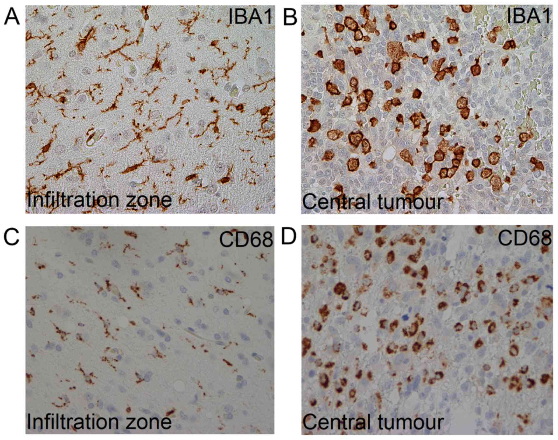
|
1
|
Louis DN, Perry A, Reifenberger G, von
Deimling A, Figarella-Branger D, Cavenee WK, Ohgaki H, Wiestler OD,
Kleihues P and Ellison DW: The 2016 World Health Organization
Classification of Tumors of the Central Nervous System: A summary.
Acta Neuropathol131. 803–820. 2016. View Article : Google Scholar
|
|
2
|
Aldape K, Zadeh G, Mansouri S,
Reifenberger G and von Deimling A: Glioblastoma: Pathology,
molecular mechanisms and markers. Acta Neuropathol. 129:829–848.
2015. View Article : Google Scholar : PubMed/NCBI
|
|
3
|
Hambardzumyan D, Gutmann DH and Kettenmann
H: The role of microglia and macrophages in glioma maintenance and
progression. Nat Neurosci. 19:20–27. 2016. View Article : Google Scholar : PubMed/NCBI
|
|
4
|
Perdiguero EG and Geissmann F: The
development and maintenance of resident macrophages. Nat Immunol.
17:2–8. 2016. View
Article : Google Scholar : PubMed/NCBI
|
|
5
|
Tremble LF, Forde PF and Soden DM:
Clinical evaluation of macrophages in cancer: Role in treatment,
modulation and challenges. Cancer Immunol Immunother. 66:1509–1527.
2017. View Article : Google Scholar : PubMed/NCBI
|
|
6
|
Domingues P, González-Tablas M, Otero Á,
Pascual D, Miranda D, Ruiz L, Sousa P, Ciudad J, Gonçalves JM,
Lopes MC, et al: Tumor infiltrating immune cells in gliomas and
meningiomas. Brain Behav Immun. 53:1–15. 2016. View Article : Google Scholar : PubMed/NCBI
|
|
7
|
Li W and Graeber MB: The molecular profile
of microglia under the influence of glioma. Neuro Oncol.
14:958–978. 2012. View Article : Google Scholar : PubMed/NCBI
|
|
8
|
Ricard C, Tchoghandjian A, Luche H, Grenot
P, Figarella-Branger D, Rougon G, Malissen M and Debarbieux F:
Phenotypic dynamics of microglial and monocyte-derived cells in
glioblastoma-bearing mice. Sci Rep. 6:263812016. View Article : Google Scholar : PubMed/NCBI
|
|
9
|
Boche D, Perry VH and Nicoll JA: Review:
Activation patterns of microglia and their identification in the
human brain. Neuropathol Appl Neurobiol. 39:3–18. 2013. View Article : Google Scholar : PubMed/NCBI
|
|
10
|
Colonna M and Butovsky O: Microglia
function in the central nervous system during health and
neurodegeneration. Annu Rev Immunol. 35:441–468. 2017. View Article : Google Scholar : PubMed/NCBI
|
|
11
|
Stensjøen AL, Solheim O, Kvistad KA,
Håberg AK, Salvesen Ø and Berntsen EM: Growth dynamics of untreated
glioblastomas invivo. Neuro Oncol. 17:1402–1411. 2015. View Article : Google Scholar : PubMed/NCBI
|
|
12
|
Stensjøen AL, Berntsen EM, Mikkelsen VE,
Torp SH, Jakola AS, Salvesen Ø and Solheim O: Does pretreatment
tumor growth hold prognostic information for patients with
glioblastoma? World Neurosurg. 101:686–694.e4. 2017. View Article : Google Scholar : PubMed/NCBI
|
|
13
|
O'Malley JT, Nadol JB Jr and McKenna MJ:
Anti CD163+, Iba1+, and CD68+
cells in the adult human inner ear: Normal distribution of an
unappreciated class of macrophages/microglia and implications for
inflammatory otopathology in humans. Otol Neurotol. 37:99–108.
2016. View Article : Google Scholar : PubMed/NCBI
|
|
14
|
Zhou W, Ke SQ, Huang Z, Flavahan W, Fang
X, Paul J, Wu L, Sloan AE, McLendon RE, Li X, et al: Periostin
secreted by glioblastoma stem cells recruits M2 tumour-associated
macrophages and promotes malignant growth. Nat Cell Biol.
17:170–182. 2015. View
Article : Google Scholar : PubMed/NCBI
|
|
15
|
Murray PJ and Wynn TA: Protective and
pathogenic functions of macrophage subsets. Nat Rev Immunol.
11:723–737. 2011. View
Article : Google Scholar : PubMed/NCBI
|
|
16
|
Sørensen MD, Dahlrot RH, Boldt HB, Hansen
S and Kristensen BW: Tumour-associated microglia/macrophages
predict poor prognosis in high-grade gliomas and correlate with an
aggressive tumour subtype. Neuropathol Appl Neurobiol. 44:185–206.
2018. View Article : Google Scholar : PubMed/NCBI
|
|
17
|
Chávez-Galán L, Olleros ML, Vesin D and
Garcia I: Much more than M1 and M2 macrophages, there are also
CD169(+) and TCR(+) macrophages. Front Immunol.
6:2632015.PubMed/NCBI
|
|
18
|
Glass R and Synowitz M: CNS macrophages
and peripheral myeloid cells in brain tumours. Acta Neuropathol.
128:347–362. 2014. View Article : Google Scholar : PubMed/NCBI
|
|
19
|
Mosser DM and Edwards JP: Exploring the
full spectrum of macrophage activation. Nat Rev Immunol. 8:958–969.
2008. View
Article : Google Scholar : PubMed/NCBI
|
|
20
|
Bieńkowski M and Preusser M: Prognostic
role of tumour-infiltrating inflammatory cells in brain tumours:
Literature review. Curr Opin Neurol. 28:647–658. 2015. View Article : Google Scholar : PubMed/NCBI
|
|
21
|
Hambardzumyan D and Bergers G:
Glioblastoma: Defining tumor niches. Trends Cancer. 1:252–265.
2015. View Article : Google Scholar : PubMed/NCBI
|
|
22
|
Schiffer D, Annovazzi L, Mazzucco M and
Mellai M: The microenvironment in gliomas: Phenotypic expressions.
Cancers (Basel). 7:2352–2359. 2015. View Article : Google Scholar : PubMed/NCBI
|
|
23
|
Yang L and Zhang Y: Tumor-associated
macrophages: From basic research to clinical application. J Hematol
Oncol. 10:582017. View Article : Google Scholar : PubMed/NCBI
|
|
24
|
Prayson RA and Cohen ML: Practical
Differential Diagnosis in Surgical Neuropathology. Humana Press;
Totowa, NJ: pp. 1772000
|