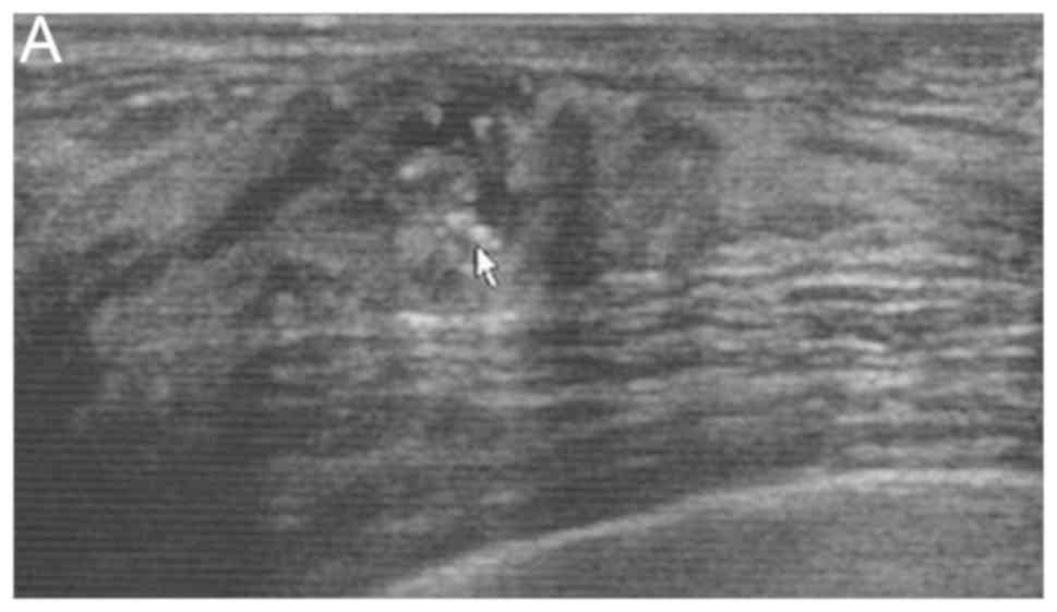
|
1
|
Schnitt SJ, Silen W, Sadowsky NL, Connolly
JL and Harris JR: Ductal carcinoma in situ (intraductal carcinoma)
of the breast. N Engl J Med. 318:898–903. 1988. View Article : Google Scholar : PubMed/NCBI
|
|
2
|
Singletary SE, Allred C, Ashley P, Bassett
LW, Berry D, Bland KI, Borgen PI, Clark G, Edge SB, Hayes DF, et
al: Revision of the American Joint Committee on Cancer staging
system for breast cancer. J Clin Oncol. 20:3628–3636. 2002.
View Article : Google Scholar : PubMed/NCBI
|
|
3
|
Wang W, Zhu W, Du F, Luo Y and Xu B: The
demographic features, clinicopathological characteristics and
cancer-specific outcomes for patients with microinvasive breast
cancer: A SEER database analysis. Sci Rep. 7:420452017. View Article : Google Scholar : PubMed/NCBI
|
|
4
|
Okumura Y, Yamamoto Y, Zhang Z, Toyama T,
Kawasoe T, Ibusuki M, Honda Y, Iyama K, Yamashita H and Iwase H:
Identification of biomarkers in ductal carcinoma in situ of the
breast with microinvasion. BMC Cancer. 8:2872008. View Article : Google Scholar : PubMed/NCBI
|
|
5
|
Vieira CC, Mercado CL, Cangiarella JF, Moy
L, Toth HK and Guth AA: Microinvasive ductal carcinoma in situ:
Clinical presentation, imaging features, pathologic findings, and
outcome. Eur J Radiol. 73:102–107. 2010. View Article : Google Scholar : PubMed/NCBI
|
|
6
|
Yao JJ, Zhan WW, Chen M, Zhang XX, Zhu Y,
Fei XC and Chen XS: Sonographic features of ductal carcinoma in
situ of the breast with microinvasion: Correlation with
clinicopathologic findings and biomarkers. J Ultrasound Med.
34:1761–1768. 2015. View Article : Google Scholar : PubMed/NCBI
|
|
7
|
Sopik V, Sun P and Narod SA: Impact of
microinvasion on breast cancer mortality in women with ductal
carcinoma in situ. Breast Cancer Res Treat. 167:787–795. 2018.
View Article : Google Scholar : PubMed/NCBI
|
|
8
|
Fang Y, Wu J, Wang W, Fei X, Zong Y, Chen
X, Huang O, He J, Chen W, Li Y, et al: Biologic behavior and
long-term outcomes of breast ductal carcinoma in situ with
microinvasion. Oncotarget. 7:64182–64190. 2016. View Article : Google Scholar : PubMed/NCBI
|
|
9
|
Gwak YJ, Kim HJ, Kwak JY, Lee SK, Shin KM,
Lee HJ, Kim GC, Jang YJ, Han MH, Park JY and Jung JH:
Ultrasonographic detection and characterization of asymptomatic
ductal carcinoma in situ with histopathologic correlation. Acta
Radiol. 52:364–371. 2011. View Article : Google Scholar : PubMed/NCBI
|
|
10
|
Breast Imaging Reporting and Data System
(BI-RADS). 5th. American College of Radiology; Reston, VA: 2013
|
|
11
|
Penault-Llorca F, André F, Sagan C,
Lacroix-Triki M, Denoux Y, Verriele V, Jacquemier J, Baranzelli MC,
Bibeau F, Antoine M, et al: Ki67 expression and docetaxel efficacy
in patients with estrogen receptor-positive breast cancer. J Clin
Oncol. 27:2809–2815. 2009. View Article : Google Scholar : PubMed/NCBI
|
|
12
|
Zhang W, Gao EL, Zhou YL, Zhai Q, Zou ZY,
Guo GL, Chen GR, Zheng HM, Huang GL and Zhang XH: Different
distribution of breast ductal carcinoma in situ, ductal carcinoma
in situ with microinvasion, and invasion breast cancer. World J
Surg Oncol. 10:2622012. View Article : Google Scholar : PubMed/NCBI
|
|
13
|
Ozkan-Gurdal S, Cabioglu N, Ozcinar B,
Muslumanoglu M, Ozmen V, Kecer M, Yavuz E and Igci A: Factors
predicting microinvasion in Ductal Carcinoma in situ. Asian Pac J
Cancer Prev. 15:55–60. 2014. View Article : Google Scholar : PubMed/NCBI
|
|
14
|
Sue GR, Lannin DR, Killelea B and Chagpar
AB: Predictors of microinvasion and its prognostic role in ductal
carcinoma in situ. Am J Surg. 206:478–481. 2013. View Article : Google Scholar : PubMed/NCBI
|
|
15
|
Lee MH, Ko EY, Han BK, Shin JH, Ko ES and
Hahn SY: Sonographic findings of pure ductal carcinoma in situ. J
Clin Ultrasound. 41:465–471. 2013. View Article : Google Scholar : PubMed/NCBI
|
|
16
|
Watanabe T, Yamaguchi T, Tsunoda H, Kaoku
S, Tohno E, Yasuda H, Ban K, Hirokaga K, Tanaka K, Umemoto T, et
al: Ultrasound image classification of ductal carcinoma in situ
(DCIS) of the breast: Analysis of 705 DCIS lesions. Ultrasound Med
Biol. 43:918–925. 2017. View Article : Google Scholar : PubMed/NCBI
|
|
17
|
Nagashima T, Hashimoto H, Oshida K, Nakano
S, Tanabe N, Nikaido T, Koda K and Miyazaki M: Ultrasound
demonstration of mammographically detected microcalcifications in
patients with ductal carcinoma in situ of the breast. Breast
Cancer. 12:216–220. 2005. View Article : Google Scholar : PubMed/NCBI
|
|
18
|
Rauch GM, Kuerer HM, Scoggins ME, Fox PS,
Benveniste AP, Park YM, Lari SA, Hobbs BP, Adrada BE, Krishnamurthy
S and Yang WT: Clinicopathologic, mammographic, and sonographic
features in 1187 patients with pure ductal carcinoma in situ of the
breast by estrogen receptor status. Breast Cancer Res Treat.
139:639–647. 2013. View Article : Google Scholar : PubMed/NCBI
|
|
19
|
Yang WT, Suen M, Ahuja A and Metreweli C:
In vivo demonstration of microcalcification in breast cancer using
high resolution ultrasound. Br J Radiol. 70:685–690. 1997.
View Article : Google Scholar : PubMed/NCBI
|
|
20
|
Gufler H, Buitrago-Téllez CH, Madjar H,
Allmann KH, Uhl M and Rohr-Reyes A: Ultrasound demonstration of
mammographically detected microcalcifications. Acta Radiol.
41:217–221. 2000. View Article : Google Scholar : PubMed/NCBI
|
|
21
|
Folkman J: What is the evidence that
tumors are angiogenesis dependent? J Natl Cancer Inst. 82:4–6.
1990. View Article : Google Scholar : PubMed/NCBI
|
|
22
|
Cao Y, Paner GP, Kahn LB and Rajan PB:
Noninvasive carcinoma of the breast: Angiogenesis and cell
proliferation. Arch Pathol Lab Med. 128:893–896. 2004.PubMed/NCBI
|
|
23
|
Dershaw DD, Abramson A and Kinne DW:
Ductal carcinoma in situ: Mammographic findings and clinical
implications. Radiology. 170:411–415. 1989. View Article : Google Scholar : PubMed/NCBI
|
|
24
|
Stomper PC, Connolly JL, Meyer JE and
Harris JR: Clinically occult ductal carcinoma in situ detected with
mammography: Analysis of 100 cases with radiologic-pathologic
correlation. Radiology. 172:235–241. 1989. View Article : Google Scholar : PubMed/NCBI
|
|
25
|
Lagios MD, Westdahl PR, Margolin FR and
Rose MR: Duct carcinoma in situ. Relationship of extent of
noninvasive disease to the frequency of occult invasion,
multicentricity, lymph node metastases, and short-term treatment
failures. Cancer. 50:1309–1314. 1982. View Article : Google Scholar : PubMed/NCBI
|
|
26
|
Berg WA, Gutierrez L, NessAiver MS, Carter
WB, Bhargavan M, Lewis RS and Ioffe OB: Diagnostic accuracy of
mammography, clinical examination, US, and MR imaging in
preoperative assessment of breast cancer. Radiology. 233:830–849.
2004. View Article : Google Scholar : PubMed/NCBI
|
|
27
|
Kolb TM, Lichy J and Newhouse JH:
Comparison of the performance of screening mammography, physical
examination, and breast US and evaluation of factors that influence
them: An analysis of 27,825 patient evaluations. Radiology.
225:165–175. 2002. View Article : Google Scholar : PubMed/NCBI
|