Introduction
Wagner-Meissner corpuscles are specialized
mechanoreceptors located in the dermal papillae that directly
connect with the basal layer of the epidermis, and are prominent in
the palms and soles (1). They show
characteristic histological features: An encapsulated round to oval
structure with central lamellation and peripherally located nuclei
of Schwann cells (1).
Wagner-Meissner corpuscles or Wagner-Meissner corpuscle-like
structures (pseudo-Meissner corpuscles) are occasionally a
component of some types of cutaneous and neurogenic tumors,
including melanocytic nevus and neurofibroma (2,3). Benign
tumorous lesions entirely composed of Wagner-Meissner corpuscles
were first described by Kaiserling and Geerts (4). They named these lesions Wagner-Meissner
neurilemmoma, and to date, only four such cases have been reported
in the English literature (4–6).
Neurofibromatosis type 1 is a relatively common
autosomal dominant disorder, characterized clinically by presence
of café-au-lait spots (7). It is
well recognized that various types of tumors, including nervous and
non-nervous systems, develop in patients with neurofibromatosis
type 1. Multiple cutaneous neurofibromas are the most frequent
tumor in patients with neurofibromatosis type 1, and patients with
this disorder have a risk of development of malignant peripheral
nerve sheath tumor (7). However, to
the best of our knowledge, occurrence of Wagner-Meissner
neurilemmoma in patients with neurofibromatosis type 1 has not been
described. Here, we report the first case of this lesion in a
patient with neurofibromatosis type 1 and discuss the
clinicopathological features.
Case report
A 16-year-old Japanese male with neurofibromatosis
type 1 presented with a tumorous lesion on the upper lip. He had
multiple café-au-lait spots in the entire body and ephelides in the
face. Moreover, he had undergone surgical resection of the
congenital melanocytic nevi of the back and thigh. Resection of the
lip tumor was performed under a clinical diagnosis of neurofibroma.
No recurrence has been observed during medical follow-up.
Formalin-fixed and paraffin-embedded specimens of
the resected tumor were processed for routine histological
examination and immunohistochemical analyses.
In this report, immunohistochemical analysis was
performed using an autostainer (Autostainer link 48; Dako
Cytomation). The primary antibody used in this report was a rabbit
polyclonal antibody against S-100 protein (Dako Cytomation).
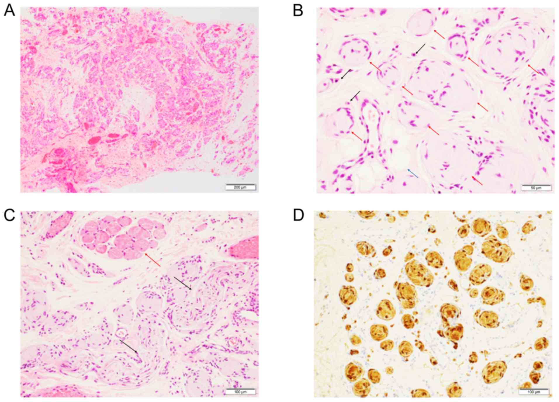
Histopathological examination revealed an
unencapsulated, poorly-circumscribed tumor located in the fatty
tissue. The tumor was comprised of abundant Wagner-Meissner
corpuscle-like structures, which were composed of 5–15 lamellated
Schwann cells containing eosinophilic cytoplasm and peripherally
located nuclei (Fig. 1A and B).
These structures were packed in several portions; however, they
were intermingled with fatty tissue and striated muscles in the
periphery of the tumor (Fig. 1A). No
mitotic figures were noted. Additionally, no spindle-shaped
neoplastic cell proliferation, as seen in conventional
neurofibroma, was observed (Fig. 1A and
B). Peripheral nerves with myxoid changes (Fig. 1C) and a few mast cells were observed
within the tumor (Fig. 1B). The
tumor extended to the margin of the resected specimen, however, no
additional resection was not performed.
Immunohistochemical analysis clearly demonstrated
that these corpuscles were diffusely positive for S-100 protein
(Fig. 1D), but S-100
protein-positive spindle cells were absent (Fig. 1D).
Based on these features, a final diagnosis of
Wagner- Meissner neurilemmoma was made.
Discussion
There has been no previous report of Wagner-Meissner
neurilemmoma occurring in a patient with neurofibromatosis type 1.
The clinicopathological features of the previously reported four
cases of Wagner-Meissner neurilemmoma, in addition to our case, are
summarized (Table I). Based on these
reports, males are preferentially affected (female:male, 1:4), and
the tumor mainly occurred in adolescents or young adults, except
for one patient (case 1). The predilection sites were the leg and
oral mucosa.
 | Table I.Clinicopathological features of
Wagner-Meissner neurilemmoma. |
Table I.
Clinicopathological features of
Wagner-Meissner neurilemmoma.
| Case no. | Age (years) | Sex |
Neurofibromatosis | Location | Capsulization | (Refs.) |
|---|
| 1 | 80 | Male | – | Thigh | Encapsulated | (4) |
| 2 | 33 | Male | – | Leg | Encapsulated | (4) |
| 3 | 24 | Female | – | Vulva | Encapsulated | (5) |
| 4 | 10 | Male | – | Cheek | Unencapsulated | (6) |
| Present case | 16 | Male | Type 1 | Upper lip | Unencapsulated |
|
The characteristic histopathological feature of the
present tumor was the presence of abundant Wagner-Meissner
corpuscle-like structures and a lack of neoplastic spindle cell
nests, as seen in conventional neurofibroma (4–6).
Therefore, we diagnosed this lesion as Wagner-Meissner
neurilemmoma, although neurofibroma with abundant Wagner-Meissner
corpuscles or pseudo-Meissner corpuscles must be differentiated.
The unique feature of the present tumor was the poor
circumscription and lack of encapsulation. The previously reported
three cases (two cases occurring in the leg and one case in the
vulva) were well-circumscribed and encapsulated (4,5);
however, a case with a poorly circumscribed and unencapsulated
tumor, percolating into the surrounding fatty tissue, resembling
the present case, occurring in the cheek of a patient has been
reported (6). The reason for this
difference remains unclear; however, it might be a reflection of
the location of the tumor (skin or mucosa). Further studies are
needed to clarify the detailed clinicopathological features of this
extremely rare tumor.
The pathogenesis of Wagner-Meissner neurilemmoma
remains unclear. The hamartomatous nature of the tumor has been
proposed to be related to its occurrence mainly in adolescents and
young adults (6). Wagner-Meissner
corpuscles are also present in the oral mucosa, where they are
termed the end-bulbs of Krause (7).
Moreover, pseudo-Meissner corpuscles are also found in cases of
traumatic neuroma (4).
Interestingly, myxoid changes in the peripheral nerves, suggestive
of nerve degeneration, were observed within the present patient's
tumor, and may be indicative of reactive proliferative changes.
Moreover, our patient was also diagnosed with neurofibromatosis
type 1. Thus, this lesion might represent an extreme form of
diffuse neurofibroma with abundant Wagner-Meissner corpuscles
associated with neurofibromatosis type 1, even though the
previously reported four patients did not have neurofibromatosis
(Table I). Further studies are
needed to clarify the pathogenesis of this extremely rare tumor,
including its association with neurofibromatosis.
Acknowledgements
Not applicable.
Funding
No funding was received.
Availability of data and materials
All data generated or analyzed during this study are
included in this published article.
Authors' contributions
CM and MI undertook study conception and design. The
acquisition and analysis of data was performed by CM, MI, YK, NM,
KK, HO, and KT. CM and MI drafted the manuscript and prepared the
figure. The final version of the manuscript has been read and
approved by all authors.
Ethics approval and consent to
participate
This study was performed according to Kansai Medical
University Hospital's ethical policy.
Patient consent for publication
Not applicable.
Competing interests
The authors declare that they have no competing
interests.
References
|
1
|
Vega JA, Haro JJ and Del Valle ME:
Immunohistochemistry of human cutaneous Meissner and pacinian
corpuscles. Microsc Res Tech. 34:351–361. 1996. View Article : Google Scholar : PubMed/NCBI
|
|
2
|
Misago N: The relationship between
melanocytes and peripheral nerve sheath cells (Part I): Melanocytic
nevus (excluding so-called ‘blue nevus’) with peripheral nerve
sheath differentiation. Am J Dermatopathol. 22:217–229. 2000.
View Article : Google Scholar : PubMed/NCBI
|
|
3
|
Watabe K, Kumanishi T, Ikuta F and Oyake
Y: Tactile-like corpuscles in neurofibromas: Immunohistochemical
demonstration of S-100 protein. Acta Neuropathol. 61:173–177. 1983.
View Article : Google Scholar : PubMed/NCBI
|
|
4
|
Kaiserling E and Geerts ML: Tumour of
Wagner-Meissner touch corpuscles. Wagner-Meissner neurilemmoma.
Virchows Arch A Pathol Anat Histopathol. 409:241–250. 1986.
View Article : Google Scholar : PubMed/NCBI
|
|
5
|
Ferrara N, Di Marino M, Rossiello L and
Baldi A: Wagner-Meissner neurilemmoma of the vulva. Int J Dermatol.
42:550–551. 2003. View Article : Google Scholar : PubMed/NCBI
|
|
6
|
Wu AJ, Jarzembowski J, Morag Y and Lucas
DR: Wagner- Meissner neurilemmoma of the right cheek. Ann Diagn
Pathol. 12:204–207. 2008. View Article : Google Scholar : PubMed/NCBI
|
|
7
|
Winkelmann RK: The erogenous zones: Their
nerve supply and significance. Proc Staff Meet Mayo Clin. 34:39–47.
1959.PubMed/NCBI
|