Introduction
Colorectal cancer is one of the most common causes of cancer mortality worldwide (1). Of the patients with advanced colorectal cancer, 8-13% develop malignant colorectal obstruction (MCRO), which requires relief of the obstruction as soon as possible (2-4). Conventionally, patients with MCRO are treated with emergency surgery (ES), which includes a variety of procedures such as Hartmann's procedure or colostomy alone. However, ES is associated with high mortality and morbidity rates compared with elective surgery (5,6).
The relief of colonic obstruction via the placement of a self-expanding metal stent (SEMS) as a palliative treatment was first described in 1991(7). SEMS as a bridge to surgery (BTS) was then described in 1994(8). Endoscopic SEMS placement for MCRO can improve luminal patency and serve as a BTS in curable patients. Systematic reviews have showed that SEMS has advantages over ES in short-term outcomes (9-11) and is used as an alternative to ES (12-16).
Despite the good short-term outcomes of SEMS placement, the European Society of Gastrointestinal Endoscopy (ESGE) clinical guidelines published in 2014 do not recommend SEMS placement as a BTS for MCRO (17). In contrast, the American Society for Gastrointestinal Endoscopy (ASGE) guidelines published in 2010 recommended SEMS placement as a BTS as a standard treatment (12). SEMS as a BTS for MCRO was reported in Japan in 1996(18); after this procedure began to be covered by Japanese health insurance in January 2012, some large-scale prospective multicenter studies on SEMS have been conducted in Japan, and SEMS placement as a BTS is considered effective and safe in the short-term (19,20).
Given the issues described above, it is clear that there is no universally accepted consensus concerning the superiority or inferiority of SEMS as a BTS compared with ES in patients with MCRO. In addition, there are few reports investigating the long-term outcomes of SEMS as a BTS in patients with MCRO. The present study aimed to clarify the efficacy of SEMS as a BTS (including long-term outcomes) in comparison with ES in patients with MCRO.
Materials and methods
Patients
A total of 3,840 patients with colorectal cancer underwent surgical resection between April 2001 and June 2016 in our institution. Of these patients, 93 had acute MCRO requiring emergency decompression at the time of diagnosis. These 93 patients were treated by either elective surgery after SEMS placement or by ES within 24 h after the initial diagnosis. Only patients in whom the colorectal lesion was ultimately resected were included in the present study. Patients who were first treated with decompression by nasal ileus tube (n=24) or trans-anal tube (n=6), and one patient with signs of peritonitis by perforation were excluded. Therefore, the present study included 62 patients: 25 in the SEMS group, and 37 in the ES group (Fig. 1).
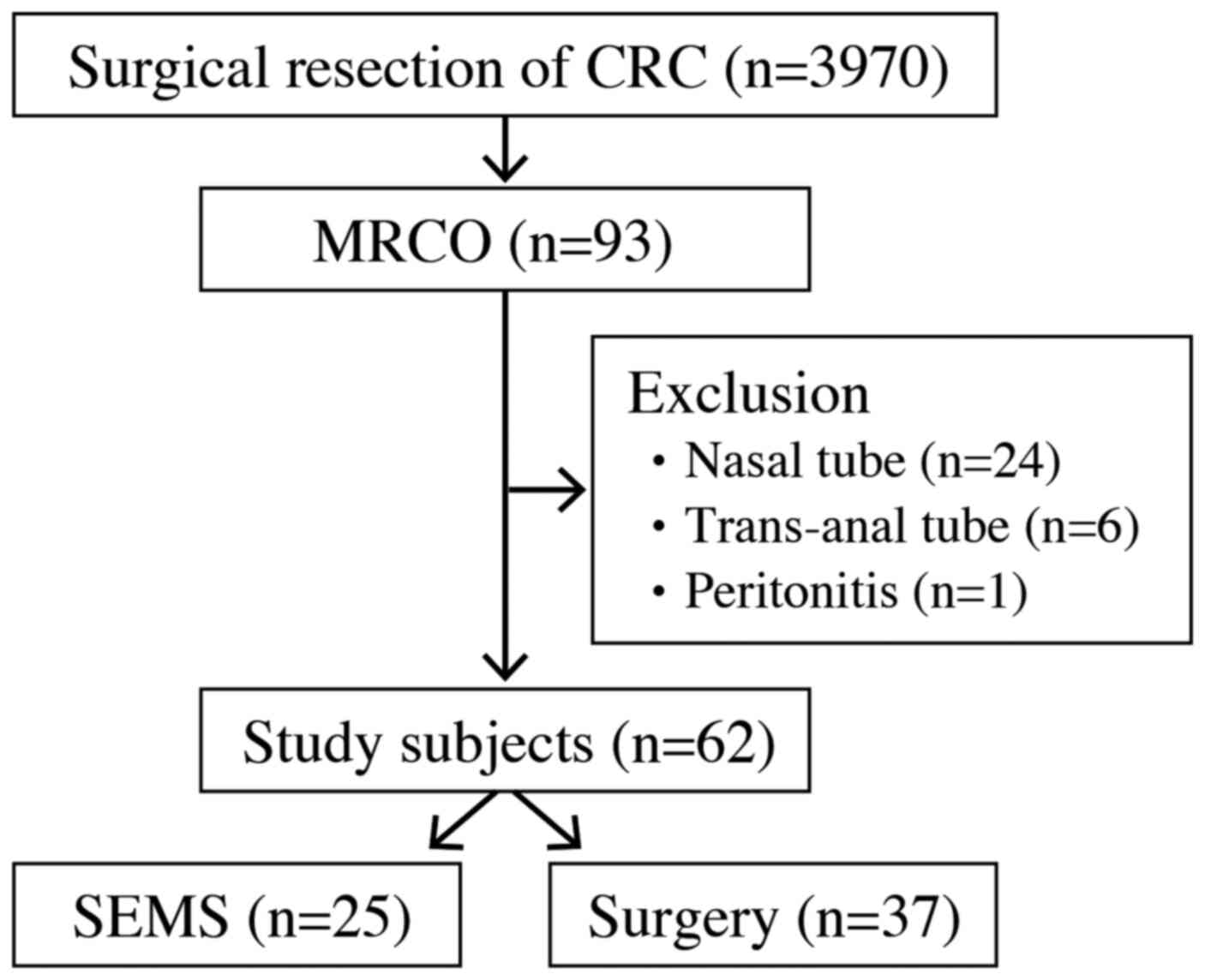 |
Figure 1
Flow diagram of the study selection process. CRC, colorectal cancer; SEMS, Self-expanding metal stent; MCRO, malignant colorectal obstruction.
|
This was a retrospective study conducted in a single institution. Data regarding the treatment method, age, sex, location, pathological stage of the tumor, treatment outcomes related to SEMS placement, treatment outcomes related to surgery, and follow-up duration were collected from the hospital records. Pathological tumor staging was done in accordance with the American Joint Committee on Cancer tumor-node-metastasis classification (7th edition) (21). Written informed consent was obtained from all patients before the procedures. The ethics committee of our institution approved the study protocol (approval no. 17H073). The present study is registered in the University Hospital Medical Network Clinical Trials Registry (UMIN R000034868).
Diagnosis of MCRO
The clinical diagnosis of MCRO was made based on the presence of nausea and vomiting, abdominal distension, abdominal pain, and inability to pass stools, and was confirmed on radiography and abdominal computed tomography (CT). After MCRO was diagnosed, a treatment plan was formed through consultation with surgeons in our institution. SEMS placement or ES was performed within 24 h after the initial diagnosis of MCRO. Thus, ES was defined as being performed within one day after diagnosis.
SEMS placement
SEMS procedures were performed by endoscopists experienced over 1,000 cases of colonoscopy, over 100 cases of endoscopic retrograde cholangiopancreatography (ERCP), and over 50 cases of esophageal, duodenal, or biliary stent placement. Bowel preparation comprised enemas only. Radiographic contrast enema was not performed before the procedures. All procedures were performed in the radiology room under fluoroscopic and endoscopic guidance, while the patient was consciously sedated with midazolam. A CF-260AI or CF-HQ290AZI colonoscope (Olympus) and CO2 gas were used in all cases. The obstructive lesion was directly confirmed and marked by a clip on the anal side. Access across the stricture was achieved with a 0.025-inch guidewire (Radifocus™; Terumo), and an ERCP catheter (MTW Co.) was then inserted to the proximal lumen. The length and degree of the stricture was measured fluoroscopically using a contrast agent, and the type of SEMS was selected. The guidewire was changed to a 0.035-inch guidewire (Jagwire™; Boston Scientific), and the SEMS was placed using the through-the-scope technique (22,23). The SEMSs used were WallFlex enteral colonic stents (Boston Scientific Corporation) and Niti-S enteral colonic stents (Tae Woong Medical). All SEMSs were uncovered stents with an inner diameter of 22 mm, and a length of 60-90 mm (WallFlex) or 60-80 mm (Niti-S). Balloon dilation was not performed in any case to minimize the risk of perforation (24). Tumor biopsy was performed just after SEMS placement. These procedures basically conform to the guidelines of the Japan Colonic Stent Safe Procedure Research Group (JCSSPRG) (http://colon-stent.com/), which received support from the Japan Gastroenterological Endoscopy Society. The position of the SEMS was confirmed on plain abdominal radiography each day. After improvement of the MCRO was confirmed, a full liquid diet was resumed within 3-5 days, and the diet was gradually progressed to include solid food. Patients whose condition improved received preoperative examinations, including total colonoscopy, and were allowed to leave the hospital until elective surgery. For patients who had undergone successful SEMS placement, elective surgery was performed about 2 weeks later. Typical cases are shown in Figs. 2 and 3.
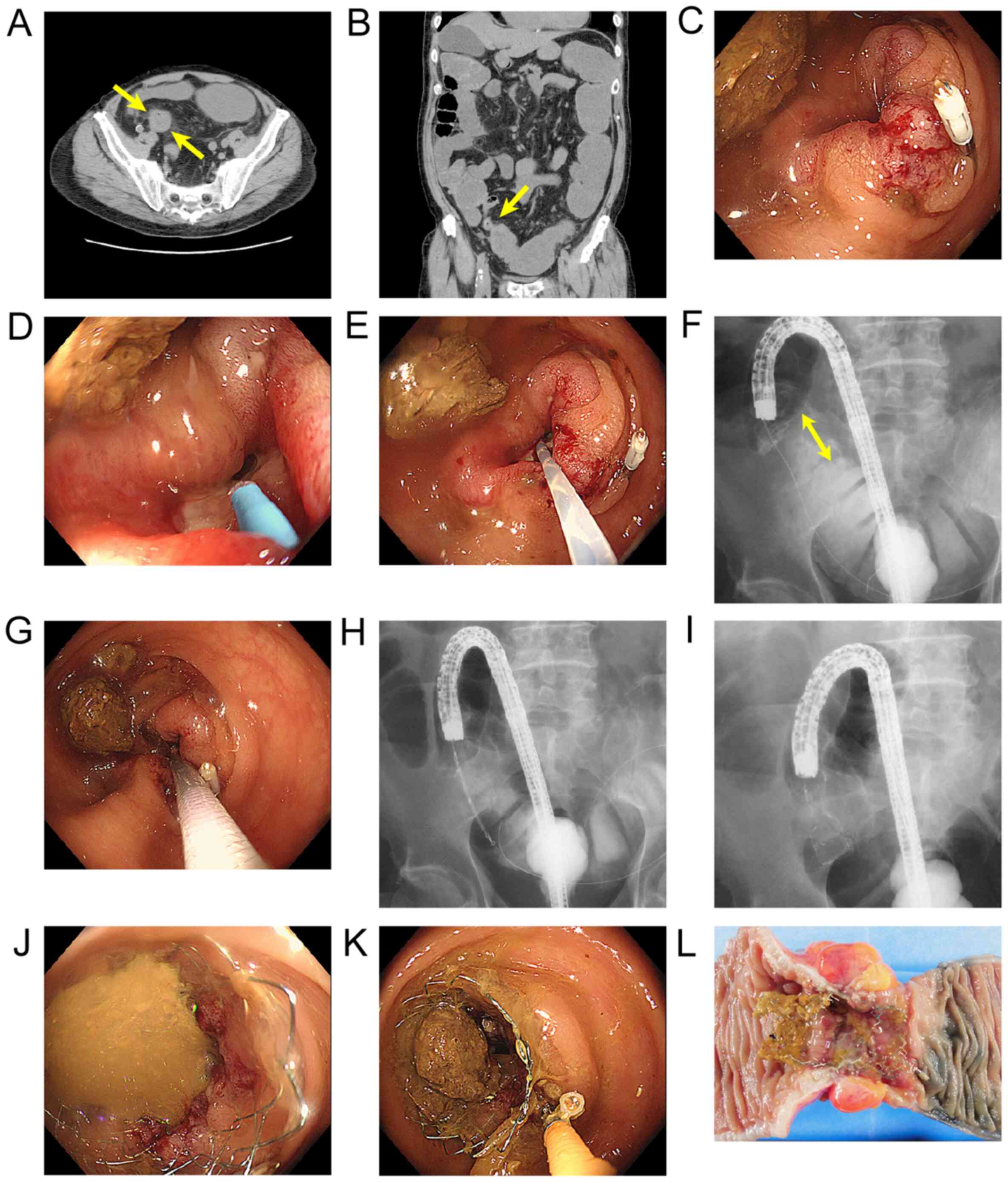 |
Figure 2
SEMS placement procedure (Case 1). (A and B) Malignant colorectal obstruction due to a sigmoid colon lesion was diagnosed using abdominal CT (axial and coronal sections). Arrows indicate the beginning of the stricture. (C) The obstructive lesion was confirmed endoscopically, and marked by a clip on the anal side. (D) An endoscopic retrograde cholangiopancreatography catheter about to be inserted across the stricture. (E) The guidewire passed the stricture through the endoscopic retrograde cholangiopancreatography catheter, and reached the oral side. (F) The length and degree of the stricture was measured fluoroscopically. The double arrows indicate the distance of the stricture. (G and H) The guidewire was changed to a 0.035-inch guidewire, and the SEMS delivery system passed the stricture using the through-the-scope technique. (I) The SEMS just after being deployed from the delivery system. (J) Stools flowed through the SEMS from the oral side. (K) Biopsy was performed from the side of the tumor just after SEMS placement. (L) The surgical specimen resected 12 days after SEMS placement as a bridge to surgery. SEMS, Self-expanding metal stent.
|
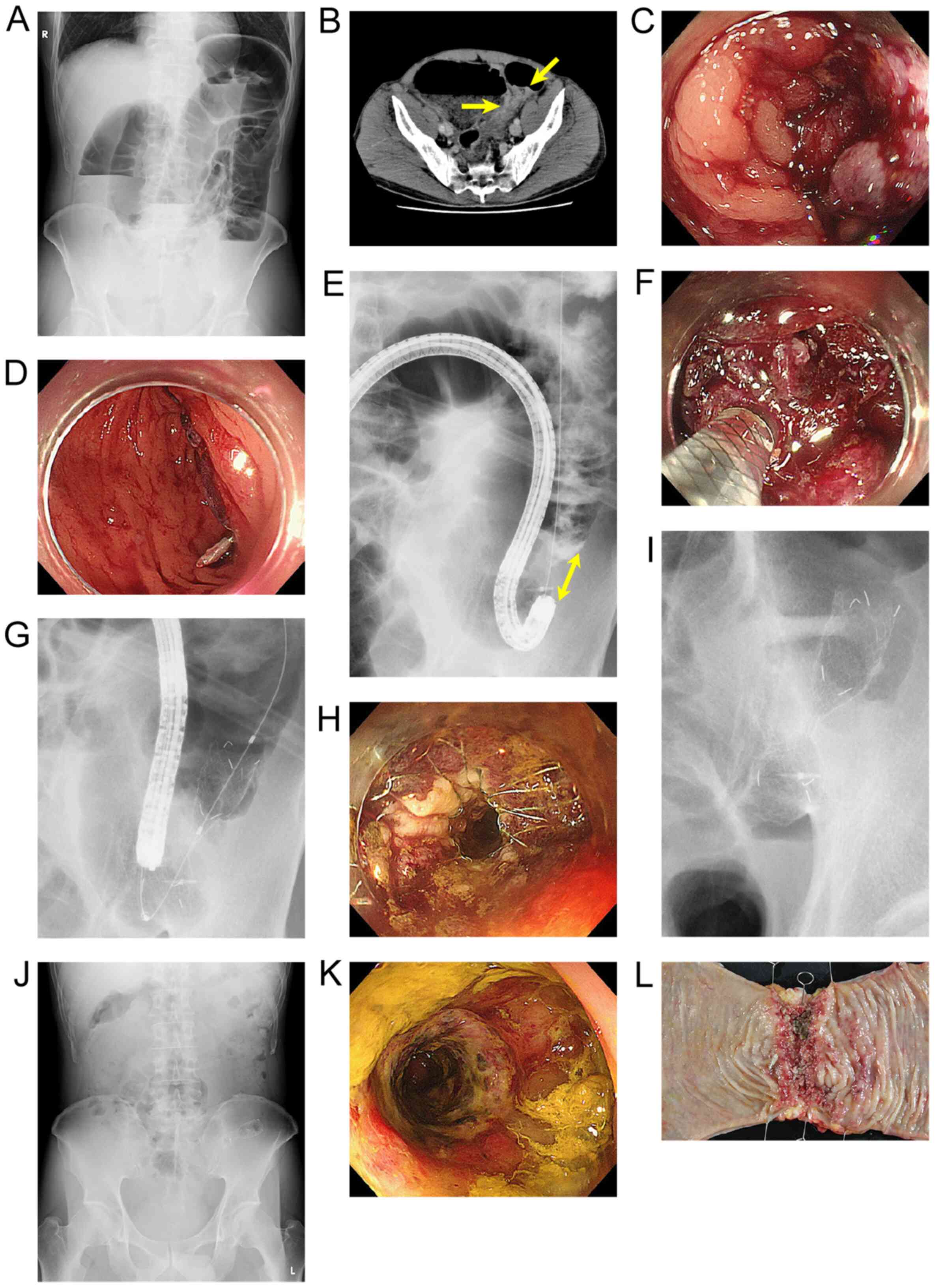 |
Figure 3
Self-expanding metal stent placement procedure (Case 2). (A) MCRO was diagnosed using radiography. (B) Abdominal CT (axial section) showing MCRO due to a sigmoid colon lesion. Arrows indicate the beginning of the stricture. (C) The obstructive lesion was confirmed endoscopically. (D) The obstructive lesion marked by a clip on the anal side. (E) The length and degree of the stricture were measured fluoroscopically. The double arrows indicate the distance of the stricture. (F) The guidewire was changed to a 0.035-inch guidewire, and the SEMS delivery system passed the stricture using the through-the-scope technique. (G) The SEMS was deployed from the delivery system. (H) The SEMS just after being deployed. (I) The SEMS position was confirmed as appropriate on a radiographic image. (J) Radiography performed the day after SEMS placement showed that the gas pattern was improved. (K) The obstructive lesion 5 days after SEMS placement. The colonoscope passed through the stricture and reached the cecum. (L) The surgical specimen resected 14 days after SEMS placement as a bridge to surgery. SEMS, Self-expanding metal stent; MCRO, malignant colorectal obstruction.
|
Study outcomes
The short-term outcomes were the rates of laparoscopic surgery, primary anastomosis, stoma formation, procedure-related adverse events, and 30-day mortality, and the number of dissected lymph nodes. Primary anastomosis was defined as main anastomosis at the first surgery with or without the formation of a diverting stoma. Stoma formation included a diverting stoma and a decompression stoma. Procedure-related adverse events were defined as adverse events associated with the SEMS placement and the surgery during all treatment periods, regardless of severity. For 30-day mortality, the period was 30 days after the diagnosis of acute MCRO. Only in the SEMS group, technical and clinical success were assessed. Technical success was defined as successful SEMS placement without adverse events during the procedure, and opening of the stent across the stricture (confirmed fluoroscopically). Clinical success was defined as resolution of symptoms and radiological relief of the obstruction within 24 h after the procedure.
The long-term outcome was disease-free survival. As it was difficult to judge whether SEMS had affected the long-term results of patients who were in stage Ⅳ (n=13) at the time of initial presentation, those who died, or those who had other advanced cancers pointed out within 1 month postoperatively (n=3), these patients were excluded. Ultimately, the disease-free survival was compared between 21 patients in the SEMS group and 25 patients in the ES group. The methods of surveillance are described below.
Follow-up
In accordance with the Japanese Society for Cancer of the Colon and Rectum (JSCCR) guidelines (25). Postoperative adjuvant chemotherapy and postoperative follow-up were considered if the clinical condition allowed. Physical examinations and blood tests, including carcinoembryonic antigen and carbohydrate antigen 19-9 levels, were performed every 3 months for the first 3 years after surgical resection, and then every 6 months for the next 2 years. In addition, CT scans of the chest, abdomen, and pelvis were performed every 6 months, and a full colonoscopy was performed every year for 5 years. Confirmation of recurrence was based on CT and endoscopic findings. Patients were followed for 5 years postoperatively, unless they ceased visiting the hospital or died.
Statistical analysis
Statistical analyses were performed using JMP Pro version 12 statistical software (SAS Institute). Continuous variables were expressed as the median (range), and categorical variables as the number (percentage). Baseline characteristics and clinical outcomes were compared using Fisher's exact test for categorical variables, and the nonparametric Wilcoxon rank sum test for continuous variables. Disease-free survival time was calculated from surgery to the date of the last visit (up to 60 months) or to death. Disease-free survival was analyzed using Kaplan-Meier survival curves, and significant differences were determined using the log-rank test. In addition, we performed multivariate Cox regression analysis for Disease-free survival in terms of prognostic factors (initial treatment, age, sex, location, stage, number of lymph nodes resected, and adjuvant chemotherapy). The location was divided into left and right side, and the cutoff value for the number of lymph nodes was set up to 25. All P values were two-sided, and results with P<0.05 were considered significant.
Results
Demographic and oncological characteristics
Baseline patient characteristics and tumor details are shown in Table I. There were no significant differences between the two groups in age, sex, tumor location, and distribution of the pathological stage.
 |
Table I
Demographics and oncological features of patients.
|
Table I
Demographics and oncological features of patients.
| Patient details |
SEMS group (n=25) |
ES group (n=37) |
P-value |
| Age in years, median (range) |
66 (39-91) |
69 (35-96) |
0.90 |
| Sex, n (%) |
|
|
0.30 |
| Male |
14 (56.0) |
15 (40.5) |
|
| Female |
11 (44.0) |
22 (59.5) |
|
| Tumor location, n (%) |
|
|
0.91 |
| Rectum |
2 (8.0) |
5 (13.5) |
|
| Sigmoid colon |
12 (48.0) |
18 (48.6) |
|
| Descending colon |
3 (12.0) |
5 (13.5) |
|
| Transverse colon |
6 (24.0) |
5 (13.5) |
|
| Ascending colon |
2 (8.0) |
3 (8.1) |
|
| Cecum |
0 (0.0) |
1 (2.7) |
|
| pStage of the tumor, n (%) |
|
|
0.78 |
| Stage II |
6 (24.0) |
8 (21.6) |
|
| Stage III |
15 (60.0) |
20 (54.1) |
|
| Stage IV |
4 (16.0) |
9 (24.3) |
|
SEMS placement outcomes
SEMS placement details are shown in Table II. The technical success rate was 92.0% (23/25). The two patients in whom SEMS placement failed underwent surgery promptly. No adverse events such as bleeding or perforation occurred during the procedures. The clinical symptoms and plain abdominal radiographs were improved in all patients who underwent successful SEMS placement. During the period from SEMS placement to elective surgery, there was one case of stent migration and one of stent re-obstruction; these two cases were improved by endoscopic intervention. Resumption of a diet was possible in all patients who had successful SEMS placement. Eight patients were able to be discharged from hospital until elective surgery. The other 17 patients were all hospitalized until surgery, even after SEMS placement. The median period from SEMS placement until elective surgery was 12.0 (4-90) days. Adverse events such as perforation did not occur between SEMS placement and elective surgery. In addition, there were no cases in which any perforation was finally found during the elective surgery.
|
Table II
SEMS placement details.
|
Table II
SEMS placement details.
| Event |
SEMS group (n=25) |
| Technical success, n (%) |
23 (92.0) |
| Inability to pass a guidewire |
1 |
| Inadequate stent placement |
1 |
| Manufacturer |
|
| Wallflex |
13 |
| Niti-S |
10 |
| Adverse events during the procedure, n (%) |
0 (0.0) |
| Clinical success, n (%) |
23 (92.0) |
| Adverse events before surgery, n (%) |
2 (8.0) |
| Perforation |
0 |
| Bleeding |
0 |
| Migration |
1 |
| Re-obstruction |
1 |
| Resumption of diet, n (%) |
23 (92.0) |
| Discharge until surgery, n (%) |
8 (32.0) |
| Preoperative period, median (range) |
12 (4-90) |
Surgical outcomes
Surgical outcomes are shown in Table III. Laparoscopic surgery was performed more often in the SEMS group than in the ES group (P<0.001). Compared with the ES group, the SEMS group had a significantly greater primary anastomosis rate (P=0.003), and a significantly lower stoma formation rate (P=0.002). The initial surgery comprised decompression stoma formation alone in 15 patients in the ES group. The SEMS group had a significantly greater number of dissected lymph nodes than the ES group (P=0.001). Although four patients in the ES group had a diverting stoma that was not closed because of progressive disease, the number of times that each patient underwent surgeries related to treatment was significantly greater in the ES group than the SEMS group (P=0.004). There was no difference between the two groups in the duration of total hospitalization.
|
Table III
Surgical outcomes.
|
Table III
Surgical outcomes.
| Outcome |
SEMS group (n=25) |
ES group (n=37) |
P-value |
| Laparoscopic surgery, n (%) |
17 (68.0) |
1 (2.7) |
<0.001 |
| Primary anastomosis, n (%) |
22 (88.0) |
19 (51.4) |
0.003 |
| Stoma formation, n (%) |
6 (24.0) |
25 (67.6) |
0.002 |
| Resection + diverting stoma |
3 |
7 |
|
| Resection + definitive stoma |
2 |
3 |
|
| Decompression stoma |
1 |
15 |
|
| Number of lymph nodes dissected, median (range) |
30 (15-71) |
18 (0-49) |
0.001 |
| Number of surgeries, median (range) |
1 (1–3) |
2 (1-3) |
0.004 |
| Total length of hospital stay in days, median (range) |
23 (11-66) |
27 (10-126) |
0.86 |
Adverse events and 30-day mortality rate
The rates of overall procedure-related adverse events and 30-day mortality are shown in Table IV. The adverse event rate was significantly lower in the SEMS group than the ES group (P=0.004). In both groups, most adverse events were improved with conservative treatment. There was one severe adverse event in the SEMS group and four in the ES group (P=0.64). Reoperation was performed due to anastomosis leakage in one patient in the SEMS group and two in the ES group. The wound infection rate was higher in the ES group (32.4%) than in the SEMS group (4.0%). There were no deaths within 30 days postoperatively in the SEMS group, but one patient in the ES group died from postoperative pneumonia and sepsis (P=1.000).
|
Table IV
Overall procedure-related adverse events and mortality.
|
Table IV
Overall procedure-related adverse events and mortality.
| Adverse events |
SEMS group (n=25) |
ES group (n=37) |
P-value |
| Minor adverse events, n (%) |
5 (20.0) |
19 (51.4) |
0.02 |
| Stent-related adverse event |
2 |
0 |
|
| Wound infection |
1 |
12 |
|
| Ileus |
1 |
2 |
|
| Intra-abdominal abscess |
0 |
2 |
|
| Peptic ulcer |
0 |
1 |
|
| Respiratory insufficiency |
0 |
1 |
|
| Hepatic insufficiency |
0 |
1 |
|
| Pancreatic fistula |
1 |
0 |
|
| Severe adverse events, n (%) |
1 (4.0) |
4 (10.8) |
0.64 |
| Anastomosis leakage |
1 |
3 |
|
| 30-day mortality |
0 |
1a |
|
| Total adverse events, n (%) |
6 (24.0) |
23 (62.2) |
0.004 |
Long-term outcomes
Data from the 46 patients with stage Ⅱ and Ⅲ tumors are shown in Table V. There were no significant differences between the two groups in age, sex, tumor location, and tumor stage. Although the rate of patients who underwent adjuvant chemotherapy tended to be higher in the ES group than in the SEMS group, this difference was not significant. Kaplan-Meier survival curves showed that the disease-free survival of the SEMS group was not inferior to that of the ES group (log-rank test, P=0.10) (Fig. 4). The result of multivariate Cox regression analysis is shown in Table VI. The choice of SEMS placement for initial treatment was found to be the only prognostic factor for disease-free survival.
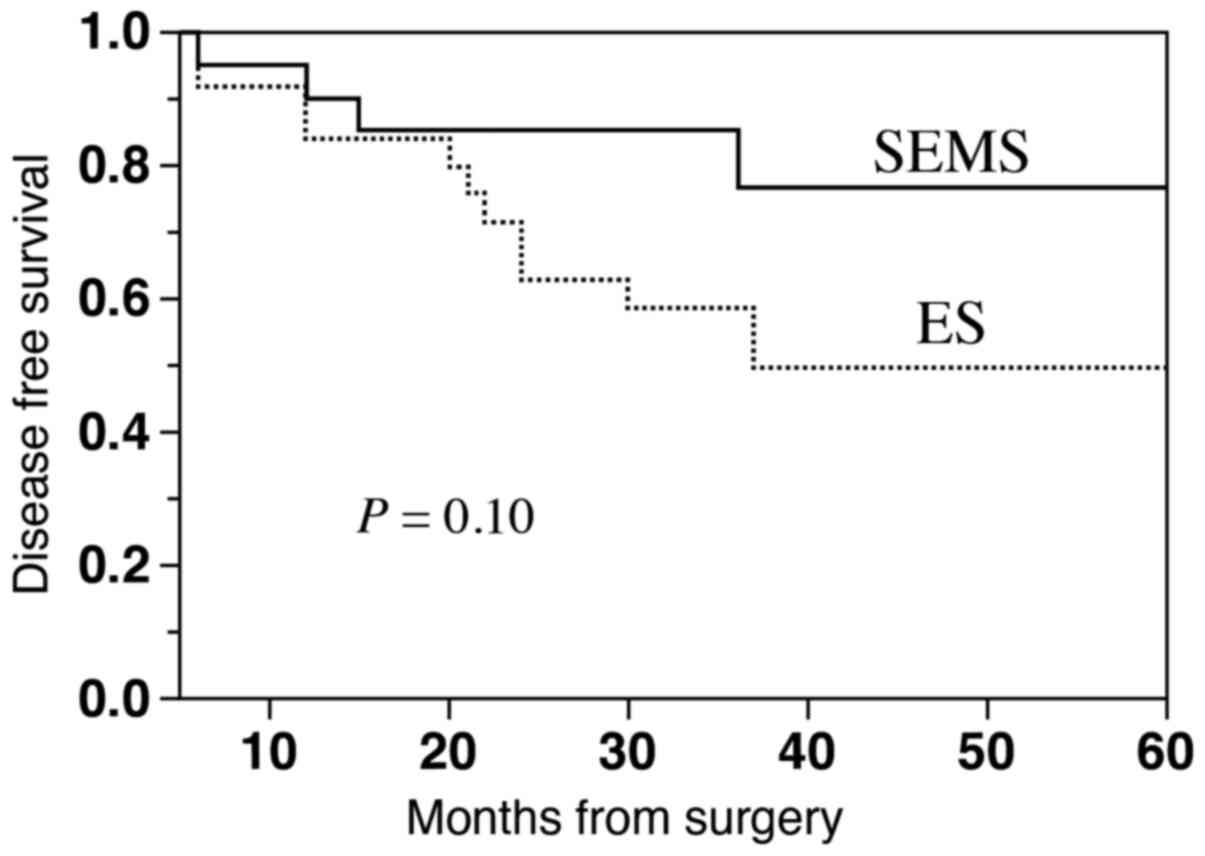 |
Figure 4
Kaplan-Meier curves of disease-free survival in the SEMS group and the ES group excluding stage IV. SEMS, Self-expanding metal stent; ES, emergency surgery.
|
|
Table V
Comparisons of background features of patients with stage II or III between the SMES group and the ES group.
|
Table V
Comparisons of background features of patients with stage II or III between the SMES group and the ES group.
| Patient detailsa |
SEMS group (n=21) |
ES group (n=25) |
P-value |
| Age in years, median (range) |
73 (48-91) |
63 (35-96) |
0.38 |
| Sex, n (%) |
|
|
0.23 |
| Male |
11 (52.4) |
8 (32.0) |
|
| Female |
10 (47.6) |
17 (68.0) |
|
| Tumor location, n (%) |
|
|
1.00 |
| Rectum |
2 (9.5) |
5 (12.0) |
|
| Sigmoid colon |
10 (47.6) |
11 (44.0) |
|
| Descending colon |
2 (9.5) |
3 (12.0) |
|
| Transverse colon |
6 (28.6) |
5 (20.0) |
|
| Ascending colon |
1 (4.8) |
2 (8.0) |
|
| Cecum |
0 (0.0) |
1 (4.0) |
|
| pStage of the tumor, n (%) |
|
|
1.00 |
| Stage II |
6 (28.6) |
8 (32.0) |
|
| Stage III |
15 (71.4) |
20 (68.0) |
|
| Number of lymph nodes resected, median (range) |
32 (15-71) |
19 (2-49) |
0.003 |
| Adjuvant chemotherapy, n (%) |
10 (47.6) |
18 (72.0) |
0.13 |
| Follow-up in months, median (range) |
33 (6-60) |
60 (12-60) |
0.02 |
|
Table VI
Results of the Cox regression analysis of prognostic factors for disease-free survival.
|
Table VI
Results of the Cox regression analysis of prognostic factors for disease-free survival.
| |
HR |
95%CI |
P-value |
| Treatment (ES/SEMS) |
4.65 |
1.23-21.12 |
0.02 |
| Age (≧65 years/<65 years) |
1.87 |
0.62-6.02 |
0.27 |
| Sex (Female/male) |
1.32 |
0.31-6.23 |
0.72 |
| Stage (Stage III/stage II) |
3.31 |
0.89-16.79 |
0.07 |
| Location (Right/left) |
1.63 |
0.54-4.96 |
0.38 |
| Chemotherapy (-/+) |
3.85 |
0.83-19.31 |
0.09 |
| Number of lymph nodes resected (<25/≧25) |
1.08 |
0.36-3.36 |
0.89 |
Discussion
The present study included patients who required emergency decompression because of acute MCRO who were treated either by SEMS placement or by ES within 24 h. Regarding short-term outcomes, the SEMS group had greater incidences of laparoscopic surgery and primary anastomosis, a greater number of dissected lymph nodes, and lower incidences of stoma formation and adverse events compared with the ES group. Regarding long-term results, disease-free survival did not significantly differ between the two groups by log-rank test, whereas the choice of SEMS placement for initial treatment was found to be the sole prognostic factor for disease-free survival by Cox regression analysis.
The present study had a technical success rate of SEMS placement of 92.0%. There were no critical events such as perforation related to SEMS placement, even in patients with MRCO requiring emergency decompression. This could be exactly why the outcomes of the SEMS group were superior to the ES group. This may be because we performed SEMS placement in accordance with the guidelines of the JCSSPRG (detailed in the Methods section). Similar favorable results for SEMS placement have been reported (19,20,23). In contrast, a previous systematic review reported a mean success rate of SEMS placement of only 76.9% (range, 46.7-100%) (10). The current success rates of SEMS placement are likely to vary depending on individual institutions or countries. The standardization of appropriate adaptation criteria and safe procedures based on accumulated experience is required in the future.
The SEMS group had superior short-term outcomes (laparoscopic surgical rate, primary anastomosis rate, number of dissected lymph nodes, stoma formation rate, and adverse event rate) compared with the ES group. Laparoscopic surgery after SEMS placement has previously been reported to be safe (26). Several studies have reported that SEMS placement is superior to ES regarding the rates of primary anastomosis, stoma formation, and adverse events (10,11,27,28). Furthermore, a previous study reported similar results to the present study regarding the number of resected lymph nodes (29). Compared with elective surgery, ES may increase the difficulty of performing sufficient lymph node dissection because of the worse patient condition and dilated intestinal tract. Although the SEMS group had a significantly greater number of dissected lymph nodes than the ES group, the number of lymph node dissections was not a significant prognostic factor by Cox regression analysis. These results might be due to the small sample size of this study. SEMS placement allows the physician to perform medical resuscitation, optimization of comorbid disorders, bowel preparation, accurate tumor staging, and preoperative total colonic examination to exclude synchronous proximal lesions (14,15). Therefore, SEMS placement as a BTS makes it possible to perform the usual elective laparoscopic surgery, even in patients with MRCO requiring emergency decompression.
The ESGE guidelines do not recommend the use of SEMS placement as a BTS for MCRO, based on the findings of a prospective cohort study and another comparative study that used propensity score matched analysis (17). The former study reported a significantly greater incidence of local disease recurrence in the SEMS group in patients aged ≤75 years, but not in the total subjects (30). The latter retrospective analysis reported a significantly lower 5-year overall survival rate and a significantly increased cancer-related mortality rate in those who underwent SEMS as a BTS compared with those who underwent ES; however, these worse outcomes were considered to have occurred due to perforation (31). Other studies that referred to the ESGE guidelines comparing SEMS placement as a BTS vs. ES also showed that impaired oncological outcomes are associated with higher perforation rates. In particular, perforation may lead an increased risk of peritoneal carcinomatosis and sepsis, which means that patients with curable colorectal cancer can become non-curable due to SEMS intervention (32-34). Moreover, follow-up data from the Stent-in 2 trial showed that the cumulative incidence of overall recurrences in patients with clinical stent-related perforation was significantly increased compared with those who underwent ES or SEMS placement without perforation (35). In the present study, no perforation occurred, and so no differences in disease-free survival were found. The higher clinical success rate in those who undergo SEMS placement without perforation would also enable curative surgery, including sufficient lymph node dissection. Furthermore, the enforced radial dilatation caused by SEMS placement may increase the risk of dissemination of cancer cells into the peritoneal cavity, surrounding lymphatic vessels, and bloodstream (36). Sabbagh et al (37) also reported that SEMS placement caused higher rates of tumor ulceration, peritumor ulceration, perineural invasion, and lymph node invasion than ES, and that these pathological alterations might worsen the oncological impact. However, several studies have recently reported no differences in long-term outcomes between the SEMS group and the ES group (28,38,39). A systematic review and meta-analysis that evaluated the long-term outcomes (overall survival, disease-free survival, and recurrence rates) of SEMS placement as a BTS and ES for MCRO also suggested that SEMS placement has no adverse impact on oncological outcomes compared with ES (40). At the very least, it cannot be concluded that SEMS placement as a BTS is inferior to ES in long-term outcomes, as long as the rate of perforation is minimized.
In evaluating both short- and long-term outcomes, it is important to assess the clinical success rate of SEMS placement without perforation. Reported risk factors for perforation include the stent design, benign etiology, stricture dilation before stent placement, and limited experience with pancreaticobiliary endoscopy (24,41-43). In addition, it is important to master the SEMS placement technique. Established procedures should always be done in the same manner; for example, in accordance with the guidelines of the JCSSPRG. If it is impossible to place the SEMS, or if perforation occurs in the period before elective surgery, it is also important to prepare a protocol for switching to surgery promptly. In the present study, adverse events related to SEMS placement before elective surgery occurred in two cases (one migration and one re-obstruction), and these adverse events were improved endoscopically. However, serious adverse events such as perforation may occasionally occur; therefore, SEMS placement as a BTS should only be performed at facilities where emergency treatment or surgery could be performed at any time.
There are some potential limitations of our study. First, the study was performed in a single center, and was retrospectively designed. Consequently, there could be a regional or institutional selection bias. In addition, although the indication of SEMS or ES was determined by a discussion between endoscopists and surgeons on the basis of individual cases, there were no specifically established criteria, which might affect the prognosis However, we registered all consecutive patients with MRCO requiring emergency decompression in the study period, and there was no deliberate selection. Second, the follow-up period in the SEMS group was shorter than that in the ES group, as colonic SEMS placement became available within the public health insurance system from 2012 in Japan. Third, it is possible that the differences in the types of adjuvant chemotherapy may have affected the prognosis. However, since 2000, we have selected regimens that included FOLFOX or FOLFILI as postoperative adjuvant chemotherapy for stage II or III colorectal cancer patients according the JSCCR guidelines (25). In Japan, molecular targeting agents have not been used for adjuvant chemotherapy, therefore, we do not believe that adjuvant chemotherapy had a significant impact on the prognosis of this cohort. Fourth, there was a patient who underwent elective surgery 90 days after the SEMS insertion. This patient was an 89-year-old woman who was deciding whether to undergo surgery and finally did. However, we always recommend that elective surgery should be performed within 2 weeks after the SEMS insertion. In real-world clinical cases, various cases were included. Large-scale, randomized controlled trials are desirable to clarify the oncological long-term effects of colonic SEMS as a BTS in the future.
In conclusion, SEMS placement as a BTS is superior to ES in several short-term outcomes. Regarding long-term outcomes, the disease-free survival of the SEMS group was not inferior to that of the ES group. As long as adverse events such as perforation are minimized, SEMS placement as a BTS could be a first treatment option for MCRO, though further study will be needed in the future.
Acknowledgements
The authors would like to thank to Miss Naomi Takenaka (Nikko Memorial Hospital, Japan) for creating the figures.
Funding
No funding was received.
Availability of data and materials
Data sharing is not applicable to this article, as no datasets were generated during the current study.
Authors' contributions
YY, HM, YM, SEK and FI conceived and designed the current study. YY, MM, YS, KK, TI, KI, TK, TH and KW acquired the data. YY, HM and TB analyzed and interpreted the data. YY performed statistical analysis. YY drafted the manuscript. SEK, TB and FI supervised the study. SEK and HM critically revised the manuscript for important intellectual content. All authors read and approved the final version of the manuscript.
Ethics approval and consent to participate
Written informed consent was obtained from all patients before the procedures. The ethics committee of our institution approved the study protocol (approval no. 17H073). The present study is registered in the University Hospital Medical Network Clinical Trials Registry (UMIN R000034868). The current study was conducted in accordance with the Declaration of Helsinki.
Patient consent for publication
Informed consent was obtained from all patients for the publication of patient information.
Competing interests
The authors declare that they have no competing interests.
References
|
1
|
Jemal A, Bray F, Center MM, Ferlay J, Ward E and Forman D: Global cancer statistics. CA Cancer J Clin. 61:69–90. 2011.PubMed/NCBI View Article : Google Scholar
|
|
2
|
Winner M, Mooney SJ, Hershman DL, Feingold DL, Allendorf JD, Wright JD and Neugut AI: Incidence and predictors of bowel obstruction in elderly patients with stage IV colon cancer: A population-based cohort study. JAMA Surg. 148:715–722. 2013.PubMed/NCBI View Article : Google Scholar
|
|
3
|
Jullumstro E, Wibe A, Lydersen S and Edna TH: Colon cancer incidence, presentation, treatment and outcomes over 25 years. Colorectal Dis. 13:512–518. 2011.PubMed/NCBI View Article : Google Scholar
|
|
4
|
Cheynel N, Cortet M, Lepage C, Benoit L, Faivre J and Bouvier AM: Trends in frequency and management of obstructing colorectal cancers in a well-defined population. Diseases of the colon and rectum. 50:1568–1575. 2007.PubMed/NCBI View Article : Google Scholar
|
|
5
|
Tekkis PP, Kinsman R, Thompson MR and Stamatakis JD: The Association of Coloproctology of Great Britain and Ireland study of large bowel obstruction caused by colorectal cancer. Ann Surg. 240:76–81. 2004.PubMed/NCBI View Article : Google Scholar
|
|
6
|
Deans GT, Krukowski ZH and Irwin ST: Malignant obstruction of the left colon. Br J Surg. 81:1270–1276. 1994.PubMed/NCBI View Article : Google Scholar
|
|
7
|
Dohmoto M: New method: Endoscopic implantation of rectal stent in palliative treatment of malignant stenosis. Endosc Dig. 3:1507–1512. 1991.
|
|
8
|
Tejero E, Mainar A, Fernández L, Tobío R and De Gregorio MA: New procedure for the treatment of colorectal neoplastic obstructions. Dis Colon Rectum. 37:1158–1159. 1994.PubMed/NCBI View Article : Google Scholar
|
|
9
|
Zhao X, Liu B, Zhao E, Wang J, Cai M, Xia Z, Xia Q, Shuai X, Tao K, Wang G and Cai K: The safety and efficiency of surgery with colonic stents in left-sided malignant colonic obstruction: A meta-analysis. Gastroenterol Res Pract. 2014(407325)2014.PubMed/NCBI View Article : Google Scholar
|
|
10
|
Huang X, Lv B, Zhang S and Meng L: Preoperative colonic stents versus emergency surgery for acute left-sided malignant colonic obstruction: A meta-analysis. J Gastrointest Surg. 18:584–591. 2014.PubMed/NCBI View Article : Google Scholar
|
|
11
|
Tan CJ, Dasari BV and Gardiner K: Systematic review and meta-analysis of randomized clinical trials of self-expanding metallic stents as a bridge to surgery versus emergency surgery for malignant left-sided large bowel obstruction. Br J Surg. 99:469–476. 2012.PubMed/NCBI View Article : Google Scholar
|
|
12
|
ASGE Standards of Practice Committee. Harrison ME, Anderson MA, Appalaneni V, Banerjee S, Ben-Menachem T, Cash BD, Fanelli RD, Fisher L, Fukami N, et al: The role of endoscopy in the management of patients with known and suspected colonic obstruction and pseudo-obstruction. Gastrointest Endosc. 71:669–679. 2010.PubMed/NCBI View Article : Google Scholar
|
|
13
|
Sebastian S, Johnston S, Geoghegan T, Torreggiani W and Buckley M: Pooled analysis of the efficacy and safety of self-expanding metal stenting in malignant colorectal obstruction. Am J Gastroenterol. 99:2051–2057. 2004.PubMed/NCBI View Article : Google Scholar
|
|
14
|
Cirocchi R, Farinella E, Trastulli S, Desiderio J, Listorti C, Boselli C, Parisi A, Noya G and Sagar J: Safety and efficacy of endoscopic colonic stenting as a bridge to surgery in the management of intestinal obstruction due to left colon and rectal cancer: A systematic review and meta-analysis. Surg Oncol. 22:14–21. 2013.PubMed/NCBI View Article : Google Scholar
|
|
15
|
Khot UP, Lang AW, Murali K and Parker MC: Systematic review of the efficacy and safety of colorectal stents. Br J Surg. 89:1096–1102. 2002.PubMed/NCBI View Article : Google Scholar
|
|
16
|
Martinez-Santos C, Lobato RF, Fradejas JM, Pinto I, Ortega-Deballón P and Moreno-Azcoita M: Self-expandable stent before elective surgery vs. emergency surgery for the treatment of malignant colorectal obstructions: Comparison of primary anastomosis and morbidity rates. Dis Colon Rectum. 45:401–406. 2002.PubMed/NCBI View Article : Google Scholar
|
|
17
|
van Hooft JE, van Halsema EE, Vanbiervliet G, Beets-Tan RG, DeWitt JM, Donnellan F, Dumonceau JM, Glynne-Jones RG, Hassan C, Jiménez-Perez J, et al: Self-expandable metal stents for obstructing colonic and extracolonic cancer: European Society of Gastrointestinal Endoscopy (ESGE) Clinical Guideline. Endoscopy. 46:990–1053. 2014.PubMed/NCBI View Article : Google Scholar
|
|
18
|
Saida Y, Sumiyama Y, Nagao J and Takase M: Stent endoprosthesis for obstructing colorectal cancers. Dis Colon Rectum. 39:552–555. 1996.PubMed/NCBI View Article : Google Scholar
|
|
19
|
Matsuzawa T, Ishida H, Yoshida S, Isayama H, Kuwai T, Maetani I, Shimada M, Yamada T, Saito S, Tomita M, et al: A Japanese prospective multicenter study of self-expandable metal stent placement for malignant colorectal obstruction: Short-term safety and efficacy within 7 days of stent procedure in 513 cases. Gastrointest Endosc. 82:697–707.e1. 2015.PubMed/NCBI View Article : Google Scholar
|
|
20
|
Saito S, Yoshida S, Isayama H, Matsuzawa T, Kuwai T, Maetani I, Shimada M, Yamada T, Tomita M, Koizumi K, et al: A prospective multicenter study on self-expandable metallic stents as a bridge to surgery for malignant colorectal obstruction in Japan: Efficacy and safety in 312 patients. Surg Endosc. 30:3976–3986. 2016.PubMed/NCBI View Article : Google Scholar
|
|
21
|
Edge SB, Byrd DR, Compton CC, Fritz AG, Greene FL and Trotti A (eds): AJCC Cancer Staging Manual. 7th editon. Springer, New York, NY, 2010.
|
|
22
|
Baron TH, Wong Kee Song LM and Repici A: Role of self-expandable stents for patients with colon cancer (with videos). Gastrointest Endosc. 75:653–662. 2012.PubMed/NCBI View Article : Google Scholar
|
|
23
|
Meisner S, González-Huix F, Vandervoort JG, Goldberg P, Casellas JA, Roncero O, Grund KE, Alvarez A, García-Cano J, Vázquez-Astray E, et al: Self-expandable metal stents for relieving malignant colorectal obstruction: Short-term safety and efficacy within 30 days of stent procedure in 447 patients. Gastrointest Endosc. 74:876–884. 2011.PubMed/NCBI View Article : Google Scholar
|
|
24
|
Small AJ and Baron TH: Comparison of Wallstent and Ultraflex stents for palliation of malignant left-sided colon obstruction: A retrospective, case-matched analysis. Gastrointestinal endoscopy. 67:478–488. 2008.PubMed/NCBI View Article : Google Scholar
|
|
25
|
Watanabe T, Muro K, Ajioka Y, Hashiguchi Y, Ito Y, Saito Y, Hamaguchi T, Ishida H, Ishiguro M, Ishihara S, et al: Japanese Society for Cancer of the Colon and Rectum (JSCCR) guidelines 2016 for the treatment of colorectal cancer. Int J Clin Oncol. 23:1–34. 2018.PubMed/NCBI View Article : Google Scholar
|
|
26
|
Shimizu H, Yamazaki R, Ohtsuka H, Osaka I, Takuma K and Morita Y: Feasibility of laparoscopic surgery after stent insertion for obstructive colorectal cancer. Asian J Endosc Surg. 11:118–122. 2018.PubMed/NCBI View Article : Google Scholar
|
|
27
|
Arezzo A, Passera R, Lo Secco G, Verra M, Bonino MA, Targarona E and Morino M: Stent as bridge to surgery for left-sided malignant colonic obstruction reduces adverse events and stoma rate compared with emergency surgery: Results of a systematic review and meta-analysis of randomized controlled trials. Gastrointest Endosc. 86:416–426. 2017.PubMed/NCBI View Article : Google Scholar
|
|
28
|
Gianotti L, Tamini N, Nespoli L, Rota M, Bolzonaro E, Frego R, Redaelli A, Antolini L, Ardito A, Nespoli A and Dinelli M: A prospective evaluation of short-term and long-term results from colonic stenting for palliation or as a bridge to elective operation versus immediate surgery for large-bowel obstruction. Surg Endosc. 27:832–842. 2013.PubMed/NCBI View Article : Google Scholar
|
|
29
|
Tung KL, Cheung HY, Ng LW, Chung CC and Li MK: Endo-laparoscopic approach versus conventional open surgery in the treatment of obstructing left-sided colon cancer: Long-term follow-up of a randomized trial. Asian J Endosc Surg. 6:78–81. 2013.PubMed/NCBI View Article : Google Scholar
|
|
30
|
Gorissen KJ, Tuynman JB, Fryer E, Wang L, Uberoi R, Jones OM, Cunningham C and Lindsey I: Local recurrence after stenting for obstructing left-sided colonic cancer. Br J Surg. 100:1805–1809. 2013.PubMed/NCBI View Article : Google Scholar
|
|
31
|
Sabbagh C, Browet F, Diouf M, Cosse C, Brehant O, Bartoli E, Mauvais F, Chauffert B, Dupas JL, Nguyen-Khac E and Regimbeau JM: Is stenting as ‘a bridge to surgery’ an oncologically safe strategy for the management of acute, left-sided, malignant, colonic obstruction? A comparative study with a propensity score analysis. Ann Surg. 258:107–115. 2013.PubMed/NCBI View Article : Google Scholar
|
|
32
|
van Hooft JE, Bemelman WA, Oldenburg B, Marinelli AW, Lutke Holzik MF, Grubben MJ, Sprangers MA, Dijkgraaf MG and Fockens P: collaborative Dutch Stent-In study group. Colonic stenting versus emergency surgery for acute left-sided malignant colonic obstruction: A multicentre randomised trial. Lancet Oncol. 12:344–352. 2011.PubMed/NCBI View Article : Google Scholar
|
|
33
|
Anwar MA, D'Souza F, Coulter R, Memon B, Khan IM and Memon MA: Outcome of acutely perforated colorectal cancers: Experience of a single district general hospital. Surg Oncol. 15:91–96. 2006.PubMed/NCBI View Article : Google Scholar
|
|
34
|
van Hooft JE, Fockens P, Marinelli AW, Bossuyt PM and Bemelman WA: Dutch Stent-In study group. Premature closure of the Dutch Stent-in I study. Lancet. 368:1573–1574. 2006.PubMed/NCBI View Article : Google Scholar
|
|
35
|
Sloothaak DA, van den Berg MW, Dijkgraaf MG, Fockens P, Tanis PJ, van Hooft JE and Bemelman WA: collaborative Dutch Stent-In study group. Oncological outcome of malignant colonic obstruction in the Dutch Stent-In 2 trial. Br J Surg. 101:1751–1757. 2014.PubMed/NCBI View Article : Google Scholar
|
|
36
|
Maruthachalam K, Lash GE, Shenton BK and Horgan AF: Tumour cell dissemination following endoscopic stent insertion. Br J Surg. 94:1151–1154. 2007.PubMed/NCBI View Article : Google Scholar
|
|
37
|
Sabbagh C, Chatelain D, Trouillet N, Mauvais F, Bendjaballah S, Browet F and Regimbeau JM: Does use of a metallic colon stent as a bridge to surgery modify the pathology data in patients with colonic obstruction? A case-matched study. Surg Endosc. 27:3622–3631. 2013.PubMed/NCBI View Article : Google Scholar
|
|
38
|
Arezzo A, Balague C, Targarona E, Borghi F, Giraudo G, Ghezzo L, Arroyo A, Sola-Vera J, De Paolis P, Bossotti M, et al: Colonic stenting as a bridge to surgery versus emergency surgery for malignant colonic obstruction: Results of a multicentre randomised controlled trial (ESCO trial). Surg Endosc. 31:3297–3305. 2017.PubMed/NCBI View Article : Google Scholar
|
|
39
|
Verstockt B, Van Driessche A, De Man M, van der Spek P, Hendrickx K, Casneuf V, Dobbels P, Van Molhem Y and Vandervoort J: Ten-year survival after endoscopic stent placement as a bridge to surgery in obstructing colon cancer. Gastrointest Endosc. 87:705–713.e2. 2018.PubMed/NCBI View Article : Google Scholar
|
|
40
|
Matsuda A, Miyashita M, Matsumoto S, Matsutani T, Sakurazawa N, Takahashi G, Kishi T and Uchida E: Comparison of long-term outcomes of colonic stent as ‘bridge to surgery’ and emergency surgery for malignant large-bowel obstruction: A meta-analysis. Ann Surg Oncol. 22:497–504. 2015.PubMed/NCBI View Article : Google Scholar
|
|
41
|
van Halsema EE, van Hooft JE, Small AJ, Baron TH, García-Cano J, Cheon JH, Lee MS, Kwon SH, Mucci-Hennekinne S, Fockens P, et al: Perforation in colorectal stenting: A meta-analysis and a search for risk factors. Gastrointest Endosc. 79:970–982.e7; quiz 983.e2, 983.e5. 2014.PubMed/NCBI View Article : Google Scholar
|
|
42
|
Small AJ, Coelho-Prabhu N and Baron TH: Endoscopic placement of self-expandable metal stents for malignant colonic obstruction: Long-term outcomes and complication factors. Gastrointest Endosc. 71:560–572. 2010.PubMed/NCBI View Article : Google Scholar
|
|
43
|
Tanaka A, Sadahiro S, Yasuda M, Shimizu S, Maeda Y, Suzuki T, Tokunaga N and Ogoshi K: Endoscopic balloon dilation for obstructive colorectal cancer: A basic study on morphologic and pathologic features associated with perforation. Gastrointest Endosc. 71:799–805. 2010.PubMed/NCBI View Article : Google Scholar
|


