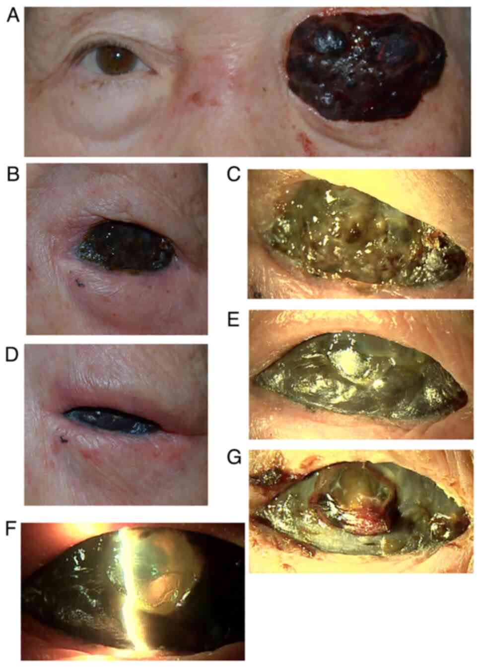
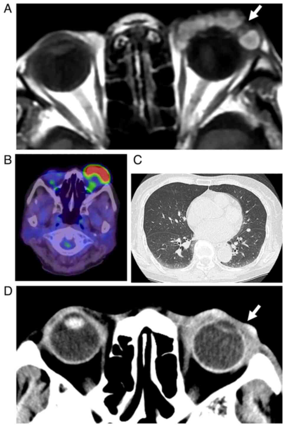
|
1
|
Matsuo T, Ogino Y, Ichimura K, Tanaka T
and Kaji M: Clinicopathological correlation for the role of
fluorodeoxyglucose positron emission tomography computed tomography
in detection of choroidal malignant melanoma. Int J Clin Oncol.
19:230–239. 2014.PubMed/NCBI View Article : Google Scholar
|
|
2
|
Matsuo T, Tanaka T and Yamasaki O:
Lacrimal sac malignant melanoma in 15 Japanese patients: Case
report and literature review. J Investig Med High Impact Case Rep.
7(2324709619888052)2019.PubMed/NCBI View Article : Google Scholar
|
|
3
|
Seregard S: Conjunctival melanoma. Surv
Ophthalmol. 42:321–350. 1998.PubMed/NCBI View Article : Google Scholar
|
|
4
|
Vora GK, Demirci H, Marr B and
Mruthyunjaya P: Advances in the management of conjunctival
melanoma. Surv Ophthalmol. 62:26–42. 2017.PubMed/NCBI View Article : Google Scholar
|
|
5
|
Jain P, Finger PT, Fili M, Damato B,
Coupland SE, Heimann H, Kenawy N, Brouwer NJ, Marinkovic M, Van
Duinen SG, et al: Conjunctival melanoma treatment outcomes in 288
patients: A multicentre international data-sharing study. Br J
Ophthalmol. 105:1358–1364. 2021.PubMed/NCBI View Article : Google Scholar
|
|
6
|
Ballo MT and Ang KK: Radiotherapy for
cutaneous malignant melanoma: Rationale and indications. Oncology
(Williston Park). 18:99–110, 113-114. 2004.PubMed/NCBI
|
|
7
|
Gorayski P, Burmeister B and Foote M:
Radiotherapy for cutaneous melanoma: Current and future
applications. Future Oncol. 11:525–534. 2015.PubMed/NCBI View Article : Google Scholar
|
|
8
|
López F, Rodrigo JP, Cardesa A,
Triantafyllou A, Devaney KO, Mendenhall WM, Haigentz M Jr, Strojan
P, Pellitteri PK, Bradford CR, et al: Update on primary head and
neck mucosal melanoma. Head Neck. 38:147–155. 2016.PubMed/NCBI View Article : Google Scholar
|
|
9
|
Grant-Freemantle MC, O'Neill BL and Clover
AJP: The effectiveness of radiotherapy in the treatment of head and
neck mucosal melanoma: Systematic review and meta-analysis. Head
Neck. 43:323–333. 2021.PubMed/NCBI View Article : Google Scholar
|
|
10
|
Wuestemeyer H, Sauerwein W, Meller D,
Chauvel P, Schueler A, Steuhl KP, Bornfeld N and Anastassiou G:
Proton radiotherapy as an alternative to exenteration in the
management of extended conjunctival melanoma. Graefes Arch Clin Exp
Ophthalmol. 244:438–446. 2006.PubMed/NCBI View Article : Google Scholar
|
|
11
|
Westekemper H, Anastassiou G, Sauerwein W,
Chauvel P, Bornfeld N, Steuhl KP and Meller D: Analysis of ocular
surface alterations following proton beam radiation in eyes with
conjunctival malignant melanoma. Ophthalmologe. 103:588–595.
2006.PubMed/NCBI View Article : Google Scholar : (In German).
|
|
12
|
Krause L, Mladenova A, Bechrakis NE,
Kreusel KM, Plath T, Moser L and Foerster M: Treatment modalities
for conjunctival melanoma. Klin Monbl Augenheilkd. 226:1012–1016.
2009.PubMed/NCBI View Article : Google Scholar : (In German).
|
|
13
|
Maschi-Cayla C, Doyen J, Gastaud P and
Caujolle JP: Conjunctival melanomas and proton beam therapy. Acta
Ophthalmol. 91(e647)2013.PubMed/NCBI View Article : Google Scholar
|
|
14
|
Scholz SL, Hérault J, Stang A, Griewank
KG, Meller D, Thariat J, Steuhl KP, Westekemper H and Sauerwein W:
Proton radiotherapy in advanced malignant melanoma of the
conjunctiva. Graefes Arch Clin Exp Ophthalmol. 257:1309–1318.
2019.PubMed/NCBI View Article : Google Scholar
|
|
15
|
Thariat J, Salleron J, Maschi C, Fevrier
E, Lassalle S, Gastaud L, Baillif S, Claren A, Baumard J, Herault J
and Caujolle JP: Oncologic and visual outcomes after postoperative
proton therapy of localized conjunctival melanomas. Radiation
Oncol. 14(239)2019.PubMed/NCBI View Article : Google Scholar
|
|
16
|
Zenda S, Kawashima M, Nishio T, Kohno R,
Nihei K, Onozawa M, Arahira S and Ogino T: Proton beam therapy as a
nonsurgical approach to mucosal melanoma of the head and neck: A
pilot study. Int J Radiat Oncol Biol Phys. 81:135–139.
2011.PubMed/NCBI View Article : Google Scholar
|
|
17
|
Fuji H, Yoshikawa S, Kasami M, Murayama S,
Onitsuka T, Kashiwagi H and Kiyohara Y: High-dose proton beam
therapy for sinonasal mucosal malignant melanoma. Radiat Oncol.
9(162)2014.PubMed/NCBI View Article : Google Scholar
|
|
18
|
Sakurai H, Ishikawa H and Okumura T:
Proton beam therapy in Japan: Current and future status. Jpn J Clin
Oncol. 46:885–892. 2016.PubMed/NCBI View Article : Google Scholar
|
|
19
|
Larsen AC, Dahmcke CM, Dahl C, Siersma VD,
Toft PB, Coupland SE, Prause JU, Guldberg P and Heegaard S: A
retrospective review of conjunctival melanoma: Presentation,
treatment and outcome and an investigation of features associated
with BRAF mutations. JAMA Ophthalmol. 133:1295–1303.
2015.PubMed/NCBI View Article : Google Scholar
|
|
20
|
Kiyohara T, Tanimura H, Miyamoto M,
Shijimaya T, Nagano N, Nakamaru S, Makimura K and Iwai H: Two cases
of BRAF-mutated, bulbar conjunctival melanoma, and review of the
published literature. Clin Exp Dermatol. 45:207–211.
2020.PubMed/NCBI View Article : Google Scholar
|
|
21
|
Matsuo T and Yamasaki O:
Vogt-Koyanagi-Harada disease-like posterior uveitis in the course
of nivolumab (anti-PD-1 antibody), interposed by vemurafenib (BRAF
inhibitor), for metastatic cutaneous malignant melanoma. Clin Case
Rep. 5:694–700. 2017.PubMed/NCBI View
Article : Google Scholar
|