Introduction
Invasive breast carcinoma (IBC) is the second most
common malignancy worldwide, accounting for 11.6% of cancer cases,
and has a mortality rate of 6.6% (1). IBC comprises a heterogeneous group of
breast malignancies with different clinical, biological and
prognostic characteristics (2).
IBC may be divided into three molecular cancer
subtypes: Luminal, HER2-enriched and basal-like. Sørlie et
al (3) further divided the
luminal subtype into luminal A and B. This is particularly relevant
in early IBC (EIBC), as hormonal therapy is usually sufficient for
luminal A tumors. By contrast, luminal B tumors benefit from more
aggressive therapeutics, including chemotherapy regimens (4-6).
MammaPrint (Agendia, Inc.) evaluates the expression
of 70 genes, which mostly have known biological functions
implicated in tumor progression and metastasis (7,8).
EndoPredict (Myriad Genetics, Inc.) is a 12-gene signature test (8
cancer-related genes, 3 normalization genes and 1 control gene)
that was designed to add clinicopathological factors such as tumor
size and nodal status to obtain the so-called EPclin score, and an
estimated risk for distant recurrence at 10 years (9,10).
The prognostic performances of these two gene molecular signature
panels have level I evidence in pre- and postmenopausal females
(11). Of note, they are
independent of other well-known prognostic tumor parameters,
including tumor size, histological grade and nodal status. The
principal implication of both molecular signature tests in clinical
management is the selection of patients that are unlikely to
benefit from conventional chemotherapy regimens (10,12).
Despite the importance of molecular signature tests
in patient management, their cost limits their routine utilization.
As a result, conventional immunohistochemistry (IHC) has been
explored as an alternative to these tests (13-15).
It has been proposed that the Ki-67 proliferative index may be
utilized in addition to the estrogen receptor (ER), progesterone
receptor (PR) and HER2 receptor to discriminate between luminal A
and B subtypes (16,17).
High values of the proliferative cell marker Ki-67
have been associated with a benefit from chemotherapy regimens in
IBC (6,18). However, establishing Ki-67 index
cut-offs for stratifying patient prognosis has proven to be a
difficult task due to the lack of assessment standardization
(19); this has been acknowledged
by the St. Gallen consensus with changes in recommendations through
time (4,20), the latest of which from 2015
suggests the median Ki-67 index internal laboratory value as the
cut-off for highly proliferative tumors (6).
In the present study, the Ki-67 index was evaluated
as an alternative to molecular signature tests to identify high
risk of recurrence in patients with hormone receptor (HR)+
EIBC.
Materials and methods
Patients
Using the breast cancer registry of San José
TecSalud Hospital (Monterrey, México), a retrospective review was
performed to identify patients with HR+ EIBC who were tested with
molecular signature tests and the Ki-67 index. The cohort was
divided according to the molecular signature test utilized. In the
EndoPredict cohort, patients were tested between June 2016 and
August 2018. This group comprised premenopausal females with HR+
EIBC, HER2 negative, T1-T2, N0-N1 and M0(21). In the MammaPrint cohort, patients
were evaluated from June 2016 to August 2018. This group included
patients with HR+ EIBC, HER2 negative, T1-T2 and operable T3, and
N0-N1 tumors according to previously utilized criteria (22). For both cohorts, age, tumor size,
TNM stage, histological subtype, Nottingham combined histological
grade (NHG) and lymphovascular invasion (LVI) data were
recorded.
IHC
In biopsies of the tumor samples, ER, PR, HER-2 and
Ki-67 were analyzed with a Ventana BenchMarck GX autostainer
(Hoffmann-La Roche, Ltd.) using the internal validated protocol.
Paraffin slides were deparaffinized using two changes of xylene for
10 min each and hydrated through an alcohol gradient and distilled
water (2 changes of 100% ethanol, 2 changes of 95% ethanol, 2
changes of distilled water). Heat-induced epitope retrieval with
citrate buffer was performed. Slides were then cooled and rinsed
with distilled water and rinsed in tris-buffered saline with
Tween-20 for 5 min. Slides were then rinsed with 3% hydrogen
peroxide, followed by a rinse with a wash buffer and covered with
300 µl of protein block (Protein block X0909; Dako; Agilent
Technologies, Inc.) for 5 min. Slides were treated with the
following antibodies for 16 min at 36˚C: Anti-estrogen receptor
(clone SP1) rabbit monoclonal primary antibody (cat. no. 790-4324;
prediluted concentration, 1 µg/ml), anti-progesterone receptor
rabbit monoclonal primary antibody (clone 1E2; cat. no. 790-2223;
prediluted concentration, 1 µg/ml), anti-HER-2/neu (clone 4B5; cat.
no. 790-100; prediluted concentration, 6 µg/ml) and anti-Ki-67
rabbit monoclonal primary antibody (clone 30-9; cat. no. 790-4286;
prediluted concentration, 2 µg/ml; all from Hoffmann-La Roche,
Ltd). Slides were then rinsed with wash buffer and incubated with
the secondary reagent, Dako Envision HRP-labeled polymer
anti-rabbit (cat. no. M3648; dilution, 1:50; Agilent Technologies,
Inc.) min at room temperature for 60 min. Subsequently,
diaminobenzidine was applied for 10 min at 36˚C and the slides were
rinsed with distilled water. Counterstaining was performed with
hematoxylin for 3 min and slides were washed in tap water. Slides
were then blued in ammonia water, rinsed in tap water, dehydrated
in an alcohol gradient (95% ethanol, 100% ethanol), cleared in
xylene (two changes) and mounted with coverslips for examination
with a microscope. All slides included an external positive tissue
control.
ER and PR were considered positive if >1% of the
neoplastic cells exhibited a nuclear stain according to the
American Society of Clinical Oncology/College of American
Pathologists (ASCO/CAP) guidelines (23). Low PR was defined as <20% of
nuclear-positive tumor cells (24). HER2 was also evaluated according to
the ASCO/CAP guidelines (25).
Ki-67 index was evaluated using a hot spots method, performed in
the area with the highest number of positive nuclei. A total of
three high-power fields using a magnification of x400 including a
hot spot were examined, as proposed by the International Ki-67 in
Breast Cancer Working group (26,27).
A cut-off of 20% was used as suggested by the St. Gallen consensus
(6).
Molecular signature tests
The EndoPredict (Myriad Genetics, Inc.) and
MammaPrint (Agendia, Inc.) molecular signature tests were performed
in a validated laboratory. An EpClin index of ≥3.3 was considered
to indicate high risk of recurrence. For the MammaPrint®
(Agendia, Inc.) assay a risk category of recurrence was assigned to
each case (LR or HR), along with the molecular subgroup (using
BluePrint assay).
Statistical analysis
An unpaired t-test, Fisher's exact and Mann-Whitney
U test were used for comparison between patients with high and low
Ki-67 index. Spearman rank-order correlation analysis for ordinal
and continuous variables. Odds ratios were determined to evaluate
the association between Ki-57 and molecular signature tests.
Receiver-operating characteristic curve (ROC) analysis was
performed for analysis of the risk of recurrence given by
MammaPrint or EndoPredict and the Ki-67 index.
Validation/association analysis was performed to test sensitivity
and specificity. The Kappa coefficient was determined to evaluate
the concordance between the Ki-67 index and the molecular signature
tests. P<0.05 was considered to indicate statistical
significance. GraphPad Prism 9.0.1 (GraphPad Software, Inc.) was
used for statistical analysis and graphics.
Results
Patient characteristics
The clinicopathological characteristics of the two
cohorts are listed in Table I. In
the MammaPrint cohort, the patients were older, had smaller tumors
and a lower stage compared with those in the EndoPredict cohort.
The proportion of patients with a high recurrence risk was higher
in the EndoPredict cohort (56.25 vs. 30.00%, P<0.001). The
median (interquartile range) Ki-67 index for the two cohorts in
patients with high and low recurrence risk was 30 (10-35) and 15
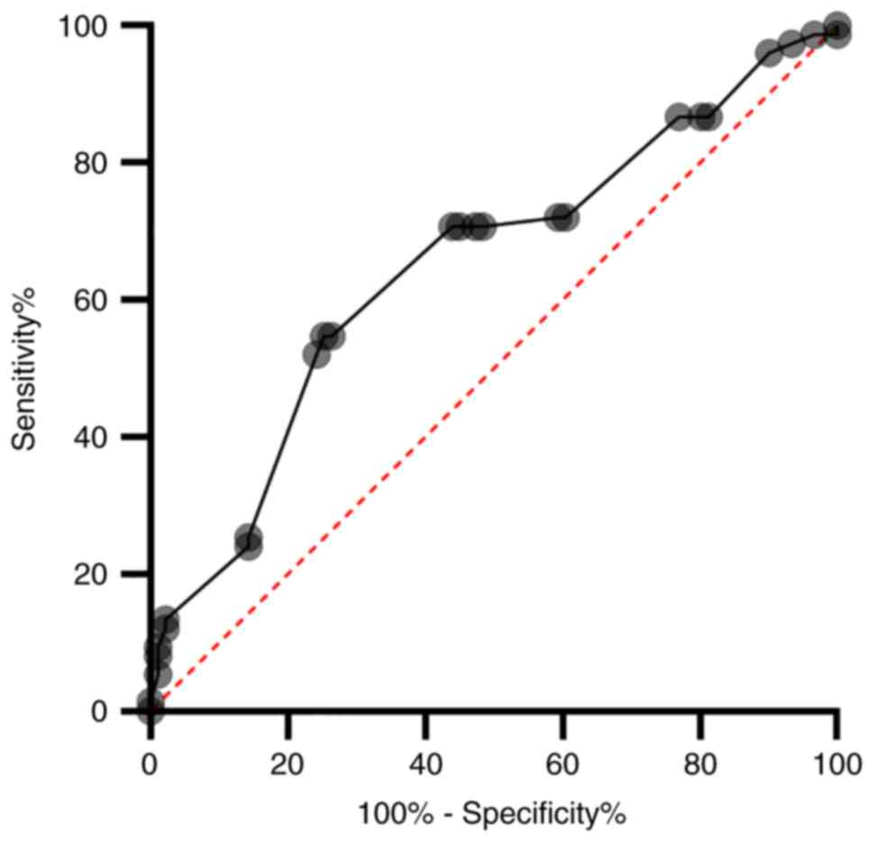
(10-25), respectively (P<0.001). In the ROC curve analysis for
the performance of Ki-67 index in the identification of all
patients at high risk of recurrence, the accuracy was 65% (P=0.001;
Fig. 1). In the ROC curve analysis
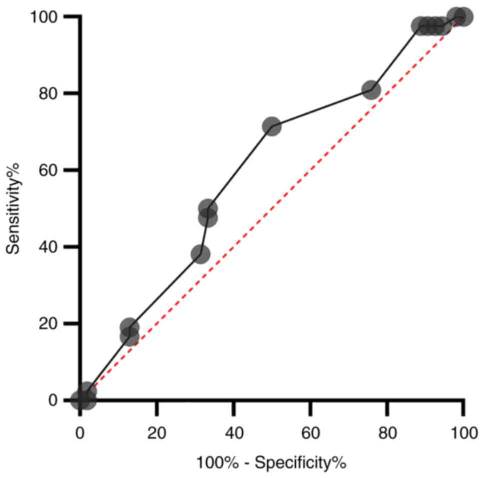
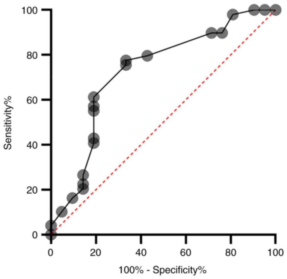
for the performance of Ki-67 index in the identification of
patients from the EndoPredict and MammaPrint cohort at high risk of
recurrence, the accuracy was 60% (P=0.110) and 70% (P=0.002)
(Figs. 2 and 3, respectively).
 | Table IPatient characteristics by test type
(n=166). |
Table I
Patient characteristics by test type
(n=166).
| Parameter | Total | EndoPredict
(n=96) | MammaPrint
(n=70) | P-value |
|---|
| Age, years | 45 (40-51) | 43 (39-46.5) | 51 (43-67) | <0.0001 |
| Tumor size, mm | 20 (13-26.5) | 22 (15-30) | 15.5 (12-25) | 0.0127 |
| TNM pathological
stage % | | | | 0.009 |
|
IA | 77(46) | 38 | 39 | |
|
IB | 3(2) | 2 | 1 | |
|
IIA | 60(36) | 34 | 26 | |
|
IIB | 26(16) | 22 | 4 | |
| Histological
subtype, % | | | | 0.19 |
|
IBC/NST | 149(90) | 89 | 60 | |
|
Lobular | 10 (6.02) | 2 | 8 | |
|
Mucinous | 3 (1.8) | 3 | 0 | |
|
Mixed | 4 (1.2) | 2 | 2 | |
| Histological grade
(Nottingham), % | | | | 0.673 |
|
G1 | 19(11) | 11 | 8 | |
|
G2 | 123(74) | 72 | 51 | |
|
G3 | 22(13) | 11 | 11 | |
| Lymphovascular
invasion, % | | | | 0.009 |
|
Yes | 101(61) | 50 | 51 | |
|
No | 65(39) | 46 | 19 | |
| Estrogen
receptor-positive tumors | 166 | 96 | 70 | >0.999 |
| Estrogen receptor
expression, % | 90 (80-100) | 98.5 (90-100) | 90 (80-100) | <0.0001 |
| Positive
progesterone receptor tumors | 158 | 91 | 67 | >0.999 |
| Progesterone
receptor expression, % | 80 (60-95) | 90 (70-100) | 80 (50-90) | 0.0064 |
| Ki-67 expression,
% | 20 (10-30) | 20 (10-30) | 20 (10-30) | 0.2512 |
| High recurrence
risk, % | 75(45) | 54 (56.25) | 21(30) | 0.001 |
Ki-67 index as a surrogate marker for
EndoPredict for recurrence risk
A total of 96 patients were included in the
EndoPredict cohort and their clinicopathological characteristics
are listed in Table II. The
median age was 43 years (range, 25-55 years). The median tumor size
was 22 mm (range, 5-50 mm). Nodal status was negative (pN0) in 69
patients (71.9%). IBC of no special type (IBC/NST) was diagnosed in
89 patients (92.7%), while 72 (76.59%) had grade 2 NHG. LVI was
present in 51 tumors (72.85%). All 96 patients (100%) were ER+ and
91 (94.8%) were PR+.
| Table IIClinicopathological characteristics
of the EndoPredict cohort according to Ki-67 expression (n=96). |
Table II
Clinicopathological characteristics
of the EndoPredict cohort according to Ki-67 expression (n=96).
| Parameter | Total | Ki-67 <20%
(n=39) | Ki-67 ≥20%
(n=57) | P-value |
|---|
| Age, years | 43 (39-46.5) | 44 (39-47) | 43 (38-46) | 0.43a |
| Tumor size, mm | 22 (15-30) | 21 (13-25) | 24 (15-30) | 0.11a |
| Nodal stage | | | | 0.82b |
|
N0 | 69 (71.87) | 29 (30.20) | 40 (41.66) | |
|
N1 | 25 (26.04) | 10 (10.41) | 15 (15.62) | |
|
N1mi | 2 (2.08) | 0 (0) | 2 (2.08) | |
| Pathological stage
TNM (AJCC) | | | |
>0.99b |
|
IA | 38 (39.58) | 15 (15.62) | 23 (23.95) | |
|
IB | 2 (2.12) | 1 (1.04) | 1 (1.04) | |
|
IIA | 34 (35.41) | 16 (16.66) | 18 (18.75) | |
|
IIB | 22 (22.91) | 7 (17.7) | 15 (15.62) | |
| Histological
subtype | | | | 0.44b |
|
IBC/NST | 89 (92.70) | 35 (36.45) | 54 (56.25) | |
|
Lobular | 2 (2.04) | 1 (1.04) | 1 (1.04) | |
|
Mucinous | 3 (3.12) | 2 (2.04) | 1 (1.04) | |
|
Mixed | 2 (2.04) | 1 (1.04) | 1 (1.04) | |
| Nottingham
histological grade | 94(100) | | | 0.43b |
|
G1 | 11 (11.70) | 6 (6.38) | 5 (3.21) | |
|
G2 | 72 (76.59) | 32 (34.04) | 40 (42.44) | |
|
G3 | 11 (11.70) | 1 (1.06) | 10 (10.63) | |
| Lymphovascular
invasion | | | | 0.01b |
|
Yes | 50 (52.98) | 14 (14.58) | 36 (37.5) | |
|
No | 46 (47.91) | 25 (26.04) | 21 (21.87) | |
| Positive estrogen
receptor | 96(100) | | | |
| % expression
(media) | 83.24±17.81 | 80.38±21.1 | 84.56±15.13 | 0.26a |
| Positive
progesterone receptor | 91 (94.79) | | | |
| Progesterone
receptor ≤20% | 11 (11.45) | | | |
| % expression
(media) | 69.16±28.77 | 66.41±29.73 | 70.44±28.24 | 0.50a |
| EPclin score | | | | 0.14b |
|
Low
risk | 42 (43.75) | 21 (21.87) | 21 (21.87) | |
|
High
risk | 54 (56.25) | 18 (18.75) | 36 (37.5) | |
|
Recurrencec,
% | 15.13±15.86 | 11.56±10.03 | 18.25±18.44 | 0.04a |
From the EndoPredict cohort, 42 patients (43.8%)
were classified as low-risk according to EPclin and 54 as
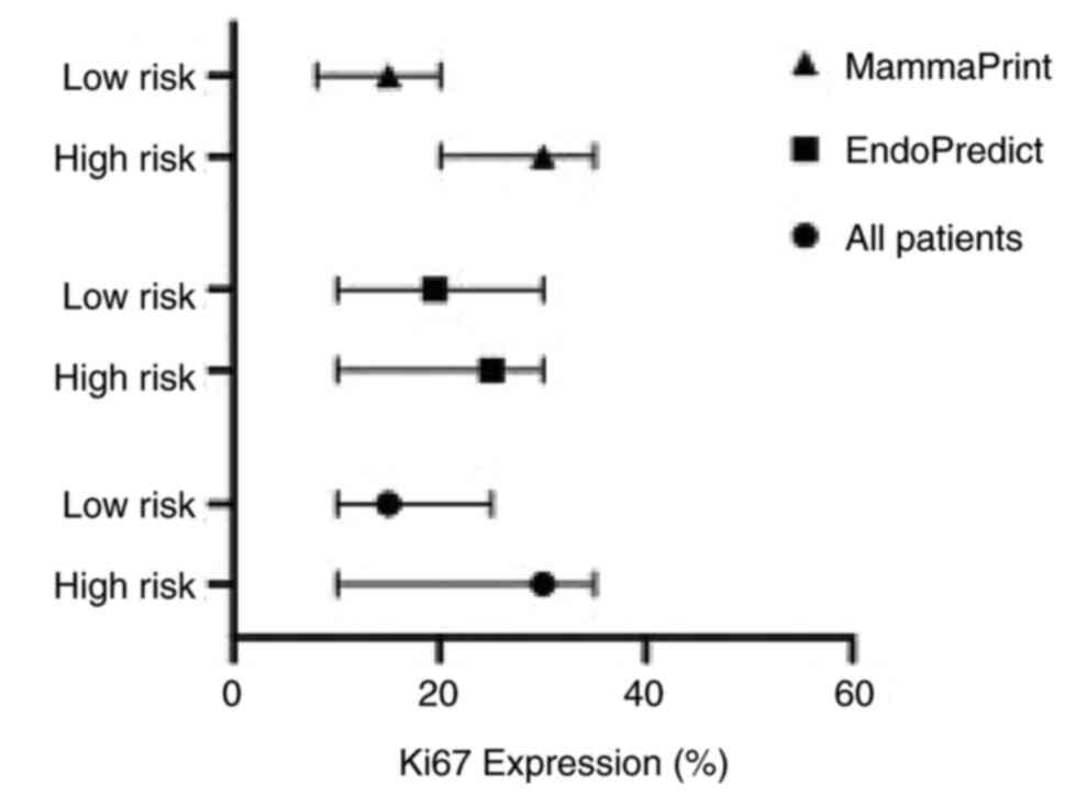
high-risk. The median Ki-67 index in the low-risk group was 19%,
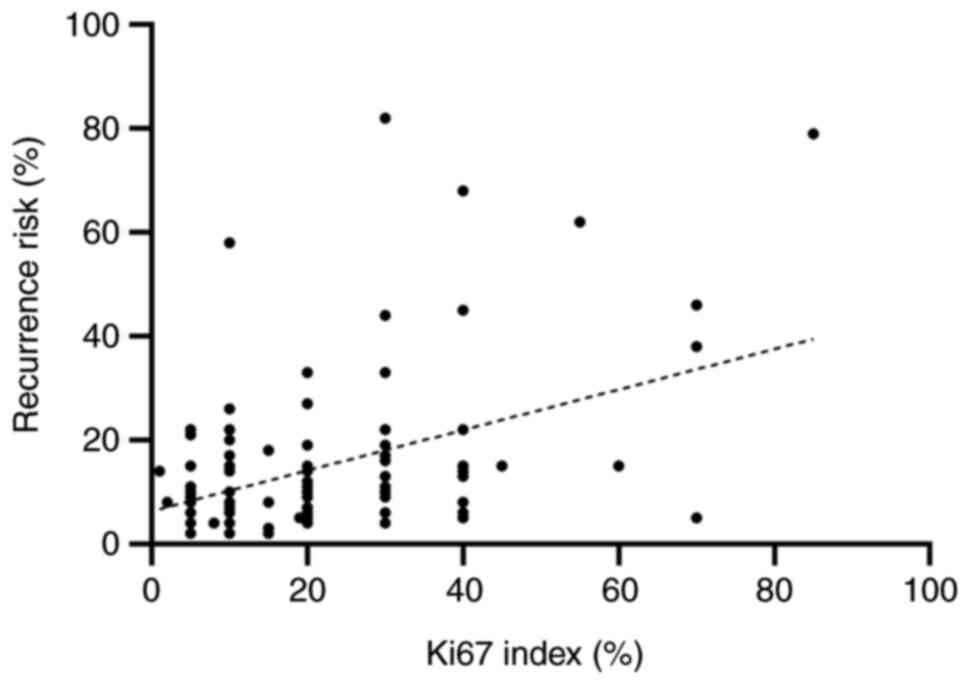
while it was 25% in the high-risk group (P=0.10, Fig. 4). No significant association was
indicated between Ki-67 index with a cutoff at 20% and EPclin risk
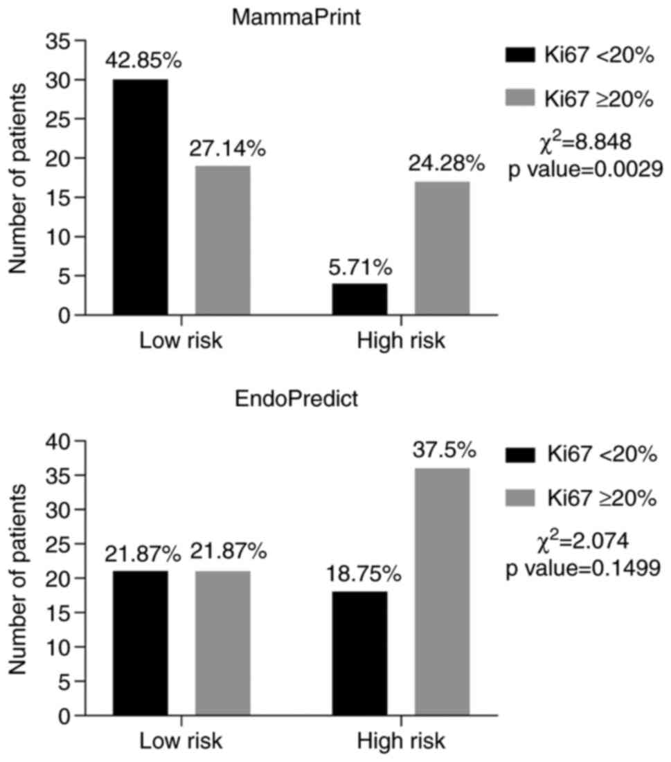
category (high vs. low) (χ2=2.07, P=0.14; Fig. 5). However, when analyzed by the
estimated risk for distant recurrence a statistically significant
correlation was observed (r2=0.2255, P=0.04; Fig. 6). Of the 42 low-risk patients, 50%
had low-risk Ki-67 index levels, while from the 52 high-risk
patients, 18 had low-risk Ki-67 index expression, resulting in an
overall concordance of 59.37% (κ=0.168; 95% CI, 0.030-0.360;
P=0.09). The association analysis [sensitivity, specificity,
positive predictive value (PPV) and negative predictive value
(NPV)] of Ki-67 index (≥20%) to predict the risk group demonstrated
a low performance (Table III).
The presence of LVI was associated with a Ki-67 index ≥20%.
| Table IIIValidation and concordance analysis
of Ki-67 with EPclin and MammaPrint. |
Table III
Validation and concordance analysis
of Ki-67 with EPclin and MammaPrint.
| Item | Ki-67 and
EPclin | Ki-67 and
MammaPrint |
|---|
| Sensitivity, % | 54 (37-70) | 88 (73-97) |
| Specificity, % | 63 (49-76) | 47 (30-65) |
| PPV, % | 50 (34-66) | 61 (46-75) |
| NPV, % | 67 (53-79) | 81 (58-95) |
| OR | 2 (0.87-4.58),
P=0.140 | 6.71 (1.96-23),
P=0.001 |
| Kappa | 0.168
(-0.03-0.36) | 0.35
(0.15-0.55) |
Ki-67 index as a surrogate marker for
MammaPrint for recurrence risk
The clinicopathological characteristics of the
MammaPrint cohort are presented in Table IV. The median age was 51 years
(range, 33-77 years). The median tumor size was 15.7 mm (range,
4-52 mm). The nodal status was pN0 in 60 patients (85.71%).
Furthermore, 60 patients (85.71%) had an IBC/NST histologic type,
51 (72.85%) had grade 2 NHG and 50 cases (72.85%) presented with
LVI. All cases were ER+ and 67 (95.71%) were PR+, all were Luminal
by BluePrint, and 21 were high-risk and 49 low-risk according to
MammaPrint.
| Table IVClinicopathological characteristics
of the MammaPrint cohort (n=70). |
Table IV
Clinicopathological characteristics
of the MammaPrint cohort (n=70).
| Parameter | Total | Ki-67 <20
(n=34) | Ki-67 ≥20%
(n=36) | P-value |
|---|
| Age, years | 51 (43-67) | 57 (48-61). | 47 (41.5-61.5) | 0.056a |
| Tumor size, mm | 15.5 (12-25) | 15 (10-25) | 17 (12.5-25) | 0.60a |
| Nodal stage | | | | 0.04b |
|
N0 | 60 (85.71) | 26 (37.14) | 34 (48.57) | |
|
N1 | 10 (14.28) | 8 (11.42) | 2 (2.85) | |
| Pathological stage
TNM (AJCC) | | | |
>0.99b |
|
IA | 39 (55.71) | 18 (25.71) | 21(30) | |
|
IB | 1 (1.42) | 1 (1.42) | 0 (0) | |
|
IIA | 26 (37.14) | 11 (15.71) | 15 (21.42) | |
|
IIB | 4 (5.71) | 4 (5.71) | 0 (0) | |
| Histological
subtype | | | | 0.04b |
|
IBC/NST | 60 (85.71) | 26 (37.14) | 34 (48.57) | |
|
Lobular | 8 (11.42) | 6 (8.57) | 2 (2.85) | |
|
Mixed | 2 (2.85) | 2 (2.85) | 0 (0) | |
| Histological grade
(Nottingham) | | | | 0.15b |
|
G1 | 8 (11.42) | 6 (8.57) | 2 (2.85) | |
|
G2 | 51 (72.85) | 26 (37.14) | 25 (35.71) | |
|
G3 | 11(15.71) | 2 (2.85) | 9 (12.85) | |
| Lymphovascular
invasion | | | | 0.79b |
|
Yes | 51 (72.85) | 24 (34.28) | 27 (38.57) | |
|
No | 19 (27.14) | 10 (14.28) | 9 (12.85) | |
| Positive estrogen
receptor | 70(100) | | | |
| % expression
(media) | 92±12.73 | 93.97±7.96 | 90.33±15.92 | 0.24c |
| Positive
progesterone receptor | 67 (95.71) | | | |
| Progesterone
receptor ≤20% | 6 (8.57) | | | |
| % expression
(media) | 78.22±28.08 | 84.53±26.17 | 72.28±28.88 | 0.07c |
| MammaPrint | | | | 0.002b |
|
Low
risk | 49(70) | 30 (42.85) | 19 (27.14) | |
|
High
risk | 21(30) | 4 (5.71) | 17 (24.28) | |
The overall median Ki-67 index was 20%. Furthermore,
the median Ki-67 index in the low-risk group was 15% and that in
the high-risk group was 30% (P=0.002; Fig. 2). The analysis indicated a
significant association between Ki-67 index and MammaPrint with a
χ2=8.85 (P=0.002l; Fig.
5). Of the 49 low-risk patients, 30 had a Ki-67 index <20%.
Furthermore, only 4 of the 21 high-risk patients had a Ki-67 index
<20% (Fig. 3). The kappa
coefficient demonstrated a fair concordance between Ki-67 index and
MammaPrint, with an overall concordance of 67.14% (κ=0.35; 95% CI,
0.15-0.55; P=0.001). The predictive accuracy analysis revealed good
sensitivity and NPV to predict the risk group. However, the
specificity and PPV were low (Table
III). In addition, the Ki-67 index was significantly associated
with the NHG and the histological type.
Discussion
Efforts have been made to match the molecular
signature tests with clinicopathological characteristics. The
ASCO/CAP associations have published guidelines for the
interpretation of HR and HER2 expression by IHC with the intent to
reduce the interobserver variability and to achieve a better
correlation with the molecular classification. However, the
capacity to discriminate between the luminal A and B subtypes by
IHC is not ideal. Even with the standardization of the technique,
there is a 30-40% discrepancy between IHC and multigene expression
assays, with a substantial impact on treatment decisions (28).
While evaluating molecular signatures, it is
important to note the relationship between Ki-67 index and the
multitude of genes tested. The Oncotype Dx gene test is a
well-known reverse transcription PCR assay of 21 genes usually
implemented to calculate the recurrence score of ER-positive breast
cancers (29,30); one of the genes assessed is the
marker of proliferation Ki-67 (MKI67), which is probably why the
Oncotype Dx assay is one of the molecular signatures with a robust
correlation with Ki-67 index (31). On the other hand, it should be
taken into consideration that neither EndoPredict nor MammaPrint
include the MKI67 gene as part of their analysis; however, they
maintain a relationship with Ki-67 significance as a proliferation
marker through other proliferation-associated genes. Bertucci et
al (32) provided a
comprehensive evaluation of the expression of genes that may be
encountered in patients stratified as high-risk by EndoPredict. The
group of genes that exhibited upregulation were involved in cell
processes such as mitotic cell cycle, proliferation and DNA
replication and division. Conversely, the ones that displayed
downregulation included genes associated with anti-apoptosis,
cell-matrix adhesion and cell cycle arrest, among others (32). The study concluded that the
upregulated genes demonstrated a correlation with proliferation
markers, such as Ki-67(32). When
analyzing MammaPrint, Tian et al (7) and others (8) were able to elucidate the genes
involved in the tumorigenesis of cancerous cells. Their results
provided different groups of genes that took part in several phases
of the cell cycle, emphasizing the upregulation of genes driving
proliferation by evading apoptosis (e.g., BCL2 binding component 3
and egl nine homolog 1, providing self-sufficiency in growth
signals [e.g., transforming growth factor beta 3 (TGFB3),
insulin-like growth factor binding protein 5 and fibroblast growth
factor 18] and insensitivity to anti-growth signals (e.g., TGFB3)
(7). With these findings they were
able to establish a connection between MammaPrint and the molecular
mechanisms of tumor growth and spread (7). Thus, a possible correlation between a
proliferation marker such as Ki-67 in IHC with genes tested in the
EndoPredict and MammaPrint molecular signatures was
demonstrated.
In the present study, the observed range of Ki-67
index was wide, with 2-70 vs. 1-85% in the EndoPredict cohort and
2-50 vs. 3-70% in the MammaPrint cohort, low-risk and high-risk,
respectively. However, the medians were slightly different for the
two risk groups. Maranta et al (33) explored the distribution of the
Ki-67 index in patients with breast cancer at their institution and
its association with other risk factors for breast cancer; their
median Ki-67 index at 22-26% was similar to that of the cohort of
the present study, acknowledging the importance for decision-making
of adjuvant therapies; however, they did not involve the use of
molecular signatures.
It is known that the Ki-67 assay has a moderate
interobserver variability (34).
The hot-spot vs. the whole-slide analysis of Ki-67 index has been
an area of controversy, with the first being more practical by
taking into account the more aggressive biology spot, acknowledging
tumor heterogeneity. Thakur et al (35) evaluated the hot-spot vs.
whole-slide Ki-67 index, identifying a strong correlation between
the two methods (r=0.938). To reduce the interobserver variability,
the International Ki-67 in Breast Cancer Working Group recommends,
if the staining is homogenous, to count at least three randomly
selected high-power fields (objective magnification, x40) and if it
is heterogenous, three fields at the tumor edge or hot spots, with
certain exceptions and scoring of preferably 1,000 cells with 500
at a minimum (27).
In the MammaPrint cohort, a low Ki-67 index
(<20%) demonstrated high sensitivity (88%) and was able to
modestly predict patients with a low risk of recurrence (PPV,
0.61%; 95% CI, 0.46-0.75). Furthermore, regarding the agreement of
the Ki-67 index and the molecular test, the MammaPrint had an
overall concordance of 67.14% and concordance index κ=0.35
(P=0.001), indicating a fair agreement. Similar to the present
results and utilizing the same Ki-67 index cutoff, Viale et
al (28) reported a
concordance of 71% (95% CI, 69-72%) between the molecular
classification of Luminal IBC and Ki-67 index in the EORTC
10041/BIG 3-04 MINDACT trial (κ=0.35; 95% CI, 0.32-0.37). In
addition, another study demonstrated comparable results (κ=0.35)
between MammaPrint and Ki-67 index in 65 patients with IBC;
however, they utilized a different cutoff for Ki-67 (14%) (36). Similar to the present study, Bösl
et al (37) compared
MammaPrint and EndoPredict with Ki-67 index, achieving a
significant correlation with MammaPrint (P=0.004) but not with the
EPclin score (P=0.09). Despite this fair concordance between Ki-67
index and MammaPrint, in the EndoPredict cohort, the Ki-67 index
overall concordance was low and did not significantly correlate
with the EPclin risk category (59.37%; κ=0.168; P=0.09). This means
that when patients were stratified by Ki-67 index, 30-40% in each
cohort were assigned to other risk categories compared to molecular
testing. The EndoPredict test gave an approximate percentage of
recurrence and this continuous variable had a positive correlation
with the Ki-67 index (P=0.04).
In clinical practice, the indication of adjuvant
chemotherapy is based on the consideration of multiple variables,
such as patient age, tumor size, histological type and grade, PR
status, LVI, and, at certain institutions, Ki-67 index. In the
present analysis, no correlation was observed between PR and Ki-67
index, EndoPredict or MammaPrint. It is worth noting that only a
small number of patients (11 and 6 patients in each cohort) had a
PR expression of <20%, highlighting the limited value of PR in
the luminal classification of EIBC compared to Ki-67 index. In
addition, a significant correlation between Ki-67 index and NHG was
observed in both cohorts (EndoPredict χ2=4.68, P=0.03;
and MammaPrint χ2=6.32, P=0.01), as has been previously
reported (38-40).
The proliferative index Ki67 is now also in use for selecting
patients who fail to achieve two weeks of Ki-67 index reduction at
<10% in the neoadjuvant endocrine therapy setting for the
addition of other therapies (34,35).
The differences in the association between Ki-67
index to MammaPrint and Ki-67 index to EndoPredict may be due to
the different patient selection criteria, clinicopathological
differences between cohorts and the acquisition of data from
multiple centers, potentially introducing interobserver variability
for Ki-67 index.
Despite the fact that molecular signature tests are
an important tool to identify patients with low risk of recurrence,
the agreement between different tests is far from perfect.
Pelaez-Garcia et al (41)
compared MammaPrint and EndoPredict and determined an overall
concordance of 72.5%, with a slight improvement using the EPclin
score to an overall concordance of 75%. Similarly, Bösl et
al (37) reported a
concordance of 66% with more patients being placed in the low-risk
category with MammaPrint.
Finally, the different molecular signature tests
have been evaluated with mixed results depending on the geographic
location. A Canadian study indicated that EndoPredict is
cost-effective with a ratio of $36,274 per quality-adjusted
life-year (QALY), with a total gain of 379 QALYs/year (42). Furthermore, in the UK, EndoPredict
was not identified as cost-effective with a threshold of
£20,000/QALY. However, it was if the incremental cost-effectiveness
ratio was £26,836/QALY (43). In
addition, a recent analysis in the UK indicated that EndoPredict
was cost-effective only if lymph node disease was present (1-3
positive nodes) with £30,000/QALY (44). On the other hand, in the USA,
MammaPrint was determined to be cost-effective at a ratio of
$10,000/QALY (45). However,
another study from the UK indicated that MammaPrint was not
cost-effective compared to current clinical practice (44). Overall, in certain countries such
as Canada and the USA, molecular signature tests are
cost-effective. The willingness of the healthcare systems of
developing countries to pay for QALYs has yet to be evaluated.
However, the cost of these tests may be onerous to healthcare
systems in precarious situations.
To the best of our knowledge, the present study was
the first to evaluate the performance of the proliferative marker
Ki-67 index for identification of high-risk patients with HR+ early
breast cancer and at the same time explore the association of Ki-67
index with two molecular signatures, MammaPrint and EndoPredict,
and risk stratification markers.
Limitations of the present study include its
retrospective nature and the potential for selection bias based on
the oncologist's selection of high clinical risk patients.
Furthermore, the groups assessed with the different molecular
signature tests were heterogeneous. However, the present study
represents a multicentric cohort of a large number of EIBC with
molecular testing that allowed the evaluation of the Ki-67 index
compared to molecular signatures tests.
In conclusion, the present study determined a
concordance between Ki-67 index and MammaPrint risk stratification
of HR+ EIBC and no concordance with the EndoPredict molecular
signature, but a positive association with the given percentage of
recurrence. Although there is no perfect molecular signature test,
these are high-value tools for therapy selection in patients with
HR+ EIBC. Cost-effectiveness analysis of these tests in developing
countries is required, and until then, the use of Ki-67 index
appears reasonable to aid in clinical decision-making together with
the other well-known clinicopathological variables.
Acknowledgements
Not applicable.
Funding
Funding: No funding was received.
Availability of data and materials
The datasets used and/or analyzed during the current
study are available from the corresponding author upon reasonable
request.
Authors' contributions
Conception and design, development of methodology,
analysis and interpretation of data and original draft preparation:
EJAG, CALG and GSGM. Development of methodology, analysis: VLR, AD
and SSF. Acquisition of data and supervision: GSGM, CALG, CVG and
ALL. Analysis and interpretation of data and supervision: CVG and
MCM. Acquisition and interpretation of data: MCM, PDCMB and ROL.
All of the authors reviewed and approved the final manuscript. The
authors GSGM and CALG approve the authenticity of the raw data.
Ethics approval and consent to
participate
Institutional review board approval was obtained
from the Ethics Committee of Research at Tecnologico de Monterrey
and the National Bioethics Commission (code ID:
CONBIOETICA19CE100820130520) and was also granted in accordance
with the Declaration of Helsinki. As the present study was
retrospective, informed consent from the subjects was not
mandatory; however, our institution requires informed consent for
any research project.
Patient consent for publication
Not applicable.
Competing interests
The authors declare that they have no competing
interests.
References
|
1
|
Bray F, Ferlay J, Soerjomataram I, Siegel
RL, Torre LA and Jemal A: Global cancer statistics 2018: GLOBOCAN
estimates of incidence and mortality worldwide for 36 cancers in
185 countries. CA Cancer J Clin. 68:394–424. 2018.PubMed/NCBI View Article : Google Scholar
|
|
2
|
Perou CM, Sørlie T, Eisen MB, van de Rijn
M, Jeffrey SS, Rees CA, Pollack JR, Ross DT, Johnsen H, Akslen LA,
et al: Molecular portraits of human breast tumours. Nature.
406:747–752. 2000.PubMed/NCBI View
Article : Google Scholar
|
|
3
|
Sørlie T, Perou CM, Tibshirani R, Aas T,
Geisler S, Johnsen H, Hastie T, Eisen MB, van de Rijn M, Jeffrey
SS, et al: Gene expression patterns of breast carcinomas
distinguish tumor subclasses with clinical implications. Proc Natl
Acad Sci USA. 98:10869–10874. 2001.PubMed/NCBI View Article : Google Scholar
|
|
4
|
Goldhirsch A, Winer EP, Coates AS, Gelber
RD, Piccart-Gebhart M, Thürlimann B and Senn HJ: Panel members.
Personalizing the treatment of women with early breast cancer:
Highlights of the St Gallen International expert consensus on the
primary therapy of early breast cancer 2013. Ann Oncol.
24:2206–2223. 2013.PubMed/NCBI View Article : Google Scholar
|
|
5
|
Gluck S, de Snoo F, Peeters J,
Stork-Sloots L and Somlo G: Molecular subtyping of early-stage
breast cancer identifies a group of patients who do not benefit
from neoadjuvant chemotherapy. Breast Cancer Res Treat.
139:759–767. 2013.PubMed/NCBI View Article : Google Scholar
|
|
6
|
Coates AS, Winer EP, Goldhirsch A, Gelber
RD, Gnant M, Piccart-Gebhart M, Thürlimann B and Senn HJ: Panel
Members. Tailoring therapies-improving the management of early
breast cancer: St Gallen International expert consensus on the
primary therapy of early breast cancer 2015. Ann Oncol.
26:1533–1546. 2015.PubMed/NCBI View Article : Google Scholar
|
|
7
|
Tian S, Roepman P, Van't Veer LJ, Bernards
R, de Snoo F and Glas AM: Biological functions of the genes in the
mammaprint breast cancer profile reflect the hallmarks of cancer.
Biomark Insights. 5:129–138. 2010.PubMed/NCBI View Article : Google Scholar
|
|
8
|
van de Vijver MJ, He YD, van't Veer LJ,
Dai H, Hart AA, Voskuil DW, Schreiber GJ, Peterse JL, Roberts C,
Marton MJ, et al: A gene-expression signature as a predictor of
survival in breast cancer. N Engl J Med. 347:1999–2009.
2002.PubMed/NCBI View Article : Google Scholar
|
|
9
|
Müller BM, Keil E, Lehmann A, Winzer KJ,
Richter-Ehrenstein C, Prinzler J, Bangemann N, Reles A, Stadie S,
Schoenegg W, et al: The endopredict gene-expression assay in
clinical practice-performance and impact on clinical decisions.
PLoS One. 8(e68252)2013.PubMed/NCBI View Article : Google Scholar
|
|
10
|
Filipits M, Rudas M, Jakesz R, Dubsky P,
Fitzal F, Singer CF, Dietze O, Greil R, Jelen A, Sevelda P, et al:
A new molecular predictor of distant recurrence in ER-positive,
HER2-negative breast cancer adds independent information to
conventional clinical risk factors. Clin Cancer Res. 17:6012–6020.
2011.PubMed/NCBI View Article : Google Scholar
|
|
11
|
Duffy MJ, Harbeck N, Nap M, Molina R,
Nicolini A, Senkus E and Cardoso F: Clinical use of biomarkers in
breast cancer: Updated guidelines from the European Group on Tumor
Markers (EGTM). Eur J Cancer. 75:284–298. 2017.PubMed/NCBI View Article : Google Scholar
|
|
12
|
Weigelt B, Reis-Filho JS and Swanton C:
Genomic analyses to select patients for adjuvant chemotherapy:
Trials and tribulations. Ann Oncol. 23 (Suppl 10):x211–x218.
2012.PubMed/NCBI View Article : Google Scholar
|
|
13
|
van Steenhoven JEC, Kuijer A, van Diest
PJ, van Gorp JM, Straver M, Elias SG, Wesseling J, Rutgers E,
Timmer-Bonte JNH, Nieboer P, et al: Conventional pathology versus
gene signatures for assessing Luminal A and B type breast cancers:
Results of a prospective cohort study. Genes (Basel).
9(261)2018.PubMed/NCBI View Article : Google Scholar
|
|
14
|
Bustreo S, Osella-Abate S, Cassoni P,
Donadio M, Airoldi M, Pedani F, Papotti M, Sapino A and Castellano
I: Optimal Ki67 cut-off for luminal breast cancer prognostic
evaluation: A large case series study with a long-term follow-up.
Breast Cancer Res Treat. 157:363–371. 2016.PubMed/NCBI View Article : Google Scholar
|
|
15
|
Dubsky P, Filipits M, Jakesz R, Rudas M,
Singer CF, Greil R, Dietze O, Luisser I, Klug E, Sedivy R, et al:
EndoPredict improves the prognostic classification derived from
common clinical guidelines in ER-positive, HER2-negative early
breast cancer. Ann Oncol. 24:640–647. 2013.PubMed/NCBI View Article : Google Scholar
|
|
16
|
Cheang MC, Chia SK, Voduc D, Gao D, Leung
S, Snider J, Watson M, Davies S, Bernard PS, Parker JS, et al: Ki67
index, HER2 status, and prognosis of patients with luminal B breast
cancer. J Natl Cancer Inst. 101:736–750. 2009.PubMed/NCBI View Article : Google Scholar
|
|
17
|
Cuzick J, Dowsett M, Pineda S, Wale C,
Salter J, Quinn E, Zabaglo L, Mallon E, Green AR, Ellis IO, et al:
Prognostic value of a combined estrogen receptor, progesterone
receptor, Ki-67, and human epidermal growth factor receptor 2
immunohistochemical score and comparison with the Genomic Health
recurrence score in early breast cancer. J Clin Oncol.
29:4273–4278. 2011.PubMed/NCBI View Article : Google Scholar
|
|
18
|
de Azambuja E, Cardoso F, de Castro G Jr,
Colozza M, Mano MS, Durbecq V, Sotiriou C, Larsimont D,
Piccart-Gebhart MJ and Paesmans M: Ki-67 as prognostic marker in
early breast cancer: A meta-analysis of published studies involving
12,155 patients. Br J Cancer. 96:1504–1513. 2007.PubMed/NCBI View Article : Google Scholar
|
|
19
|
Focke CM, van Diest PJ and Decker T: St
Gallen 2015 subtyping of luminal breast cancers: Impact of
different Ki67-based proliferation assessment methods. Breast
Cancer Res Treat. 159:257–263. 2016.PubMed/NCBI View Article : Google Scholar
|
|
20
|
Goldhirsch A, Wood WC, Coates AS, Gelber
RD, Thürlimann B and Senn HJ: Panel members. Strategies for
subtypes-dealing with the diversity of breast cancer: Highlights of
the St. Gallen International expert consensus on the primary
therapy of early breast cancer 2011. Ann Oncol. 22:1736–1747.
2011.PubMed/NCBI View Article : Google Scholar
|
|
21
|
Villarreal-Garza C, Lopez-Martinez EA,
Deneken-Hernandez Z, Maffuz-Aziz A, Muñoz-Lozano JF,
Barragan-Carrillo R, Ramos-Elias P, Moreno B, Diaz-Perez H,
Peña-Curiel O, et al: Change in therapeutic management after the
EndoPredict assay in a prospective decision impact study of Mexican
premenopausal breast cancer patients. PLoS One.
15(e0228884)2020.PubMed/NCBI View Article : Google Scholar
|
|
22
|
Cardoso F, van't Veer LJ, Bogaerts J,
Slaets L, Viale G, Delaloge S, Pierga JY, Brain E, Causeret S,
DeLorenzi M, et al: 70-Gene signature as an aid to treatment
decisions in early-stage breast cancer. N Engl J Med. 375:717–729.
2016.PubMed/NCBI View Article : Google Scholar
|
|
23
|
Allison KH, Hammond ME, Dowsett M,
McKernin SE, Carey LA, Fitzgibbons PL, Hayes DF, Lakhani SR,
Chavez-MacGregor M, Perlmutter J, et al: Estrogen and progesterone
receptor testing in breast cancer: ASCO/CAP guideline update. J
Clin Oncol. 38:1346–1366. 2020.PubMed/NCBI View Article : Google Scholar
|
|
24
|
Prat A, Cheang MC, Martín M, Parker JS,
Carrasco E, Caballero R, Tyldesley S, Gelmon K, Bernard PS, Nielsen
TO and Perou CM: Prognostic significance of progesterone
receptor-positive tumor cells within immunohistochemically defined
luminal A breast cancer. J Clin Oncol. 31:203–209. 2013.PubMed/NCBI View Article : Google Scholar
|
|
25
|
Wolff AC, Hammond MEH, Allison KH, Harvey
BE, Mangu PB, Bartlett JMS, Bilous M, Ellis IO, Fitzgibbons P,
Hanna W, et al: Human epidermal growth factor receptor 2 testing in
breast cancer: American society of clinical oncology/college of
American pathologists clinical practice guideline focused update. J
Clin Oncol. 36:2105–2122. 2018.PubMed/NCBI View Article : Google Scholar
|
|
26
|
Penault-Llorca F and Radosevic-Robin N:
Ki67 assessment in breast cancer: An update. Pathology. 49:166–171.
2017.PubMed/NCBI View Article : Google Scholar
|
|
27
|
Dowsett M, Nielsen TO, A'Hern R, Bartlett
J, Coombes RC, Cuzick J, Ellis M, Henry NL, Hugh JC, Lively T, et
al: Assessment of Ki67 in breast cancer: Recommendations from the
International Ki67 in breast cancer working group. J Natl Cancer
Inst. 103:1656–1664. 2011.PubMed/NCBI View Article : Google Scholar
|
|
28
|
Viale G, de Snoo FA, Slaets L, Bogaerts J,
van 't Veer L, Rutgers EJ, Piccart-Gebhart MJ, Stork-Sloots L, Glas
A, Russo L, et al: Immunohistochemical versus molecular (BluePrint
and MammaPrint) subtyping of breast carcinoma. Outcome results from
the EORTC 10041/BIG 3-04 MINDACT trial. Breast Cancer Res Treat.
167:123–131. 2018.PubMed/NCBI View Article : Google Scholar
|
|
29
|
Paik S, Shak S, Tang G, Kim C, Baker J,
Cronin M, Baehner FL, Walker MG, Watson D, Park T, et al: A
multigene assay to predict recurrence of tamoxifen-treated,
node-negative breast cancer. N Engl J Med. 351:2817–2826.
2004.PubMed/NCBI View Article : Google Scholar
|
|
30
|
Sahebjam S, Aloyz R, Pilavdzic D, Brisson
ML, Ferrario C, Bouganim N, Cohen V, Miller WH Jr and Panasci LC:
Ki 67 is a major, but not the sole determinant of Oncotype Dx
recurrence score. Br J Cancer. 105:1342–1345. 2011.PubMed/NCBI View Article : Google Scholar
|
|
31
|
Ahmed W, Malik MFA, Saeed M and Haq F:
Copy number profiling of Oncotype DX genes reveals association with
survival of breast cancer patients. Mol Biol Rep. 45:2185–2192.
2018.PubMed/NCBI View Article : Google Scholar
|
|
32
|
Bertucci F, Finetti P, Viens P and
Birnbaum D: EndoPredict predicts for the response to neoadjuvant
chemotherapy in ER-positive, HER2-negative breast cancer. Cancer
Lett. 355:70–75. 2014.PubMed/NCBI View Article : Google Scholar
|
|
33
|
Maranta AF, Broder S, Fritzsche C, Knauer
M, Thürlimann B, Jochum W and Ruhstaller T: Do YOU know the Ki-67
index of your breast cancer patients? Knowledge of your
institution's Ki-67 index distribution and its robustness is
essential for decision-making in early breast cancer. Breast.
51:120–126. 2020.PubMed/NCBI View Article : Google Scholar
|
|
34
|
Gluz O, Nitz UA, Christgen M, Kates RE,
Shak S, Clemens M, Kraemer S, Aktas B, Kuemmel S, Reimer T, et al:
West German study group phase III PlanB trial: First prospective
outcome data for the 21-Gene recurrence score assay and concordance
of prognostic markers by central and local pathology assessment. J
Clin Oncol. 34:2341–2349. 2016.PubMed/NCBI View Article : Google Scholar
|
|
35
|
Thakur SS, Li H, Chan AMY, Tudor R, Bigras
G, Morris D, Enwere EK and Yang H: The use of automated Ki67
analysis to predict Oncotype DX risk-of-recurrence categories in
early-stage breast cancer. PLoS One. 13(e0188983)2018.PubMed/NCBI View Article : Google Scholar
|
|
36
|
Nguyen B, Cusumano PG, Deck K, Kerlin D,
Garcia AA, Barone JL, Rivera E, Yao K, de Snoo FA, van den Akker J,
et al: Comparison of molecular subtyping with BluePrint,
MammaPrint, and TargetPrint to local clinical subtyping in breast
cancer patients. Ann Surg Oncol. 19:3257–3263. 2012.PubMed/NCBI View Article : Google Scholar
|
|
37
|
Bösl A, Spitzmüller A, Jasarevic Z, Rauch
S, Jäger S and Offner F: MammaPrint versus EndoPredict: Poor
correlation in disease recurrence risk classification of hormone
receptor positive breast cancer. PLoS One.
12(e0183458)2017.PubMed/NCBI View Article : Google Scholar
|
|
38
|
Trihia H, Murray S, Price K, Gelber RD,
Golouh R, Goldhirsch A, Coates AS, Collins J, Castiglione-Gertsch
M, Gusterson BA, et al: Ki-67 expression in breast carcinoma: Its
association with grading systems, clinical parameters, and other
prognostic factors-a surrogate marker? Cancer. 97:1321–1331.
2003.PubMed/NCBI View Article : Google Scholar
|
|
39
|
Marwah N, Batra A, Marwah S, Gupta V,
Shakya S and Sen R: Correlation of proliferative index with various
clinicopathologic prognostic parameters in primary breast
carcinoma: A study from North India. J Cancer Res Ther. 14:537–542.
2018.PubMed/NCBI View Article : Google Scholar
|
|
40
|
Shokouh TZ, Ezatollah A and Barand P:
Interrelationships between Ki67, HER2/neu, p53, ER, and PR status
and their associations with tumor grade and lymph node involvement
in breast carcinoma subtypes: Retrospective-observational
analytical study. Medicine (Baltimore). 94(e1359)2015.PubMed/NCBI View Article : Google Scholar
|
|
41
|
Pelaez-Garcia A, Yébenes L, Berjón A,
Angulo A, Zamora P, Sánchez-Méndez JI, Espinosa E, Redondo A,
Heredia-Soto V, Mendiola M, et al: Comparison of risk
classification between EndoPredict and MammaPrint in
ER-positive/HER2-negative primary invasive breast cancer. PLoS One.
12(e0183452)2017.PubMed/NCBI View Article : Google Scholar
|
|
42
|
Hannouf MB, Zaric GS, Blanchette P,
Brezden-Masley C, Paulden M, McCabe C, Raphael J and Brackstone M:
Cost-effectiveness analysis of multigene expression profiling
assays to guide adjuvant therapy decisions in women with invasive
early-stage breast cancer. Pharmacogenomics J. 20:27–46.
2020.PubMed/NCBI View Article : Google Scholar
|
|
43
|
Hinde S, Theriou C, May S, Matthews L,
Arbon A, Fallowfield L and Bloomfield D: The cost-effectiveness of
EndoPredict to inform adjuvant chemotherapy decisions in early
breast cancer. Health Policy and Technology. 8:75–83. 2019.
|
|
44
|
Harnan S, Tappenden P, Cooper K, Stevens
J, Bessey A, Rafia R, Ward S, Wong R, Stein RC and Brown J: Tumour
profiling tests to guide adjuvant chemotherapy decisions in early
breast cancer: A systematic review and economic analysis. Health
Technol Assess. 23:1–328. 2019.PubMed/NCBI View Article : Google Scholar
|
|
45
|
Chen E, Tong KB and Malin JL:
Cost-effectiveness of 70-gene MammaPrint signature in node-negative
breast cancer. Am J Manag Care. 16:e333–e342. 2010.PubMed/NCBI
|